OUTCOME OF ILIZAROV FIXATOR IN
INFECTED NONUNION TIBIA
A dissertation submitted to the Tamil Nadu Dr.M.G.R.
Medical University in partial fulfillment of the requirement for
CERTIFICATE
This is to certify that this dissertation titled “OUTCOME OF ILIZAROV FIXATOR IN INFECTED NONUNION TIBIA ” is a bonafide work done by Dr. SHYAMASUNDAR. L.G , in the Department of Orthopaedic Surgery, Christian Medical College and Hospital, Vellore in partial fulfillment of the rules and regulations of the Tamil Nadu Dr. M.G.R. Medical University for the award of M.S. Degree (Branch-II) Orthopaedic Surgery under the supervision and guidance of Prof. Vernon N. Lee during the period of his post-graduate study from March 2007 to February 2009.
This consolidated report presented herein is based on bonafide cases, studied by the candidate himself.
Prof. Vernon N. Lee
D.Ortho., M.S.Ortho. M.Ch.Orth.(L’Pool) Professor of Orthopaedics
CERTIFICATE
This is to certify that this dissertation titled “OUTCOME OF ILIZAROV FIXATOR IN INFECTED NONUNION TIBIA” is a bonafide work done by Dr. SHYAMASUNDAR. L.G, in the Department of Orthopaedic Surgery, Christian Medical College and Hospital, Vellore in partial fulfillment of the rules and regulations of the Tamil Nadu Dr. M.G.R. Medical University for the award of M.S. Degree (Branch-II) Orthopaedic Surgery during the period of his post-graduate study from March 2007 to February 2009.
This consolidated report presented herein is based on bonafide cases, studied by the candidate himself.
Prof. Vrisha Madhuri
D.Ortho., M.S.Ortho., M.Ch.Orth.(L’Pool) Professor & Head
ACKNOWLEDGEMENT
I have great pleasure in thanking my teacher Prof. Vernon.N.Lee, Professor and Head of the Department of Orthopaedic Surgery – Unit I, Christian Medical College for permitting me to use the clinical materials and for guiding me through the preparation of the dissertation.
I owe my sincere thanks to Dr. Vinoo Mathew Cherian, Associate professor, Dr. Manasseh Nithyananth, Associate professor and Dr. Abhay Gahukamle, Lecturer, Department of Orthopedic Surgery – Unit I for their guidance and encouragement throughout this study.
My sincere thanks to my teachers Prof. G.D. Sunderaraj,
Prof. Samuel Chittaranjan, Prof. Ravi Jacob Korula and Prof. Vrisha Madhuri who gave valuable advise, support and encouragement throughout the preparation of this thesis.
I sincerely acknowledge the help rendered by Mr. Prasanna Samuel, Department of Biostatistics in performing the statistical analysis of data.
S. No Title Page No
1 Aims and objectives 2
2 Literature review 3
3 Materials and methods 54
4 Result 63
5 Discussion 71
6 Conclusion 92
7 Bibliography 93
AIMS AND OBJECTIVES
To study patients with infected nonunion of the tibia who were treated with the Ilizarov fixator.
To study the outcome of infected nonunion of tibia treated with Ilizarov ring fixator .
To study factors affecting the outcome.
LITERATURE REVIEW INTRODUCTION-
DEFINITION
Brinker1 defined nonunion as a fracture that according to treating doctor , has no possibility of healing without surgical intervention.
The Food and drug administration definition for nonunion – The fracture that is over 9months old and that has not shown radiographic signs of progression toward healing for 3 consecutive months.
Out of several parameters that help to identify infected nonunion of a tibial fracture as cited by Toh and Jupiter, exposed bone that has been devoid of vascularized periosteal coverage for more than six weeks and purulent
drainage were considered to make a diagnosis of infected nonunion2.
Infected nonunion of the tibia is a posttraumatic bony wound3 and not equivalent to hematogenous osteomyelitis. The latter disease is a bacterial intramedullary abscess usually occurring through a hematogenous route into the metaphyseal region of the involved bone. If neglected, the condition creates necrotic bone, which requires surgical intervention and skeletal reconstruction. Apart from the presence of pus, the lesion of hematogenous osteomyelitis of childhood has little in common with that which is caused by trauma.
compromised, this creates an ideal milieu for bacterial proliferation4 .
Whereas successful management of hematogenous osteomyelitis is often the result of an appropriate choice of parenteral antibiotic, a successful outcome to posttraumatic wound of bone requires surgical revision of the wound to prevent further bacterial growth.
Several parameters will help identify infected nonunion of a tibial fracture 3 . 1. Exposed bone that has been devoid of vascularized periosteal coverage for more than 6 weeks,
2. Purulent drainage,
3. Positive bacterial culture from the depths of the wound, and 4. Histological evidence of necrotic bone containing empty lacunae.
CAUSES 5
Infected nonunion can develop – 1. After an open fracture
2. After a previous open reduction and internal fixation (ORIF) 3. Sequelae to chronic hematogenous osteomyelitis.
The open fracture is the most common cause of infected nonunion and the tibia is the most commonly involved bone in the infected nonunion after an open fracture. Because of increased trend for operative fracture surgery,infected nonunion after implant surgery has increased in incidence 4. PATHOPHYSIOLOGY OF POSTTRAUMATIC
OSTEOMYELITIS 6,7,8
Osteomyelitis is a frequent complication of trauma induced open fractures. This is especially true of fractures associated with severe soft tissue injury. Osteomyelitis is notorious for its tendency to recur, even after years of quiescence. It has been proposed that cure is not an appropriate word to describe the outcome of treatment of osteomyelitis, that the best one can hope to accomplish is arrest.
The stages in the pathophysiology are- 1. Entry of pathogens
3. Interference in host reaction to infection 4. Damage to the host
5. Persistence of infection. 1. ENTRY OF PATHOGENS
To accomplish this microbial pathogen must breach the mechanical barriers, the skin and mucus membranes, that normally protect the host. Without a break in the skin, as is the case in closed fractures,
osteomyelitis is an infrequent complication. The incidence of
osteomyelitis is much higher in open fractures when bacteria have the opportunity to enter the wound directly from the environment.
Clostridium perfringens associated with farm injuries, Pseudomonas aeruginosa and Aeromonas hydrophila after injury in fresh water, and Vibrio and Erysipelothrix infections with salt water
contamination. However, although most wounds are contaminated with bacteria at the time of injury, most cases of posttraumatic osteomyelitis are caused by hospital acquired pathogens such as
2. ESTABLISHING INFECTION
The presence of bacteria in a wound is not sufficient to cause infection. Approximately 60% to 70% of open fractures are contaminated by
bacteria, but a much smaller percentage go on to infection. Factors that help in establishment of infection are-
1. Damage to the local soft tissue & bone –There is 30%-40% risk of infection in type 3B open fractures . Traumatized soft tissue and bone expose potential binding sites for bacteria such as collagen. Staphylococcus aureus, the most common cause of posttraumatic osteomyelitis, has receptors for numerous host proteins including collagen.
2. Compromised blood supply- Necrosed tissue act as foreign body . 3. Implants used for stabilization serve as additional foci for colonization
of bacteria.
4. The instability of fracture
inhibits phagocytosis, impedes the ingress of antibodies, and impairs T lymphocyte and B lmphocyte function.
3. INTERFERENCE IN HOST REACTION TO INFECTION
The initial host response to infection is an acute inflammatory reaction, which fight against the bacteria. But trauma has been reported to delay the inflammatory response, depress cell mediated immunity, the function of polymorphonuclear leukocytes, including chemotaxis, superoxide production, and microbial killing.
The presence of a foreign body at the site of infection, usually in the form of a metallic fixation device or prosthetic implant, can inhibit the efforts of host defense mechanisms. Metals used in these devices stimulate production of cytokines and impair lymphocyte activity.
Dean et al found that Cr and Ti inhibit mitogen stimulated T cell and B cell proliferation,and inhibited the production of IL-2 and INF-gamma. 4. DAMAGE TO HOST
Although the primary goal of initial inflammatory response is to destroy bacteria and contain the spread of infection, proteolytic enzymes released by phagocytes also can damage surrounding tissue. Certain bacterial
Bone resorption is also a feature of osteomyelitis. Osteopenia and weakened bone may predispose the patient to pathologic fractures. If a fracture already is present, bone resorption may delay or prevent its healing. Bacterial products, such as lipopolysaccharide of Gram negative bacteria and surface associated protein of Staphylococcus aureus are stimulators of bone resorption.
Proinflammatory cytokines - IL 1beta, IL-6 and TNF-alpha are released in the acute inflammatory response to injury and infection , and are the most important mediators of bone resorption. Systemic effects of osteomyelitis also may arise, but are not common. Adjacent joints may become infected by contiguous spread, or bacteremia can carry infection
to metastatic sites. Staphylococci and Streptococci also have the ability to cause toxic shock syndrome.
5. PERSISTENCE OF INFECTION
INVESTIGATIONS
The following investigations will help in the diagnosis of infected nonunion HEMATOLOGIC INVESTIGATION
An elevated leukocyte level or Westergren erythrocyte sedimentation rate can be consistent with ongoing deep bony infection, although they are not necessarily diagnostic 9. Normal levels can be found even with active drainage in which there is little surrounding cellulitis or systemic involvement. Metabolic parameters such as an evaluation of glucose tolerance, renal function, and liver functions (particularly protein levels) should be considered before beginning operative reconstruction.
RADIOGRAPHIC STUDIES
Several radiographic features, such as cortical irregularity, perisosteal reaction, alteration in normal mineralization, or the presence of a bony sequesturm have been associated with deep bony infection, in the presence of an ununited fracture (and especially in association with previously implanted metal internal fixation devices) 10.
BONE SCAN
The 67-gallium citrate isotope may accumulate at the site of infection or inflammation by virtue of an increase in the permeability of the local vascular structures, the isotope being taken up by microorganisms and some elements present in the inflammatory exudates. Because any process leading to reactive bone formation will result in increased uptake of 67-gallium citrate, the scan suggests a deep bony infection only when the focal uptake is inconsistent with that seen on the
technetium 99-MDP scan. If a similar pattern exists, the 67-gallium scan must be substantially greater in uptake than that of the technetium-99 MDP scan to be considered suggestive of deep bony infection. The accuracy rate of this approach has been disappointing, varying from 50% to 60%. 12
An additional diagnostic approach has been to label leukocytes with 111-indium. In a study comparing indium to sequential technetium and gallium scans, the overall sensitivity rate was 83%, with a specificity rate of 86% and an accuracy rete of 83%, yet false results have been attributed to problems with the technique of leukocyte labeling.
TISSUE BIOPSY
CLASSIFICATION OF INFECTED NONUNION TIBIA
Classification must be made after soft tissue & skeletal debridement. Several classifications exist-
1. Weiland’s 13 (on extent of infection)-
Type 1- Bone exposed and soft tissue infection present.
Type 2- Circumfrential cortical & endosteal infection present. Type 3- - Circumfrential cortical & endosteal infection with segmental bone loss present.
2. Gordon & Chiu’s (on severity of underlying bone damage.) 14 A- Tibial defect & nonunion without substantial bone loss (< 3cm)
B- Tibial defect >3cm with intact fibula C- Tibial defect >3cm with nonunion fibula
3.University of Texas classification-(location of infection and immune competence of the host)
1- Intramedullary ; 2-Superficial; 3- Local; 4- Diffuse with segmental boneloss.
Type A- Healthy with adequate soft tissue cover; Type B- With local or systemic compromise;
Type C-Severely compromised, contraindicated for surgical reconstruction.
4. May et al 3 -
Type 1- An intact tibia & fibula that can withstand functional loads. The skeletal involvement is unicortical & debridement can be done without any threat to skeletal integrity.
Type 2- An intact tibia with bone graft needed for structural support. Type 3- A tibial defect </= 6cm with intact ipsilateral fibula.
Type 4- A tibial defect > 6cm with intact ipsilateral fibula.
(The defect > 6cm can not be bridged successfully by autograft alone) Type 5- A tibial defect > 6cm & no usable fibula.
5 Dror Paley-
A1- Mobile atrophic, Bone loss <1cm ,
B1- Osseous defect >1cm, length maintained B2- Bone loss >1cm, shortening present
B3- Both osseous defect & bone loss with shortening. 5. Jain et al 5 -
Type A - infected nonunion of long bones with nondraining (quiescent) infection, with or without implant in situ.
Type B - infected nonunion of long bones with draining (active) infection. Both are classified further into two subtypes: 1) nonunion with a bone gap smaller than 4 cm or 2) nonunion with a bone gap larger than 4 cm.
TACTICS FOR RECONSTRUCTION
The overall goal in the reconstruction of an infected, ununited tibial fracture involves more than control of the infection and includes creation of a healed, aligned, and drainage –free limb that is functionally better than that which could be achieved by amputation and prosthetic fitting. Several
Once the decision has been made to reconstruct, the first step is to eradicate infected and avascular bone with surrounding compromised soft tissue. The extent of bony and soft tissue debridement is defined by the presence of punctate bleeding .
The experience with the posterolateral bone graft between the fibula and tibia has suggested that in some cases active infection will become quiescent once the tibia is inherently stable.
Skeletal Stabilization
Soft tissue reconstruction- Important issues to be considered-
1. As a general rule, use of parenteral antibiotics should be delayed until the time of soft tissue coverage. This will minimize the possibility that the organisms identified at the initial debridements will become resistant to the culture-specific antibiotic. 17
2. Timing- In most instances, soft tissue coverage is delayed until the extent of the bony and soft tissue infection has been identified and the base of the wound has become vascular. This is best defined as a wound that will support granulation tissue.
The possibilities are from skin graft to free tissue transfer. Several local pedicled muscle and myocutaneous flaps exist in and about the tibia,
including the gastrocnemius and soleus flaps in the leg 18. At the distal third of the leg, flaps such as the extensor hallucis longus and peroneus brevis have been developed, although they are not used widely. Free muscle flaps, including the latissimus dorsi, gracilis, tensor fascia lata, and rectus
The experience of several authors suggest a high rate of success using
microvascular free tissue transfers, and these have become in many cases the preferred method for soft tissue reconstruction, in contrast to pedicle flaps, cross-leg flaps, or both. 14,19
In the presence of large bony defects that require reconstruction,
antibiotic-impregnated methylmethacrylate beads can be used to fill the dead space, in which later bony reconstruction can be done.
Bony reconstruction
1 Open cancellous bone grafting-
Papineau technique 20 - consists of packing a skeletal defect with minced cancellous bone without overlying soft tissue coverage. most applicable when treating patients who have a metaphyseal skeletal defect associated with a limited zone of soft tissue loss.The method will be
successful only if the wound is sufficiently cleaned and well vascularized to support the growth of granulation tissue.
graft incorporation and its remodeling to develop the intrinsic strength to withstand functional loading.
2. Posterior bone grafting and tibiofibular synostosis- 21
Advantages - a relatively straightforward surgical approach that avoids the zone of trauma and previous infection and incorporates an intact fibula into the overall structural integrity of the lower limb. Autogenous cancellous grafts can be harvested from the iliac crest and placed in a relatively well-vascularized environment beneath the posterior muscles of the lower limb, facilitating rapid revisualization and graft incorporation.
The disadvantages associated with the posterolateral approach include potential injuries to either the peroneal or posterior tibial artery. Because many of these limbs may have been injured by high-energy trauma, they may have only one remaining arterial pedicle. Thus when considering this approach, the physician might consider obtaining a preoperative arteriogram. 3 Anterior cancellous bone graft beneath a flap
must know the location of the vascular pedicle to the muscle flap in this approach.
The advantage of the anterior cancellous graft beneath a flap is
primarily that the bone graft can be placed directly in the defect. Its ability to revascularize and reincorporate is enhanced by the overlying
well-vascularized muscle bed.
The disadvantages of this approach involve primarily the danger to the overlying muscle flap.
4 Fibula-pro-tibia transfer 22
The fibula can be raised on its vascular pedicle and transferred as a vascular zed graft in a single stage. The advantages of this procedure are that the ipsilateral fibula can be used without violating the contralateral leg. The fibula is a straight cortical bone and is often long enough to bridge most defects of the tibia. A fibula transferred with its vascular pedicle intact will hypertrophy when subjected to greater loading stresses. Finally, the cortical structure of the fibula allows bony screws and plates to be used to securely fix the bone in these unstable limbs.
5 Vascularized autogenous bone graft
The donor sites from which vascularzed bone grafts can be obtained to reconstruct the tibia include the fibula, the iliac crest, and the ribs.Although the iliac crest offers some theoretical advantages in that it can be elevated as a composite osteocutaneous or osteomyocutaneous flap, difficulties with its shape, size, and strength have limited its application.
Yajima et al 23 in their series of use of vascularised bone graft stated bone union to occur in 7months for femur and 6months for tibia. The disadvantages being donor site morbidity and graft can only bridge the gap but shortening is not addressed. In other series, the osseous union has occurred between 16 to 30 weeks after transfer. Given its technical
complexity, however, application of the technique is limited to centers with experienced microvascular surgeons.
6 Nonvascular autogenous cortical bone grafting
7. Transplant of allograft
The disadvantages of the allograft transfer in a patient with a history of infection are those primarily related to the inherent potential for the allograft to become a sequestrum should residual organisms remain in the recipient site.
8. Distraction Histogenesis
The technique involves corticotomy and slowly transporting the segment along with soft tissue envelope, supported by flexible ring external fixator.
The advantages of this technique, if it is successful, are that the
reconstruction can require only one surgical procedure. It can restore skeletal length and alignment, osseous union, and functional rehabilitation while the external ring frame is in place. A contracted, avascular soft tissue envelope may not be a contraindication to this approach, although it has been found that the procedure can be made substantially easier if the traumatized soft tissue is replaced by a muscle flap before bony reconstruction.
withstand functional loading. The device is cumbersome, technically difficult to apply, and often is uncomfortable for the patient.
The use of the concept of bone transport stems from earlier attempts by surgeons to overcome limb length discrepancy by limb lengthening. In the late 19th century Alessandro Codivilla pioneered the concept of limb
lengthening but his methods were primitive and fraught with problems. His successor Vittorio Putti devised a relatively safe procedure for limb
lengthening. he fashioned an instrument called osteoton for operative lengthening of the femur . He achieved this by producing a fracture of the shaft and used two large pins in the proximal and distal fragments. In addition he used a telescoping tube which could be attached to the pins by metal sockets and these tubes were used to provide traction on the pins with a powerful spring. Two scales divided in millimetres gave the surgeon the measure of traction and the length distracted. A screw was used to operate the spring. Putti performed Z – Shaped osteotomies similar to that done for tendon lengthening and fixed the traction apparatus and progressively
callus in all and neurogenic pain in one patient in the sciatic and tibial nerve dermatomes 24.
Patterson 25 in his review on leg lengthening highlights the work of Anderson. Boyd et al and surgeons like Wagner, Western and Kawamura, which led to the rekindling of interest in limb lengthening. This led to the concept of bone transport by callus distraction.
Wagner 26 performed limb lengthening in 58 patients under the age of 17y during 1972 to 1978. He developed an external fixator which could be used to distract the femur and the tibia after an osteotomy. He performed satisfactory lengthening up to 7cm and published his results in his review.
TREATMENT BASED ON CLASSIFICATION
Accoring to Jain et al 5 -
Type A1 (Quiescent infection with bone defect less than 4cm)- Single-stage debridement and bone grafting with fracture stabilization are the methods of choice.
Type B1 (Draining infection with bone defect less than 4cm)- Adequate debridement, fracture stabilization, and second-stage bone grafting gives desirable results .. Type A2 (Quiescent with bone defect > 4cm) and Type B2 (Draining infection with bone defect > 4cm) – Debridement and
According to Dror Paley’s classification- (Modified by Maurizio Catagni 27
Classifn. PATHOLOGY TREATMENT
A1 Mobile atrophic nonunion with bone loss less than 1cm.
Excision of the atrophic ends & BIFOCAL osteosynthesis
A2.1 Stiff hypertrophic without deformity & bone loss less than 1cm.
MONOFOCAL osteosynthesis
A2.2 Stiff hypertrophic with deformity & bone loss less than 1cm.
MONOFOCAL osteosynthesis and correction of deformity B1 Osseous defect more than
1cm with length maintained
B1.1 Defect 1cm- 5cm BIFOCAL osteosynthesis
B1.2 Defect more than 5cm TRIFOCAL osteosynthesis
B1.3 Defect more than 8-10cm TRIFOCAL osteosynthesis & transport using crossed
longitudinal olive wires. B2 Bone loss more than 1cm &
shortening present
B2.1 Shortening less than 5cm BIFOCAL osteosynthesis of tibia with fibular osteotomy. B2.2 Shortening more than 5cm TRIFOCAL osteosynthsis with
fibular osteotomy. B3 Both osseous defect and bone
loss with shortening
Bone transport is done till
ILIZAROV FIXATOR AND DISTRACTION HISTONEOGESIS: HISTORICAL REVIEW
Prof. Gavriel Ilizarov was born on 15 june 1921 in Russia .He graduated from medical school in 1944. Without any practical training he was sent to a War torn region of Kurgan in north Siberia .There were many soldiers who had posttraumatic osteomyelitis with nonunion of long bones. In 1951, out of sheer necessity he developed a revolutionary technique and called it a RING FIXATOR .With this fixator he saved limbs and lives of many
soldiers. Word of his success was largely ignored by the mainstream medical authorities in Russia until in 1967, Valery Brumel, an Olympic champion high-jumper and national hero had an infected tibial nonunion after a motorcycle accident. After 14 failed operations, he was finally referred to Dr. Ilizarov. Brumel was treated with the Ilizarov technique and one year later jumped 2 meters in a high jump competition. After this, word spread like wildfire. Ilizarov was granted permission and funding in 1971 to build the Institute of Orthopaedics in Kurgan. The center is now known as the Russian Ilizarov Scientific Centre for Restorative Traumatology and Orthopaedics.
which inhibit union, damages local circulation and leads to formation of bone through fibro cartilage. Stable fixation, active muscle function and weight bearing enhance local circulation and shorten the period of osseous callus formation and remodeling. Axial distraction of callus produces osteoneogenesis similar to membranous ossification.
Biomechanically 28 axial stiffness (the ability of the fixator to resist gap closure between bone ends) showed the greatest difference between Ilizarov and conventional external fixators. The Ilizarov fixator showed low axial stiffness to axial loading (75% less compared to conventional) and high axial stiffness on bending loading. Centering of the wires was associated with lower axial stiffness than offset configuration.
The reason 1.5 or 1.8mm wires are used is to optimize the low stiffness property while maintaining sufficient strength to resist breakage or plastic deformation.
Bagnoli showed that for Ilizarov wires yield point is 120Kg/sq mm , that is 210Kg for 1.5mm wires and 305Kg for 1.8mm wires. Usually the optimum tension applied is 105Kg for 1.5mm and 150kg for 1.8mm. The Lecco group (Head quarter of Italian A.S.A.M.I) routinely use 130kg for full ring and 90Kg for half rings.
I Apparatus related (Extrinsic) II Intrinsic factors
I. Apparatus related (Extrinsic)- Factors increasing the stability are 1. Increase in number, diameter and tension of the wires. 2. The angle between the wires- spread of wires approaching 90 degree.
3. Increase in number of rings.
4. Decreased ring size (wire span distance of 2-3cm around the limb)
5. Close positioning of center rings to fracture or nonunion site. 6. Use of olive (stop) wires.
II. Intrinsic factors-
1. Area of tissue contact between the bone ends. 2. Modulus of elasticity of tissue between bone ends 3. Length of gap between bone ends.
4. Tension of soft tissue surrounding bone.
The stability of the fixator depends on the understandings of biomechanical principles important for the correct application of the
but also causes pain and pin related sepsis. A frame producing pain leads to decreased functional use, limitation of joint movement and less weight bearing in the limb, all of which result in progressive osteoporosis.
Increasing fixation instability further inhibits functional limb use, creating a cycle of discomfort and disuse that characterizes reflex sympathetic
dystrophy: altered vascularity, stiffness and osteoporosis. Principles in the method of application of ring fixator-
The wires are introduced taking in to consideration the topography of
vessels, nerves and tendons. The first principle of wire insertion technique is to prevent thermal injury to skin, soft tissue and bone when drilling a wire through cortical bone. Heat build up can be reduced by using a bayonet shaped wire tip. To further reduce thermal damage the wire should be introduced in a start and stop fashion while the wire is being irrigated to conduct away the heat. The wire should be introduced through the soft tissue straight to bone before drilling and similarly after the tip emerges through the opposite cortex, the wire should be driven by hammering on the blunt end to prevent tissue being caught by the spinning tip 29.
pass through extensor surface of the limbs and extended as the same wire traverses the flexor musculature. Adjustments must be made in the skin position before wire insertion. The skin should be displaced away from the fracture site to provide maximum skin for elongation. While securing wires to the rings, the ring must be fixed to the wire and not the wire to the ring. If the wires are bent to attach them to the frame they will throw abnormal stress on the bone, which will cause displacement of the fragments and soft tissue. Tension in the wires may decrease after surgery due to deformation, osteoporosis and other causes. Periodic retensioning of wires may be
necessary especially during compression osteosynthesis 29 .
Distraction osteogenesis- The concept of callotasis (callus distraction) or distraction osteogenesis was studied in depth by Ilizarov and his associates by performing animal experiments. Its application in infected nonunion was worked out in his institute. His work on this concept led to the use of the Ilizarov fixator and its many ensuing modifications in the treatment of difficult nonnions. The advantages of the system as worked out and theorised by him are a follows 29,31.
rigidity of the system while still providing for axial loading which greatly enhances the chance of union at the site of the nonunion and prevents osteoporosis of immobilization.
2. The ideal rate of distraction osteogenesis was ingeniously worked out and found to yield best results at 1mm per day in multiple fractions during the day. He designed an auto distractor which could distract the callus upto 60 times a day over 1 mm distance to achieve a uniform and good quality callus. For best clinical results he recommended distraction in 4 installments of 0.25 mm each. Doubling the rate of distraction requires that two corticotomy sites be used, proximal and distal. At this rate the regenerative potential for the bone may exceed that for surrounding tissues. The tendency is to develop musculotendinous contracture and joint contracture, paraesthesia due to peripheral nerve stretching and rarely traction injury to vessels resulting in ischemia. These complications can be avoided if patient maintains normal function during procedure 3. His experiments on the effects of sustained graduated traction on
noununion. This obviates the need for complex soft tissue transfers that would otherwise be necessary.
4. Ilizarov also described techniques by which commonly associated problems like bone defect, angular deformities, rotational deformities and limb length discrepancy could be overcome with his ring fixator simultaneously thereby providing a single solution to this multifaceted problem of infected nonunion.
5. Probably the most significant but inadequately recognized contribution of the Ilizarov system to the problem of nonunion associated with infection is the vascular response of the bone and the limb to distraction osteogenesis. Whether this vascular response of the limb is the cause or the effect of the callotasis is not known, but the benefits of this probably is vital in the fight against infection. This is also probably the reason why Ilizarov reiterates the fact that antibiotics were never needed and that “infection dies in the fire of distraction osteoneogenesis”.
also advantageous for union as well as preventing osteoporosis associated with immobility.
The one other contribution of his work in this field, though of
controversial significance, is the technique of corticotomy (compactotomy), which he recommended. He stresses on the maintainance of the integrity and continuity of the periosteum and the endosteal blood supply. However this theory has been countered by various subsequent experiments and
observations. Those who advocate an osteotomy 31 state that -
1. The period of delay in distraction is sufficient for re-establishment of the intramedullary vasculature.
2. Often attempts at corticotomy leads to inadvertent disruption of the periosteum especially in the posterior portions and often ends as an osteotomy.
3. Some investigators have found no significant difference in the quality of the regenerate following corticotomy and osteotomy.
submitted the callus to varying rates of distraction and compared their microscopic qualities and published a series of articles on the tension-stress effect 29,31.
His observations can be summarized as follows. When callus is distracted at 0.5mm a day at frequency of 0.125 mm every 6 hours, osteogenesis overtook the speed of distraction causing premature consolidation. A rapid rate of distraction of 2 mm per day retarded osteogenesis and also caused detrimental changes in the soft tissue
ossifies in both the proximal and distal directions from a central growth zone during distraction. Under ideal conditions of fixation and distraction,
neo-osteogenesis proceeds directly from marrow tissue without the formation of an intervening cartilagenous layer. In this sense, the growth zone of the distraction regenerate resembles intramembranous ossification. Morphologically the parallel columns of osseous tissue and vascular
channels resemble the zone of primary osteoid in the physis 29. Radiographic classification of regenerate-(Maurizio Catagni) 32
1. Normotrophic- Early radiodense new bone formation occurring approximately 20days after corticotomy. Definite columns of longitudinally oriented new bone appear extending from the corticotomy surface toward a central, transverse radiolucent area approximately 4mm in height.
2. Hypertrophic – The regenerate appears before 20 days after
corticotomy. Cross-sectional diameter of regenerate exceeds that of corticotomy site . Premature consolidation occurs if distraction is done at 1mm/day. Factors leading to hypertrophic regenerate are- young age, more active and with good local blood supply within
3. Hypotrophic – Regenerate does not appear even by 30 days , the bone columns themselves contain multiple breaks(radiolucencies) and the overall shape of regenerate is hourglass appearance. Factors causing this include prior surgery in corticotomy site with known vascular deficit, local scarring or swelling which constricts the new tissue formation and lack of function or weight bearing by patient. In these cases rate of distraction has to be slowed down.
Ilizarov also studied the electron microscopic structure, cellular morphology and biochemistry of the regenerate. The first response to the corticotomy is an inflammatory reaction similar to that seen in fracture healing. Once distraction begins, fibroblast–like cells appear in the gap with their long axis parallel to the vector of elongation. Under electron
conditions there is a zigzag area of about 2 to 4 mm width which is relatively avascular at the middle of the distraction zone. During the neutral fixation period that follows distraction, the central growth zone gradually ossifies while simultaneously the regenerate in the gap forms a cortex that blends with and eventually becomes indistinguishable from the original cortex 29. Ilizarov studied the effect of distraction on nonosseous tissue 31. Fascia has a wavy appearance under light microscope. Distraction at the rate
of 1 mm in one step every 24 hours caused fascial collagen fibers to lose their normal wavy structure by 14 days and stain unevenly due to
pronounced swelling and focal homogenization. When distracted in 4 steps at the rate of 1 mm per day, by 14 days, slight swelling in some fibres and less waviness than in fibres of undistracted control limbs is seen. Along the periphery small accumulations of undifferentiated fibroblast–like cells appear, indicating stimulation of tissue growth. In animals, fascia distracted at 1 mm per day in an auto distracter they appeared almost normal and showed greater accumulation of undifferentiated cells .
Arterioles in the distraction region showed marked increase in
enriched with organelles and in the length of intracellular contents, both characteristic of actively growing vessels. With ideal distraction rates, marked hypertrophy of organelles was noticed within the cytoplasm of vascular smooth muscle.
Nerves -When distracted with autodistractor at 0.017mm every 24minutes ,
nerves appeared similar to that of developing foetal nerve trunk. When distracted once a day by 1mm, uneven diameters of axons and irregular accumulation of cytoplasm were seen. These changes were less pronounced at a rate of 2mm per day in 4 steps.
Skeletal muscle- During elongation under the influence of tension
stress effect skeletal muscle demonstrated ultra structural changes in both energy supplying and protein synthesizing systems. The energy providing mitochondria is hypertrophied and displayed an enlarged amount with multiple cristae especially in sections obtained from the ends of muscle fibers and from the subsarcolemmal regions where actin and myosin myofilaments were being synthesised on the polysomes. The functional activity of the nuclei were enhanced and characterized by hypertrophy of the nucleoli and the appearance of deep karyolamellar invaginations. Muscle growth under the influence of tension stress occurred not only by
new muscle tissue as demonstrated by the increased number of satellite cells, the appearance of myoblasts and their fusion into myotubes. Within the newly formed muscle fibers active formation of myofibrils and sarcomeres took place.
Skin- The cellular elements of the skin also showed signs of activation as a
result of tension stress mainly in the basal cell layers of the epidermis. Basal cells acquire a highly cylindrical shape with their long hyperchromatic nuclei oriented towards the long cellular axis. As a result of this
proliferation, the number of basal cell layers and consequently the thickness of the skin increased considerably up to 10 layers compared to 3 to 5 layers in control limbs. Skin appendages were also activated as demonstrated by the increased number of hair follicles, sweat and sebaceous glands.
There is reason to believe that all is not well at distraction rates of 1 mm per day in 4 or more increments. In experiments conducted on rabbits, Simpson et al 33 - Using Orthofix application on the medial side of tibia with mid-diaphyseal osteotomy, found that muscles respond adversely to
these investigators found greater loss of dorsiflexion than plantar flexion. Histological examination of these muscles showed significant abnormalities in muscles lengthened at rates higher than 0.4 and 0.7 mm per day with a strong correlation between rates of distraction and histological appearance. In a cross section of the muscle, the percentage of completely damaged fibers rose in an exponential manner with an increasing rate of distraction. The main abnormalities were whorled fibers and centralization of nuclei. These indicate abnormalities of the contractile thickening of the
endomysium and perimysium at rates of 1 mm per day and above. At more rapid rates such as 2.7mm per day there were gross changes with necrosis and disorganization of muscle structure.
Bone marrow was found to be the largest contributor to the amount of interfragmentary callus.
Yasui, Kojimoto and Saski34 with the help of micro
angiographic studies were able to demonstrate a complete restitution of the intramedullary vessels after complete osteotomy. They performed this study because of the difficulty in achieving a genuine corticotomy with the
preservation of intramedullary blood vessels. They emphasize, “no matter how carefully the anterior and the mediolateral cortex is cut, the process of manual fracture of the posterior cortex could easily damage the endosteal blood vessels”. They performed osteotomies in 32 Japanese white rabbits, stabilized with ring fixators and distracted at varying rates after a delay of 10days. Microangiography was performed periodically. They found
adequate intramedullary vasculature after 10days and during various stages of distraction. Continuity in vasculature was present throughout distraction when the distraction rate was 0.7 mm every 12 hours. They also found no significant difference in callus formation in rabbit bones, which underwent osteotomy and corticotomy.
results demonstrate that endosteum and bone marrow are not the only contributors to callus formation.
THE ILIZAROV FIXATOR- USE IN INFECTED NONUNION TIBIA :
There are many reports of treatment of infected nonunion of long bones using Ilizarov fixator available in the literature. There are many methods to deal with this condition but none of the techniques except the Ilizarov ring fixator have the ability to correct deformities, eliminate long duration antibiotics, regenerate new bone without the use of bone graft, progressively lengthen the extremity and to allow weight bearing
simultaneously during the treatment.
In 1988 Dror Paley, Catagini, Catanco et al36 published their results. They reported 25 cases, 13 of which were infected. All patients in the study achieved union. One patient had tibio-fibular synostosis, which when
result. 7 were good, one was fair and one was poor. This study however included patients who had no evidence of infection also.
Green et al37 reported a series of 17 patients of which 9 had septic nonunions in 1991. Sixteen of these patients eventually united. Seven patients required bone grafting, 6 at the nonunion site and one at the corticotomy site. One patient had amputation for persistent nonunion.
In 1991 Cataneo, Cataginin and Johnson38 reported 28 patients with infected non-union of the tibia. Six of these patients had hemi
circumferential bone loss and were treated with bone transport following anterior hemi circumferential corticotomy. Infection was eradicated in 23 of the 28 patients. All fractures united but three had refractures at the nonunion site due to premature removal of the frame. They were successfully treated with re-corticotomy and circular fixator. No Patient required bone graft, microsurgery or skin grafting. Twenty-one had good functional rating and others had fair rating.
considered as quiescent - when wound was not draining for at least 3m, active but no drainage- when there was abscess or fever and active with drain - when the wound continued to discharge.
Cierny et al 40 studied treatment of segmental tibial defects comparing conventional (debridement and bone grafting) and Ilizarov methodologies. They noticed that Ilizarov method had several advantages –
1. The bone regenerate is exactly the right size for the anatomic site. Massive cancellous grafts undergo a 20 to 40% volume loss in B-hosts(like infected nonunion tibia), fibular transfers require years of hypertrophy to reach a volume match.
2. The wound margins can be approximated after debridement and
shortening in Ilizarov methodology.77% of the conventional group required soft-tissue reconstructions but only 14% of the Ilizarov patients required coverage at the docking site.
3. The transfusions and ancillary procedures were few in the Ilizarov group.
4. Long duration pareteral antibiotics were needed in the conventional group.
They concluded that segmental tibial defects are successfully reconstructed using conventional and Ilizarov methodologies. The final result in the two treatment groups were the same. The Ilizarov group proved faster, safer in B-hosts (Infected nonunion), less expensive, and easier to perform.
In a study by Bobroff et al 41 treatment of large tibial defect (mean 9.45cm) treated by Ilizarov bone transport with single corticotomy from 1990-1999 revealed External fixation index of 2months per cm (longer in smokers) and complication rate of 3 per patient. The bone and functional results were good to excellent.
which compared use of only Ilizarov versus intramedullary nail and external fixator showed
the same results as Mehmet et al but the cost and blood loss were higher in lengthening over IM nail.
But in a study by Kristiansen et al 44 for lengthening of tibia over an IM nail using Ilizarov fixator for constitutional shortness found increased number of major complications (deep intramedullary infection) and slow consolidation (which they thought it to be a result of reduced endosteal blood supply after reaming and lack of soft tissue cover anteromedially for tibia as compared to femur). For these reasons they prefer the traditional callotasis lengthening technique.
In 2007 El-Rosasy et al 45 studied 21 patients with fractures of tibia
complicated by bone and soft tissue loss (10 open fractures and 11 infected nonunions) and managed by debridement , acute shortening and
relengthening after corticotomy.The mean bone loss was 4.7cm (3-11cm). The relatively safe limits for acute shortening were estimated as: upper third of the leg-3cm; middle third - 3 cm to 5 cm and lower
third- less than 6 cm . During shortening, if the distal blood flow was
shortened to the safe limit and the remaining gap gradually closed at a rate of 2 mm to 3 mm per day. In their
series all fractures united with well aligned limb with no deep infection. In 1995 Saleh et al 46 compared the results of the treatment of bone defects by bone transport with those of acute limb shortening followed by
lengthening. They obtained excellent results in 12 patients (75%) and good in 4 (25%). They found a shorter treatment time and fewer complications with limb shortening and relengthening .
They recommended that acute shortening should be considered for tibial defects of less than or equal to 3cm and femoral defects of less than or equal to 5cm.
sympathetic dystrophy. Hypertension and depression have also been reported during bone transport.
Paley 47 has classified undesirable side effects of this procedure into
problems, obstacles and complications. Problems were defined as difficulties that arise during the course of treatment that were fully resolved without operative treatment by the end of treatment period. An obstacle was defined as one, which was resolved completely by the end of treatment period with operative intervention. A true complication was a local or systemic effect that remained at the end of treatment or occurred after the end of the
MATERIALS & METHODS
This is a retrospective study done on patients who underwent treatment with the Ilizarov ring fixator system for infected nonunion of the tibial shaft. The nonunion was defined according to Brinker’s definition which is as follows- A fracture according to the treating doctor has no possibility of healing without surgical intervention. All the patients had a discharging sinus for a period of at least 3months.
Inclusion criteriae were-
Diaphyseal & Metaphyseal fracture Psychologically stable patients
Persistent pus drainage for atleast 3months Brinker’s definition for nonunion
The required information was collected from out patient charts, discharge summaries & inpatient charts. From 2000 to 2005 – 56 patients had Ilizarov fixator application for infected nonunion tibia, and 39 were followed up. Patient information-
There were 39 patients (39 tibia infected nonunions). All were male.
Their ages ranged from 22y to 74y (mean of 45.5y). The mode of injury was as follows- 35 had motor vehicle accidents, two were due to wall collapse, one had a blast injury and one a fall from a height.
19 patients had initial treatment in our hospital .Among these 13 had a debridement and external fixator application as initial treatment , four had external fixator application with flap cover and two had closed fracture , were treated by plate osteosynthesis . Two of the local patients had native treatment initially and came with pus discharge .There were 19 smokers and 12 with co-morbid conditions (most common was diabetes mellitus).
The parameters studied were-
Parameter Parameter No. of patients
Proximal third 2 Middle third 19 Site of fracture
Lower third 18
Open 34 Type of fracture
Closed 5 Active 21 Type of infection
Quiescent 18 Fair 29 Condition of soft tissue
Poor 10
Type A 27
Type of nonunion
In the preoperative assessment patients were examined
clinically and roentgenographically and cultures were taken whenever there was a discharge. The patients with a discharging sinus at presentatation were grouped as having active infection and those with at least 3m of discharging sinus initially which later became quiescent were grouped as having
quiescent infections .12 had Methicillin resistant Staphylococcus aureus infection (MRSA) . The condition of the soft tissue cover was considered fair if patient had discharging sinuses & scars of previous surgeries along with edema, poor if along with a sinus there was a flap cover/ Split thickness skin graft /adherent scar. 4 patients had common peroneal nerve injury. Six patients had a valgus deformity.
The average number of previous surgeries were two per patient.
The duration of chronicity (time from fracture to Ilizarov ring application) was 3months-48months (mean 25m). The patient who came after 48m, initially had debridement then after few months external fixator application and later internal fixation with plate osteosynthesis , which got infected. 14 had associated injuries and six had ipsilateral injuries.
which includes bone loss less than 1cm and type B which includes bone loss more than 1cm.
Preoperatively the type of Ilizarov frame construct was prepared after assessing the patient and the radiograph.
Surgical technique-
At surgery under appropriate anaesthesia the nonunion was exposed and radically debrided inclusive of skin, fascia and muscles. Sequestrae and implants were removed and granulation curetted out .The ends of the bone were excised till fresh bleeding bone was visible all around. Whenever the gap was less than 2cm, acute docking was done. During this the angulation if present was corrected. The frame was fixed to bone with atleast 2 K-wires for each ring.
All K-wires were tensioned before fixing to the ring. Additional Schanz pins were used to improve the stability whenever necessary. Whenever nonunions were close to the ankle joint, joint spanning ring through the calcaneum was applied. The corticotomy was done at the level of
metaphyses, carefully separating the periosteum from bone and
when required. While inserting K-wires care was taken to abide by the safe zones and were stimulated by low voltage current to detect proximity of wires to nerves.
Type of treatment given-
For the purpose of treatment three groups of patients were identified 1. Unifocal osteosynthesis which involved debridement & Ilizarov ring application, 2. Bifocal osteosynthesis with only bone transport and 3.
Bifocal osteosythesis where in distraction at corticotomy site was combined with gradual docking at fracture site. 23 had unifocal osteosynthesis, 8 had gradual docking & distraction, 8 had only lengthening.
17 had corticotomy at the time of Ilizarov ring application but one did not distract. 29 had 4 ring construct , 6 had 3 ring, 2 had 4 ring with foot ring and rest 2 had 5 ring construct. The type of regenerate (consolidation) was classified according to Maurizio Catagni – there were 14 patients with normal type and 2 had hypotrophic regenerate. Five patients had bone grafting or bone marrow injection.
Postoperative protocol-
regenerate and communition during osteotomy were advised to distract 0.5 mm per day. When distraction was combined with gradual docking, the docking at fracture was done at the rate of about 1cm in three days interval keeping a watch on distal neurovascularity.
Post operatively one of the patients had soft tissue defect for which regular dressing & Papineau bone grafting was done. No long term antibiotic treatment was given (average duration of about 2 weeks even in MRSA infection). Short courses of antibiotics & pin track injections using
Gentamycin were given for pin track infections. Weight bearing ambulation was taught using crutches in a week time and were encouraged to bear full weight during the treatment.
During the bone transport regular radiographs were done after a week of distraction to confirm the movement & later again repeated after 3 to 4weeks to assess the type of regenerate .After the completion of distraction the ring was kept for about twice the time taken for distraction, with X-rays taken regularly. After consolidation the ring fixator was removed & below knee Patella tendon bearing walking cast applied for 4-6 weeks.
During the treatment patients were encouraged to move the proximal & distal joint. If there is evidence of equinus deformity, tendoachilles
there is evidence of fracture union the dynamization of the ring fixator was done & patient was advised to continue full weight bearing walkig.
The total duration of treatment averaged 15months. The complications were divided in to obstacles, problems & true complications.
During follow up the outcomes analyzed were 1. Union, 2. Complications, 3. EFI (External Fixiation Index), 4. RCI (Radiological Consolidation Index), 5. Bone Results and 6. Functional Results.
1. Union- The fracture was considered to be united when it appeared so roentgenographically , when there was no motion at the site of
fracture after loosening of the connecting rods and when the patient was able to walk without pain and had a feeling of solidity of the limb. In patients with transport the union time was considered from the time the fracture ends approximated.
2. Complications- Problem was defined as the difficulty which resolved completely before the fixator removal by non operative means.
Obstacle was defined as difficulty which resolved completely before the fixator removal by operative means. True complication was
3. EFI - The EFI is defined as duration of external fixation in days/amount of lengthening in cm.
4. RCI- RCI is defined as time to appearance of consolidation of atleast 3 cortices in AP & Lat in days/ amount of lengthening in cm.
5. Bone results- According to ASAMI protocol bone results were
divided in to - Excellent , good ,Fair and poor considering the union, effectively controlled infection, deformity less than 7degree and limb length descrepency less than 2.5cm.
Excellent- united, no infection, deformity less than 7degree,LLD less than 2.5cm
Good - united, any two of the other three criteria Fair -union and one of the other criteria
Poor - Non union or refracture; Union but none of the three criteria.
6. Functional results- According to ASAMI protocol functional results were divided in to - Excellent , good ,fair and poor considering- 1. Noteworthy limp,
4. Pain that reduced activity or disturbed sleep
5. Inactivity (unemployment or an inability to return to daily activities )
Excellent-Active & no other criteria
Good -Active but one or two criteria were applicable Fair - Active but three or four criteria are applicable Poor -Inactive regardless of other criteria
RESULTS
Out of 56 patients 39 patients (69.6%) were followed up. The duration of follow up ranged from 3 y – 8 y (5years 6 months). The total amount of lengthening ranged 3 – 10 cm (mean 6 cm) .
The outcomes analyzed were 1. Union, 2. Complications, 3. EFI (External Fixiation Index), 4. RCI (Radiological Consolidation Index), 5. Bone Results and 6. Functional Results.
1. UNION
37 (95%) fractures united. Two patients had nonunion .The union time ranged 3 – 17 months (mean 9.5, median 6).
TIME TO UNION
The patients were grouped as one in whom the union time was less than or equal to 6 months and other group more than 6 months. 22 patients (56.4%) united within 6 months.
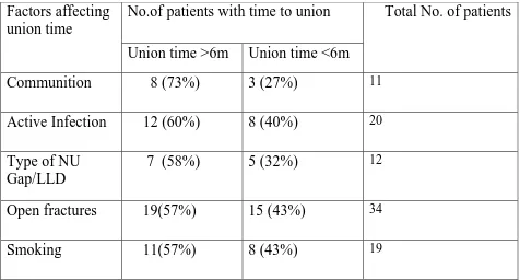
Out of the factors analyzed we observed communition, active infection, type of non union (with gap/limb length discrepancy), open
Table 1– Factors observed to affect the union time No.of patients with time to union Factors affecting
union time
Union time >6m Union time <6m
Total No. of patients
Communition 8 (73%) 3 (27%) 11 Active Infection 12 (60%) 8 (40%) 20 Type of NU
Gap/LLD
7 (58%) 5 (32%) 12
Open fractures 19(57%) 15 (43%) 34 Smoking 11(57%) 8 (43%) 19
The type of treatment – between unifocal and bifocal groups there was no difference in union time (50% in both the groups united within 6
months). On comparing patients with only transport versus gradual docking and lengthening, 5 out of 8 (63%) in the earlier group had union time more than 6 months where as 3 out of 8 (37%) in later group had union time more than 6 months. Early docking of fracture site was observed to shorten the union time.
2. COMPLICATIONS
9(23%) had true minor complication. All patients in minor complications had no dorsiflexion but a plantigrade foot.
There were five patients with major complications 1. Reinfection—one patient (Case no. 7)
2. Non union - two patients (Case no. 11 & 17) 3. Refracture – one patient (Case no. 33)
4. Joint stiffness ( equinus) and refracture - one patient (Case no 6) Apart from this, one patient had knee stiffness following transarticular external fixator application for soft tissue injury involving quadriceps mechanism. This patient continued to have knee flexion deformity of 15 degrees after Ilizarov fixator removal.
All the patients with minor complications were among the patients where lengthening or transport was done; this also caused distal joint stiffness.
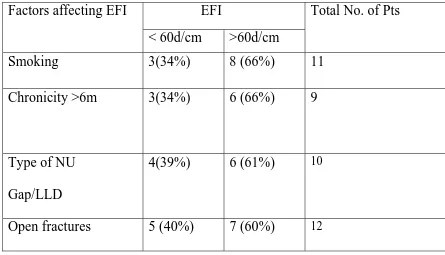
3. External fixation index- (EFI)
There were 16 patients who had distraction osteogenesis. They were grouped as –
The range was 33-100 days/cm (69days/cm or 2.3m/cm). 9 out of 16 (56%) had an EFI of more than 60d/cm. Of these 9, 8were from a far locality who did not come for regular follow up.
Table 2 -Factors affecting EFI
EFI Factors affecting EFI
< 60d/cm >60d/cm
Total No. of Pts
Smoking 3(34%) 8 (66%) 11
Chronicity >6m
3(34%) 6 (66%) 9
Type of NU Gap/LLD
4(39%) 6 (61%) 10
Open fractures 5 (40%) 7 (60%) 12
As smoking was a factor that influenced the outcome, the EFI was compared between smokers and non smokers. EFI in smokers was 2.5months/cm
where as in nonsmokers 1.75m/cm.
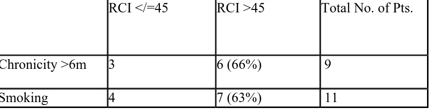
4 . Radiographic consolidation index-(RCI) The patients were grouped as-
1. RCI less than 45days/cm and
2. RCI more than 45 days/cm. There were 9 patients in the earlier group and 7 in the later group. The range was 20-70days/cm(mean 45d/cm or
1.5m/cm).Two patients had a hypotrophic regenerate .
[image:67.612.89.390.390.497.2]The factors observed to delay the RCI were -Non compliance of patient, violent corticotomy causing communition , chronicity and smoking. Effect of smoking on RCI is shown in table 3.
Table 3- Effect of smoking on RCI
Our study
RCI in smokers- 1.6 m/cm (48days/cm) RCI in nonsmokers- 0.9 m/cm (28days/cm)
Effect of chronicity and smoking on RCI is shown on table 4. Table 4 -Effect of chronicity and smoking on RCI
RCI </=45 RCI >45 Total No. of Pts.
Chronicity >6m 3 6 (66%) 9
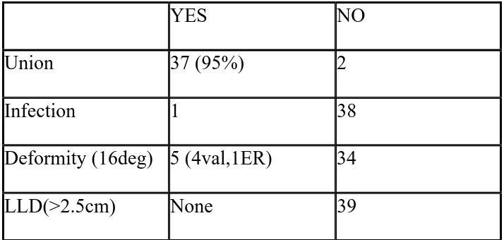
[image:67.612.92.525.593.703.2]5. Bone results-
24 patients (62%) had excellent, 12(30%) had good and 3(8%) had poor results. As ASAMI protocol does not consider bone union obtained after bone grafting as excellent, one patient who had excellent result was considered to have a good result.
Out of the 3 who had poor results two had nonunion and one had refracture. Union was achieved in 37 of 39 patients. One had persistent infection, 4 had valgus deformity (8-25deg), one had 10 deg external rotation deformity and none had LLD more than 2.5cm. However the LLD ranged from 0.5-2cm (mean 1cm) was observed in 17 patients.
[image:68.612.87.444.455.625.2]The bone results are shown in table 5. Table 5- The bone results
YES NO
Union 37 (95%) 2
Infection 1 38
Deformity (16deg) 5 (4val,1ER) 34
The factors observed to affect the deformity are - Communition (3 out of 4 patients with communition had deformity), inadequate stabilization during transport (2 out of 4 had deformity), improper ring application (3 out of 4 had deformity).
6. Functional results-
7 patients (18%) had excellent, 29 patients (76%) had good and 2 patients (6%) had fair results.
The problems observed after functional assessment were- Noteworthy limp- 3 patients
Joint stiffness- 1 had knee stiffness 9 had ankle stiffness 1 had equinus
Soft tissue dystrophy- 7 pts had edema, adherent scar & scars of multiple surgeries.
Pain- 2 had persistent pain. Inactivity- None.
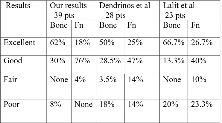
The comparison with other studies is shown in table 6.
Table 6- showing the comparision of the bone and functional results
Our results 39 pts
Dendrinos et al 28 pts
Lalit et al 23 pts Results
Bone Fn Bone Fn Bone Fn Excellent 62% 18% 50% 25% 66.7% 26.7%
Good 30% 76% 28.5% 47% 13.3% 40%
Fair None 4% 3.5% 14% None 10%
DISCUSSION
Infected nonunion is still an extremely difficult clinical problem despite major advances in the fixation technique, soft tissue management and antibiotic therapy. The infection of the fracture site not only prevents stable internal fixation but also delays fracture healing. The combination of mechanical instability and infection of a fractured bone provides an unfavorable condition for fracture healing. If the infection is not controlled, the fracture healing processes are eventually arrested.
Debridement is the most important step in the treatment of the infected elements. After debridement bony reconstruction can be done by different methods of bone grafting or by using the Ilizarov fixator.
This study describes the outcome of infected nonunions of the tibial shaft treated with an Ilizarov ring fixator. We have analyzed the outcome by studying the following factors.
1. Union time. 2. Complications.
3. External fixation index
Union time: Union time is not the part of ASAMI protocol. We have studied the time to union and also analyzed the factors responsible for prolonging the healing time. The union time ranged 3–17months. The patient with union time of 3months had active drainage for more than 3months before becoming quiescent. He had hypertrophic non union with loss of 3cm of bone. After adequate stabilization the nonunion healed after 3months. Lengthening was done by distraction through a proximal corticotomy.
Union time for most patients ranged between 5-9months. The result was comparable to other results .The mean union time for infected fractures of tibia nonunion treated by the Ilizarov fixator was nearly 6months in the study by G.K. Dendrinos et al 39 in 1995 and Marsh et al 51 in 1997.
The patient with the longest union time of 17months had an interesting finding to note. There was an avascular bone fragment that prevented fracture site from aproximating . Other factors were the unstable fracture pattern which needed readjustment of the frame to prevent translation.
application, there was 3cm gap between the fracture ends. Once the Ilizarov fixator was applied he required corticotomies twice because of premature consolidation of the corticotomy and patient’s non-compliance. In view of this the non union site was bonegrafted. After about 5months of bone-grafting the Ilizarov fixator was removed because of persistant pin site infection and patient’s non compliance, even though the radiological union seen was a very sparse attempt at union. At the time of follow up it was noted that the patient had a hypertroptic non-union a hypertrophied fibula. Although his radiological outcome was poor, his functional outcome was good.
Complications
The common complications were pin tract infection and transient edema which resolved by non-operative means. The pin track infections were treated by local injection with gentamycin and regular dressings. The major complications that affected the outcome were joint stiffness, refracture, reinfection and non-union.
The reasons for the true complications observed were – 1. Joint stiffness- Noncompliance of patients
Initial injury
Distraction osteogenesis 2. Reinfection- Poor soft tissue condition Persisting cavity
3. Refracture- Smoking, Co morbidity, Premature removal of ring, Ununited fibula fracture at the same level, Equinus deformity.
Premature removal of the fixator and with-holding bone-grafting for a fracture with gap in anterior cortex was the reason for another refracture (case no. 33). The fracture united after intramedullary nailing.
In all the patients the infection was controlled except for one (Case no.7) who had recurrent pus discharge because of the persisting cavity within the bone. All the patients who underwent distraction osteogenesis had distal joint stiffness because the muscles respond poorly to the distraction rate of 1mm/day and as the gastrosoleus forms the main bulk in the calf, it resulted in ankle stiffness. The stiffness was overcome to some extent by regular physiotherapy and at follow-up patients with no dorsiflexion but a plantigrade foot had difficulty in squatting but could manage by modification of posture. Another cause of ankle stiffness was foot drop that resulted from initial injury to tendons (No.49). After regular physiotherapy he was able to have plantigrade foot.
In the study by G.K Dendrinos et al 39 on 28 infected nonunions of tibia, three had nonunion, one had refracture, thirteen had joint stiffness and eleven had axial deviation as true major complications.
EFI
Observed reasons for the increased EFI were – 1. Non compliance of patient
2. Smoking 3. Chronicity 4. Type of NU 5. Type of fracture