DIRECT IMMUNOFLUORESCENCE STUDY OF
25 CASES OF SUBEPIDERMAL BLISTERS
Dissertation Submitted in
fulfillment of the university regulations for
MD DEGREE IN
DERMATOLOGY,VENEREOLOGY AND LEPROSY
(BRANCH XII A)
MADRAS MEDICAL COLLEGE
THE TAMILNADU Dr.M.G.R. MEDICAL UNIVERSITY
CHENNAI
CERTIFICATE
This is to certify that this dissertation entitled “DIRECT IMMUNOFLUORESCENCE STUDY OF 25 CASES OF SUBEPIDERMAL BLISTERS” is a bonafide work done by Dr.VIJAY
SINGHAL, Post Graduate in M.D. Dermatology, Venereology and Leprosy,
Madras Medical College, Chennai- 600 003, during the academic year 2006-2009. This work has not been formed previously the basis for the award of any degree.
Prof. Dr.B.PARVEEN, M.D., D.D.
Professor and Head Department of Dermatology & Leprosy Madras Medical College Chennai - 600 003
Prof. Dr.T.P. Kalaniti, M.D., Dean
DECLARATION
I, DR.VIJAY SINGHAL, solemnly declare that this dissertation titled
“DIRECT IMMUNOFLUORESCENCE STUDY OF 25 CASES OF
SUBEPIDERMAL BLISTERS” is a bonafide work done by me at Madras
Medical College during 2006-2008 under the guidance and supervision of
Prof. Dr. B. Parveen, M.D., D.D., Professor and Head, Department of
Dermatology, Madras Medical College, Chennai - 600 003.
This dissertation is submitted to The Tamilnadu Dr. M.G.R Medical University, towards partial fulfillment of requirement for the award of M.D. Degree in Dermatology, Venereology and Leprosy (Branch XII A).
Place: Chennai.
SPECIAL ACKNOWLEDGEMENT
My sincere thanks to
Prof. Dr.T.P. Kalaniti, M.D.
DEAN
Madras Medical College
ACKNOWLEDGEMENT
I am gratefully indebted to Prof.Dr.B.Parveen M.D., D.D., Professor
and Head, Department of Dermatology and Leprosy for her invaluable guidance, motivation and help throughout the study. I would like to express my sincere and heartful gratitude to Prof.Dr.N.Kumar, M.D., D.V.,DMRD.,
Additional Professor, Institute of Venereology.
I wish to thank Prof.Dr.V.S.Dorairaj, M.D., D.V., Former Director,
Institute of Venereology for his constant support and motivation.
I express my gratefulness to Prof.Dr.D.Prabhavathy,M.D.,D.D.,
Professor and Head of Department of Occupational Dermatology and Contact Dermatitis for her constant motivation and guidance. I thank
Prof,Dr.V.Somasundaram M.D.,D.D., Additional Professor, Department of
Occupational Dermatology and Contact Dermatitis for his benevolent help and support.
I am very grateful to Prof.Dr.S.Jayakumar M.D.,D.D., Additional
Professor, Department of Dermatology for his invaluable guidance and help. I sincerely thank Prof.Dr.C.Janaki, M.D.,D.D., Additional Professor,
Department of Dermatology (Mycology) for her priceless support.
I express my sincere gratitude to Prof.Dr.Jeyakumari Jeevan, M.D,
D.D., Professor of Leprosy and Dr.R.Arunadevi, M.D., D.D., Reader,
I am thankful to my guide Dr. Afthab Jameela Wahab, M.D., D.D.,
Assistant Professor, Department of Occupational Dermatology and Contact Dermatitis for her immense help and continued encouragement at every stage of this study.
I incline to thank Dr.V.Anandan, M.D (Derm); D.C.H; D.N.B., Dr.G.K.Tharini, M.D., Dr.N.Hema, M.D (D.V.L)., Dr.Samuel Jayavel
Daniel, M.D (D.V.L)., Dr.S.Anupama, D.D., Assistant Professors,
Department of Dermatology for their kind support and encouragement.
I thank Dr.Hameedulla, M.D., D.D., Dr.S.Kumaravel, M.D., D.D.,
and Dr.J.Manjula, M.D., D.N.B., Assistant Professors, Department of
Occupational Dermatology and Contact Dermatitis for their support and help.
My sincere thanks to Dr.V.Thirunavukkarasu, M.D., D.V., Dr.K.Venkateswaran, M.D., D.V., Dr.S.Thilagavathy, M.D., D.V.,
Dr.P.Mohan, M.D., D.V., Dr.S.Arunkumar, M.D., Dr.S.Kalaivani, M.D.,
D.V., Dr.S.Prabhakar, M.D (D.V.L)., Dr.V.N.S.Ahamed Sheriff, M.D
(D.V.L)., Assistant Professors, Institute of Venereology for their help and
suggestions.
I am also thankful to Dr.R.Priyavathani, M.D., for her continuing
guidance and support.
I am thankful to Prof.Dr.C.Balachandran, M.D., D.V.D., Professor
College, Manipal, for his willing help in the study of direct immunofluorescence.
I duly acknowledge the paramedical staff and my collegues for their help and favour.
Last but not the least I am profoundly grateful to all patients for their cooperation and participation in the study.
CONTENTS
Sl.No. Title Page No.
1. INTRODUCTION 1
2. REVIEW OF LITERATURE 3
3. AIM OF THE STUDY 33
4. MATERIALS AND METHODS 34
5. OBSERVATIONS AND RESULTS 37
6. DISCUSSION 46
7. CONCLUSION 54
8. BIBLIOGRAPHY 56
9. ANNEXURES
PROFORMA
ETHICAL COMMITTEE APPROVAL
INTRODUCTION
Several skin diseases are characterized by the presence of blisters.
In some, this may be the only morphologic clue to the diagnosis,
whereas in other disease, blisters may be one of the several
manifestations.
The immunobullous disorders are a group of autoimmune
diseases in which components of the epidermis and basement membrane
zone are the focus of attack, resulting in the formation of cutaneous and
mucosal blisters. The autoimmune bullous diseases result from an
immune response to molecular components of intercellular adhesion
molecules in desmosomes or the basement membrane zone (BMZ). The
diagnosis of autoimmune bullous diseases is based on the evaluation of
clinical findings, histopathology, direct immunofluorescence (DIF), and
indirect immunofluorescence (IIF). The autoimmune blistering diseases
may be subdivided into intraepidermal (pemphigus) and subepidermal
(pemphigoid) blistering disorders on the basis of the level at which
blistering occurs.
Prior to the early 1950s, most generalized bullous diseases were
considered to be variants of pemphigus, regardless of the level of skin
pemphigoid, the two most common autoimmune bullous disorders, were
first well separated by Lever in the early 1950s1
With the development and application of immunofluorescence
microscopy in the mid 1960s further distinctions were made not only
between pemphigus and pemphigoid but among other autoimmune
diseases also. Immunofluorescence testing is of utmost value in
confirming a diagnosis that is suspected by clinical or histologic
examination. This is especially true in subepidermal bullous diseases
REVIEW OF LITERATURE
A blister is a fluid filled cavity formed with in or beneath the
epidermis. The fluid consists of tissue fluid, plasma and variable amount
of inflammatory cells. Artificial distinction has been made into small
(vesicles, <0.5cm) and large blisters (bullae,>0.5cm). Blisters in the skin
can occur at any age and may be caused by common infections, rare
genetic skin diseases or auto immunobullous diseases.
Depending on the level of blister in the skin, immunobullous
diseases can be classified into intraepidermal or subepidermal.
Intraepidermal bullous disorders include:
Pemphigus vulgaris
Variant: pemphigus vegetans
Pemphigus foliaceous
Variant: pemphigus herpetiformis
Variant: pemphigus erythematosus
Intercellular IgA dermatosis
Subepidermal bullous disorders include:
Bullous pemphigoid
Variant: pemphigoid nodularis
Variant: pemphigoid vegetans
Variant: lichen planus pemphigoides
Mucous membrane pemphigoid
Pemphigoid gestationis
Linear IgA disease
Variant: chronic bullous disease of childhood
Variant: linear IgA disease of adults
Epidermolysis bullosa acquisita
Bullous systemic lupus erythematosus
Dermatitis herpetiformis
Subepidermal autoimmune blistering disorders arise due to antibodies
targeting various antigens located in the basement membrane zone
Structure Of Skin Basement Membrane Zone (BMZ) /
Dermoepidermal Junction (DEJ)
Skin basement membrane zone (BMZ) is an ultrastructurally
defined area situated between the epidermis and the dermis. Skin BMZ
is 0.5-1.0 mm-thick band-like structure that is positively stained by
periodic acid-Schiff (PAS) stain. The major function of skin BMZ is to
serve as an adherent connection between the epidermis and the dermis.
The skin BMZ can be divided into four ultrastructurally distinct areas:
1. Hemidesmosome
2. Lamina lucida
3. Lamina densa
4. Sub-lamina densa.
Hemidesmosome
It contains BP Ag1 and 2, α6β4 integrin and plectin. The BPAg1
(BP 230) is located intracellularly whereas the BPAg2 (BP180;
collagenXVII) is a transmembranous protein that contains an
intracellular domain, a transmembranous segment, and an extracellular
domain that projects into the lamina lucida. The extracellular domain of
BP180 protein contains a collagenous domain interrupted by 16 small
non-collagenous domains. The largest of these 16 non-collagenous
domains, NC16A, is located adjacent to the transmembranous segment.
In addition, a member of the integrin family, α6β4, and plectin (a
cytoskeleton-associated attachment protein), are also located in this
area. Anchoring filaments originate at hemidesmosomes and insert into
the lamina densa.
Lamina lucida
Situated between the hemidesmosome and the lamina densa, the
lamina lucida is electron-lucent under electron microscope.
Nevertheless, fine filamentous structures are observed in this area and
are termed anchoring filaments. Anchoring filaments extend from the
basal keratinocyte hemidesmosomes to the lamina densa, thus traversing
the lamina lucida. One of the anchoring filament components is a
BM600).Other lamina lucida located antigens include entactin/nidogen
and fibronectin. The laminin family consists of a group of heterotrimers
of various combinations of three chains,α, β and γ, and are synthesized
and secreted by keratinocytes. The lamina lucida appears to be the
weakest zone of the DEJ. It separates easily with heat and suction, with
treatment with salt solutions and proteolytic enzymes, and in diseases.
Lamina densa
Lamina densa, named according to its electron dense appearance
under electron microscope, is 35-45 nm-thick. The BMZ components
that are located in this area include: type IV collagen, perlecan (heparan
sulphate proteoglycan), and laminin-6.Type IV collagen is considered to
be the major component in this area.
Sub-lamina densa
Below the lamina densa, there are fibrillar structures that connect
the lamina densa onto the dermal plaque-like structures. These fibrillar
structures have been named anchoring fibrils. Type VII collagen is the
major component of the anchoring fibrils. Type VII collagen is a
290-kDa protein synthesized and secreted by both keratinocytes and
fibroblasts. Other minor fibers that connect to the area beneath lamina
Mucosal BMZ also contain identical components as skin BMZ.
Different antigens have been discovered in the four zones of the BMZ,
which are the targets for various diseases. Autoantibodies targeting skin
and/or mucosal BMZ components, like genetic mutations of BMZ
components, result in histopathologically defined subepidermal
blistering diseases. Regardless of which BMZ antigen is targeted by
autoantibodies, the histopathological findings of all autoimmune
subepidermal blistering diseases are similar. Although the inflammatory
cell infiltrate may provide clues in the diagnosis of certain diseases,
such as eosinophilic infiltrate in the diagnosis of BP, they are not always
reliable and cannot be used as definitive diagnostic criteria. Therefore,
more specific techniques are required for accurate diagnosis like direct
and indirect immunofluorescence.
IMMUNOFLUORESCENCE
Immunofluorescence is a laboratory technique for demonstrating
the presence of antibodies in tissues or body fluids. The use of
immunofluorescence in both diagnosis and research is well recognised
in dermatology, dating from the description of granular deposits of IgG
and C3 along the dermoepidermal junction in the lesions of lupus
technique to demonstrate what was then considered to be intercellular
substance of stratified squamous epithelium in the serum of patients of
pemphigus.3 Later, in 1967 Beutner and Jordon described the
immunological basis of pemphigoid.
Historically, Coons et al were the first group to work on the use of
chemically labeled antibodies as reagents for the detection of antigens in
mammalian tissue. The same authors in 1942 demonstrated the
pneumococcal antigen in murine tissue by using this technique.4They
improved the method using fluorescein as the reagent because of the
brilliance of its fluorescence and the wavelength of its emitted light
close to the maximum sensitivity of the human retina. In 1963 a report
of dermoepidermal fluorescent band in the lesion of lupus
erythematosus was published.2
A year later Beutner demonstrated an intercellular substance in
stratified squamous epithelium using IIF technique. Since then there
have been many refinements and improvements in the techniques.
The next decade saw the use of immunoelectron microscope
(IEM) in understanding vesicobullous lesions of the skin. Subsequently
during the 1980’s western blotting, immunoprecipitation and cell culture
are targeted by the autoimmune phenomenon in various bullous
disorders.
IMMUNOFLUORESCENCE TECHNIQUES
Specimen processing
DIF is performed on normal-appearing skin immediately adjacent
to a lesion (perilesional skin). If the patient does not have blisters, the
specimen may be obtained from skin adjacent to erythematous or
urticarial plaques because the immune deposits are partially or
completely degraded in inflamed or blistered skin, and DIF may be
falsely negative. For IIF, blood, blister fluid or urine may be used.
Skin specimen usually obtained by using a punch biopsy or
surgical biopsy from the perilesional skin. Then it is immediately snap
frozen for best preservation of the tissue which is done with liquid
nitrogen and maintained at -80°C. If delay is inevitable due to want of
transportation, the specimen is placed in a special transport medium like
Michel’s medium without loss of antigenic expression.5
Michel’s medium consists of liquid fixative in the form of
ammonium sulphate which prevents degradation of tissues with immune
reactants. Use of transport medium maintains a neutral pH; this is best
phosphate buffered saline (PBS) to remove blood and serum proteins.
The biopsy specimen stays in this medium for up to 8 weeks without the
loss of specific fluorescence.6
After the biopsy specimen is received at the laboratory liquid
fixative if any is washed off in neutral buffer. It is then snap frozen and
stored at-70°C to -20°C .The method of freezing varies from place to
place. (Table 1). This can be done using liquid nitrogen or slow freeze in
cryostat.
Sections are obtained of the unfixed frozen specimen by cutting in
a cryostat equipped with an anti role device to correct the tendency of
the specimen to curl. Four to six microns thick sections are cut and four
sections are placed on each slide by bringing it close to the knife surface
so that the sections thaw and become firmly bound to the glass. The
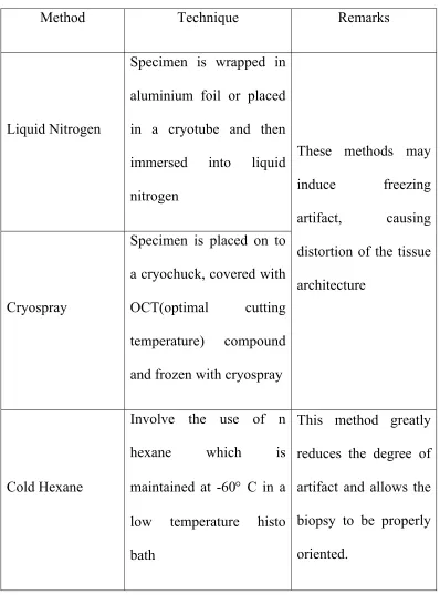
Table 1. Methods for freezing skin biopsy specimen
Method Technique Remarks
Liquid Nitrogen
Specimen is wrapped in
aluminium foil or placed
in a cryotube and then
immersed into liquid
nitrogen
Cryospray
Specimen is placed on to
a cryochuck, covered with
OCT(optimal cutting
temperature) compound
and frozen with cryospray
These methods may
induce freezing
artifact, causing
distortion of the tissue
architecture
Cold Hexane
Involve the use of n
hexane which is
maintained at -60° C in a
low temperature histo
bath
This method greatly
reduces the degree of
artifact and allows the
biopsy to be properly
Sections on each slide are incubated with FITC labeled antisera
against IgG, IgA, IgM, C3, and fibrinogen. Each reagent is usually
incubated for 30 minutes. The non specifically bound reactants are
removed by washing the slides in a bath of PBS (pH 7.4). Washing
should be carried out with all reagents for a minimum of thirty minutes.
Finally stained preparations are mounted in buffered glycerol and
examined with a fluorescent microscope.7Provided the sections are
stored in the dark at 4°C; the IF is retained for 48-72 hours.
Principles of Immunofluorescence 8, 9
Fluorescent techniques involve the emission of light of one
wavelength/colour from a substance being irradiated with the light of
different wavelength, the emitted light showing a low energy level. The
fluorochromes used routinely in dermatopathology are fluorescein
isothiocynate (FITC) and tetra methyl rhodamine isothiocynate
(TRITC). FITC has an emission wavelength of 525nm and a maximum
absorption wavelength of 495 nm. It emits apple-green fluorescence.
TRITC has an emission wavelength of 580nm and a maximum
Spectrofluorometric characteristics
Substrate Absorption maximum Emission maximum
FITC conjugate 495nm (blue) 525nm (apple-green)
TRITC conjugate 555nm (green) 580nm (orange-red)
To visualize the characteristic emission of the dye, different
excitation and barrier filters must be used. The excitation or primary
filter is placed between the light source and the tissue section to screen
out the wavelengths other than those near the excitation maximum of the
dye and the barrier filter removes all but the emitted wavelength.
Fluorochrome conjugated antibody preparations are commercially
available. These have to be stored in dark containers frozen at -200°C.
The classical fluorescent microscope has a mercury vapour lamp
that generates a light beam which is reflected by a concave mirror. This
is projected through collecting lenses to the excitation filter which emits
a fluorescent light beam. The beam then passes through a condenser on
to the specimen with the help of a reflecting mirror. This emitted light
passes through a barrier filter. The resultant pattern of fluorescence is
There are three basic types of IF used in dermatology:
Direct Immunofluorescence (DIF)
It is a one-step procedure used to detect and localize
immunoreactants deposited in vivo in the patient's skin or mucosa. The
immunoreactants include antibodies, complement components and
fibrinogen. The direct test for the tissue fixed antibody uses a frozen
section of patient’s skin on which is overlaid fluorescein or rhodamine
conjugated antibody to immunoglobulins and complements. If the tissue
contains an antibody e.g., IgG, then fluorescein labeled antihuman IgG
will bind to it when the frozen tissue section is overlaid with a solution
containing this reagent. Under fluorescent microscope the fluorescein
attached to antibody is activated and emits an apple green colour. The
degree of fluorescence (weak, moderate, strong) can only be
approximated by the microscopist. Antibody titre cannot be determined
by direct IF.
Indirect Immunofluorescence (IIF)
It is a two-step procedure used to identify circulating auto
antibodies to cutaneous or mucosal structures in a patient's serum. These
antibodies are most commonly of IgG or IgA classes. Indirect IF is a
used in the detection of circulating antibodies in bullous diseases
include human skin,10monkey esophagus,11 guinea pig lip or esophagus,
and salt-split human skin.12 The sensitivity and specificity of the
substrates may vary for the various bullous diseases.13 Frozen tissue
section or cell suspensions of this normal tissue are placed on
microscope slides. Sections are overlaid with appropriate dilutions of
serum or other body fluids so that any antibody against normal tissue
components will bind to them. The final steps of indirect IF are identical
to DIF. Fluorescein labeled antihuman immunoglobulin or complement
is overlaid on the tissue section. Sections are washed and examined
under fluorescent microscope. Titers of antibodies in fluids can be
determined by IIF. The titer is the last dilution of serum where antibody
can be detected.
SUBSTRATES USED IN IIF
Antibody type Substrates
Intercellular Normal skin
Monkey or guinea pig oesophagus Rat bladder
Antibody type Substrates
Anti-nuclear Human spleen Rat spleen
Rat liver Hep2 cells
Complement indirect IF:
It is a modification of indirect IF for demonstrating complement
fixing antibodies in fluids. It may be more sensitive than usual indirect
IF in certain instances. A single IgM antibody or two IgG antibodies
bound to antigen can generate many molecules of C3. Complement IIF
makes use of this amplification principle to increase the sensitivity of
the assay system. It attempts to demonstrate the antibody in serum or
other tissue fluids. Normal tissue substrate is overlaid with serum or
other tissue fluid that has been heated to 56°C for 30 min to destroy
complement fixing activity. Antibody against tissue binds during this
step. In the second step, the tissue sections are incubated with a source
of complement such as fresh human serum.
The complement fixing IgG or IgM antibody that attached to the
antigen in the first step can now activate complement in the second step
of antigen-antibody binding in the tissue. In the third step, sections are
incubated with fluorescein labeled antihuman C3 antibodies. They bind
to C3 molecules generated in the second step. Sections are washed and
examined under fluorescent microscope. In certain situations, few IgG
or IgM antibodies bind to tissue antigen that they cannot be detected by
classic indirect IF. However, since these molecules generate many C3
complement molecules, complement indirect IF may show fluorescence
at the site of antigen antibody binding.
Uses of Immunofluorescence in dermatology for the diagnosis of:
• Pemphigus vulgaris and its variants
• Bullous pemphigoid and its variants
• Pemphigoid (herpes) gestationis
• Epidermolysis bullosa acquisita
• Linear IgA bullous dermatosis (LAD ) & CBDC
• Dermatitis herpetiformis
• Discoid lupus erythematosus
• Systemic lupus erythematosus
• Lichen planus
• Porphyria
• Vasculitis
Ideal Site for Taking Skin Biopsy for IF in Various Skin Conditions
Condition Biopsy site(s)
Pemphigus (all forms) Perilesional
Uninvolved (buttock)
Pemphigoid (all forms) Perilesional
Uninvolved (buttock)
Pemphigoid (herpes) gestationis Perilesional
Epidermolysis bullosa acquisita Perilesional
Linear IgA bullous dermatosis (LAD )& CBDC
Perilesional
Uninvolved (buttock)
Dermatitis herpetiformis Uninvolved (buttock)
Discoid lupus erythematosus Lesional
Systemic lupus erythematosus Lesional, Sun exposed uninvolved, Non sun exposed uninvolved
Lichen planus Lesional
Porphyria Lesional
Vasculitis Lesional
Several authors have described the positive effect of saline in
increasing the sensitivity of IF analysis. Judd and Lever showed that
skin biopsies stored for 24 hours in 0.15 M phosphate buffered saline
prior to freezing gave a very high incidence of positive readings in
direct IF.14 It has been suggested that the increase of IF sensitivity by
saline incubation is due to improved exposure of epitopes and/or by a
decrease of background staining.15
Indirect Split Skin Immunofluorescence
This method relies on splitting normal skin through the lamina
lucida. Split normal skin is used as a substrate in an indirect IF method
to differentiate between antibodies that bind to antigens in the upper
lamina lucida or hemidesmosomes and to those that bind to antigens in
the lamina densa or sublamina densa
Procedures For Splitting The Skin
1.Incubation with cold 1M NaCl solution
2.Treatment with proteolytic enzymes
Direct Split Skin Immunofluorescence
Direct IF is performed on patient’s split skin to localise the site of
antibody deposition.
Immunofluorescence In Autoimmune Bullous Diseases
The autoimmune bullous diseases result from an immune
response against adhesion molecules of the epidermis and basement
membrane zone (BMZ).16The pemphigus group of diseases is associated
with antibodies to desmosomal proteins.17, 18 The antibodies in each type
of pemphigus are directed against a unique desmosomal protein or a
specific combination of desmosomal proteins .
Intraepidermal bullous disorders with their target antigen
Pemphigus type Target desmosomal protein
Pemphigus vulgaris Desmoglein 3 and desmoglein 1
Pemphigus foliaceous Desmoglein 1
Paraneoplastic pemphigus Desmoglein 3, desmoplakin 1, desmoplakin 2, BP 230, envoplakin, periplakin
There is strong direct experimental evidence that antibodies in
pemphigus vulgaris (PV) and pemphigus foliaceous (PF) cause
acantholysis and blister formation 17, 19, 20 by directly interfering with
desmosomal function.21
The subepidermal bullous diseases are associated with antibodies
against one or more components of the BMZ.22, 23
Subepidermal bullous disorder and their target antigen
Bullous disease Targeted molecule
BP BP 180, BP 230 (hemidesmosome and lamina lucida)
HG BP 180, BP 230 (hemidesmosome and lamina lucida)
CP BP 180, laminin V
(hemidesmosome and lamina lucida)
EBA Type VII collagen (anchoring fibrils)
Bullous SLE Type VII collagen (anchoring fibrils)
LAD (adults and children) LAD antigen (BP 180) (hemidesmosome and lamina lucida)
The differential diagnosis of a DIF test depends on 4 features:
1. The primary site of immune deposition
2. The class of immunoglobulin or other type of immune deposit
3. The number of immune deposits and, if multiple, the identity of
the most intense deposits
4. Deposition in other sites besides the main site.
With above parameters accurate diagnosis can be made in the
majority of cases24
A. “Intercellular space” deposition
The intercellular space (ICS) fluorescence pattern results from
binding of antibodies to desmosomal proteins around the keratinocyte
cell surface and is characteristic of the pemphigus group of disorders.
1. IgG deposition in the ICS only
This pattern is characteristic of all types of pemphigus except IgA
pemphigus. DIF is positive in 90% to 100% of patients
The pattern of fluorescence appears continuous around individual
higher magnification. The latter pattern reflects binding of antibodies to
desmosome associated proteins. Complement component C3 may be
seen in a pattern similar to that of IgG.24The frequency and, usually, the
intensity of C3 deposition are lower than those of IgG.25
2. IgG deposition in the ICS and BMZ
The combination of ICS and BMZ deposition may be seen in two
settings, PE26 and PNP.27
3. IgA deposition in the ICS
It is characteristic of IgA pemphigus, also known as
“subcorneal pustular dermatosis with intercellular IgA deposition”
and “intraepidermal neutrophilic dermatosis with intercellular IgA
deposition.” Because antibodies are directed against desmosomal
proteins, the term pemphigus is appropriate for the condition.
BMZ deposition
The detection of immune deposits at the BMZ by DIF is
characteristic of the subepidermal bullous diseases. There are several
parameters to evaluate for the accurate interpretation of BMZ
1. The type of immune deposit (including class of
immunoglobulin)
2. The number of immune deposits, namely, whether the
deposition is of one immunoreactant versus multiple
immunoreactants
3. The morphology of the fluorescence at the BMZ e.g.
continuous, discontinuous, linear, granular, and homogeneous28
4. Evaluation for fluorescence in any other site besides the BMZ,
such as dermal blood vessels.
Diseases with immune deposits at dermoepidermal junction29, 30, 31, 32
• Lupus erythematosus
• Dermatomyositis
• Leucocytoclastic vasculitis
• Bullous pemphigoid
• Herpes gestationis
• Epidermolysis bullosa acquisita
• Dermatitis herpetiformis
• Porphyria cutanea tarda
• Lichen planus
• Rosacea
There are clues that are helpful in the differential diagnosis.
Deposition of C3 with significantly higher intensity than IgG strongly
favors the pemphigoid group of diseases (BP, mucosal pemphigoid, and
HG). C3 may be the exclusive immunoreactant at the BMZ in patients
with HG and occasionally BP. The pattern of deposition in BP and HG
has been described as linear, wavy, tubular, and granular. The variation
in pattern may result from variations in the angle at which the
cryosections are made, the intensity of deposition, and the site of
biopsy.39 In specimens that contain adnexal structures, a similar
deposition may be seen along the BMZ of follicular and sweat gland
epithelium. Differentiation between BP and HG is not possible by
immunofluorescence or histopathology. There is ample evidence
confirming that HG is a variant of BP induced by pregnancy.35
If the intensity of IgG deposition at the BMZ is significantly
higher than that of C3, EBA and bullous SLE are more likely than
pemphigoid. The differential expression of intensity between IgG and
Deposition of IgG and C3 seen in EBA can be differentiated from
BP by salt split skin technique.41 In BP immunoreactants are deposited
in epidermal side alone or both epidermal and dermal side whereas in
EBA they are deposited in dermal side. IgG will localize to the roof of
the split in the majority of patients, to both roof and floor in 10% but
occasionally to the floor alone.42,43 C3 will always bind to both roof and
floor.44 Deposition in BP is within the lamina lucida. This site of
deposition corresponds to the location of the extracellular domain of
BP180 antigen that contains the dominant epitopes recognized by the
pathogenic BP antibodies.45 In contrast, deposition of antibodies in EBA
is in the sublamina densa area where the target antigen, type VII
collagen within the anchoring fibrils, is present.46 Exclusive deposition
on the dermal side may also be seen in antiepiligrin disease, also
referred to as antiepiligrin CP.47
Pang BK et al has shown that Floor-pattern salt-split skin cannot
distinguish bullous pemphigoid from epidermolysis bullosa
acquisita.48BP can present with a floor pattern fluorescence on salt-split
skin. But toad skin can be used to differentiate the two. Only BP will
give positive immunofluorescence with toad skin because it has BP and
2. Multiple deposits at the BMZ
This pattern of deposition strongly favors EBA and bullous SLE
over the pemphigoid group of diseases. In EBA, intense IgG deposition
is almost consistently present.37The intensity of C3 deposition is usually
less than that of IgG. Deposition of IgA is present in approximately two
thirds of cases and deposition of IgM in approximately one half of
cases.37The morphologic pattern of deposition is usually homogeneous,
thick, and broad.49The BMZ of adnexal epithelia reveals similar
deposition.
In bullous SLE, approximately 60% of cases reveal BMZ
deposition indistinguishable from that of EBA.50In the remaining cases,
the deposition is granular and mimics that seen in cases with nonbullous
SLE. Compared with nonbullous SLE, bullous SLE is more frequently
associated with deposition of IgA.50 In the absence of clinical history, it
is not possible to distinguish EBA, bullous SLE, and nonbullous SLE
with certainty. Since most patients with bullous SLE have detectable
antibodies to type VII collagen that is also the target of EBA
antibodies,51differentiation between bullous SLE and EBA is based on
Revised diagnostic criteria for bullous SLE52
1. A diagnosis of SLE based on ARA criteria.
2. Vesicles and bullae arising on, but not limited to, sun exposed
skin.
3. Histopathology compatible with DH.
4. Negative or Positive IIF for circulating BMZ antibodies, using
separated human skin as substrate.
5. DIF revealing IgG and/or IgM and often IgA at BMZ.
3. Deposition of IgA at BMZ
Linear deposition of IgA at the BMZ is characteristic of LAD53, 54
The so-called chronic bullous disease of childhood reveals identical
findings and represents the childhood form of LAD.55, 56 Deposition of
C3 is present less frequently and with lower intensity compared with
IgA.
C. Deposition at the BMZ and blood vessel walls
Homogeneous deposition of immunoreactants (usually multiple)
within superficial dermal blood vessel walls, in addition to BMZ
D. Papillary dermal deposition
Granular deposition of IgA and C3 in the papillary dermis and
along the BMZ is diagnostic of DH58
False-negative DIF in occurs in approximately 10% of
specimens59and may result from
1. Technical error (e.g., by using wrong or weak antisera)
2. The presence of clinical or subclinical inflammation and early
blister formation within the biopsy specimen (this is especially
true in cases with PNP)
3. The use of a limited panel of antisera that does not include IgA
Imunofluorescence Findings In Various Sub Epidermal Bullous
Disorders
Disease Findings
Bullous pemphigoid Linear BMZ IgG/C3 ~ 80%
IgA/IgM ~ 20%
Cicatricial pemphigoid Linear BMZ IgG/C3 ~ 80%
IgA~ 20%
Epidermolysis bullosa acquisita Linear BMZ IgG ~ 80%
IgA ~ 30%
Linear IgA bullous dermatosis (LAD & CBDC)
Linear BMZ IgA ~ 90%
IgG/C3 ~ 20%
Dermatitis herpetiformis Granular deposits in dermal papillae IgA ~ 100%
C3/fibrinogen ~ 40%
IgM ~ 10%
Bullous systemic lupus erythematosus
Linear or granular BMZ IgG/C3
IgM ~ 50% ; IgA 60%
Bullous lichen planus Colloid bodies IgM/IgA/C3
Colloid bodies can also be seen in bullous pemphigoid. These
homogeneous, fibrillar bodies are histologically, immunohistologically
and ultra structurally indistinguishable from the colloid bodies found in
the lesional skin of lichen planus, lupus erythematosus, dermatomyositis
and several other dermatoses.
Bullous Disorders with Negative/Nonspecific IF
Hailey-Hailey disease
Bullous impetigo
Grover’s disease
Acantholytic PR
Bullous insect bite
Bullous drug eruption
Drug induced lichenoid photodermatitis
AIM OF THE STUDY
1. To find out the incidence of various subepidermal bullous
disorders
2. To study the age wise distribution
3. To study the sex distribution
4. To study the disease association
5. To study the immunoreactant pattern of subepidermal blisters on
MATERIALS AND METHODS
In the present study, total number of bullous disorder patients
who attended the dermatology OPD, Government General Hospital,
Chennai from June 2006 to June 2008 were 107. Of these, 70 patients
had intraepidermal blistering disease and 37 patients had various
subepidermal bullous disorders. Out of these 37 patients, twenty five
cases of subepidermal autoimmune bullous disorders (based on clinical
findings) were included in this study.
In all the cases a detailed history was taken. Patients were asked
about the duration of the disease, the extent of skin involvement and the
presence of oral and other mucous membrane involvement. History of
urticarial weals and generalized pruritus was noted. History of drug
intake prior to the onset of lesions was taken. Precipitating or
aggravating factors were noted. History suggestive of gluten sensitive
enteropathy like abdominal pain, diarrhoea and constipation was asked.
History of fever, joint pain and photosensitivity was noted. History of
blistering on trivial trauma was taken. History of lesions suggestive of
lichen planus was also taken. They were enquired about loss of weight,
loss of appetite, malena and hemetemesis to rule out internal
malignancy. Treatment history, personal history and family history of
Each patient was carefully examined .The various clinical
parameters studied include:
1. Distribution of vesicles/bullae, number, size, shape, content of
bulla: hemorrhagic or clear fluid
2. Presence of bulla on erythematous or non erythematous skin
3. Oral or other mucous membrane involvement
4. Erosions and crusting
5. Hair and nail involvement
6. Palms and soles involvement
7. Nikolsky and Asboe Hansen sign
All the clinical findings were recorded in a standard study
proforma. All the patients enrolled in the study were investigated as
outlined in the proforma. This study was approved by the Ethical
Committee.
Perilesional skin biopsy was performed under strict aseptic
precautions, under local anaesthesia using 0.5 ml of 2% lignocaine. A 5
mm disposable punch (Fig.1) was inserted gently into the skin up to the
gently squeezed out and detached from its socket by sharp scissors. The
biopsy specimen was washed in normal saline before putting into the
Michel’s transport medium (Fig.1). It was labeled and sent to the
Department of Dermatology, Kasturba Medical College, Manipal for
direct immunofluorescence.
In the laboratory, biopsy specimens were washed in buffer for 30
minutes and then snap frozen. Four micron thick sections were cut in a
cryostat (Fig.2), set at -18°C. Sections were washed in PBS (phosphate
buffer saline) for 10 minutes and fan dried for another 10 minutes.
Sections were then covered with diluted FITC labeled antisera (Fig.3)
and then incubated in a moist chamber at 37°C for 30 min. Subsequently
specimens were washed with three changes of PBS over a period of 30
minutes. Sections were dried, mounted in buffered glycerol and
examined with fluorescent microscope (Fig.4).
All the patients were given appropriate treatment including drugs
such as systemic steroids, dapsone, doxycycline and nicotinic acid.
Healed lesions were noted for post inflammatory hyper or
OBSERVATIONS AND RESULTS
Age & Sex Incidence
Of the 25 cases, 13 were males and 12 females. The age of the
patient varied from 16 years to 80 years. 18 patients had bullous
pemphigoid. Of these 18 BP patients 12 were males and 6 females.
Males’ age ranged from 16 to 80 years with average age of 47 years.
Females’ age range varied from 40 to 72 years with average age of 61
years. One patient had linear IgA disease, one patient had Chronic
bullous dermatosis of childhood (CBDC), two patients had bullous SLE,
one patient had pemphigoid vegetans, one patient had bullous lichen
planus and one patient had lichen planus pemphigoides.
Disease wise distribution of cases
Diagnosis Total no of
cases
Bullous pemphigoid 18
Bullous SLE 2
Linear IgA disease 1
CBDC 1
Bullous LP 1
Age wise distribution of bullous pemphigoid patients
Age of
patient(years)
Bullous pemphigoid
Percentage
(%)
0-9 0 0
10-19 2 11.1
20-29 2 11.1
30-39 0 0
40-49 3 16.6
50-59 1 5.5
60-69 6 33.3
70-79 3 16.6
80-89 1 5.5
Sex Wise Distribution of bullous pemphigoid patients
Age of
patient(years)
Bullous pemphigoid
Male female
0-9 0 0
10-19 2 0
20-29 2 0
30-39 0 0
40-49 2 1
50-59 1 0
60-69 3 3
70-79 1 2
80-89 1 0
0
10
20
30
40
50
60
70
B. SL E ( M ) B. SL E (F) L INE A R I g A( F ) C BDC (F ) LP P ( F ) BUL L O US L P PEM PH IG O ID VEG E T A N SAge wise distribution
Family History
None of the 25 cases gave family history of similar illness in the
family.
Clinical Manifestations
The main presenting complaints were blisters and erosions over
the skin. Pruritus was the commonest symptom in all the cases. Out of
weals prior to the onset of blisters. Duration between the prodromal
symptoms and appearance of blisters varied from 1 month to two years.
Mucous Membrane Involvement
Six cases of bullous pemphigoid had oral mucosal erosion apart
from skin lesions. Both the bullous SLE patients had oral erosions.
Cutaneous Manifestations
All cases presented with tense bullae and vesicles, except one
patient of pemphigoid vegetans who presented with vegetative plaques.
Hemorrhagic bullae were seen in 2 BP patients. Lesions occurred over
both normal and erythematous skin in 8 cases and over normal skin
alone in rest of the 17 cases. The lesions were distributed over chest,
abdomen, back, upper limb, thighs and legs. One case presented with
only few lesions localised to hands and abdomen. Typical ‘string of
pearls’ appearance was seen in linear IgA disease and CBDC. In 18
patients lesions resolved with post inflammatory hypopigmentation
where as in others it resolved by post inflammatory hyperpigmentation.
Nikolsky sign was negative in all the cases. In 7 patients, bulla
Disease Association
Out of 18 bullous pemphigoid patients 10 patients had various
associated diseases. 4 patients had diabetes, 5 patients had hypertension,
1 patient had carcinoma prostate, 4 patients had cystic lesions of various
organs (kidney, uterus, lung, and liver), 1 patient had renal calculus, and
1 patient had interstitial lung disease.
Investigations
Tzanck smear was negative in all the cases. Five patients had
elevated blood sugar. Antinuclear antibody test was positive for SLE
patients. Ultrasonography abdomen showed cystic lesions of various
organs in four bullous pemphigoid patients. Cysts were seen in lung,
liver, uterus and kidney.
Histopathology
Skin biopsy was done in all the cases from an early blister. All
cases showed subepidermal bulla. Bulla cavity contained eosinophils
and fibrin. In the dermis, inflammatory infiltrate consisted of
eosinophils, lymphocytes and few neutrophils. Skin biopsy was also
done for other subepidermal bullous disorders and histopathology was
Immunofluorescence
Bullous Pemphigoid
All the 18 patients of bullous pemphigoid patients showed C3
deposition at BMZ. C3 was deposited linearly in 17 patients (Fig.5); 4
patients showed strong, 11 patients showed moderately strong and 2
patients showed weak C3 deposition, whereas in one case it was
deposited in a weak granular pattern. IgG was present in 11 out of 18
cases i.e. in 61% of cases along with C3 deposition (Fig.6). It was
present in a linear fashion in all the cases. Two cases showed IgM
deposition (one linear and the other granular pattern) along with IgG and
C3. IgA deposition was present in one case of bullous pemphigoid
(5.5% of cases) in moderately strong discontinuous pattern. Fibrinogen
deposit was present in 3 out of 18 BP cases (16.6% of cases). One case
showed colloid bodies with IgM deposition.
LINEAR IgA DISEASE AND CBDC
Linear IgA disease patient showed deposition of IgA at BMZ in
linear pattern (Fig.7). In CBDC patient, all the five immunoreactants
were present at BMZ i.e. IgA, IgG, IgM, C3 and fibrinogen, but
deposition of IgA was stronger as compared to IgG (Fig.8). C3 was
BULLOUS SLE
One Bullous SLE patient showed moderately strong deposition of
IgG in linear pattern, moderately strong deposition of C3 in granular
pattern which was present at the floor in the split skin specimen (Fig.9),
weak linear deposition of IgM, moderately strong linear deposition of
IgA (Fig.10) and fibrinogen at BMZ. Second patient of Bullous SLE
showed deposition of IgG and IgM in moderately strong granular
pattern (Fig.11), linear weak deposition of C3 and moderately strong
deposition of IgA at BMZ. In this patient blood vessel wall deposits
composed of fibrinogen were present.
Bullous lichen planus
Bullous lichen planus patient showed deposition of strong cluster
of colloid bodies stained with IgM, also few colloid bodies were present
which were stained with IgG, C3 and IgA. There was strong ragged
fibrinogen band present at BMZ (Fig.12).
Lichen planus pemphigoides
Lichen planus pemphigoides patient showed moderately strong
colloid bodies stained with IgM and IgA were also present. Also there
was weak deposition of fibrinogen at BMZ.
Pemphigoid vegetans
Pemphigoid vegetans patient showed weak deposition of C3
(Fig.14) and IgG along with focal deposition of IgA and fibrinogen at
BMZ.
Treatment
Patients were given appropriate treatment to control the disease.
Course and prognosis
All the cases responded very well to the treatment and showed excellent
remission within a month. No mortality was recorded. The disease free
DISCUSSION
Pemphigus vulgaris has been reported to be the most common
autoimmune blistering disease in eastern countries, such as Malaysia,
China and India. Whereas in Western Europe, bullous pemphigoid is the
most common immunobullous disorder. During the study period bullous
pemphigoid was the commonest subepidermal autoimmune blistering
disorder .It constituted 30 out of 107 cases (28%). But overall
pemphigus vulgaris was the commonest blistering disorder (70 out of
107 cases).
The mean age of onset of bullous pemphigoid was 56 years in
males. The age range varied from 16 years to 80 years. Whereas in
females age range varied from 40 years to 70 years with a mean age of
61 years. Although the age range for female patients was narrow, they
had higher mean average age of presentation of disease. This is
consistent with other studies by lever W F60 and Amos B et al.61
The youngest patient was 16 year male in our study. Although
bullous pemphigoid is a disease of the elderly, it can occur in
individuals less than 40 years. Several cases have been described in
children. Few cases has also been reported in infants by Oranje AP and
Out of 30 bullous pemphigoid patients 20 were males and 10
were females, constituting male to female ratio of 2:1. Incidence of
bullous pemphigoid is more in males as compared to females and
similar studies were reported by Jung M et al.62
Out of 18 BP patients 6 had oral lesions (33%).According to the
literature oral lesion can occur in 10-30 % of patients. Similar studies
has been reported by Person JR et al.63
Bullous pemphigoid can be associated with malignancy. Although
in many studies there is no increase in incidence of malignancy in
bullous pemphigoid patients, gastric carcinoma is the most common
malignancy reported by Ogawa H et al 64 in their series of more than
1000 BP patients. In our study one patient had prostate carcinoma. This
rare association of prostate adenocarcinoma with bullous pemphigoid
has been reported by Oztürkcan S et al.65
Four out of 18 patients (22%) of bullous pemphigoid showed
cystic lesion of various organs including liver, kidney, lung and uterus.
DIF
C3 deposition was seen in 100% of BP cases, linear deposition in
17 out of 18 patients and weak granular fashion in 1 patient. IgG
deposition was seen in 66% of patients.ie 12 out of 18 patients, linear in
4 cases and discontinuous in 8 patients. Chan YC et al have shown in
their study linear deposits of IgG and C3 along the dermoepidermal
junction in twenty-one of 23 (91%) patients.66 Similar results have been
reported by Chang YT et al in their study of 88 patients.67 Harrist TJ and
Mihm MC have shown that C3 is present in 100% and IgG in 65% to
95% of patients.
Positive deposition of C3 but negative staining for IgG on direct
immunofluorescence (DIF) studies has been noted in some patients.68
Following factors contribute to false-negative staining for IgG on DIF in
some BP patients:
1. Sub threshold IgG in skin specimens
2. Limited reactivity of commercial antihuman IgG conjugates to the
IgG4 subclass
3. Decreased sensitivity of DIF compared with double antibody
The use of sandwich double antibody immunofluorescence
methods to test for IgG and/or IgG subclasses may be helpful in
definitively diagnosing BP in patients with negative IgG and positive C3
staining on DIF68
One patient of bullous pemphigoid showed colloid bodies stained
by IgM. Similar colloid bodies has been described by Y. Horiguchi et al
in 8 out of 18 patients in their studies.69 These homogeneous, fibrillar
bodies were histologically, immunohistologically and ultrastructurally
indistinguishable from the colloid bodies found in lesional skins of
lichen planus, lupus erythematosus, dermatomyositis and several other
dermatoses.69 In BP, degenerated keratinocytes adjacent to the blister
roof, may, after undergoing a filamentous change, drop off into the
dermis and subsequently form homogeneous, fibrillar bodies in the
uppermost dermis when re epithelization is completed. Some findings
that may help favor lichen planus include the tendency for cytoid bodies
in LP to cluster in groups, to be present in high number, to be larger, to
have higher fluorescent intensity, and to contain multiple immune
deposits.70
Two cases of BP also showed weak deposition of IgM along with
from other immunoglobulin and C3. Similar results have been shown by
Ahmed AR et al, 71 and Cormane RH.72 They have shown the deposition
of IgM, IgA, IgD and IgE at the dermoepidermal junction. This is also
consistent with the findings of Provost TT and Tomasi TB who have
shown that IgE, alternate pathway components and fibrinogen can be
deposited along DEJ in bullous pemphigoid patients.73
Linear IgA disease patient showed deposition of only IgA at BMZ
in linear pattern. This is consistent with the diagnosis. Studies by
Wojnarowska F, Bhogal B and Black MM have shown deposition of
other immunoreactants also (IgM, IgG and C3) in 20-30% of
patients.74,75
In CBDC patient all the five components were present i.e. IgA,
IgG, IgM, C3 and fibrinogen, but deposition of IgA was stronger as
compared to IgG. C3 was present in moderately strong linear fashion
which is consistent with the diagnosis of CBDC. According to Petersen
MJ et al that if both IgA and IgG are present and deposition of IgA is
more intense than IgG, and C3 deposition is strong, than it should be
considered as a case of linear IgA dermatosis.76 There have been many
case reports in which there is deposition of only IgG and IgA. These
al77 has described a patient of Linear IgA dermatosis with IgA and IgG
deposition only at BMZ. Similar deposition has been described by
Watanabe M et al78 and Kersting E et al.79
Bullous SLE patient showed moderately strong deposition of IgG
in linear pattern, moderately strong deposition of C3 in granular pattern
which was present at the floor in the split skin specimen, deposition of
IgM in weak linear, and IgA and fibrinogen were present in moderately
strong linear pattern. Another patient of Bullous SLE showed deposition
of IgG and IgM in moderately strong granular pattern, weak deposition
of C3 and moderately strong deposition of IgA.
Similar results have been reported by Gammon WR et al in their
studies.80They have shown that IgG is always present and IgA and IgM
are also frequently deposited at the BMZ. The pattern of deposition may
be granular (60%), linear (40%) or rarely fibrillar.80 A linear rather than
granular pattern along the BMZ is associated with the presence of higher
titer of circulating autoantibodies.80
Both of the Bullous SLE patients showed deposition of IgA.
Gammon WR et al have shown that Bullous SLE is associated with a
higher incidence of IgA deposition (76%) than other forms of SLE
Lichen planus pemphigoides patient showed moderately strong
deposition of IgG and strong deposition of C3 at BMZ. Few colloid
bodies stained with IgM and IgA were also present in the superficial
dermis. There was also weak deposition of fibrinogen at BMZ.
Lichen planus pemphigoides is a rare clinical variant of bullous
pemphigoid and DIF findings are similar to BP. Okochi H et al have
drawn conclusion from their study that the presence of C3 alone or with
IgG along the dermoepidermal junction is confirmatory on DIF82 Vibhu
Mendiratta et al have shown deposition of IgG and C3 at BMZ in lichen
planus pemphigoides patient.83
Bullous LP patients showed strong cluster of IgM colloid bodies
in superficial dermis. There were also colloid bodies with weak
deposition of IgG, C3 and IgA. Also there was strong deposition of
ragged fibrinogen band along DEJ. A Sandra et al have shown in their
study of 18 bullous lichen planus patients similar findings.84 In their
study ragged fibrinogen band was present in all the patients whereas
colloid bodies demonstrating IgG, C3, IgM and IgA were seen in 88%
of patients. In bullous lichen planus patients immune deposits are
present within cytoid bodies in the superficial dermis, as well as along
fibrinogen.85, 86 Deposition of IgG, IgA, and C3 is less frequently
present.87, 88
Gawkrodger DJ et al compared two patients with lichen planus
pemphigoides and two with bullous lichen planus and showed direct
immunofluorescence was positive in lichen planus pemphigoides and
negative in bullous lichen planus.89
Pemphigoid vegetans patient showed C3 and IgG deposition with
weak deposition of IgA and fibrinogen. Chan LS et al described a
patient of pemphigoid vegetans with deposition IgG at BMZ.90 They
considered it to be a variant of bullous pemphigoid. Marie Ogasawara et
al described a patient of pemphigoid vegetans with IgA, IgG and weak
C3 BMZ deposition.91 Lawrence S Chan et al reported a patient of
pemphigoid vegetans with linear IgG deposition at BMZ.92 IgG, IgM,
C3 and fibrin deposition has also been described by Winkelmann RK
and Su WPD who first described this clinical entity.93 Y. Ueda et al
CONCLUSION
1. Bullous pemphigoid was the most common subepidermal blistering
disease.
2. Bullous pemphigoid was twice as common in males as in females.
3. Rare association of cystic disease of various organs was noted with
bullous pemphigoid group of patients.
4. Linear pattern deposition of C3 at BMZ was seen in 17 patients and
granular pattern in one patient.
5. Colloid bodies were also seen in one BP patient, they are not specific
for any disease.
6. Deposition of IgM and fibrinogen, apart from other immunoreactants
were seen in bullous pemphigoid patient.
7. Linear IgA patient showed linear IgA deposition at BMZ.
8. In CBDC patient IgA, IgG, IgM, C3 and fibrinogen deposition was
seen.
9. Multiple immunoreactant deposition was seen in bullous SLE
10.In bullous SLE, immunoreactant deposition was seen in both
granular and linear patterns.
11.Lichen planus pemphigoides patient showed IgG and C3 deposition
at BMZ and few colloid bodies stained with IgM and IgA.
12.Bullous lichen planus patient showed colloid bodies stained by IgM,
IgG, IgA and C3. Strong ragged fibrinogen band was seen at BMZ.
13.Pemphigoid vegetans patient showed C3, IgG, IgA and fibrinogen
BIBLIOGRAPHY
1. Jo – David fine : bullous disease, In : moschella and hurley
dermatology, 3rd ed, Philadelphia, W.B. saunders company, 1992,
pp 655-697
2. Burnham TK, Neblett TR, Fine G. The application of the
fluorescent antibody technique to the investigation of lupus
erythematosus and various dermatoses. J Invest Dermatol. 1963
Dec;41:451-6.
3. Beutner, EH; Jordon, RE. Demonstration of skin antibodies in
sera of pemphigus vulgaris patients by indirect
immunofluorescent staining. Proc Soc Exp Biol Med. 1964
Nov;117:505–510
4. Coons, A.H., Creech, H.J., & Jones, R.N.The demonstration of
pneumococcal antigen in tissues by the use of fluorescent
antibody. 1942. J. Immunol. 45:159-179.
5. Michel B, Milner Y, David K. Preservation of tissue-fixed
immunoglobulins in skin biopsies of patients with lupus
erythematosus and bullous diseases-preliminary report. J Invest
Dermatol 1973;59:449-52.
6. Vaughan Jones SA, Salas J, McGrath JA, Palmer I, Bhogal BS,
immunoreactants from skin biopsies maintained in Michel's
medium. Dermatol 1994;189 (suppl 1):131-2.
7. Nousari HC, anahalt GJ. skin diseases. In : rose NR, Hamilton
RG, detrick B, eds. Manual of clinical laboratory immunology.
Washington, DC: ASM press, 2002: 1032
8. Huligol SC, Bhogal BS, Black MM. Immunofluorescence of the
immunobullous disorders: Part one: Methodology. Indian J
Dermatol Venereol Leprol 1995;61:187-95.
9. Vassileva S. Immunofluorescence in Dermatology. Int J Dermatol
1993;32:153-61.
10. Katz SI, Halprin KM, Inderbitzin TM. The use of human skin for
the detection of anti-epithelial autoantibodies: a diagnostic and
prognostic test. J Invest Dermatol 1969;53:390-9.
11. . Kumar V, Beutner EH. Monkey esophagus: a unique antigenic
substrate in immunodermatology. In: Beutner EH, Chorzelski TP,
Kumar V, editors. Immunopathology of the skin. 3rd ed. New
York: John Wiley & Sons; 1987. p. 65-90.
12. Woodley DT, Sauder D, Talley MJ, Silver M, Grotendorst G,
Quarnstrom E. Localization of basement membrane components
after dermal-epidermal junction separation. J Invest Dermatol
13. Acosta E, Ivanyi L. Comparison of the reactivity of various
epithelial substrates for the titration of pemphigus antibodies by
indirect immunofluorescence. Br J Dermatol 1982;107:537-41.
14. Judd KP, Lever WF: Correlation of antibodies in skin and serum
with disease severity in pemphigus. Arch Dermatol 1979,
115:428-432.
15. Gammon WR, Kowalewski C, Chorzelski TP, Kumar V,
Briggaman RA, Beutner EH: Direct immunofluorescence studies
of sodium chloride-seprated skin in the differential diagnosis of
bullous pemphigoid and epidermolysis bullosa acquisita. J Am
Acad Dermatol 1990, 22:664-670.
16. Burgeson RE, Christiano AM. The dermal-epidermal junction.
Curr Opin Cell Biol 1998;9:651-8
17. Roscoe JT, Diaz L, Sampaio SA, Castro RM, Labib RS,
Takahashi Y, et al. Brazilian pemphigus foliaceus autoantibodies
are pathogenic to BALB/c mice by passive transfer. J Invest
Dermatol 1985;85:538-41
18. Eyre RW, Stanley JR. Identification of pemphigus vulgaris
antigen extracted from normal human epidermis and comparison
19. Amagai M, Klaus-Kovtun V, Stanley JR. Autoantibodies against
a novel epithelial cadherin in pemphigus vulgaris, a disease of
cell adhesion. Cell 1991;67:869-77.
20. Hu CH, Michel B, Schlitz JR. Epidermal acantholysis induced in
vitro by pemphigus autoantibody. Am J Pathol 1978;90:345-51.
21. Mahoney MG, Wang Z, Rothenberger K, Koch PJ, Amagai M,
Stanley JR. Explanations for the clinical and microscopic
localization of lesions in pemphigus foliaceus and vulgaris. J Clin
Invest 1999;103:461-8
22. Diaz LA, Giudice GJ. End of the century overview of skin
blisters. Arch Dermatol 2000;136:106-12.
23. Stanley JR. Pemphigus and pemphigoid as paradigms of
organ-specific, autoantibody-mediated diseases. J Clin Invest
1989;83:1443-8.
24. Jordon RE, Triftshauser CT, Schroeter AL. Direct
immunofluorescent studies of pemphigus and bullous
pemphigoid. Arch Dermatol 1971;103:486-91.
25. Jordon RE, Schroeter AL, Rogers RS III, Perry HO. Classical and
alternate pathway activation of complement in pemphigus
26. Chorzelski TP, Jablonska S, Blaszczyk M. Immunopathological
investigations in the Senear-Usher syndrome (coexistence of
pemphigus and lupus erythematosus). Br J Dermatol
1968;80:211-7.
27. Anhalt GJ, Kim S, Stanley JR, Korman NJ, Jabs DA, Kory M, et
al. Paraneoplastic pemphigus: an autoimmune mucocutaneous
disease associated with neoplasia. N Engl J Med
1990;323:1729-35.
28. Mutasim DF, Anhalt GJ, Diaz LA, Patel HP. Linear
immunofluorescence staining of the cutaneous basement
membrane zone produced by pemphigoid antibodies: the result of
hemidesmosome staining. J Am Acad Dermatol 1987;16:75-82.
29. Dahl MV, Gilliam JN. Direct immunofluorescence in lupus
erythematosus. In: Beutner EH, Chorzelski TP, Kumar V, editors.
Immunopathology of the skin. 3rd ed. New York: John Wiley &
Sons; 1987. p. 499-518.
30. Dahl MV. Usefulness of direct immunofluorescence in patients
with lupus erythematosus. Arch Dermatol 1983;119:1010-7.
31. Gilliam JN. The significance of cutaneous immunoglobulin
deposits in lupus erythematosus and NZB/NZW F, hybrid mice. J
32. Monroe EW. Lupus band test. Arch Dermatol 1977;113:830-4.
33. Gammon WR. The immunopathology of bullous pemphigoid
antibodies. In: Beutner EH, Chorzelski TP, Kumar V, editors.
Immunopathology of the skin. 3rd ed. New York: John Wiley &
Sons; 1987. p. 322-36.
34. Bean SF. Cicatricial pemphigoid: immunofluorescent studies.
Arch Dermatol 1974;110:552-5.
35. Morrison LH, Anhalt GJ. Herpes gestationis. Autoimmunity
1991;4:37-45.
36. Schornick JK, Bangert JL, Freeman RG, Gilliam JN. Herpes
gestationis: clinical and histologic features of twenty-eight cases.
J Am Acad Dermatol 1983;8:214-24.
37. Woodley DT, Gammon WR, Briggaman RA. Epidermolysis
bullosa acquisita. In: Jordon RE, editor. Immunologic diseases of
the skin. Norwalk (CT): Appleton & Lange; 1991. p. 321-33.
38. Barton DD, Fine JD, Gammon WR, Sams WM Jr. Bullous
systemic lupus erythematosus: an unusual clinical course and
detectable circulating autoantibodies to the epidermolysis bullosa
acquisita antigen. J Am Acad Dermatol 1986;15:369-73.
39. Mutasim DF, Anhalt GJ, Diaz LA, Patel HP. Linear
membrane zone produced by pemphigoid antibodies: the result of
hemidesmosome staining. J Am Acad Dermatol 1987;16:75-82.
40. Gammon WR, Inman AO III, Wheeler CE Jr. Differences in
complement-dependent chemotactic activity generated by bullous
pemphigoid and epidermolysis bullosa acquisita immune
complexes: demonstration by leukocytic attachment and organ
culture methods. J Invest Dermatol 1984;83:57-61.
41. Gammon WR, Kowalewski C, Chorzelski TP, Kumar V,
Briggaman RA, Beutner EH. Direct immunofluorescence studies
of sodium chloride-separated skin in the differential diagnosis of
bullous pemphigoid and epidermolysis bullosa acquisita. J Am
Acad Dermatol 1990;22:664-70.
42. Provost TT, Tomasi TB. Immunopathology of bullous
pemphigoid: basement membrane deposition of IgE, alternate
pathway components and fibrin. Clin Exp Immunol
1974;18:193-200.
43. Gammon WR, Fine J-D, briggaman RA. Immunofluorescence on
split skin for the detection and differentiation of basement
membrance antibodies. J Am Acad Dermatol 1992;27:79-87.
44. Smoller BR, Woodley DT. Differences in direct