STUDY OF LIVER FUNCTION ABNORMALITIES IN THE
TUBERCULOSIS PATIENTS UNDERGOING RNTCP-DOTS
IN TB CLINIC MADURAI
Dissertation Submitted for
MD Degree (Branch I) General Medicine
March- 2009
The Tamilnadu Dr.M.G.R.Medical University
Chennai – 600 032.
CERTIFICATE
This is to certify that this dissertation titled “STUDY OF LIVER FUNCTION
ABNORMALITIES IN THE TUBERCULOSIS PATIENTS UNDERGOING
RNTCP-DOTS IN TB CLINIC MADURAI submitted by DR. C. THOMAS
KINGSLEY to the faculty of General Medicine, The Tamil Nadu Dr. M.G.R. Medical
University, Chennai in partial fulfillment of the requirement for the award of MD
degree branch I General Medicine, is a bonafide research work carried out by him under
our direct supervision and guidance.
DR. M. MUTHIAH, M.D. DR.A.AYYAPPAN, M.D.
Professor of Medicine, Professor and Head, Chief, VI Medical Unit, Department of Medicine, Department of Medicine, Madurai Medical College, Madurai Medical College, Madurai.
DECLARATION
I, Dr.C.Thomas Kingsley, solemnly declare that the dissertation titled “Study of
Liver Function Abnormalities in the tuberculosis Patients Undergoing
RNTCP-DOTS in TB Clinic Madurai” has been prepared by me.
This is submitted to The Tamil Nadu Dr. M.G.R. Medical University, Chennai,
in partial fulfillment of the rules and regulations for the award of MD degree (branch I)
General Medicine.
Place: Madurai
ACKNOWLEDGEMENT
At the outset, I wish to thank our Dean Dr. S.M.SIVAKUMAR, M.S., for
permitting me to use the facilities of Madurai Medical College and Government Rajaji
Hospital to conduct this study.
My beloved Head of the Department of Medicine, PROF.A.AYYAPPAN, M.D.
has always guided me, by example and valuable words of advice and has always given
me his moral support and encouragement throughout the conduct of the study and also
during my post graduate course. I owe my sincere thanks to him.
I also owe my sincere thanks to my unit chief and my guide PROF. M.
MUTHIAH, M.D., for his guidance and advice throughout the study.
My sincere thanks to the Professor and Head, Department of Medical
Gastroenterology, PROF. L.THAYUMANAVAN M.D., D.M., for his support and
valuable suggestions.
I also wish to thank the Professor and Head, Department of Thoracic Medicine
PROF.C.RAMESH M.D. (CHEST), for permitting me to utilize the clinical material
and for his valuable support.
Knowledge and kindness abounds my beloved teachers, Dr. M.Kamaraj M.D.,
Dr. Daniel. K .Moses M.D., Dr.S.Vadivelmurugan M.D., Dr.D.D.Venkatraman
M.D., Dr. V.T.Premkumar M.D.,
Dr. P.Thirumalaikolundusubramanian M.D., Dr.Nalini Ganesh M.D., Dr.
I offer my heartfelt thanks to my Assistant Professors
Dr. M.Sooriyakumar M.D., Dr.D.Ganesapandian M.D., Dr. M. Natrajan M.D., Dr.
R.Prabhakaran M.D., Dr.G.Gurunamasivayam M.D., for their constant
encouragement, timely help and critical suggestions throughout the study and also for
making my stay in the unit both informative and pleasurable.
I profusely thank the Biochemistry Department for their cooperation and support.
My family and friends have stood by me during my times of need. Their help and
support have been invaluable to the study.
My patients, who form the most integral part of the work, were always kind and
cooperative. I cannot but pray for their speedy recovery and place this study as a tribute
to them and to the numerous others likely affected.
CONTENTS
S NO. CONTENTS PAGE NO
1. INTRODUCTION 1
2. REVIEW OF LITERATURE 3
3. AIMS AND OBJECTIVES 29
4. MATERIALS AND METHODS 30
5. RESULTS AND ANALYSIS 34
6. DISCUSSION 50
7. SUMMARY 57
8. CONCLUSION 58
9. APPENDIX
BIBLIOGRAPHY
PROFORMA
MASTER CHART
INTRODUCTION
Tuberculosis has proved to be a menace for the human population in general and
to the developing countries in particular as a widely prevalent infectious disease. WHO
has declared that Tuberculosis is a global emergency. An effective control has been
achieved by the widespread use of anti tuberculosis drugs. However, despite their
efficacy, superadded problems have to be faced in terms of long duration of treatment,
emergence of MDR strains and certain adverse effects ascribed to these drugs. Among
these adverse effects hepatotoxicity is a well known complication of Anti Tuberculous
Therapy (ATT).
The severity ranges from alteration in liver enzymes, chronic active hepatitis and
picture of acute hepatitis, occasionally complicated by acute liver failure carrying very
high mortality unless transplanted. It is common with Isoniazid especially when given in
combination with Rifampicin and Pyrazinamide. Fifteen to 20 percent of patients
receiving Isoniazid as a single agent for prophylaxis against tuberculosis may have
increased serum alanine and aspartate aminotransferase levels, but only one percent
develop hepatic necrosis severe enough to require the withdrawal of the drug. The
clinical, biochemical and histopathological features of drug induced hepatotoxicity
Early identification and modification of treatment regimen are required for
patients who are at increased risk of anti tuberculous drug induced hepatotoxicity and
hence reducing the morbidity and mortality.
Reported risk factors for hepatotoxicity include: older age, female sex, poor
nutritional status, high alcohol intake, pre-existing liver disease, HIV co-infection,
hepatitis B carriage, increased prevalence of viral hepatitis in developing countries,
hypoalbuminaemia, advanced tuberculosis, inappropriate use of drugs and acetylator
status.
Despite the above known risk factors, there is a subset of patients who develop
fulminant hepatic failure as an idiosyncratic reaction to isoniazid.
There has been an increase in the incidence of hepatotoxicity in the short course
REVIEW OF LITERATURE
TUBERCULOSIS
DEFINITION
Tuberculosis is a bacterial infection caused by the acid fast bacillus
Mycobacterium tuberculosis. The principal lesions are found in the lungs although other
organs like lymph nodes, abdomen, meninges, bones and joints could be involved by
dissemination. The major portal of entry is by droplet inhalation except in bovine
tuberculosis where the organism enters by the oral route.
Tuberculosis affects humans in mainly two forms
Primary TB: is common among children and usually not transmissible
Secondary (postprimary) TB: happens in adults which is often infectious
PATHOGENESIS
This is determined by both the bacterial and host factors. The tubercle bacillus has three
important factors which distinguish it from other organisms and help in the pathogenetic
process:
1. Slow generation time
2. Lack of exotoxin or endotoxin
PHARMACOTHERAPY OF TUBERCULOSIS
For many years, the only drug used to treat tuberculosis was isoniazid and the
duration of therapy was nearly 18 months. In 1972, Wallace fox and his colleagues
showed that the addition of rifampicin, and pyrazinamide to regimens containing
isoniazid made it possible to reduce the duration of treatment.
Advantages of the short course regimen include rapid bacteriological conversion,
lower failure rates, better patient compliance and reduction in the frequency of
emergence of drug resistant bacilli, the only disadvantage being the high cost.
There are now a number of short course regimens of six months duration which
are highly effective, less toxic and well tolerated. The regimens used under the Revised
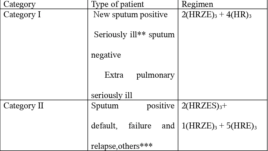
[image:10.612.51.479.478.719.2]National Tuberculosis Control Program (RNTCP) are shown in table 1.2
TABLE 1: CATEGORIES OF ATT UNDER RNTCP
Category Type of patient Regimen Category I New sputum positive
Seriously ill** sputum
negative
Extra pulmonary
seriously ill
2(HRZE)3 + 4(HR)3
Category II Sputum positive
default, failure and
relapse,others***
2(HRZES)3+
Category III Sputum negative not
seriously ill
Extra pulmonary not
seriously ill
2(HRZ)3 + 4(HR)3
The numbers before the letters denote the number of months of treatment.
The subscript after the letters denotes number of doses per week.
The strengths of the drugs are as follows: isoniazid (H)-600mg, rifampicin(R)-450mg,
Ethambutol (E)-1200mg, pyrazinamide (Z)-1500mg, streptomycin(S)-750mg.
Patients who weigh 60kg or more receive additional 150mg rifampicin.
Patients who are more than 50yrs receive 500mg streptomycin.
Patients who weigh < 30kg receive drugs as per body weight.
Patients in category I and II who remain sputum positive at the end of the intensive
phase are given one more month of intensive therapy.
Patients in categories I and II who have positive sputum smear at the end of the initial
intensive phase receive an additional month of intensive phase treatment.
** Seriously ill also includes, any patient, pulmonary or extra pulmonary who is HIV
positive
***In exceptional cases, patients who are sputum smear negative or extra pulmonary
can have relapse or failure and categorized as, “others”
Examples of extra pulmonary seriously ill include meningitis, pericarditis,
with neurological complications. 3
CLASSIFICATION OF ANTITUBECULAR AGENTS
Traditionally anti TB agents have been classified into first line and second line
agents as follows:
First line essential: isoniazid, rifampicin, pyrazinamide.
First line supplementary: ethambutol and streptomycin.
Second line: older agents (paraaminosalicylic acid, ethionamide and cycloserine), newer
agent (rifapentine), kanamycin, amikacin, capreomycin and newer quinolones like
[image:12.612.53.507.370.722.2]gatifloxacin and moxifloxacin.1
TABLE 2: SIDE EFFECTS OF ANTI TB DRUGS
Isoniazid Common Anorexia, nausea, vomiting, fever
Rare Rash, peripheral neuropathy, hepatotoxicity, optic neuritis, hyperglycemia, hemolytic anemia Rifampicin Common Orange coloration of urine, nausea, diarrhea,
rash
Rare Hepatotoxicity, flu like syndrome (intermittent regimen), thrombocytopenia, interstitial nephritis, menstrual disturbances
Pyrazinamide Common Nausea, vomiting, fever
Uncommon Hepatitis, urticaria, skin rash, arthralgia
Rare Photosensitivity, gout, sideroblastic anemia, aggravation of peptic ulcer
Ethambutol Common Optic neuritis, arthralgia, color blindness
Streptomycin Common Vertigo, tinnitus, cutaneous hypersensitivity, deafness
Rare Renal damage, agranulocytopenia, aplastic anemia, neuromuscular blocking action1
ISONIAZID
Isoniazid is still considered the main drug in any anti TB regimen. Its primary
mechanism of action is inhibition of synthesis of mycolic acids which are important
constituents of the bacterial cell wall. 90% of the drug is excreted within 24 hours in the
urine and the excretory products are mainly due to acetylation. The acetylation is
significantly altered by the acetylator status of the individual. The status is measured by
estimating free and total sulfa dimidine in blood and urine. Several risk factors for
isoniazid toxicity have been identified which include acetylator status, old age,
alcoholism and malnutrition. Jaundice occurs in 0.6% to 1% of patients whereas enzyme
increase occurs in 10-20% of the patients.
RIFAMPICIN
Rifampicin inhibits DNA dependent RNA polymerase of mycobacterium. It is
bactericidal for both intracellular and extra cellular organisms and is widely distributed
in all body fluids including CSF. Hepatotoxicity from rifampicin rarely occurs in
patients with normal liver function. Risk factors include old age, chronic liver disease
and alcoholism. Increase in bilirubin and alkaline phosphatase are characteristic of
rifampicin toxicity. Clinical hepatitis occurs in 0.6% to 2% whereas enzyme increase
appears to be enhanced by the addition of isoniazid.
PYRAZINAMIDE
It is a synthetic pyrazine derivative of nicotinamide. Target appears to be
mycobacterial mycolic acid I gene which is involved in mycolic acid synthesis. When a
dose of 40-50mg/kg is administered orally, hepatotoxicity is seen in 15% of patients and
jaundice in 2-3%. Recent regimens are much safer as they employ lower doses. A large
Indian study showed that in regimens containing isoniazid, rifampicin and pyrazinamide,
there was no additional hepatoxicity contributed by pyrazinamide.
ETHAMBUTOL
It is a bacteriostatic drug with no reports of hepatotoxicity. Three fourths of the
ingested dose is excreted unchanged through the kidneys.
BACTERIAL POPULATIONS AND ACTIONS OF DRUGS
A model proposed for explaining the early bactericidal and special sterilizing
properties of drugs includes:
Population A: actively dividing organisms, killed mainly by INH.
Population B: semi dormant organisms mainly seen in acid pH inhibited by
pyrazinamide.
Population C: semi dormant organisms with spurts of activity killed mainly by
rifampicin.
DRUG INDUCED LIVER INJURY
Drug-induced liver injury (DILI) has been a long-standing concern in the
treatment of tuberculosis (TB) infection. The liver has a central role in drug metabolism
and detoxification, and is consequently vulnerable to injury.4
Drug-induced liver injury (DILI) is ultimately a clinical diagnosis of exclusion.
Other causes of liver injury, such as acute viral hepatitis, should be methodically sought
and their absence makes the diagnosis plausible. Usually, the time of onset to acute
injury is within months of initiating a drug. Rechallenge with the suspected offending
agent with more than twofold serum alanine aminotransferase (ALT) elevation and
discontinuation leading to a fall in ALT, is the strongest confirmation of the diagnosis.
Rechallenge may, in some instances, endanger the patient and is usually confined to
essential drugs or used when multiple potentially hepatotoxic drugs have been
administered concomitantly.
DILI may result from direct toxicity of the primary compound, a metabolite, or
from an immunologically mediated response, affecting hepatocytes, biliary epithelial
cells and/or liver vasculature. In many cases, the exact mechanism and factors
contributing to liver toxicity remain poorly understood.
The two major classes of DILI are the predictable and unpredictable responses.
Predictable DILI is generally characterized by certain dose-related injury in
experimental animal models, has a higher attack rate and tends to occur rapidly.
arterioles, where metabolism is greatest and antioxidant detoxifying capacity is the least.
Unpredictable or idiosyncratic reactions comprise most types of DILI. These
hypersensitivity or metabolic reactions occur largely independent of dose and relatively
rarely for each drug, and may result in hepatocellular injury and/or cholestasis.
Hepatocyte necrosis is often distributed throughout hepatic lobules rather than being
zonal, as is often seen with predictable DILI. In hypersensitivity reactions, immunogenic
drug or its metabolites may be free or covalently bound to hepatic proteins, forming
haptens or neoantigens. Antibody-dependent cytotoxic, T-cell and occasionally
eosinophilic hypersensitivity responses may be evoked. Released tumor necrosis factor
(TNF), interleukin (IL)-12 and IFN promote hepatocellular programmed cell death
(apoptosis), an effect opposed by IL 4, IL-10, IL-13, and monocyte chemotactic
protein-1.
Metabolic idiosyncratic reactions may result from genetic or acquired variations
in drug biotransformation pathways, with synthesis or abnormally slow detoxification of
a hepatotoxic metabolite. Metabolic idiosyncratic reactions may have a widely variable
latent period, but recur within days to weeks after re-exposure.
Types of DILI
A variety of clinical syndromes may be seen with DILI, even with a single drug.5
Hepatic adaptation
Exposure to certain drugs may evoke physiologic adaptive responses. The
and antiapoptotic pathways, may attenuate toxin-related injurious responses. Such injury
may also stimulate hepatocyte proliferation and protective adaptation. Asymptomatic,
transient elevations of ALT may reflect slight, nonprogressive injury to hepatocyte
mitochondria, cell membranes or other structures. Such injury rarely leads to
inflammation, cell death or significant histopathologic changes. The induction of hepatic
microsomal (cytochrome P450) enzymes, capable of metabolizing the inducing
medication, is another form of hepatic adaptation.
Drug-induced acute hepatitis or hepatocellular injury
A transaminase threshold for clinicopathologically significant drug-induced
hepatitis has not been systematically determined for most medications. Patients with
acute hepatocellular injury may be asymptomatic or may report a prodrome of fever and
constitutional symptoms, followed by nausea, vomiting, anorexia and lethargy.
Histopathology may reveal focal hepatic necrosis, with bridging in severe cases.
Markedly increased transaminase concentrations followed by jaundice imply severe
liver disease with a 10% possibility of fulminant failure, as said by the late hepatologist
and DILI expert Hyman Zimmerman. Coagulopathy may develop 24 to 36 hours after
onset, although this can subsequently resolve. Coagulopathy persisting beyond 4 days is
a poor prognostic sign in acetaminophen-related hepatotoxicity.
Nonalcoholic fatty liver disease
Steatosis or simple fatty liver is most commonly caused by obesity, insulin
highly active antiretroviral therapy (HAART) are associated with the development and
exacerbation of non alcoholic fatty liver disease. Constitutional symptoms, nausea,
vomiting or abdominal pain are uncommon. Laboratory findings in severe cases include
hypoglycemia, increased serum transaminase concentrations, prolonged coagulation
time and metabolic acidosis. Most instances of drug-induced steatosis are reversible, if
the offending agent is stopped. Persistent steatotic injury may progress to steatohepatitis,
characterized histopathologically by hepatic inflammation, fatty infiltration and by a
subsequently higher risk of cirrhosis.
Granulomatous hepatitis
Granulomata are common, nonspecific findings in liver histology and are
potentially related to infectious, inflammatory or neoplastic etiologies. Hypersensitivity
reactions to drugs, such as allopurinol, quinidine, sulfonamides and pyrazinamide are a
common cause of this type of lesion. Patients may have fever, lethargy, myalgias, rash,
lymphadenopathy, hepatosplenomegaly with increased serum ALT concentration and
even vasculitis.
Cholestasis
Bland cholestasis, typically reported with estrogen treatment, consists of
asymptomatic, usually reversible increases in serum alkaline phosphatase and bilirubin
concentration caused by a failure of bilirubin transport. There is a lack of inflammation
in liver tissue.
Ethanol induces cytochrome P450 2E1, which promotes metabolism of ethanol
itself, acetaminophen and others. Ethanol metabolism yields acetaldehyde, which
contributes to glutathione depletion, protein conjugation, free radical generation and
lipid peroxidation. Chronic ethanol abuse activates hepatic collagen-producing
sinusoidal (stellate) cells, potentially contributing to fibrosis.
Pre-existing liver disease
Abnormal baseline transaminases are an independent risk factor for DILI. Patients
with HIV and hepatitis C however, appear to have increased frequency of antiretroviral
medication related DILI. The severity of DILI, when it occurs, may be greater in patients
with underlying liver disease, likely reflecting a summation of injuries.
Clinical syndromes associated with Drug induced hepatotoxicity include:
1. Liver enzyme elevation in asymptomatic patients
2. Acute viral hepatitis like picture
3. Fulminant hepatic failure
4. Sub acute hepatic failure
5. Cholestatic hepatitis
6. Acute hepatic venous outflow obstruction
7. Autoimmune hepatitis like picture
8. Chronic hepatitis
MECHANISM OF HEPATOTOXICITY DUE TO ANTI TB AGENTS
ISONIAZID
Reactive metabolites of MAH (mono acetyl hydrazine) are probably toxic to
tissues through free radical generation. The antioxidant N-acetyl-cysteine, a substrate for
glutathione synthesis, inhibits isoniazid-induced liver injury in pretreated rats, with
unknown relevance in humans.
Additional metabolic idiosyncratic mechanisms appear to be operative. Slow
acetylators appear to be particularly susceptible to hepatotoxicity. The isoniazid
metabolite acetyl-hydrazine covalently binds to liver macromolecules, a process
mediated by microsomal enzymes. The exact mechanism is still elusive.1
RIFAMPICIN
Conjugated hyperbilirubinemia probably is caused by rifampin inhibiting the
major bile salt exporter pump. Asymptomatic elevation of bilirubin may also result from
dose-dependent competition with bilirubin for clearance at the sinusoidal membrane or
from impeded secretion at the canalicular level. Rare hepatocellular injury appears to be
a hypersensitivity reaction and it may be more common with large, intermittent doses.
Hypersensitivity reactions have been reported in combination with renal dysfunction,
hemolytic anemia or flu like syndrome. Studies have proved that rifampicin induced
hepatitis occurs earlier than isoniazid. There is also evidence that the addition of
rifampicin to isoniazid causes enzyme induction and enhanced production of its toxic
PYRAZINAMIDE
Pyrazinamide may exhibit dose dependent and idiosyncratic hepatotoxicity.
Several decades ago, daily doses of pyrazinamide at 40 to 50 mg/kg commonly caused
hepatotoxicity, and a relationship to dose was noted. There may be shared mechanisms
of injury for isoniazid and pyrazinamide, because there is some similarity in molecular
structure. Patients who previously had hepatotoxic reactions with isoniazid have had
more severe reactions with rifampin and pyrazinamide given for latent tuberculosis
infection. Pyrazinamide may induce hypersensitivity reactions with eosinophilia and
liver injury or granulomatous hepatitis.
RIFABUTIN
At the usual doses (150-200 mg/day), hepatotoxicity is uncommon. There is less
induction of hepatic microsomal enzymes than with rifampin. Elevated transaminases
have been reported with high-dose (600 mg/day) rifabutin treatment in combination with
macrolides.
HEPATOTOXICITY DURING TREATMENT OF TB
Overall, the risk of TB DILI in various studies ranges from 5% to as high as 33%.
The possible risk factors for TB DILI are discussed subsequently.5
Age over 35
Several studies suggest that increasing age is a risk factor for TB DILI, but often
statistical significance was not achieved or hepatotoxicity was not treatment limiting.
average of 5%.Other studies have reported that hepatotoxicity ranges from 22 to 33%in
those older than 35 years, compared with 8 to 17% in those younger than 35 years.7
Children
There are varied reports of increased hepatotoxicity amongst children treated with
anti TB therapy which was especially high in children with neurotuberculosis.8
Sex
For women, several studies report increased risk of hepatotoxicity, but this was
not always treatment limiting, or did not achieve statistical significance. One study did
show a four times higher risk of treatment limiting hepatotoxicity in women, but with an
overall incidence of only 2%. Two other studies showed no increased risk in women.9
Cofactors
Several studies have indicated that alcohol use was a significant predictor of TB
DILI, whereas two studies found no association.10
Abnormal Baseline Transaminases
One study found an increased risk of hepatotoxicity during treatment of TB
disease in individuals with abnormal baseline transaminases.11
Acetylator Status
When acetylation rate has been determined by phenotypic assays, slow acetylators
Other Factors
Malnutrition or hypoalbuminemia was associated with TB DILI in several studies
from India.12 The presence of HLA-DQB1*0201 is an independent risk factor for the
development of TB DILI.13 Gene polymorphisms at loci of genes coding for cytochrome
P4502E1 and for glutathione S-transferase have also been associated with
hepatotoxicity.14
Disease severity
Extensive TB disease itself may be a risk factor for TB DILI, although
confounding factors are impossible to exclude.12 Liver transplant patients with TB
appear to have a high rate of hepatotoxicity, with five of six treated patients developing
this complication, confirmed by liver biopsy and three of six suffering graft rejection.
Regimen
In a meta-analysis, the presence of rifampicin in a multidrug treatment regimen
increased the incidence of significant hepatotoxicity for adults from 1.6 to 2.55% and in
children from 1.0 to 6.9%.15 The influence of pyrazinamide on TB DILI is ambiguous;
some studies indicate little to no increased rate of hepatotoxicity, whereas others point to
it as a contributor to increased incidence or severity of hepatotoxicity, although dosing
variations and patient selection biases may have contributed to these results.
HIV-infected Individuals
to 1994, many who used intravenous drugs, 13 to 15% of patients had transaminase
increases of at least three times the ULN in the first 2 months. Hepatoxicity was
attributed to isoniazid in 55%of those with hepatitis. In a study of HIV, hepatitis C and
TB treatment, HIV infection independently increased the risk fourfold of serum
transaminase increase to 120 IU/L or of total bilirubin to at least 1.5 mg/dl.
Approximately 27%of HIV infected individuals developed hepatotoxicity, compared
with 12% among HIV-uninfected individuals. Nearly 80% of the patients in this study
had a history of alcohol abuse, although random testing did not reveal active drinking,
patients had not consumed alcohol in the 10 days before study entry, and all had normal
baseline hepatic transaminases.16
Hepatitis B
Several studies from Asia have addressed DILI during treatment of TB disease in
patients with hepatitis B infection. Additional studies are needed, but the limited data
leave sufficient concern that hepatitis B maybe a risk factor for more frequent or severe
hepatotoxicity during treatment of TB disease.
Hepatitis C
Hepatitis C was an independent risk factor for the development of hepatotoxicity,
elevating the risk fivefold of transaminase elevation of at least 120 U/L, or of serum
bilirubin of at least 1.5 mg/dl. Co infection with both hepatitis C and HIV elevated the
risk of hepatotoxicity more than 14-fold.
Hepatotoxicity has been recognized to occur in about 2% of patients treated with
ethionamide or prothionamide and in 0.3% of patients treated with para-aminosalicylic
acid17, 18. Cycloserine does not appear to be associated with hepatotoxicity, but should be
used with caution in patients at risk for alcohol withdrawal seizures.
Singh et al19 reported an overall mortality of 12% in patients with ATT induced
hepatotoxicity while it was 75% in patients with acute and sub acute liver failure.
In a case control study from AIIMS New Delhi, Pande et al20 proposed the
following diagnostic criteria for ATT induced
hepatotoxicity-1. Symptoms and signs of icteric hepatitis (anorexia, nausea, jaundice)
2. Rise in liver transaminases to more than 3 times the upper limit of normal on 3
occasions or a single value of >250IU/L.
3. Increase in serum bilirubin >1.5mg/dl
4. Absence of serological evidence of viral hepatitis.
In a retrospective study of 519 patients in Germany receiving isoniazid,
rifampicin and pyrazinamide, hepatotoxicity was observed in 11% of patients9.
In another study of 456 patients from Argentina receiving all 5 first line drugs,
liver injury was seen in 9.9% of patients21.
MANAGEMENT OF TB DILI
Pretreatment Clinical Evaluation
1. A standardized history form is recommended, which includes risk factors for
2. The physical examination should include evaluation for signs of liver disease, such as
liver tenderness, hepatosplenomegaly, jaundice, caput medusa, spider angiomata, ascites,
and edema.
3. Previous laboratory values should be reviewed when available.
4. Screening for viral hepatitis should be considered for the following patients
- Individuals who inject drugs;
- were born in endemic areas of Asia, Africa, the Pacific Islands, Eastern Europe,
or the Amazon Basin;
- are HIV infected;
- may have had sexual or household contact with chronically infected individuals;
- may have had occupational exposure to infected blood;
- are chronic hemodialysis patients;
- are recipients of clotting factors before 1987;
- have undiagnosed liver disease
5. Voluntary HIV counseling and testing are recommended for all patients with TB
disease.5
Patient Education
1. Printed instructions should include clinic telephone numbers, include explicit
instructions for after-hours care, and utilize patient preferred language at a readable
level.
vomiting, abdominal discomfort, or unexplained fatigue and to contact the clinic for
further evaluation.
3. Patients should attend clinic follow-up visits for monitoring and reinforcement of
education.
4. Patients should be warned about concomitant alcohol and hepatotoxic
over-the-counter, and alternative and prescription medication use.
5. Patients should inform their health care providers of anti-TB medications prescribed
drug adverse events.
Baseline laboratory testing and monitoring:
1. Baseline measurements of serum transaminases, bilirubin, alkaline phosphatase,
creatinine and a blood platelet count are recommended for all adults beginning treatment
for TB disease.
2. For patients with preexisting severe liver disease, some clinicians also recommend
periodic measurement of prothrombin time and INR to assess hepatic synthetic function.
3. Routine measurements during treatment are recommended when baseline
abnormalities are present and for patients, who chronically consume alcohol, take other
potentially hepatotoxic medications or who have viral hepatitis or history of liver
4. In patients with abnormal baseline transaminases, the range of their prior fluctuations
may be of assistance in interpreting results of biochemical monitoring of treatment.
5. Some providers prefer to monitor ALT in women or older adults being treated for TB
disease
7. ALT is preferred for detecting and tracking hepatocellular injury in those who develop
symptoms of hepatotoxicity.
8. Measurements of AST, bilirubin and alkaline phosphatase are adjunctive for
monitoring chronic liver disease, cholestasis or severe hepatocellular injury.
9. The ULN used should be that of the laboratory performing the assay.
10. Optimally, reference limits for enzymes should be adjusted for age in children and in
adults older than 60 and for sex in adults, if available.
11. Hepatitis B surface antigen seropositive individuals with elevated ALT should have
HBeAg testing. If positive, rifampin may be preferred over isoniazid. A hepatologist
should be consulted regarding further testing and possible pretreatment in individuals
with an ALT at least two times the ULN and who are HBeAg seropositive. In
HBeAg-seropositive individuals, clinical and ALT monitoring should occur every 2 to 4 weeks.
12. Patients with baseline transaminases more than three times the ULN should have
ALT retested along with bilirubin, as well as screening for viral or other causes of
hepatitis, including alcohol and hepatotoxic drugs.
Treatment of TB Disease
The crucial efficacy of isoniazid and particularly rifampin warrants their use and
retention, if at all possible, even in the face of preexisting liver disease. Several
regimens are recommended if baseline serum ALT is more than three times the ULN and
TB is not believed to be the cause.
1. Treatment without pyrazinamide might utilize isoniazid and rifampin for 9 months
with ethambutol until drug susceptibility testing of the M.tuberculosis isolate is
completed.
2. In patients with cirrhosis, rifampin, Ethambutol and treatment with levofloxacin,
moxifloxacin, gatifloxacin or cycloserine for 12 to 18 months may be considered.
3. For patients with encephalopathic liver disease, Ethambutol combined with a
fluoroquinolone, cycloserine and capreomycin or aminoglycoside for 18 to 24 months
may be an option. However, these regimens have not been tested systematically.
4. Some providers avoid aminoglycosides in severe, unstable liver disease due to
concerns about renal insufficiency or bleeding from injected medication in patients with
thrombocytopenia and/or coagulopathy.
Interventions for hepatotoxicity.
The first-line anti-TB drugs, especially rifampin, should not be discontinued for
mild gastrointestinal complaints, which may be relatively frequent in the initial weeks of
anti-TB treatment. If serum transaminase concentrations are more than five times the
ULN (with or without symptoms)or more than three times the ULN with jaundice and/or
immediately and the patient evaluated promptly. Serologic tests for hepatitis A, B, C and
E viruses should be obtained, and the patient should be evaluated for biliary disease, use
of alcohol and other hepatotoxic drugs. Some experts recommend interrupting treatment
for lesser increases in patients with cirrhosis or encephalopathy. Until the specific cause
of abnormalities can be determined, clinicians should treat with at least three anti-TB
agents that are less likely to cause hepatotoxicity namely streptomycin, Ethambutol and
quinolones.
Rechallenge
The risk of reintroducing of a TB medication could be hazardous and should be
considered relative to its potential benefit. Rechallenge is considered when it is unclear
which medication was the cause of symptoms or of transaminases increases.
Rechallenge also may be considered if an increase in transaminases concentration did
not reach the usual treatment limiting threshold. Rechallenged patients who had reached
a treatment limiting threshold should have clinical and biochemical monitoring at 2-to
4-week intervals. Rechallenged patients should be told to stop medication in case of
hepatitis symptoms.
After the enzymes return to normal, the drugs are introduced sequentially.
Isoniazid is introduced first at a dose of 50mg/day and increased to 300mg/day over the
next 2-3 days if well tolerated.2
Rifampicin is introduced after another 3-4 days at doses of 75mg/day and increase
250mg, increasing to 1000mg over the next few days.
Studies show that the recurrence of hepatotoxicity is very less after reintroduction
of ATT. Singh et al reported a recurrence rate of 6.8% in their study. Telman et al
reported a recurrence rate of 6.3%.11
A high recurrence rate was noted in one study in Copenhagen10 (26%). Further
AIMS AND OBJECTIVES
The aim of the study is to analyze the
following-1. To study the incidence of hepatotoxicity in patients taking antituberculous drug
therapy under the RNTCP short course schedule in Madurai district
MATERIALS AND METHODS
THE STUDY GROUP
The study was conducted on patients coming to the chest clinic in Govt. Rajaji
hospital, Madurai. Approval from ethical committee was obtained. The study was a
prospective study conducted for a period of one year between June 2007-2008.
The inclusion criteria
were-1. Patients prescribed to receive ATT for confirmed pulmonary or extra pulmonary
tuberculosis under the RNTCP schedule.
2. Patients coming from in and around Madurai city, to minimize the rate of dropouts.
The exclusion criteria
were-1. Patients not receiving isoniazid or rifampicin as a part of therapy.
2. Patients with preexisting acute or chronic liver disease.
3. Patients with fatty liver as diagnosed by ultrasound examination of the abdomen.
4. Baseline transaminases more than twice the upper limit of normal.
5. Chronic alcohol intake.
6. Patients with previous history of hepatotoxicity due to ATT.
METHODS
All the patients had pretreatment evaluation clinically especially for evidence of
liver disease, history of alcoholism or concomitant drug therapy and systemic illness in a
prepared proforma, a copy of which is annexed. Baseline laboratory evaluation was done
and ultra sonogram of the abdomen and HIV status. Body mass index (BMI) was
calculated as follows2
-BMI = weight (kg)/height (metre2)
Malnutrition - <18.5kg/m2
Normal – 18.5-24.9kg/m2
Obese - > 25kg/m2
Normal values for LFT were as
follows-Serum albumin >3.5grams/dl – normal
Serum albumin <3.5grams/dl – hypoalbuminemia
Serum bilirubin 0.2-0.8 mg%--normal
AST 15-40 iu/l--normal
ALT 15-40 iu/l--normal
Anemia was defined as the hemoglobin of less than 13 g/dl in males and less than 12
g/dl in females.22
Mild anemia was defined as hemoglobin level of 12.9 g/dL in males and
10-11.9 g/dL in females, moderate anemia was defined as hemoglobin level of 7-9.9 g/dL
and severe anemia was defined as hemoglobin level of less than 7g/dL both among
males and females.22, 23
Viral markers were done to exclude chronic viral hepatitis at the start of therapy.
Presence of fatty liver was excluded on the basis of ultrasonography.
type of disease, the details of which were explained previously.
Doses of the drugs were as
follows-Isoniazid-600mg
Rifampicin-450mg (600mg if >60kg)
Pyrazinamide-1500mg
Ethambutol- 1200mg
Every effort was taken to maintain compliance to the drug therapy.
Patients were informed about the side effects of medications and were asked to
report immediately if they developed any of these symptoms. Patients with minor
symptoms were treated symptomatically. Those with major symptoms suggestive of
hepatotoxicity were hospitalized and evaluated.
Diagnosis of Drug Induced Hepatotoxicity:
Hepatotoxicity was defined as the presence of at least one of the following criteria:
(1) Appearance of jaundice.
(2) a rise of at least five times the upper limit of normal levels (40 IU/L) of serum
aspartate aminotransferase (AST) and/or alanine aminotransferase (ALT) or >250IU/L
on any one occasion without symptoms or more than three times ULN with symptoms.
(3) A rise in the level of serum total bilirubin > 1.5mg/dl.24
Liver enzymes (AST/ALT) were estimated using the UV kinetic International
Federation of Clinical Chemistry method.
and after the completion of ATT (six months).
If the patients developed evidence of hepatotoxicity, viral markers (hepatitis A, B,
C) were again performed to rule out acute viral hepatitis.
The information collected regarding all the selected cases were recorded in a
Master Chart. Data analysis was done with the help of computer using Epidemiological
Information Package (EPI 2002).
Using this software, frequencies, percentages, means, standard deviations, chi
square and ‘p’ values were calculated. Kruskul Wallis chi-square test was used to test the
significance of difference between quantitative variables and Yate's test for qualitative
variables. A 'p' value less than 0.05 is taken to denote significant relationship.
RESULTS AND ANALYSIS OF OBSERVED DATA
EPIDEMIOLOGY
The study group was divided into various age groups which ranged between 15
and 78 years. A total of 166 patients were included in the study of which 156 patients
were followed up till six months of treatment. Total number of dropouts from the study
at the end of six months was 10 out of which seven patients dropped out at the end of
two weeks and three at the end of three weeks. Most patients were from in and around
Madurai city.
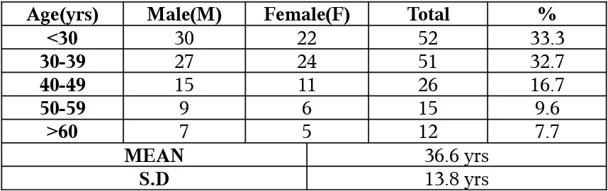
The mean age of the population was 36.6+13.8 years. Out of 156 patients, 88
were male (56.5%) and 68 were female (43.5%). Most of the patients were in the ages
[image:37.612.50.479.442.577.2]between 15 and 39 years (66%).
TABLE 3: AGE DISTRIBUTION OF PATIENTS
Age(yrs) Male(M) Female(F) Total %
<30 30 22 52 33.3
30-39 27 24 51 32.7
40-49 15 11 26 16.7
50-59 9 6 15 9.6
>60 7 5 12 7.7
MEAN 36.6 yrs
SYMPTOMATOLOGY
There was a higher proportion of pulmonary tuberculosis (n=90) than the extra
pulmonary form (n=66). Pleural effusion was the most common form of extra
pulmonary tuberculosis seen in 31 patients (19.9%) followed by neurotuberculosis in 16
[image:38.612.50.498.248.367.2]patients (10%).
TABLE 4: SITE OF DISEASE
Site M F Total %
Pulmonary 50 40 90 57.7
Pleural effusion 20 11 31 19.9
Lymph node 5 8 13 8.3
Abdominal 2 2 4 2.6
Skeletal 2 0 2 1.3
NeuroTB 9 7 16 10.2
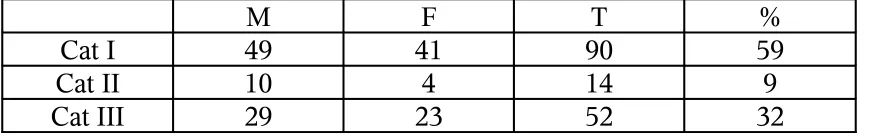
Ninety patients were registered under category I ATT (59%) followed by 52
patients in category III (32%) and 14 patients in category II (9%) as shown in table- 5.
TABLE 5: CATEGORY OF ATT
M F T %
Cat I 49 41 90 59
Cat II 10 4 14 9
Cat III 29 23 52 32
Eighty six patients were sputum positive (55%) out of which 56% were males and
[image:38.612.50.485.496.565.2]TABLE 6: BODY MASS INDEX (BMI)
BMI M F T %
<18.5 20 15 35 22.4
18-24.9 59 47 106 68
>25 10 5 15 9.6
MEAN 20.5
S.D 3.1
The mean BMI was 20.5+3.1kg/m2. Body mass index was <18.5kg/m2 in 35
patients (22.4%).
The mean hemoglobin values were 10.4+1.8gms/dl. It was found that 49 patients
[image:39.612.51.510.571.732.2](31.4%) had a hemoglobin level of <9.9grams/dl, signifying moderate to severe anemia.
TABLE 7: HEMOGLOBIN (Hb) LEVELS
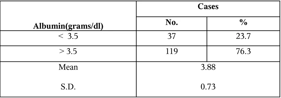
Serum albumin levels were < 3.5 grams/dl in 37 patients (23.7%). The mean
albumin values were 3.88+0.73grams/dl.
TABLE 8: SERUM ALBUMIN LEVELS
Albumin(grams/dl)
Cases
No. %
< 3.5 37 23.7
> 3.5 119 76.3
Mean
S.D.
3.88
[image:39.612.52.509.573.732.2]Nineteen patients (12%) had systemic disease in the form of diabetes mellitus or
chronic renal failure.
Twenty two patients (14.1%) of patients tested positive for HIV.
RESULTS OF LFT
Overall, 24 patients out of 156 had some abnormality of liver function (15.4%).
The most common form of hepatotoxicity was asymptomatic rise in transaminases
(n=16) followed by acute hepatitis like picture with jaundice (n=8). All 16 patients who
developed increase in enzymes were asymptomatic.
Eight patients developed jaundice. There was no incidence of fulminant hepatic
failure or chronic hepatitis. All patients who developed liver injury were investigated for
viral hepatitis and none of the patients tested positive for any of the viral markers
performed namely, IgM antibody for HAV, HbSAg, and anti HCV. Most of the
derangements occurred within the first two weeks of starting therapy and subsided
spontaneously on stopping the drug within 4-6 weeks. The results are summarized in
TABLE 9: BILIRUBIN ABNORMALITIES
Bilirubin at
< 1.5mg% > 1.5mg%
Mean S.D.
No. % No. %
0 week 156 100 - - 0.81 0.19
2 weeks 150 94.9 6 5.1 1.01 0.37
4 weeks 154 98.7 2 1.3 0.85 0.19
24 weeks 156 100 - - 0.78 0.16
It was observed that the mean bilirubin levels were 1.01+0.37mg% in the second
week and dropped down to 0.85+0.19mg% at the end of four weeks. Six patients had
jaundice at the end of two weeks and two patients had persistent jaundice at four weeks
that recovered within the next two weeks.
The abnormalities in liver enzymes were analyzed in relation to the time of onset
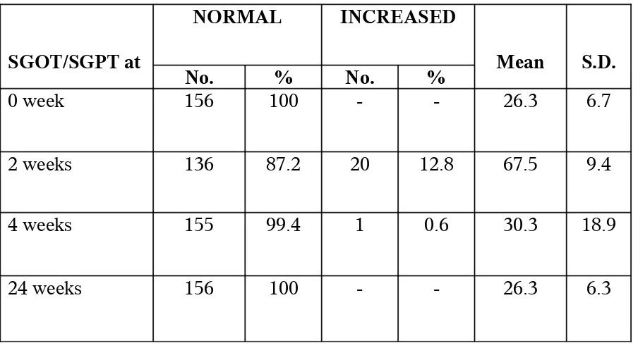
TABLE 10: ENZYME ABNORMALITIES
SGOT/SGPT at
NORMAL INCREASED
Mean S.D.
No. % No. %
0 week 156 100 - - 26.3 6.7
2 weeks 136 87.2 20 12.8 67.5 9.4
4 weeks 155 99.4 1 0.6 30.3 18.9
24 weeks 156 100 - - 26.3 6.3
It was observed that most of the enzyme abnormalities occurred within the
first 2 weeks. Twenty patients (12.8%) developed enzyme elevation within two weeks
of starting therapy and only one patient had persisting abnormality at the end of four
weeks.
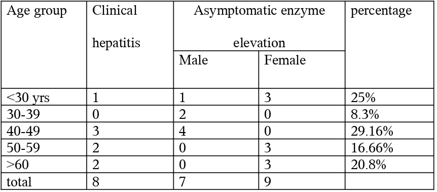
The patterns of hepatotoxicity were analyzed in relation to symptoms and LFT
TABLE 11: PATTERN OF HEPATOTOXICITY
Age group Clinical
hepatitis
Asymptomatic enzyme
elevation
percentage
Male Female
<30 yrs 1 1 3 25%
30-39 0 2 0 8.3%
40-49 3 4 0 29.16%
50-59 2 0 3 16.66%
>60 2 0 3 20.8%
total 8 7 9
It was observed that asymptomatic enzyme elevation was the most common form
of hepatotoxicity seen in 16 patients (66.6%) followed by clinical hepatitis with jaundice
in eight patients (33.3%). There was near equal incidence of toxicity in males (n=7) and
females (n=9).
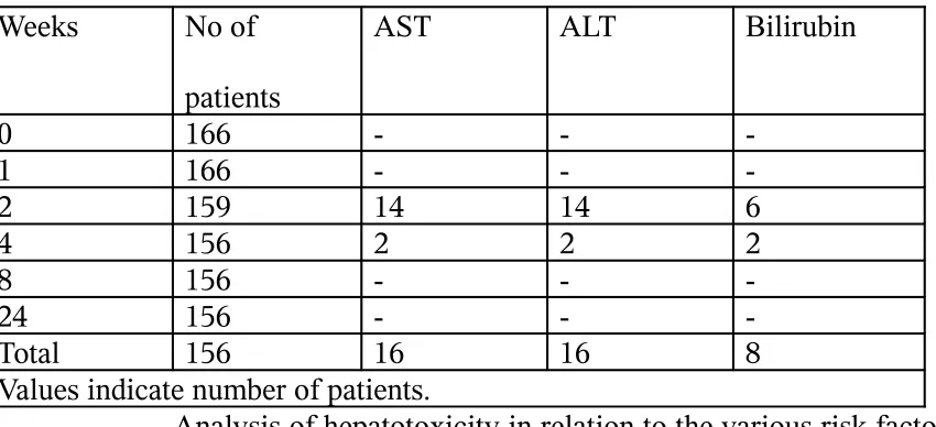
TABLE 12: INITIAL ELEVATION OF LFTs
Weeks No of
patients
AST ALT Bilirubin
0 166 - -
-1 166 - -
-2 159 14 14 6
4 156 2 2 2
8 156 - -
-24 156 - -
-Total 156 16 16 8
Values indicate number of patients.
Analysis of hepatotoxicity in relation to the various risk factor parameters
was done and the following observations were made-
1. Category of ATT :
It was observed that six patients (42.9%) started on category II developed toxicity
in comparison to 13 patients (14.4%) on category I and five patients (9.6 %) on category
III.
TABLE 13: CATEGORY OF ATT VERSUS HEPATOTOXICITY
Category of ATT
LFT abnormality
Present Absent
No. % No. %
I (90) 13 14.4 77 85.6
II (14) 6 42.9 8 57.1
[image:44.612.48.483.519.675.2]2. Age group:
It was observed that five out of 12 patients above the age of 60 years developed
abnormalities of LFT (41.7%).
TABLE 14: AGE AND HEPATOTOXICITY
The value was statistically significant (p=0.0027), implying that the elderly
patients were more susceptible to liver damage due to ATT.
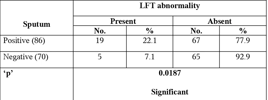
3. Sputum positivity:
It was observed that out of 86 patients who were sputum positive, 19 developed
[image:45.612.51.512.428.600.2]hepatotoxicity (22.1%) in comparison with only five sputum negative patients (7.1%).
TABLE 15: SPUTUM POSITIVITY VERSUS HEPATOTOXICTY
Sputum
LFT abnormality
Present Absent
No. % No. %
Positive (86) 19 22.1 67 77.9
Negative (70) 5 7.1 65 92.9
‘p’ 0.0187
Significant
80% of patients with hepatotoxicity were sputum positive, possibly indicating
severe disease as a risk factor for hepatotoxicity. The value was statistically significant
4. Hemoglobin:
Out of 49 patients who had moderate to severe anemia, 17 developed liver injury
(34.7%). In contrast, only seven patients who had hemoglobin levels above 9.9grams%
developed toxicity (6.5%).
TABLE 16: ANEMIA VERSUS HEPATOTOXICTY
70% of patients with toxicity had hemoglobin levels <9.9gm% and the value was
statistically significant (p=0.0001). Moderate to severe anemia directly correlated with
hepatotoxicity.
5. Body mass index:
It was observed that 19 out of 35 patients (54.3%) with BMI <18.5kg/m2 had LFT
[image:46.612.54.511.512.732.2]abnormalities.
TABLE 17: MALNUTRITION AND HEPATOTOXICITY
BMI (kg/m2)
LFT abnormality
Present Absent
No. % No. %
< 18.5 (35) 19 54.3 16 45.7
> 18.5 (121) 5 4.1 116 95.9
Mean
S.D.
17.13
1.39
21.09
2.9
‘p’ 0.0001
80% of patients with toxicity had BMI values of <18.5kg/m2 and the value was
statistically significant (p=0.0001), implying that malnutrition may be a significant risk
factor for toxicity.
6. Serum albumin:
It was observed that 45.9% of patients with hypoalbuminemia (n=17) had toxicity.
[image:47.612.48.503.337.595.2]The value was statistically significant (p=0.0001) as shown in the table below.
TABLE 18: HYPOALBUMINEMIA AND HEPATOTOXICITY
Serum albumin
(grams/dl)
LFT abnormality
Present Absent
No. % No. %
< 3.5 (37) 17 45.9 20 54.1
> 3.5 (119) 7 5.9 112 94.1
Mean
S.D.
3.14
0.84
4.01
0.61
‘p’ 0.0001
7. HIV status:
Out of 22 patients who tested positive for HIV, 11 (50%) developed
[image:48.612.53.504.200.386.2]hepatotoxicity.
TABLE 19: HIV STATUS AND HEPATOTOXICITY
HIV
LFT abnormality
Present Absent
No. % No. %
Positive (22) 11 50 11 50
Negative (134) 13 9.7 121 90.3
‘p’ 0.0001
Significant
The value obtained was statistically significant (p=0.0001) indicating that HIV
infection may be a significant risk factor for ATT induced hepatotoxicity.
8. Site of disease:
Hepatotoxicity was more in abdominal (25%) and pulmonary tuberculosis
TABLE 20: SITE OF DISEASE AND HEPATOTOXICITY
Site of disease
LFT abnormality
Present Absent
No. % No. %
AB (4) 1 25 3 75
LN (13) - - 13 100
P (90) 20 22.2 70 77.8
PE (31) 3 9.7 28 90.3
SK (2) - - 2 100
DISCUSSION
The wide prevalence of tuberculosis through out the world makes it a social and
economical burden especially for developing countries and the use of anti tuberculous
drugs is an optimistic approach for this problem. However certain reservations
associated with its use need to be properly evaluated especially ATT induced liver injury
and the predisposing factors that add to this hepatotoxicity.
This study was conducted to study the incidence of ATT induced hepatotoxicity in
RNTCP clinic, Madurai and to assess the role of age, sex, severity of the disease,
nutritional status, hypoalbuminemia, sputum positivity and HIV status as risk factor for
ATT induced hepatotoxicity.
The reported incidence of ATT induced hepatotoxicity is different in various
countries though not fully understood but could be due to the characteristics and the risk
factors of the population studied, the different diagnostic criteria used to define
hepatotoxicity, different geographical areas, tests carried out during follow ups and the
type of monitoring.25
Why only some patients who receive ATT develop hepatitis is not clear and several studies searched for host factors, environmental factors or some interaction among various factors. While some papers have focused on genetic factors, such as HLA typing, Cytochrome P450 2E1 or acetylator status, others have primarily
studied clinical factors.
and belonged to different treatment categories. Male: female ratio was almost equal
(53.5%:48.5%). Age group of the patients ranged from 15-78 years.
Various studies report different incidence rates of hepatotoxicity due to anti
tuberculous therapy. A higher risk of hepatotoxicity has been reported in Indian patients
than in their Western counterparts.19, 26 the risk of hepatotoxicity based on data from four
prospective Indian studies was 11.5% compared with 4.3% in Western publications.15
In our study 15.4% of the patients developed ATT induced hepatotoxicity that almost
overlaps the other studies conducted world wide. The incidence of hepatotoxicity due to
combination chemotherapy ranges from 1-39%. The following is a list of incidence by
some workers:
1. Parthasarathy et al 27 TBM: 16-39%, PT: 2-3%
2. Schberg et al 9 11%
3. Devoto et al 21 9.9%
4. Steele et al 15 2.6%
5. Dossing et al 10 8%
6. Kamat et al28 18%
7. Sivaraman et al29 7%
The TRC, Chennai published its first report on hepatotoxicity induced by
antituberculous drug therapy and the incidence was 2.5%.
In a study done in Pakistan, 19.76% of the patients developed ATT induced
hepatotoxicity that almost overlapped the study conducted at Japan.30,31 A study
conducted in Nepal32 resulted in 8% and 13% in Hong Kong Chinese patients.33 In the
analysis done by Col AC Anand et al, the incidence of hepatotoxicity among patients on
ATT was 10.1%,34
In our study we analyzed the possible predisposing factors for hepatotoxicity
induced ATT. A significant proportion of patients older than 60 years developed
hepatotoxicity (p=0.0027). Although the number of patients above the age of 60 years
receiving ATT was less (n=12), nearly half of these patients developed liver function
abnormalities.
Some studies have reported that the risk of ATT-induced hepatitis increases with
advancing age, the highest incidence being in individuals older than 50 years as shown
by Gangadharan et al.35
In a study done by Col AC Anand et al in 2006, no significant correlation of age
with ATT-induced hepatotoxicity was found. However, once hepatotoxicity developed,
fatal out come was much more likely among the older patients (mean age 47.1 years as
compared to 38.9 years in non fatal cases).34 Studies from Pande et al and other workers
In a study done by Khalid Mohammad et al, older age group was affected more as
compared to younger one (25.8% vs. 14.4%).30
Although previous studies have quoted an increased risk of hepatotoxicity for
females, it was found to be almost equal in this study. There was a marginal increase in
sub clinical hepatitis in females in accordance with many other studies (nine in females
vs. seven in males). Vulnerability of females could be due to variations in
pharmacokinetics and slow acetylating enzymatic pattern, resulting in hepatotoxicity.20, 38
Anand AC et al did not find any difference in the incidence of hepatotoxicity in
their study.34 This has also been reported in a study done by Taneja et al.39
Nutritional status of our patients was very poor. BMI were below 18.5 (kg/m2) in
23% of patients and 23.4% of the patients had hypoalbuminemia. In our study there was
a significant relationship of hepatotoxicity to low serum albumin and low BMI as noted
in the previous studies done by Pande et al20. Nearly 72% of patients with hepatotoxicity
had serum albumin <3.5 grams%.
Krishnasamy says that under nutrition contributes to drug toxicity by various
mechanisms. 40, 41 Toxicity and over dosage is much more likely to occur even with
normal dosage of medicine in the presence of normal serum albumin.42 A study from
Pakistan shows significant correlation between the two variables.30
The possible explanation of ATT induced hepatotoxicity in malnutrition is
nutritional status is considered to be one of the factors contributing to relatively high
incidence of ATT-related hepatitis in studies from developing countries. Drug
metabolism pathways including acetylation pathway have been shown to be deranged in
states of protein energy malnutrition.43, 44
A direct correlation was also obtained between low BMI and hepatotoxicity
(p=0.0001) and this was in concordance with the previous studies done by Shakya et al.7
A similar significant relationship was noted between hemoglobin levels and
hepatotoxicity (p=0.0001). Most of the patients with hepatotoxicity had severe anemia.
Nineteen patients (80%) were sputum smear positive and they were severely
affected indicating the extensiveness of the disease also as a risk factor as noted in
previous studies done by Pande et al20 (p=0.001)and Devoto et al 21(p=0.02). Severity of
the disease in sputum smear positive patients could be secondary to more tubercular
bacilli in smear positive patients as compared to smear negative patients.
In our study, there was direct association between sputum positivity and
hepatotoxicity (p=0.0187). In the study done by Khalid Mohammad, forty patients
(59.70%) were sputum smear positive and they were severely affected indicating the
extensiveness of the disease as a risk factor.30, 45, 10, 20
In our study, HIV infection was found to be a significant risk factor for TB DILI
(p=0.0001). The patients were not on antiretroviral therapy at the start of ATT, thus
ruling out antiretroviral therapy as the cause of liver function abnormalities. This was in
where it was found that patients with HIV and TB had significant risk of hepatotoxicity
irrespective of whether the patient was on ART or not.16
None of the patients in this study who developed toxicity had viral hepatitis
though there is evidence to indicate that patients with viral hepatitis B or hepatitis C had
a higher risk of drug toxicity than general population.46, 47, 48
The frequency of self limiting asymptomatic enzyme elevation raises the question of
whether the drugs should be stopped; and if so, at what levels they should be stopped.
Mild transient self limiting transaminase rise occurs early during the course of therapy
irrespective of the regimen used and this should not be used as a criterion for stopping
therapy. Onset of hepatotoxicity occurred within one month of start of therapy in our
study. Usually pyrazinamide produced a delayed onset of hepatotoxicity whereas early
toxicity is produced by isoniazid and rifampicin.
Parthasarathy et al concluded that acute hepatitis is nearly always associated with
jaundice.27 In our study also, patients who developed jaundice has an enzyme elevation
of >200 IU/L. after withdrawing the drugs, all levels returned to normal within four
weeks.
Some workers say it may not be advisable to stop all drugs at a time but since the
possibility of fulminant hepatic failure always arises, it is advisable to stop all drugs till
reintroduced according to the British Thoracic society guidelines24. None of the patients
developed recurrent hepatotoxicity.
The onset of drug induced hepatotoxicity cannot be exactly predicted but
measures to prevent it can be taken well in advance. High protein diet, abstinence from
alcohol and smoking, good supportive medications like vitamin B6 and vitamin C have
shown to reduce the incidence of hepatotoxicity. Well educated patients and skilled,
alert, treatment supervisor can reduce the hepatotoxicity, fulminant hepatitis and its
SUMMARY
In this study, 156 patients belonging to different categories of RNTCP-DOTS
were followed up for a period of six months. There were 88 Males and 68 females.
15.4% of patients developed liver function abnormalities of which asymptomatic
enzyme elevation was the most common feature. There was no incidence of fulminant
hepatic failure or chronic hepatitis.
Transient self limiting rise of transaminases occurred early during the course of
therapy which subsided on stopping drug over a period of four to six weeks.
Reintroduction of ATT was tolerated well and all had completed treatment. Advancing
age, malnutrition, anemia, advanced disease with sputum positivity, hypoproteinemia
and HIV positivity directly correlated with drug induced hepatotoxicity.
Identification of these risk factors may be helpful in predicting hepatotoxicity and
correction of the modifiable risk factors prior to start of therapy may prevent liver
CONCLUSIONS
The following conclusions were made from our
study-1. Liver function abnormalities occurred in 15% of the study group under
RNTCP which is a significant number.
2. Asymptomatic enzyme elevation is the most common abnormality in
our study.
3. Direct correlation existed between increasing ages, malnutrition, anaemia, advanced
disease with sputum positivity and hypoproteinemia.
Correction of the modifiable risk factors can lead to decrease in hepatotoxicity.
4. Hepatotoxicity is usually self limiting and treatment need not be discontinued
permanently.
5. Serial monitoring of liver function tests will help in early identification of drug
induced hepatotoxicity and prevention of fulminant hepatic failure.
6. Further research is needed to find out the exact mechanisms of hepatotoxicity due to
FIGURE 1
[image:59.612.60.492.267.573.2]Illustration of the proposed mechanism of DILI, which involves drug metabolism, hepatocyte damage, activation of innate immune cells, and production of tissue-damaging and tissue-protective mediators. CYP - cytochrome P450; IFN- interferon; IL- interleukin; NK-natural killer cell; NKT- natural killer T cell; TNF- tumor necrosis factor.
32%
33% 10%
8%
17%
FIGURE 3: BILIRUBIN ABNORMALITIES
FIGURE 4: SGPT ABNORMALITIES
156 0 148 8 154 2 156 0 0% 20% 40% 60% 80% 100%
0 2 4 24
Bilirubin at weeks
Less than 1.5 More than 1.5
156 0 136 20 156 0 156 0 0% 20% 40% 60% 80% 100%
0 2 4 24
SGPT at weeks
FIGURE 5: SGOT ABNORMALITIES
FIGURE 6: CATEGORY OF ATT VERSUS HEPATOTOXICITY
156 0 136 20 155 1 156 0 0% 20% 40% 60% 80% 100%
0 2 4 24
SGOT at weeks
normal increased 13 77 6 8 5 47 0% 10% 20% 30% 40% 50% 60% 70% 80% 90% 100%
I II III
CATEGORY OF ATT
FIGURE 7: AGE VERSUS HEPATOTOXICITY
5 47 2 49 8 18 4 11 5 70% 20% 40% 60% 80% 100%
FIGURE 8: SPUTUM POSITIVITY VERSUS HEPATOTOXICITY
19 67
5 65
0%
20%
40%
60%
80%
100%
LFT ABNORMALITY
P
O
S
IT
IV
E
N
E
G
A
T
IV
E
S
P
U
T
U
M