STUDY OF THE PATTERNS OF FAILURE IN
CARCINOMA ESOPHAGUS
A DISSERTATION SUBMITTED TO THE
TAMIL NADU DR MGR MEDICAL UNIVERSITY
IN PARTIAL FULFILMENT OF THE REQUIREMENTS FOR
THE AWARD OF THE DEGREE OF
CERTIFICATE
This is to certify that this dissertation titled “STUDY OF THE
PATTERNS OF FAILURE IN CARCINOMA ESOPHAGUS” is a
bonafide record of the work done by Dr RATNAGIRI RANGANATH
in the Division of Surgical Oncology, under our guidance and supervision during the period of his post graduate study for M.Ch (Branch VII) Surgical Oncology from March 2004 to February 2007 in the College of Oncological Sciences, Cancer Institute (WIA), Chennai 20.
DR R RAVI KANNAN DR TG SAGAR PROFESSOR AND HEAD PRINCIPAL
DIVISION OF SURGICAL ONCOLOGY COLLEGE OF ONCOLOGICAL SCIENCES
CANCER INSTITUTE (WIA) CANCER INSTITUTE (WIA) [GUIDE]
DECLARATION
I hereby declare that this dissertation titled “STUDY OF THE
PATTERNS OF FAILURE IN CARCINOMA ESOPHAGUS” has been
prepared by me under the guidance of Dr R Ravi Kannan, Professor & Head, Division of Surgical Oncology, College of Oncological Sciences, Cancer Institute (WIA), Chennai 20, as part of my study for the award of Degree of M.Ch. (Branch VII) Surgical Oncology from 2004 to 2007 of the Tamil Nadu Dr. MGR Medical University. It has not been submitted previously for the award of any degree or diploma from any university.
Dr Ratnagiri Ranganath
ACKNOWLEDGEMENTS
This dissertation owes its conception and execution to the unrelenting
guidance and encouragement of Dr R Ravi Kannan, who took a personal
interest in every step of the formulation of this work.
Dr Vikash Mahajan offered his trademark pragmatic suggestions and shaped
the dissertation into this finished product.
I gratefully acknowledge the valuable suggestions and advice of the entire
faculty of the Division of Surgical Oncology.
I would like to thank all my colleagues, senior and junior, whose cheerful
acceptance of additional responsibilities enabled the completion of this
study.
I express my gratitude to the Executive Chairman, the Advisor, the Director
and the Principal for permitting me to conduct the study in this hospital.
I humbly acknowledge the courage and fortitude of my patients and pray for
their future well being.
Last but not the least, none of this would have been possible without the
“Surgeons must be very careful
When they take the knife
Underneath their fine incisions
Stirs the culprit –Life!”
CONTENTS
Page No.
INTRODUCTION … 1
OBJECTIVES OF THE STUDY … 4
MATERIALS AND METHODS … 5
REVIEW OF LITERATURE … 8
RESULTS … 20
DISCUSSION … 42
SUMMARY … 49
BIBLIOGRAPHY
Carcinoma esophagus is the fifth most common malignancy world wide and occurs at a crude incidence rate of 5.5 per 100000 population 1. It is unique among cancers of the gastrointestinal tract in that it traverses three anatomical compartments: the neck, thorax and the abdomen, and encompasses two different primary histologies: adenocarcinoma and squamous cell carcinoma.
Natural history data and patterns of failure after specific treatment modalities provide an insight into the biologic tendencies of esophageal malignancy and suggest potential therapeutic avenues to explore.
were shown to be positive for tumor cells by immunohistochemistry and polymerase chain reaction (PCR) studies in up to 90% of the patients sampled 4. The clinical significance of these findings is not known, but probably indicates the need to focus on systemic therapy in addition to loco regional treatment.
Median survival after esophagectomy for patients with localized disease is 15 to 18 months with a 5-year overall survival rate ranging from 27 % to 30%. The patterns of failure are influenced by the site of the tumor and the histology. Loco regional recurrences predominate in tumors of the upper and middle third whereas distant failure is more common with adenocarcinomas arising in the lower esophagus
3
.
The addition of chemotherapy, chemo radiotherapy or radiotherapy to surgery may alter patterns of failure but the reported results are not consistent. Incidence of distant metastases does not seem to be affected by any of these combinations, and thus improvement in survival rates will need a further stress on systemic modalities of therapy.
delineate the natural history of the malignancy by means of a retrospective analysis of the data on these patients. We hope that this study would lead to further research in decreasing the morbidity associated with treatment and contribute to a better understanding of the biology of carcinoma esophagus.
1. To study the patterns of failure in patients of carcinoma esophagus treated with various modalities.
2. To identify the treatment related morbidity and the means to reduce the same.
A total of 818 patients of carcinoma esophagus were seen in the outpatient department of our Institute during a ten year period from 1995-2004.
Of these 818 patients, only about 368 were considered suitable for some form of treatment after clinical assessment of the metastatic nature of the disease and performance status.
A retrospective review of the records of all these patients was done. Data for 346 patients was available in full and was considered for analysis.
Information regarding the demographic characteristics of the patient and the possible risk factors was collected.
The treatment modality chosen, the complications because of the treatment given and the response of the tumor to therapy was also documented. In patients who underwent surgery, the approach chosen, the extent of lymphadenectomy and the pathologic tumor and nodal status were recorded.
The site of first failure and secondary treatment if given was also established.
Sentinel node biopsy:
The feasibility of a sentinel node biopsy was proposed to be prospectively examined in 6 patients of carcinoma esophagus. However, because of logistic reasons it could be done only in one patient. About 5 to 7 ml of patent blue dye was injected intra operatively, before mobilization of the esophagus was performed. As the diffusion of the dye would occur within two to three minutes, transmural injection of the dye into four quadrants at the level of the tumor was done.
the surgery was completed as per the pre operative plan with the requisite lymph node dissection. The non-sentinel nodes which were found to be metastatic were marked on the same chart with a different colour.
Statistical methods used:
The data was analysed using the SPSS software. Frequency tables were generated by the software and used to convert the data into relevant clinical conclusions. Kaplan Meier curves were used to assess the expected survival of the different subsets within the cohort. The Cox regression test was used to determine statistically significant factors which affected survival.
Epidemiology and biologic factors
Carcinoma of the esophagus is the sixth leading cause of death from cancer worldwide 5. According to the Madras metropolitan tumor registry (MMTR), it is the fourth most common cancer in males and sixth most common in females. The crude incidence rate is 5.5 per 100000 men and 4.1 per 100000 women 1. More than 90% of esophageal cancers are either squamous cell carcinomas or adenocarcinomas. Malignancies arising in the upper or mid thoracic esophagus are usually squamous cell carcinomas, whereas adenocarcinomas arise more commonly in the distal esophagus 6. The lifetime risk of esophageal cancer is 0.8% for men and 0.3 % for women, and increases with age with a mean age at diagnosis of 67 years 7.
success. The esophagus lacks a serosa, and once the tumor penetrates the muscular layer, it can invade any of the surrounding structures. The submucosa is rich in lymphatics which spread longitudinally as well as laterally, and so submucosal spread of tumor especially proximally is common 9. The longitudinal network of lymphatics allows spread of the tumor to the neck, thorax and abdomen irrespective of the location of the tumor. Once a tumor has breached the muscular layer, the incidence of lymph node involvement exceeds 75% 9. Definitive therapy therefore should aim not only at loco regional control but also systemic control of disease.
Natural history
This has however not been converted into survival benefit as patients present with recurrent disease following apparently curative surgery. Hence attempts have been made to improve the survival by increasing the radicality of the lymphadenectomy or by combining surgery with other treatment modalities. An understanding of the sites, causes and timing of the recurrences will therefore point us in the right direction of attempting to improve the survival of these patients.
Mariette et al detected loco regional recurrences in more than 50% of their patients who had undergone apparently curative esophagectomy within the first 3 years after surgery with an overall 5 year survival rate of 41% 3. In their study distant failures were more common with malignancies of the lower third of the esophagus. Tumor depth appeared to be the only significant factor predictive of loco regional or distant failure.
at the time of diagnosis and that these metastases grow more rapidly after the primary is resected 11.
Some authors also believe that lymphatic and hematogenous metastases occur independent of each other, and this accounts for the distant failure in about 40% of patients who are lymph node negative 12. One prospective study demonstrated an incidence of 88% micrometastases in the ribs and 15% in iliac bone marrow specimens of patients with localized carcinoma esophagus who underwent surgery 4.
Hence there are two groups of patients who need to be identified: one, those patients with metastatic disease which is not picked up by the current modalities of investigation and two, those with micrometastases. The former group can be excluded from curative treatment options whereas the second group needs to be enrolled in trials of systemic therapy.
Evolution of surgery for carcinoma esophagus
approaches, in providing local tumor control with durable relief of dysphagia, and the potential for a prolonged disease free survival. Selection of the optimal approach depends on the tumor location, histology, extent of local resection and lymphadenectomy, anastomotic site, performance status and most importantly the surgeon’s experience. No prospective trial has shown a survival advantage to any one approach 13.
Transhiatal esophagectomy (THE) entails mobilization and resection of the intra thoracic esophagus and limited nodal dissection through an abdominal and neck incision, without a thoracotomy. This procedure requires less operating time than the trans thoracic approach and avoids the complications of a thoracotomy. As initially described by Denk 14 and re introduced by Orringer and Sloan 15, this approach does not afford direct visualization of middle or proximal third tumors thus limiting the ability to perform a complete intra thoracic lymphadenectomy and increasing the potential for injury to the intra thoracic structures. Reported survival rates range from 22% to 27%.
a thoracotomy and laparotomy incision. Upper and mid thoracic lesions are approached through a right thoracotomy whereas lower esophageal cancers can be approached through the left side. Initially described by Lewis 16 in 1946, this technique offers excellent exposure and theoretically allows for a more definitive oncologic procedure. The three incision approach of McKeown 17 with a cervical incision, right thoracotomy and laparotomy combines the advantages of a cervical anastomosis with the exposure of an Ivor- Lewis procedure. The most common post operative complications include respiratory compromise (atelectasis, pneumonia and empyema); anastomotic leak with mediastinitis and wound infections 18.
En bloc esophagectomy was initially proposed by Logan in 1963 and modified by Skinner in 1969 19. It entails en bloc resection of the thoracic esophagus with the azygous vein, thoracic duct, mediastinal pleura and pericardium through a thoracotomy. Local recurrence rates are less than 10% and when compared to limited resections, en bloc esophagectomy has provided improved survival 20.
significantly higher operative mortality among patients resected by TTE (9.5% v/s 6.3%). A higher anastomotic leak rate was reported for THE; however, when a leak occurred in a patient who had undergone trans thoracic esophagectomy, the mortality was much higher because of the associated mediastinitis. Recurrent laryngeal nerve palsy was found to be higher with the transthoracic approach. Cancer related survival appeared to be the same with both approaches 21, 22 A standard lymphadenectomy involves the removal of the periesophageal and perigastric nodes. In addition to a thorough mediastinal dissection extending from the carina to the hiatus, an upper abdominal dissection along the hepatoduodenal ligament, celiac axis, left gastric and splenic arteries is accomplished in a two-field lymph node dissection 23. A three-field lymphadenectomy
standard resection group 25. A group at Cornell University examined 80 patients who underwent three field lymphadenectomy. Over all 30 day mortality was 5% with a respiratory complication rate of 16%, anastomotic leak rate of 11% and a 9% incidence of recurrent laryngeal nerve palsy. Over all 5 year survival was 51%. The incidence of cervical nodal metastases was about 36% and the survival in this sub group was only 25% 26.
Radiation therapy (RT) and ChemoRT in the treatment of
carcinoma esophagus
chemotherapy was 14.9 months and for those treated with surgery was 16.1 months 27.
Clark et al demonstrated a survival of16.8 months for patients who received pre operative chemotherapy when compared to 13.3 months for patients who did not 28.
There have been numerous trials of chemoradiation versus surgery. Walsh et al reported the results of an Irish trial in which patients were treated with surgery or preoperative chemoradiation. Median survival was 16 versus 11 months in favor of the chemoradiation arm 29.
The EORTC (European Organisation for Research and Treatment of Cancer) conducted a multi center trial comparing preoperative chemoradiation to surgery alone in patients with Stage I and II squamous cell carcinoma. There was no survival difference between the two arms with a median survival of 18.6 months and a 3 year survival of 36% 30.
Urba et al were also unable to demonstrate any significant survival advantage for chemoradiation 31.
randomized to receive treatment with radiation alone versus those who received radiation with two courses of concurrent chemotherapy followed by two more courses of chemotherapy. The median survival was 14.1 months and 5 year survival was 27% in the chemoradiation arm; median survival was 9.3 months with no patients alive at 5 years in the radiation alone arm 32.
Hence, surgery remains the standard of care for patients with resectable disease. Definitive chemoradiation is the treatment of choice for patients considered unfit for surgery. There is no definitive evidence that chemoradiation plus surgery is superior to surgery alone.
Role of sentinel node biopsy in carcinoma esophagus
Therefore, determination of optimal extent of lymph node dissection based on actual node status is required. A sentinel node sampling is a step in this direction.
A sentinel node is defined as the first draining node from the primary lesion and the first possible site of metastasis 33. Orderly progression of lymph node metastases has been well documented in breast cancer and melanoma. The validity of the sentinel node concept for gastro intestinal cancers has not however been verified.
Kitagawa et al described their technique of detection of sentinel nodes in carcinoma esophagus using radioactive technetium 99m- tin colloid. The tracer was injected sub mucosally through an endoscope at the site of the tumor about seven hours prior to surgery, and a hand held gamma probe was used intra operatively to localize the sentinel node. Sentinel nodes were located in the first nodal basin based on anatomic classification only in 15% of the patients. The detection rate was 91% with a sensitivity of 86% 34.
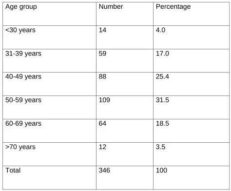
Table 1: Demographic profile: Age wise distribution (N=346)
Age group Number Percentage
<30 years 14 4.0
31-39 years 59 17.0
40-49 years 88 25.4
50-59 years 109 31.5
60-69 years 64 18.5
>70 years 12 3.5
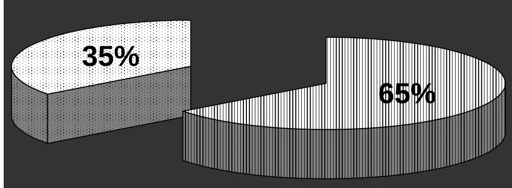
Figure 1: Gender wise distribution
65%
35%
Male
Female
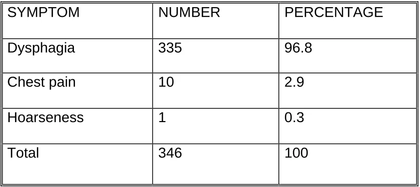
Table 2: Clinical features – symptomatology (N=346)
SYMPTOM NUMBER PERCENTAGE
Dysphagia 335 96.8
Chest pain 10 2.9
Hoarseness 1 0.3
[image:33.612.83.492.119.302.2]Total 346 100
Table 3: Risk factor- smoking (N=346)
SMOKING NUMBER PERCENTAGE
Yes 189 54.6
No 157 45.4
Table 4: Risk factor- alcohol intake (N=346)
ALCOHOL NUMBER PERCENTAGE
Yes 79 22.8
No 267 77.2
Total 346 100
Table 5: Risk factor- family history (N=346)
FAMILY HISTORY NUMBER PERCENTAGE
Present 37 10.7
Absent 309 89.3
Total 346 100
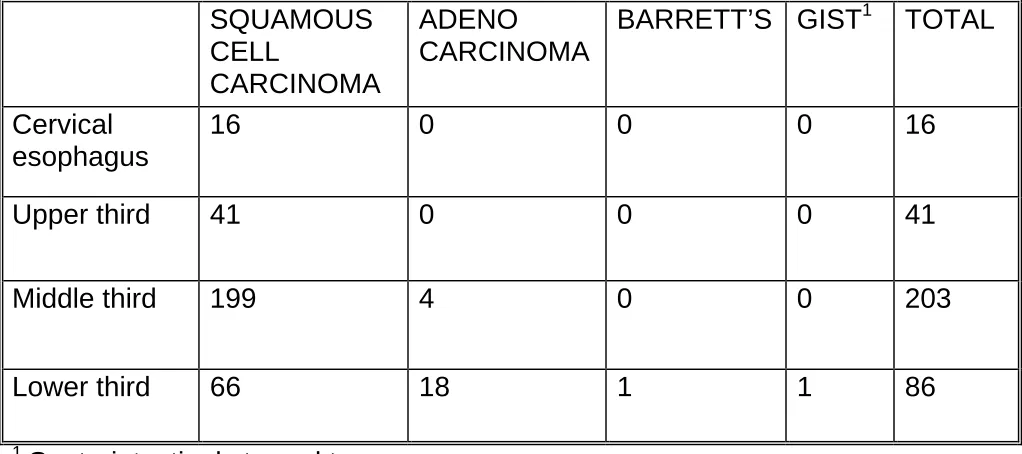
Table 6: Tumor profile according to histology and site
1
Gastrointestinal stromal tumor
Squamous cell carcinoma was the most common histology constituting 92.7% of all the tumors. The mid thoracic esophagus was the commonest site (58.6%).
SQUAMOUS CELL
CARCINOMA
ADENO CARCINOMA
BARRETT’S GIST1 TOTAL
Cervical esophagus
16 0 0 0 16
Upper third 41 0 0 0 41
Middle third 199 4 0 0 203
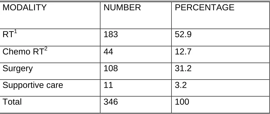
Table 7: Primary treatment offered (N=346)
MODALITY NUMBER PERCENTAGE
RT1 183 52.9
Chemo RT2 44 12.7
Surgery 108 31.2
Supportive care 11 3.2
Total 346 100
1
External beam radiation therapy 6500 cGy
2
Concurrent chemoradiation with cisplatin and 5 FU
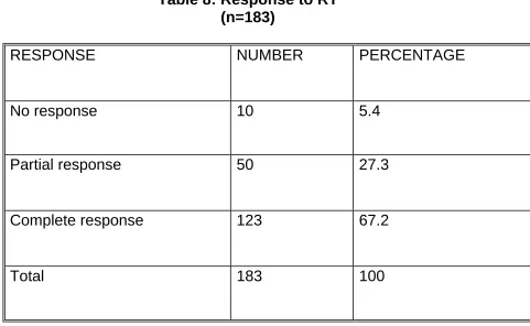
Table 8: Response to RT (n=183)
RESPONSE NUMBER PERCENTAGE
No response 10 5.4
Partial response 50 27.3
Complete response 123 67.2
Total 183 100
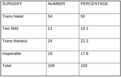
Table 9: Type of surgery performed (n=108)
SURGERY NUMBER PERCENTAGE
Trans hiatal 54 50
Two field 11 10.1
Trans thoracic 24 22.2
Inoperable 19 17.6
Total 108 100
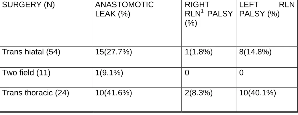
Table 10: Major complications of surgery (n=89)
SURGERY (N) ANASTOMOTIC LEAK (%)
RIGHT
RLN1 PALSY (%)
LEFT RLN PALSY (%)
Trans hiatal (54) 15(27.7%) 1(1.8%) 8(14.8%)
Two field (11) 1(9.1%) 0 0
Trans thoracic (24) 10(41.6%) 2(8.3%) 10(40.1%)
1
Recurrent laryngeal nerve
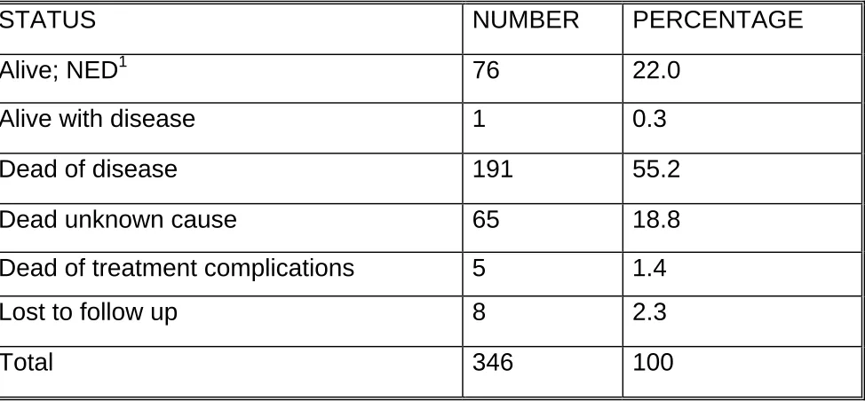
Table 11: Status of the cohort as of June 2006 (N=346)
STATUS NUMBER PERCENTAGE
Alive; NED1 76 22.0
Alive with disease 1 0.3
Dead of disease 191 55.2
Dead unknown cause 65 18.8
Dead of treatment complications 5 1.4
Lost to follow up 8 2.3
Total 346 100
1
No evident disease
Table 12: Sites of failure in the cohort (n=338)
FAILURE SITE NUMBER PERCENTAGE
NED1 76 22.5
Esophagus 148 43.7
Mediastinum 8 2.3
Neck nodes 15 4.4
Liver 9 2.6
Peritoneal deposits 6 1.7
Skeletal metastases 5 1.4
Brain metastases 1 0.3
Pulmonary metastases 2 0.6
Skin nodules 1 0.3
Second primary 3 0.9
Unknown 64 18.9
Total 338 100
1
No evident disease
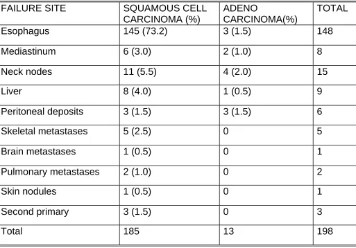
Table 13: Correlation between histology and site of failure (n=198)
FAILURE SITE SQUAMOUS CELL CARCINOMA (%)
ADENO
CARCINOMA(%)
TOTAL
Esophagus 145 (73.2) 3 (1.5) 148
Mediastinum 6 (3.0) 2 (1.0) 8
Neck nodes 11 (5.5) 4 (2.0) 15
Liver 8 (4.0) 1 (0.5) 9
Peritoneal deposits 3 (1.5) 3 (1.5) 6
Skeletal metastases 5 (2.5) 0 5
Brain metastases 1 (0.5) 0 1
Pulmonary metastases 2 (1.0) 0 2
Skin nodules 1 (0.5) 0 1
Second primary 3 (1.5) 0 3
Table 14: Correlation between primary treatment and site of failure (n=271)
FAILURE SITE RT1 CHEMO RT2
SURGERY TOTAL (N)
NED3 19 11 46 76
Esophagus 115 16 17 148
Mediastinum 1 1 6 8
Neck nodes 6 3 6 15
Liver 2 3 4 9
Peritoneal deposits
0 0 6 6
Skeletal metastases
5 0 0 5
Brain
metastases
1 0 0 1
Pulmonary metastases
1 0 1 2
Skin nodules 1 0 0 1
1
External beam radiotherapy
2
Concurrent chemo radiotherapy
3
Table 15: Correlation between primary treatment and present status (N=346) PRESENT STATUS RT1 (%) CHEMO RT2 (%)
SURGERY (%)
SUPPORTIVE CARE (%) Alive; NED 19
(10.4)
11 (25) 46 (42.6) 0 Alive with
disease
0 0 1 (0.9) 0
Dead of disease 121 (66.1)
21 (47.7) 42 (38.8) 7 (63.6) Dead unknown
cause
41 (22.4)
10 (22.7) 14 (12.9) 0 Dead of
treatment complications
2 (1.1) 2 (4.5) 1 (0.9) 0
Total 183 44 108 11a
a
Data unavailable for 4 patients in this group
1
External beam radiation therapy
2
Table 16: Correlation between pathologic tumor stage and present status (n=94)
STATUS T1 (%) T2 (%) T3 (%) T4 (%)
Alive; NED 1(100) 14(77.7) 28(46.6) 2(13.3)
Alive with disease 0 0 1 (1.6) 0
Dead of disease 0 2 (11.1) 20(33.4) 12 (80) Dead unknown cause 0 2 (11.1) 10(16.7) 1(6.7) Dead of treatment
Complications
0 0 1 (1.6) 0
Total 1 18 60 15
Table 17: Correlation between pathologic nodal status and present status (n=91)
STATUS N01
(%) RLN2 (%) PERI ESOPHAGEAL (%) PERI GASTRIC (%)
Alive; NED 26 (59.1)
5 (41.7)
13 (43.3) 2 (40)
Alive with Disease
1 (2.2) 0 0 0
Dead of disease 9 (19.8)
5 (41.7)
12 (40) 3 (60)
Dead unknown cause 7 (15.4) 2 (16.6)
5 (16.6) 0
Dead of treatment complications
1 (2.2) 0 0 0
Total 44 12 30 5
1
Node negative
2
Table 18: Correlation between perinodal spread of tumor and present status (n=88)
STATUS ABSENT (%) PRESENT (%)
Alive; NED 39 (57.3) 6 (35)
Alive with disease 1 (1.4) 0
Dead of disease 16 (23.5) 11(55)
Dead unknown cause 11 (16.1) 2 (10) Dead of treatment complications 1 (1.4) 0
Total 68 20
Figure 2: Factors influencing survival – pathologic tumor status (univariate analysis)
Survival Function
STATUS1 = alive
DFS1 160 140 120 100 80 60 40 20 0 -20 Cu m S u rv iva l 1.2 1.0 .8 .6 .4 .2 0.0 -.2
Pathologic tumor sta
T4
T3
T2
T1
DFS – disease free survival in months
Figure 3: Factors influencing survival – histology (univariate analysis)
Survival Function
STATUS1 = dead
DFS1 140 120 100 80 60 40 20 0 -20 Cu m S u rv iva l 1.2 1.0 .8 .6 .4 .2 0.0 -.2 HISTOL adenocarcinoma SCC
DFS – disease free survival in months.
Figure 4: Factors influencing survival – Nodal status (univariate analysis)
Survival Function
STATUS1 = dead
DFS1 140 120 100 80 60 40 20 0 -20 Cu m S u rv iva l 1.2 1.0 .8 .6 .4 .2 0.0 -.2 PN1 node positive node negative
DFS – disease free survival in months
Figure 5: Factors influencing survival – perinodal spread (univariate analysis)
Survival Function
STATUS1 = dead
DFS1 140 120 100 80 60 40 20 0 -20 Cu m S u rv iva l 1.2 1.0 .8 .6 .4 .2 0.0 -.2 Perinodal spread present absent
DFS – disease free survival in months
a
Data available for 346 patients.
Total number of patients seen in the OP N=818
Number of patients treated N = 368a
[image:52.612.99.562.137.515.2]RT n= 183 Surgery n= 108 Supportive care n=11 Complete response n=123 Partial response n=50 No response n=10 Alive n=20 Dead n=103 Alive n=2 Dead n=48 Dead n=10 Dead n=61 Alive n=46
Esophageal cancer is a malignancy where refinements of surgical technique and the evolution of more radical procedures have not resulted in a proportionate increase in survival. As surgeons become more radical in their approach, the morbidity of the procedures increases and the quality of life of the patients decreases. Hence the thrust of future research will be on minimally invasive surgery, tailoring the nodal dissection according to the nodal status and newer adjuvant and neo adjuvant therapies.
The age and gender distribution of our cohort roughly parallels that of other studies and the report of the IARC (International Association for Research against Cancer)1, 2. Most patients are male and the single largest group belonged to the sixth decade. Also these males who developed esophageal cancer were long term smokers (55%), but the consumption of alcohol (22%) was surprisingly low in contrast to other reports on the etiology of esophageal malignancy 7.
(45%) patients were accepted for treatment. Studies by Pisani et al and data from the National Cancer Institute reflect the same trend wherein about 50% of newly diagnosed patients of esophageal cancer are found to be unsuitable for definitive treatment either due to metastatic disease or due to loco regionally advanced tumors 5, 7. Patients were considered good candidates for surgery on the basis of their performance status (0 or 1) and an objective assessment of their pulmonary function. Those patients who were not willing to undergo surgery were offered radiation therapy or concurrent chemo radiotherapy. As the data covers the period between 1995 and 2004, and the benefit of concurrent chemo radiotherapy had not yet been convincingly demonstrated in the earlier half of the study period, there is a relatively smaller number of patients (12.7%) in the concurrent chemo radiotherapy group.
Third, because some patients treated without surgery are approached with a palliative intent, sub optimal doses of radiation or chemotherapy may have been used.
Many studies have reported results using external beam radiation therapy alone. Most include patients with unfavorable features like T4 disease and multiple positive nodes. The use of radiation as a potentially curative modality requires doses of at least 5000 c GY at 180 to 200 c GY per fraction. Shi and colleagues reported a 33% 5 year survival rate with the use of accelerated fractionation to a total dose of 68.4 Gy 37. However, in the radiation therapy alone arm of the RTOG 85-01 trial (Radiation Therapy Oncology Group), all patients were dead of disease by three years 32.
Though only a small number of patients were treated with concurrent chemo radiation therapy (44), the 5 year survival of this group (25%) correlates well with the results of Herskovic et al(5 year survival of 27%) 32.
The nature of surgery offered was decided by the site of the tumor, loco regional extent, the performance status and the pulmonary function. More than 50% of the patients underwent a trans hiatal esophagectomy, with trans thoracic esophagectomy being performed in 22%.
Hulscher et al randomized patients into two surgical arms: trans hiatal versus trans thoracic. They found that the ICU stay and the hospital stay in the post operative period was significantly more in the trans thoracic group. However, the 5 year overall survival between the two arms was not significantly different (29% versus 39%) 38.
underwent a trans hiatal esophagectomy and in 40% of patients who underwent a trans thoracic esophagectomy.
These percentages are much higher than the published rates of 10-15% of anastomotic leak and about 12-14% recurrent nerve injury in various studies, and can only be attributed to the surgeons’ learning curve.
In spite of the higher incidence of post operative morbidity compared to other published studies, the 5 year overall survival of the surgical arm of the study population is about 42%, which compares well with the best of studies.
A study published by the MD Anderson Cancer Center attempted to determine the difference in natural history and prognosis between squamous cell carcinoma and adenocarcinoma of the esophagus. They found that the incidence of nodal spread and distant metastases was slightly higher in patients with adenocarcinoma. However, the disease free survival and the overall survival was the same in both groups 39.
Mariette et al pointed out that in their study the only factor which predicted disease recurrence and survival was the pathologic tumor status. However they were unable to demonstrate the effect of nodal positivity on survival 3. Tabira et al in their study of patients who underwent trans thoracic esophagectomy determined age, T4 tumors and number of metastatic nodes as the factors which influenced survival 40.
In our study, pathologic tumor status, nodal positivity and peri nodal spread were the factors which affected survival on univariate analysis. However, on multivariate analysis, only peri nodal status seemed to be statistically significant.
The esophagus, mediastinum and the supra clavicular nodes were the most common sites of failure irrespective of the histology and the treatment modality.
explained by the absence of peri nodal spread conferring a relative survival advantage in this subset of patients 41.
The demonstration of an improvement in survival of patients who underwent three field lymphadenectomy by Japanese surgeons needs to be carefully balanced by the additional morbidity of increased vocal cord paralysis.
A total of 818 patients of carcinoma esophagus were seen in the out patient department of our Institute in the period 1995-2004.
Of these, about 368 patients (44.9%) were offered treatment of some kind.
65% of these patients were male and about 31.5% belonged to the sixth decade.
About 97% of the cohort presented with dysphagia.
55% of the patients were smokers whereas only about 23% gave history of long term alcohol intake.
52.6% of all tumors were located in the mid thoracic esophagus and 92.7% of all tumors were squamous cell carcinomas.
31.2% of the cohort were considered suitable for surgery.
Of the 227 patients who were offered radiotherapy or chemo radiotherapy, 157 (69.1%) had a complete response.
Only 13.2% of the patients treated with radiation therapy were alive at 5 years.
129 patients (82.1%) of those who had a complete response were dead at 5 years.
28% of the patients who underwent trans hiatal esophagectomy and 41% of the patients in the trans thoracic esophagectomy group developed an anastomotic leak.
14% of the patients in the trans hiatal group and 41% of the trans thoracic group had vocal cord palsy.
26.7% of the patients in the surgery arm developed loco regional recurrences. Most of these recurrences were in the first three years after the surgery.
About 42.6% of the surgery arm as a whole was alive at 5 years.
78% of patients with pT2 tumors were alive at 5 years when compared to 48% of patients with pT3 tumors and 13% of pT4 tumors.
The median survival of patients with node negative disease was 50 months whereas that of node positive patients was 31 months.
65% of patients with perinodal spread of tumor were dead at 5 years.
This retrospective study of 346 patients of esophageal cancer
demonstrates the dismal survival rates achieved with radiation
therapy alone, which seem to be improved dramatically with the
made on this front however, in view of the small number of patients
administered chemoradiation therapy. There was a high rate of
complications with the surgical arm, though the 5 year survival
achieved approximated the best in available literature. Pathologic
tumor stage, nodal positivity and peri nodal spread were found to be
prognostic on univariate analysis, however only peri nodal extension
of tumor was the only factor found to be statistically significant on
multivariate analysis. Hence a balance needs to be struck between
the survival benefit of extended lymph node dissections and the
consequent complications. Sentinel node biopsy offers a simple and
efficient means of achieving this and further research should be in
1. Madras Metropolitan Tumor Registry, Cancer Institute (WIA). Swaminathan, Shanta V, Rama et al
2. Daly JM, Karnell LH, Menck HR. National cancer database report on esophageal carcinoma. Cancer 1996; 78: 1820.
3. Mariette C, Balan JM, Piessen G et al. Pattern of recurrence following complete resection of esophageal carcinoma and factors predictive of recurrent disease. Cancer 2003; 97: 1616. 4. O’Sullivan, Sheehan D, Clarke A et al. Micrometastases in
esophagogastric cancer: high detection rates in resected rib segments. Gastroenterology 1999; 116: 543.
5. Pisani P, Parkin DM, Bray F, Perlay J. Estimates in worldwide mortality from 25 centers in 1990. Int J Cancer 1999; 83: 870 – 873.
6. Siewert JR, Stein HJ, Feith M, Bruecher BL. Histologic tumor type is an independent prognostic parameter in esophageal cancer. Ann Surgery 2001; 234: 360 – 367.
7. Rier AG, Eisner MP, Kosary C. SEER cancer statistics review 1973 – 1999. Bethesda, Md: NCI 2002.
9. Rice TW, Zuccaro G, Adelstein DJ et al. Esophageal carcinoma: depth of tumor invasion is predictive of lymph node status. Ann Thorac Surg 1998; 65: 787 – 792.
10.Triboulet JP, Mariette C, Chevalier D, Amrount H. Surgical management of carcinoma hypopharynx and cervical
esophagus analysis of 209 cases. Arch Surg 2001; 136: 1164 – 1170.
11.Van Lanschot JJ, Tilanus HW, Voormden MH, Van Deelen RA Recurrence pattern of esophageal carcinoma after limited resection does not support wide local excision with extensive lymph node dissection. BJS 1994; 81: 1320 – 1323.
12. Matsubara T, Ueda M, Katsaki S et al. Localisation of initial lymph node metastases from carcinoma thoracic esophagus. Cancer 2000; 89: 1869 – 1873.
13. Goldmine M, Maddern G, Le Prise E et al. Esophagectomy by transhiatal approach or thoracotomy: a prospective
randomized trial. BJS 1993; 80: 367.
14. Denk W. Radical surgery for carcinoma esophagus (German). Journal Surgery 1913; 40: 1065.
JTCVS 1978; 76: 643.
16. Lewis I. The surgical treatment of carcinoma esophagus. BJS 1946; 36: 18.
17. Mc Keown KC. Total three stage esophagectomy for carcinoma esophagus. BJS 1976; 63: 259.
18. Law S, Wang J. What is appropriate treatment for carcinoma thoracic esophagus. WJS 2001; 25: 189.
19. Skinner DB. En bloc resection for neoplasms of esophagus and cardia. JTCVS 1983; 85: 59.
20. Altorki NK, Skinner DB. Should en bloc esophagectomy be the standard of care for esophageal carcinoma. Ann Surg 2001; 234: 581.
21. Hulscher JB, Tiffsen JG, Obertop H et al. Transthoracic esophagectomy versus transhiatal esophagectomy for carcinoma esophagus – a meta analysis. Ann Thorac Surg 2001; 72: 306 – 313.
22. Rindani R, Martin CJ, Cox MR. Transhiatal esophagectomy versus Ivor Lewis esophagectomy: is there a difference? Aust NZ JS 1999; 69 (3): 187 – 194.
control trial of extended cervical and superior mediastinal lymphadenectomy for carcinoma thoracic esophagus. Ann J Surg 1998; 175: 47.
24. Akiyama H, Tsurumaru M, Udagawa H et al. Radical lymph node dissection for carcinoma thoracic esophagus. Ann Surg 1994; 220 – 364.
25. Altorki NK, Girardi L, Skinner DB. En bloc esophagectomy improves survival for stage three esophageal cancer. JTCVS 1997; 114: 948.
26. Altorki NK, Kent M, Ferrara C et al. Three field lymph node dissection for squamous cell carcinoma and adenocarcinoma of esophagus. Ann Surg 2002; 236: 177.
27. Kelsen DP, Ginsberg R, Pajak TF et al. Chemotherapy followed by surgery compared with surgery alone for esophageal cancer. NEJM 1998; 339: 1979 – 1984.
28. Clark P. Clinical Trials Unit Medical Research Council. Surgical resection with or without preoperative chemotherapy in
esophageal cancer. Proceedings of American Society of Clinical Oncology 2001; 20: 502.
multimodal treatment and surgery for esophageal adenocarcinoma. NEJM 1996; 335: 462 – 467.
30. Basset JF, Gignoux M, Triboulet JP et al. Chemoradiotherapy followed by surgery compared with surgery alone in squamous cell carcinoma of the esophagus. NEJM 1997; 337: 161 – 167. 31. Urba SG, Orringer MB, Turissi et al. Randomized trial of
preoperative chemoradiotherapy versus surgery alone in patients with locoregional esophageal carcinoma. JCO 2001; 19: 305 – 313.
32. Herskovic A, Martz K, Al -Sarraf M et al. Combined
chemotherapy and radiotherapy compared with radiotherapy alone in patients with carcinoma esophagus. NEJM 1992; 326: 1593 – 1598.
33. Morton DL, Wen DR, Waz JH et al. Technical details of intra – operative lymphatic mapping for early stage melanoma. Arch Surg 1992; 127: 392 – 399.
34. Ketagawa Y, Fujii H, Mukai M et al. The role of the sentinel lymph node in gastrointestinal cancer. SCNA 2000; 80: 1799 – 1809.
Barett’s and cardia cancer. Ann Surg Oncol 2004; 11 (3): 255 – 258.
36. Kato H, Miyazaki, Nakajuma et al. Sentinel lymph node with Tc 99m colloidal Rhenium sulfide in patients with esophageal cancer. Cancer 2003; 98: 932 – 939.
37. Shi X, Yao W, Liu T. Late course accelerated fractionation in radiotherapy of esophageal carcinoma. Radiother Oncol 1999; 51: 21.
38. Hulscher, Johana, Van Sandich et al. Extended transthoracic resection compared with limited transhiatal resection for adenocarcinoma esophagus. NEJM 2002; 347: 1662 – 1669. 39. Rohatgi PR, Swisher SG, Correa AM et al. Comparison of stage, therapy response and patient outcome between
squamous cell carcinoma and adenocarcinoma of esophagus. Int J Gastroint Cancer 2005; 36 (2): 69 – 76.
40. Yoichi Tabira, Toshitada Okuma, Keiichiro Kondo et al. Indications for three field dissection followed by
esophagectomy for advanced carcinoma of the thoracic esophagus. JTCVS 1999; 117: 239 – 245.
esophagectomy with two – field lymphadenectomy. BJS 2000; 87: 1426 – 1433.