CERTIFICATE
This is to certify that the dissertation titled “Hepatotoxicity in patients receiving HAART therapy: Clinical Profile & Risk factors” is a genuine work done by Dr. P. Karthikeyan, Post graduate in Medical Gastroenterology under my supervision between June 2006 to February 2007 and is being submitted in partial fulfillment of the requirement for the awarding of D.M. (Medical Gastroenterology) degree by the Tamil Nadu Dr. M.G.R. Medical University, Chennai.
Dean
Madras Medical College, Chennai – 600 003
Prof. S. Barnaba Durairaj, MD., DM Department of Medical Gastroenterology Madras Medical College,
ACKNOWLEDGEMENT
I thank Dr.Kalavathy Ponniraivan B.Sc., M.D., former Dean, Madras Medical College, for giving the permission to carryout the study.
ACKNOWLEDGEMENT
I am extremely thankful to my guide and mentor Dr. S. Barnaba Durairaj, M.D. D.M., Professor and Head, Department of Medical Gastroenterology for his valuable comments, constant enthusiasm, and support he has shown me through out the study period
I thank Prof. P. Padmanaban, M.D., D.M., Additional Professor for his expert guidance, suggestions and constructive criticism which were invaluable for my study. My special thanks to him for his painstaking effort in correcting the manuscript in a short period of time.
My sincere thanks to my Assistant professors of the department Dr. P. Ganesh, Dr.K. Narayanaswamy, Dr. K. Premkumar, Dr.K.Caroline Selvi for their words of encouragement and support they offered during difficult periods.
I express my gratitude to Mrs. Supriya Sahu I.A.S, Project Director, TNSACS, Chennai for her appraisal of the study and permission to carry out the study.
I thank Dr.V.Raji M.D., Former Professor of Medicine, and Institute of Internal Medicine for her permission for the utilization of ART clinic records and services. I also thank Dr. Muthuselvan M.D, Dr. Sekar in- charge of ART clinic for their help during the study.
I thank my colleagues Dr. Kandasamy @ Kumar, Dr. G.Ramkumar, Dr. Rema Krishnakumar, Dr. Antony Joe and Dr.Mahadevan for their cooperation, enthusiasm and support. I thank my friends Dr.Deena Sangeetha and Dr. Paranthaman Sethupathi for their help. I thank Mr. Venkatesan for his help in statistical analysis
CONTENTS
CHAPTER
TITLE
P.No.
1. INTRODUCTION 1
2. REVIEW OF LITERATURE 12
3. AIM OF STUDY 29
4. MATERIALS & METHODS 30
5. RESULTS 36
6. DISCUSSION 41
7. CONCLUSION 46
8. SUMMARY 47
9. APPENDIX 48
1. INTRODUCTION
HIV infection/AIDS is a global pandemic with cases reported from virtually every country. The current estimate of cases among adults is estimated to be around 37 million. Two thirds of them live in Sub Saharan Africa and 50% are women. The prevalence ranges from 0.6% to 8% in different parts of the world.1
Gastroenterological manifestations are quite common in advanced HIV infection/ AIDS and are some times initial manifestations or AIDS defining illness. Liver involvement is unique since it can be because of direct involvement, opportunistic infections (OI ) with r co-infection of one or more hepatotrophic viruses, or because of toxicity due to retroviral drugs.2
HEPATIC MANIFESTATION OF HIV/AIDS
Presence of liver disease is a frequent finding in AIDS. Hepatomegaly may be detected on examination in most patients. Hepatomegaly is usually associated with one or more liver chemistry test abnormalities, although significant jaundice due to parenchymal disease is uncommon. As with other organ systems, the spectrum of hepatic infections in patients with HIV evolves as immunocompromise advances. Clinical manifestations of hepatobiliary disease can vary from no symptoms to liver failure.
Drug-induced liver injury has emerged as the most prevalent cause of liver test
abnormalities and is related to the increasing array of antiretroviral medications. Use of prescription or nonprescription drugs as well as herbal remedies should also be considered a cause of abnormal liver test results in the HIV-infected patient. Before HAART, drug hepatotoxicity was most commonly due to sulfonamides, and the increased frequency of adverse reactions to these medication is well recognized in AIDS.
Well established syndromes characterized by marked hepatomegaly, steatosis, lactic acidosis, and liver failure has been increasingly recognized.Hepatic steatosis is usually evident on imaging of the liver. Although reversal has occurred in some patients following drug withdrawal, most patients have worsening disease and death. Liver transplantation is curative.
The incidence of drug induced hepatotoxicity, its mechanisms, evaluation, management, analyses of various studies on the risk factors and prognosis are described in detail in literature review. Other causes of liver dysfunctions are reviewed below and should be kept in mind before evaluating these patients.2
INFECTIONS
Mycobacterium avium intercellulare complex (MAC) is consistently the most frequent
Mycobacterium. tuberculosis, in contrast to MAC, may occur before HIV-infected
patients are profoundly immunocompromised. Extrapulmonary tuberculosis is common in patients with HIV infection, especially in patients with prior OIs and those whose risk behavior is injection drug use. Hepatic disease as part of miliary tuberculosis has been noted. Rarer manifestations include tuberculous abscesses and bile duct tuberculomas. The diagnosis of hepatic tuberculosis is made by culture of the organism from liver tissue obtained by percutaneous or laparoscopic biopsy. PCR may be helpful.
CMV is the most frequent infectious pathogen in AIDS, and the liver is involved in 5% to
25% of liver biopsies. However, its discovery in the liver antemortem is extremely unusual. Typical viral inclusions are usually identified in Kupffer cells .
Clinical manifestations and histologic features of viral hepatitis from HBV, HCV, or
hepatitis D virus are altered in the presence of HIV coinfection but in remarkably different ways for each virus.
Clinical and autopsy studies in AIDS patients have reported up to a 90% seroprevalence of hepatitis B markers indicating past or present infection.
Concurrent HIV and HBV infections lead to alterations of HBV antigen-antibody display, viral replication, and clinical consequences. Several reports have described reappearance of hepatitis B surface antigen (HBsAg) in HIV-infected patients previously thought to be immune to hepatitis B virus as indicated by the presence of anti-HBs.5
anti-HBs even in those patients who remain anti-HBsAg negative. With loss or reduction in immunity to HBV, there is an increased prevalence of hepatitis B e antigen expression, elevated mean levels of DNA polymerase, and increased titers of anti–hepatitis B core antigen.6
Acquisition of the chronic carrier state is also much more likely in the HIV-infected patient, especially if infection occurs when immunodeficiency is more advanced. Thus, a larger proportion of patients with HIV and hepatitis B infections have a chronic carrier state, with highly infectious serum and body fluids, compared with those who are HIV negative
Although HIV infection leads to more prevalent chronic HBV carriage, it appears to attenuate the severity of biochemical and histologic liver disease in most, but not all1 patients. In one study, the mean alanine aminotransferase (ALT) level correlated with CD4 lymphocyte count. The mechanism for reduced hepatitis B virus–related liver injury following HIV infection is not certain but has been attributed to a diminution in lymphocyte-mediated hepatocellular injury as a result of HIV effects on lymphocytes. In those patients without serologic evidence of past or present hepatitis B virus and HIV infection, vaccination appears to be ineffective, regardless of the stage of immunocompromise.
lamivudine, which has potent antiviral effects on hepatitis B virus, in the HAART regimen may reduce the likelihood of acute hepatitis B.6
The consequences of HIV infection on delta hepatitis appear similar to those of HBV, although far fewer patients have been studied.
The prevalence of HCV in those with HIV infection depends on the risk group evaluated and the assay used. Prevalence is highest in injection drug users (52% to 89%)7,8 and
hemophiliac patients with HIV, whereas in military populations and non–drug users, the prevalence is much lower, ranging from 1% to 11%. Assaying antibodies to hepatitis C virus alone, rather than hepatitis C virus RNA, may underestimate the true prevalence, because loss of antibody may occur with progression of immunodeficiency.5, 6
The effect of HAART on hepatitis C viral dynamics and liver injury is variable. Some studies have found attenuation of disease, whereas others had documented exacerbations reflected by increases in serum transaminases. Hepatitis C viral load has also been variably affected. 8
The role of interferon therapy for HIV/HCV coinfected patients remains unsettled. α-Interferon is less effective for treating hepatitis C virus liver disease in coinfected patients . More recently, combination therapy of Peg-interferon and ribavirin has shown promise. 10
Fungal infections of the liver are not unusual when immunocompromise is advanced.
Histoplasmosis of the liver may be seen in patients with disseminated fungal disease,
predominantly but not exclusively in regions of high prevalence of the organism. Biopsies of the liver may also show caseating granulomas containing fungal organisms. Culture of hepatic tissue, blood, or bone marrow can confirm the diagnosis, but several weeks may be required for the organism to grow in culture.
Pulmonary disease is seen in most patients at diagnosis.Cryptococcus may infect the
liver in the setting of disseminated infection. Typically the organism is found in the sinusoids and is associated with a poor inflammatory response. Similarly, coccidioidomycosis can involve the liver as part of a systemic infection, especially in endemic regions. The organisms appear as spherules within a fibrosing granulomata.Candida infection of the liver is rare, in contrast to its high prevalence in
Kaposi's sarcoma, which is caused by infection with human herpesvirus 8 (HHV-8) has a
predilection for periportal regions of the liver and is seen in 10% to 15% of liver biopsies.
Tumor nodules appear grossly as violaceous or hemorrhagic masses within hepatic parenchyma. Microscopically, the characteristic spindle cells and vascular slits of Kaposi's sarcoma usually directly abut normal-appearing liver tissue.
Hepatic involvement by non-Hodgkin's lymphoma may be the index manifestation of
AIDS in homosexual men and may be the primary site of the neoplasm. The lesions are usually focal and may be large. In addition, Hodgkin's disease in the AIDS patient tends to be more aggressive histologically and clinically, spreading rapidly to extranodal sites, making liver involvement more likely.
Isolated cases of P. carinii pneumonia (PCP) hepatitis have been described and are
attributable to the use of inhaled pentamidine, which fails to protect extrapulmonic sites from PCP. In addition to PCP, the liver may be the site of infection by the protozoa
Cryptosporidium, Microsporidium, or Dicrocoelium dentriticum or by other multicellular
organisms.
Bacillary peliosis hepatis, may be caused by either Bartonella henselae or Bartonella Quintana.
the findings are similar to those seen in patients with kwashiorkor. Massive steatosis has also been observed from antiretroviral drug use (lipodystrophy syndrome).
Biliary tract involvement in AIDS may result in marked liver test abnormalities and right
upper quadrant symptoms; jaundice is unusual. A syndrome resembling sclerosing cholangitis with papillary stenosis is well recognized and has been termed AIDS cholangiopathy. Patients characteristically develop significant upper abdominal pain in
association with marked elevation of alkaline phosphatase, and minimal elevations of bilirubin, AST, and ALT.
Ductular changes may consist of either papillary stenosis alone, sclerosing cholangitis– like lesions alone, a combination of the two, or long extrahepatic strictures. Most series have found papillary stenosis with intrahepatic disease as the most common findings. Ultrasonography or CT detects ductular abnormalities in 77% of those with cholangiographically proven disease, implying that a negative imaging study does not definitively exclude the diagnosis. The etiology in most cases is infectious because
Cryptosporidium, CMV, or Microsporidium may be found in bile, duodenal, or biliary
epithelium. For patients with predominantly papillary stenosis, sphincterotomy results in a symptomatic improvement in most patients; alkaline phosphatase may continue to rise, however, probably reflecting progression of associated intrahepatic disease.
EVALUATION
The initial decision in evaluating the AIDS patient with jaundice, hepatomegaly, or both, is to determine whether the findings are due to intrahepatic or extrahepatic disease. Simultaneous disease in both sites must also be considered. A history of mild jaundice, often in association with fever and constitutional symptoms, is more consistent with intrahepatic disease, whereas symptoms of deep jaundice associated with pain of relatively acute onset suggest extrahepatic disease. Careful review of medications, both prescription and nonprescription, is essential.
Because the clinical history and the finding of symptomatic hepatomegaly are nonspecific, further evaluation is always necessary. Elevations of ALT or AST or both, are common, but neither the pattern nor the extent of elevation of these tests appears to correlate with specific findings in the liver. Significant elevation of the transaminases favors a drug-induced or viral cause. In contrast, marked elevation of alkaline phosphatase correlates statistically with the presence of MAC infection in the liver in AIDS when extrahepatic obstruction is absent. CT scan and ultrasonography should be employed early because they are especially useful in identifying ductal dilation, gallbladder pathology, and focal hepatic lesions.
The indications for liver biopsy for the patient in whom intrahepatic disease is suspected are not well defined. Biopsy is appropriate when symptomatic, treatable disease of the liver is suspected and when a specific diagnosis of hepatic disease is needed.
Table 1.1 : Major cause of liver injury in HIV infection Drugs
NRTI, NNRTI, PI Anti microbials
Antituberculosis , INH, rifampicin
Macrolides : clarithromycin, azithromycin Antifungal : KTZ, itraconazole, fluconazole
Anti pneumocystitis carini drugs : TMP-SMX, dapsone Infections
Viral hepatitis
HAV, HBV, HCV, CMV, HSV, EBV
Mycobacteria,Mycobacterium avium, M tuberculosis Fungal
Cryptococcus, histoplasma, coccidioides, candidia Protozoa
Pneumocystitis, toxoplasma,microsporidia, cryptosporidium
Biliary infections
HIV cholangiopathy Acalculouscholecystitis
Neoplasm and vascular lesions Kaposi sarcoma
Lymphoma Peliosis hepatitis
Steatosis and lipodystrophy HCV co-infection
2. REVIEW OF LITRETURE
Since its introduction over 10 years ago, highly active antiretroviral therapy (HAART) has dramatically changed the course of human immunodeficiency virus (HIV) infection by decreasing morbidity and mortality as a result of opportunistic infections. However, HIV-infected patients are now experiencing a wide array of adverse events attributed to the drugs themselves. Liver toxicity is an important example because it carries its own morbidity and mortality. Perhaps more importantly, it often leads to HAART discontinuation
HAART INDUCED LIVER DISEASE
Highly active antiretroviral therapy (HAART) has decreased the morbidity and mortality derived from classical opportunistic infections. As a counterweight to this positive impact, antiretroviral therapy (ART) carries along undesirable effects, which challenge the management of HIV-infected patients to a great extent. Among these, liver toxicity deserves a special attention since it often leads to HAART discontinuation; particularly in hepatitis C (HCV) and/or hepatitis B (HBV) co-infected patients. The mechanisms involved in HAART-derived liver toxicity are not well understood, which makes its management more difficult
Possible pathogenic mechanisms involved in hepatotoxicity are multiple, including direct drug toxicity, immune reconstitution in the presence of HCV and/or HBV co-infections, hypersensitivity reactions with liver involvement, and mitochondrial toxicity. Other pathogenic pathways may be involved, such as insulin resistance caused by several antiretrovirals, which may contribute to the development of steatohepatitis. The management of liver toxicity is based mainly on its clinical impact, severity and pathogenic mechanism.
CLINICAL IMPACT
With the widespread use of HAART and the availability of more drugs, some of them perhaps more hepatotoxic, HAART-linked hepatotoxicity has been made evident over the past few years. Liver toxicity generates medical visits, work-up exams, and frequent hospital admissions, all of which increase expenses. In addition, hepatotoxicity hampers the maintenance of HIV suppression over time.
In a recent American study, which evaluated the causes of death of HIV-infected individuals, discontinuation of ART due to hepatotoxicity increased from 6% in 1996 to 31.8% in 1998-1999 among those mortalities . More recently, Kramer and colleagues have highlighted the increase in the number of cases of fulminant liver failure in HIV/HCV-coinfected individuals during the HAART era, even after excluding patients with advanced liver disease and adjusting by alcohol intake.
those with prior liver disease, most cases of liver toxicity is mild-to-moderate and asymptomatic .
Drug liver toxicity has impacted on the recommendations for antiretroviral therapy in certain scenarios. Thus, the use of nevirapine (NVP) has been recommended to be avoided as part of post-exposure prophylaxis regimens. The reason for that was the occurrence of fulminant hepatitis in two cases and severe liver toxicity in 12 other healthy subjects who received a NVP-including HAART regimen after HIV exposure. However, NVP seems to be safe when administered to mother and child as a single dose for prevention of mother-to-child HIV transmission . 3,4
DEFINITION OF LIVER INJURY
The clinician thinks of liver damage when abnormalities in the liver tests are seen. There is a broad variability among studies in the criteria to categorize the severity of hepatotoxicity. the most accepted one, the AIDS Clinical Trials Group scale of liver toxicity .
DEFINITIONS OF HAART-ASSOCIATED HEPATOTOXICITY:
15The AIDS Clinical Trials Group currently uses the following toxicity grading scale:
Patients with normal pretreatment ALT/AST:
Grade 0 hepatotoxicity <1.25 times the ULN (upper limit of normal)
Grade 1 hepatotoxicity 1.25 to 2.5 times the ULN
Grade 2 hepatotoxicity 2.5 to 5 times the ULN
Grade 3 hepatotoxicity 5.1 to 10 times the ULN
Grade 4 hepatotoxicity >10 times the ULN
There is a separate grading scale for the HAART-associated cholestasis:
Grade 0 cholestasis <1.1 times the ULN
Grade 1 cholestasis 1.1 to 1.5 times the ULN
Grade 2 cholestasis 1.6 to 2.9 times the ULN
Grade 3 cholestasis 3 to 5 times the ULN
Grade 4 cholestasis >5 times the ULN
Severe hepatotoxocity is defined as grade 3 or 4 change in transaminase levels, while severe cholestasis, analyzed independent of transaminase levels, is defined as grade 3 or 4 change in total bilirubin.
Many drugs increase γ-glutamyltranspeptidase (GGT) levels. This is often misinterpreted as a marker of liver damage, but the isolated elevation of this enzyme actually reflects enzyme induction. Only when associated with a proportional increase in alkaline phosphatase levels should it be considered as a cholestatic lesion. Bilirubin should not be considered itself as indicator of liver toxicicity it can be elevated due to a variety of reasons, such as hemolysis, fasting and certain drugs (e.g. indinavir and atazanavir). In addition to HAART-derived hepatotoxicity, some other conditions or drugs used in HIV infection, can cause elevations in the levels of liver enzymes and should be ruled out.
INCIDENCE AND RISK FACTORS
The reported incidence of severe liver toxicity after initiating HAART ranges from 2 to 18% . Differences in the study populations, as well as in the methods used probably account for the wide range. In Tables 1 and 2, which summarize the main trials assessing liver toxicity in patients taking antiretroviral therapy, the risk factors are recorded.
Hepatitis B and C co-infections 14
higher response to interferon (IFN) and ribavirin (RBV), anti-HCV treatment should be given if no major contraindication is present.
ANTIRETROVIRALS
The results of the studies that have evaluated the risk for liver toxicity associated with the use of particular antiretroviral drugs or families are conflicting. The unbalanced and often insufficient representation of some antiretrovirals in these series, make it difficult to determine with accuracy the role of each particular drug in the development of liver toxicity. In addition, the use of several antiretrovirals combined makes it difficult to ascribe the elevation of transaminases to single drugs. 16
Protease inhibitors
The phenomenon of hepatotoxicity became more evident after the introduction of ART of high activity, which initially included invariably a protease inhibitor (PI). However, none of the studies has been able to prove the higher potential for liver toxicity of this particular family of drugs. Among the PI, in some studies full-dose ritonavir (RTV) has been found to be more hepatotoxic .In certain cases, RTV has caused fatal acute hepatitis. Several cases of liver toxicity associated with the use of indinavir (IDV) and saquinavir (SQV) have also been reported . Nelfinavir was found to be less hepatotoxic than the other PI anlyzed .
more hepatotoxic, most probably because it is given with higher doses of RTV (400mg/day).
Nucleoside analogues reverse transcriptase inhibitors (NRTI)
Some authors have found a lower incidence of hepatotoxicity with lamivudine (3TC) and tenofovir . However, the majority of the NRTI can induce mitochondrial damage, and, therefore, have a potential for the development of liver injury, as it will be explained below . Cases of hepatic failure have been reported in patients taking zidovudine, but didanosine and stavudine have been most often involved in severe hepatotoxicity . Abacavir (ABC) and tenofovir (TDF), with low potential for mitochondrial damage, seem to have a safer profile regarding the liver. In patients with chronic hepatitis B, the removal of 3TC may be accompanied by a flare of HBV replication, translated into an increase in transaminases.
Non-nucleoside analogues reverse transcriptase inhibitors
with an increase in the cumulative incidence over time . Therefore, it looks like there is a second mechanism through which NVP causes liver toxicity, much more common than the hypersensitivity syndrome.
Table 2.1 : Liver toxicity of commonly used anti- HIV medications
Dug type Drug name Pattern of injury Comments Protease inhibitor Indinavir
Saquinavir Nelfinavir Ritonivir Hepatocellular, distinct histologic pattern including hepatocyte ballooning,Kuppfer cell activation, pericellular zone 3 fibrosis
< 10% of patients have transaminases > 5 ULN Ritonavir inhibits P 450
NRTI ddC d4T ddl AZT Microvesicular steatosis Mitochondrial toxicity manifexting as lactic asidosis
NNRTI Nevirapine Efavirenz
Hepatocellular NVP associated
with grade 4 toxicity; FDA alert
Other factors
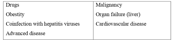
Table 2.2 : Common causes and risk factors Drugs
Obestity
Coinfection with hepatitis viruses Advanced disease
Malignancy
Organ failure (liver) Cardiovascular disease
MECHANISMS OF LIVER TOXICITY
Despite the numerous published studies on antiretrovirals and hepatotoxicity, many unanswered questions still remain, in particular those related to the mechanisms involved. The possible mechanisms involved in the development of hepatotoxicity associated with the use of antiretrovirals are summarized in Fig. 1. It is probable that multiple pathogenic pathways simultaneously concur in some patients, being difficult to identify the exact mechanisms involved in the development of hepatotoxicity.
Table 2.3 : Mechanisms of hepatotoxicity
Direct toxicity
Hypersensitivity reaction Mitochondrial toxicity Metabolic abnormalities
Direct toxicity
Antiretrovirals, as any other drug, can induce direct toxicity in the liver. Drugs metabolized in the liver through the cytocrom pathways may cause liver toxicity when there are polymorphisms in the enzymes . Since many of the antiretrovirals are metabolized in the liver through the cytocrome pathways, idiosyncratic polymorphisms of the enzymatic complexes might lead to significant heterogeneity in drug metabolism, predisposing to the development of hepatotoxicity in certain individuals. Some drugs may potentiate the activation of death receptors and/or intracellular stress pathways.
Hepatocytes promote mechanisms of cytoprotection against the oxidative stress caused by drug metabolism. Heat-shock proteins, induced by various forms of stress including drugs, may exert cytoprotective functions helping to tolerate potentially damaging toxicants . An increase in heat-shock proteins in individuals with polymorphisms may help the liver adapt to and minimize drug toxicity. Anti-oxidation stress mechanisms might explain the spontaneous normalization in the levels of transaminases despite maintenance of HAART, as it occurs with INH. Although still early in its development, pharmacogenomics is a new approach, which may be very valuable to predict the risk for hepatotoxicity in each individual after initiation of ART .
Hypersensitivity reactions
In patients taking NVP, the incidence of symptomatic events involving the liver has been reported to be of 4.9%. In some occasions fatal outcomes were linked to the use of NVP in women with CD4 counts >250cells/mm within 6 weeks after initiation of treatment, due to hypersensitivity reactions. According to a recent analysis, a low body mass index is an independent risk factor for this type of events.
Immune mediated drug reactions seem to involve the generation of neoantigens formed by the reaction of liver proteins with reactive drug metabolites . Assays examining in vitro activation of peripheral blood mononuclear cells against a drug or its metabolites are currently under research. These assays are a promising approach to identify susceptible individuals . On the other hand, individuals with the HLA-DRB1*0101 marker, especially if they have <25% CD4 cells, have been shown to be at greater risk of developing NVP-induced hypersensitivity reactions.
Hypersensitivity reactions have been reported relatively often with NVP and abacavir (ABC), both in infected patients and in subjects receiving prophylaxis after HIV-exposure , but also with other antiretrovirals such as zalcitabine (ddC) .
Mitochondrial toxicity
lesion . The ability of NRTI to inhibit mitochondrial DNA synthesis in vitro is in the following order: ddC, ddI, d4T, AZT, 3TC=ABC=TDF . Hydroxyurea, used as coadjuvant treatment with ddI to enhance its activity, seems to increase the toxic effect of some NRTI.
Metabolic abnormalities
Steatohepatitis may cause hypertransaminasemia. Insulin resistance is believed to be the metabolic hallmark of predisposition to non-alcoholic steatohepatitis (NASH). HAART may cause, in the context of the lipodystrophy syndrome, marked abnormalities in the metabolism of both lipids and glucose, including insulin resistance . Mild-to-moderate degrees of steatosis have been found in the liver of patients experiencing HAART-derived hepatotoxicity . Thus, in some patients receiving HAART, insulin resistance and NASH may contribute to the development of liver toxicity. 18
Studies in HIV-negative individuals find rosiglitazone and pioglitazone can reduce risks associated with metabolic syndrome and can reduce risks for cardiovascular disease associated with insulin resistance and diabetes, as well as in non-diabetics. Preclinical studies suggest that atazanavir may reduce risk for glucose anmormalities in HIV+ patients who have not yet developed glucose abnormalities compared to other PIs associated with risk for glucose abnormalities.
Immune reconstitution in HCV and/or HBV-infected patients
response to HCV and/or HBV antigens exposed in the liver cell is also restored. Thus, HAART therapy may induce the development of hypertransaminasemia and even symptomatic hepatitis in patients with HCV or HBV co-infection.
[image:29.612.162.454.433.631.2]The immune reconstitution syndrome as a mechanism of liver toxicity is controversial, but there are several data supporting it. Markers of HCV-specific immune responses (HCV core-specific IGG antibody), T-cell activation and inflammation, have been found to correlate with liver damage and immune reconstitution. Some authors have identified an increase in >50 CD4 cells per mm3 after initiating HAART as an independent risk factor for hepatotoxicity . Liver histology has shown exacerbated viral hepatitis in some patients developing severe hypertransaminasemia while on HAART .
FIG. 2.1 : MECHANISMS OF HEPATOTOXICATY
ALT
HBV HCV
METHODONE
ALCOHOL
STEATOSIS
MITOCHONDRIAL TOXICITY
ART
HIV
Insulin resistance Immune
reconstitution
hypersensitivity
Mitochondrial injury
THERAPEUTIC MANAGEMENT
The three main considerations necessary for the management of transaminase elevation after the introduction of HAART are severity, clinical impact and etiologic mechanisms. Suspicion of hypersensitivity reactions or lactic acidosis, and the presence of liver decompensation, are all reasons to stop treatment]. Severe liver toxicity (grades 3-4), even in the absence of symptoms warrants discontinuation of the antiretroviral therapy.
FIG 2.2 : THERAPEUTIC ALGORITHM
Management
Management of HAART-related hepatotoxicity is complicated by the desire of the practitioner to minimize adverse effects while at the same time relying on the chronic use of potentially hepatotoxic agents to prevent life threatening complications from immunodeficiency. Early withdrawal of therapy is recommended for patients who develop symptomatic or severe hyperlactatemia because of a nearly linear association between serum lactate values and mortality. There are anecdotal reports of the use of antioxidants such as riboflavin, L-carnitine, and coenzyme Q-10 (ubiquinone). No controlled studies are available to evaluate these reports. After serum lactate levels and LFTs normalize, reinitiation of retroviral therapy without NRTIs or utilizing NRTIs which have a lower likelihood of causing lactic acidosis, such as lamivudine, abacavir, and tenofovir.18
Liver enzymes At 4-6 weeks
high normal
>10X ULN <10
Symptoms of acute hepatitis, Rash NVP ABC
Yes No
Stop immediately Continue With monitoring
Repeat every 3 months
>10X ULN or
Lactate > 5 mmol/L XULN<10
Stop
immediately alternate regimen Continue with
ASSESSMENT OF LACTIC ACIDOSIS IN PATIENTS WITH HIV Table 2.4 : Signs and symptoms
Nausea Vomiting Weakness Abdominal pain Diarrhea
Malaise Anorexia Dyspnea
[image:32.612.154.477.144.299.2]Cardiac arrhythmia (fatal)
Table 2.5 : Management
Discontinue ART
Deliver adequate oxygen to tissues Reduce O2 demand
Mechanical ventilation
3. AIM OF THE STUDY
The study was conducted with the objective of
i. estimate the incidence of drug induced hepatotoxicity in patient receiving HAART therapy for HIV/AIDS
4. MATERIALS & METHODS
About 1523 patients who receive ART were screened and patients with evidence of liver dysfunction were isolated. Sample population was selected as follows.
50 adult patients of both sexes infected with HIV and fulfill the WHO criteria for clinical AIDS receiving HAART for a period of more than 1 month were included in the study.
PATIENT SELECTION
Inclusive criteria
Patients with HIV infection
i. who receive HAART therapy for > 1 month
Exclusion criteria
i. Patients with base line LFT abnormal
ii. No evidence of extrahepatic cause of jaundice iii. No past history of jaundice
iv. No clinical evidence of liver disease at the institution of HAART v. Antenatal mothers
PROTOCOL
1. All patients who receive HAART therapy who met the above criteria were included in the study
2. The following were noted in each patient
i. Age
ii. Sex iii. BMI
iv. Alcohol usage v. Smoking
vi. H/o jaundice in the past vii. Drug allergy
viii. Diabetes
ix. Tuberculosis (pulmonary/ extra pulmonary) x. Mode of acquisition of HIV
xi. Nadir CD4 count xii. Latest CD4 count xiii. Socio economic status xiv. Dose of each drug
xv. Duration of therapy of each drug xvi. Drug withdrawal
xvii. Other adverse reaction with the drug xviii. Anti tubercular therapy (ATT) xix. Anti fungals
3. Base line LFTs were measured
4. Those who have abnormal LFT following were noted
i. USG
ii. HBsAg, anti HCV antibodies iii. Prothrombin time
iv. Chest X ray
v. Complete blood count vi. Bleeding time
THE STUDY
Study design Retrospective Analytical Study Venue GGH, Chennai
Duration 6 months
Collaborating departments
ART Clinic, Govt. General Hospital, Chennai
ART Clinic, Govt. Hospital for Thoracic Medicine, Tambaram ART Clinic, Kilpauk Medical College Hospital, Chennai Liver Clinic, Govt. General Hospital, Chennai
STASTICAL ANALYSIS
RISK FACTORS FOR HEPATOTOXICITY IN PATIENTS ON
HAART
PROFOMA
S.NO.
NAME AGE SEX UNIT
OCCUPATION ADDRESS
CONTACT NO.
HOSPITAL NO
SYMPTOMS
PRIMARY SYMPTOMS GI SYMPTOMS
Fever Jaundice Loss of appetite Loss of weight Pruritus GI bleed
PAST HISTORY Jaundice
Smoking
Tobacco usage of other means IVDU
Drug abuse Marital status
Promiscuity M2F / M2M
SIGNS General
Weight height Nutrition BMI Pallor
Oral lesions Jaundice
Evidence of liver cell failure Ascites
Cutaneous bleed Hepatic encephalopathy
DURATION OF HAART
NO OF DRUGS DOSE
Drugs Started on Stopped on Dose
ATT
Drugs Dosage Duration Comments
OTHER ANTIVIRALS ANTIFUNGAL
ANTIBIOTICS
OTHER DRUGS
CAM
PAST H/O JAUNDICE
ALCOHOL Y/N DETAILS SMOKING
IV DRUG ABUSE
NUTRITIONAL STUATUS BMI
PRETREATMENT LFT SERIAL LFT
HBsAg Anti HCV
5. RESULTS
Baseline data
Total screened : 1253
Sample size : 50 hepatotoxicity : 3.9% Males : 35 Females : 15 Age : 17 to 49 years Mean age : 31 BMI : 16 - 30 Mean : 21.2 CD4 count : 10 - 185 Mean : 122.5
LFT
Pretreatment LFT Post treatment LFT
Range Mean Range Mean
0.7-1.1 0.9 T.BIL 0.9-24.5
D.BIL 0.8-20.4
15 – 42 25.6 AST 77 – 1048 280.1
15 – 43 27.4 ALT 88 – 1138 342
PT 0 – 10 1.4
HBV co-infection : 10 (20%) M : F 8:2 HCV co-infection: 2 (4%)
Alcohol abuse: 16
Table 5.1
REGIMEN
Oneway ANOVA F_test
ALN SLN SLE
Mean SD Mean SD Mean SD
Age 30.86 7.58 31.95 9.22 26.22 7.01 F=1.55 P=0.22 BMI 21.23 2.88 20.00 3.32 21.89 4.43 F=1.17 P=0.31 CD4nadir 122.45 28.36 99.00 33.81 92.22 61.93 F=2.89 P=0.07 duration 6.64 4.04 5.21 3.54 3.78 1.64 F=2.24 P=0.11 bilirubin 1.26 1.51 .92 .12 .91 .15 F=0.71 P=0.49 AST 25.64 7.05 25.42 7.43 27.11 7.20 F=0.18 P=0.83 ALT 27.45 7.93 28.32 4.03 30.89 5.21 F=0.97 P=0.39 ALP 130.05 26.69 133.58 29.40 130.22 30.82 F=0.09 P=0.91 Albumin 3.07 .21 3.11 .22 3.17 .28 F=0.55 P=0.58
Considering associations between various treatment regimens REGIMEN 1 : Zidovudine + Lamivudine + Nevirapine (ALN) REGIMEN 2 : Stavudine + Lamivudine + Nevirapine (SLN) REGIMEN 3: Stavudine + Lamivudine + Efavirenz (SLE) and Liver Function Test (TB,AST,ALT,ALP,PT).
Table 5.2
REGIMEN
Oneway ANOVA F_test
ALN SLN SLE
Mean SD Mean SD Mean SD
T_BIL 2.93 1.53 5.88 5.73 3.90 4.52 F=2.62 P=0.08 AST1 234.27 153.14 350.00 296.04 286.00 265.46 F=1.21 P=0.30 ALT1 318.68 184.87 416.79 334.13 292.44 247.54 F=0.99 P=0.38 ALP1 196.59 49.90 242.95 160.40 186.22 86.59 F=1.21 P=0.30 PT_N 1.36 1.84 1.89 2.31 1.11 1.27 F=0.62 P=0.55
Considering Age as an independent factor for elevated liver enzymes, 3 age groups were analysed ( A < 30 yrs, B 31- 40, C >40). There was no ststistical significance between age and Bilurubin level, AST, ALT, ALP and Prothrombin time (PT). p 0.22
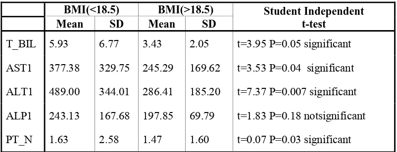
BODY MASS INDEX (BMI)
[image:42.612.116.517.493.647.2]Analysis of data with BMI as an independent variable showed strong correlation with elevation of Total Bilirubin, AST, ALT, ALP and PT (p<0.05)
Table 5.3
BMI(<18.5) BMI(>18.5) Student Independent t-test
Mean SD Mean SD
SEX
Comparing the means of liver function tests with Sex did not show statistically significant correlation (p =0.13) (Table A.)
CD4 Count
[image:43.612.105.557.340.514.2]The association of hepatotoxicity with different CD4 counts catagories was analyzed. Patients belonging to CD4 count < 100 have significant correlation with TB, AST, ALT, and PT. (p 0.04). The risk of hepatotoxicity correlates well with low CD4 count whereas in groups with CD4 count between 100-150 and > 150 did not show any correlation.
Table 5.4
CD4nadir
Oneway ANOVA F_test
<100 101-150 >150
Mean SD Mean SD Mean SD
T_BIL 6.12 6.02 3.23 2.33 2.49 0.79 F=3.40 P=0.04 significant AST1 383.89 306.57 242.17 179.88 181.71 70.87 F=3.12 P=0.05
significant ALT1 420.00 336.85 311.83 194.66 299.71 221.30 F=1.07 P=0.35
Not significant ALP1 241.00 155.71 199.63 71.35 178.14 62.77 F=1.12 P=0.03
significant PT_N 1.89 2.33 1.50 1.79 .57 .79 F=1.21 P=0.01
significant
ALCOHOL
HEPATITIS B
Coinfection with HBV increases the risk of hepatotoxicity.The data analyzed show positive correlation with HBV positivity with TB, AST, ALT, PT (p0.001). Patients with HBV infection had higher levels of T.B, AST, ALT and Prothrombin compared to negative group.
[image:44.612.114.519.287.453.2]On combining HBV coinfection and alcohol abuse there was a positive correlarion with TB, AST, ALT, PT. (p < 0.03). (Table 5.4)
Table 5.4
HBV
Student Independent t-test
No Yes
Mean SD Mean SD
T_BIL 3.80 4.50 5.92 2.73 t=1.89 P=0.04 significant AST1 229.83 203.43 518.50 233.25 t=3.90 P=0.001 significant ALT1 301.38 239.93 550.70 260.58 t=2.89 P=0.006 significant ALP1 202.30 114.86 252.50 88.23 t=1.29 P=0.22 not significant PT_N 1.13 1.79 3.10 1.79 t=3.12 P=0.002 significant
6. DISCUSSION
There is an increase in the reporting of drug induced liver injury following ART therapy since 1995 following wide spread usage of antiretrovirals.12
The incidence of drug hepatotoxicity has been variously reported. It ranged from 5% to 30% in different series (17 cohorts and 2 metanalyses). Less often they cause steatosis, Lactic acidosis and encephalopathy with mortality rates between 0.1 to 7%
Our incidence is likely to be around 3.9%when grade 3 or 4 injury is taken as cut off limit.
We compared our results with that of the literature available on this issue. Much of the data came from large trials like Amsterdam, CHORUS, ICONA and TARGET which involved more than 5100 patients.3,4,14,15,16,17
AGE, SEX AND BMI
Age was not considered to be an individual risk factor in most of the published series. Large trials like Saves17 failed to demonstrate any correlation with age. Our series also found no correlation to the incidence of hepatotoxicity with age.
Obese patients had a higher risk of hepatic steatosis and liver injury (Carr A et al). Other studies have shown that malnutrition and low BMI are also contributory factors in hepatic injury in Asian and African populations (Sampras K et al). In our series there is a strong correlation with liver injury (Bilirubin level, AST, ALT ) with low BMI (< 18.5), but did not have impact on normal BMI. Our study population did not include obese BMI patients and thus could not be compared.
DRUGS AND REGIMENS
Previously number of reports of increased liver injury were attributed to certain drugs like Zidovudine, Nevirapine , full dose Ritonavir. Review of 17 clinical trials between 1991 to 2001 in FDA database attributes risk of liver injury for Nevirapine (NVP) and Efavirenz (EFZ). In 2NN study, post exposure prophylaxis of NVP was associated with severe liver injury and it was recommended to exclude NVP from PEP programs.
Our series included 3 standard regimen (AZT + Lamivudine + Nevirapine , Stavudine + Lamivudine + Nevirapine, Stavudine + Lamivudine + Efavirenz). When these were compared with the incidence of liver injury there was no correlation. All regimens had equal incidence of hepatotoxicity following therapy.20,25
ALCOHOL
ALP1 ALT1
AST1 1,200
1,000
800
600
400
200
0
[image:47.612.114.495.64.374.2]0.00
fig 6.1:Alcohol+ HBV Coinfection
HEPATITIS B AND HEPATITIS C
There are atleast 10 studies which show consistent association of liver toxicity with HBV infection. Studies by Saves, Sulkowski13, Den Brinker, D’Armino, Aceti26, Wit16, De Maat have shown that HBV is an individual risk factor.
The risk increased with high viral load, HBeAg positivity and raised baseline AST and ALT. Co infection with HCV is also identified as a contributing factor in these studies. Withdrawal of Lamivudine in HBV positive patients also found to have higher incidence of hepatotoxicity.
ALP1 ALT1
AST1 1,200
1,000
800
600
400
200
[image:48.612.116.522.65.399.2]0
fig 6.2:HBV Co-infection
CD4 COUNT
The correlation with CD4 count and hepatotoxicity was found in our study. Patients with CD4 < 100 cells ran a higher risk of hepatotoxicity and death. This is attributed to profound state of immune suppression and loss of liver regeneration functions. Studies by Saves had similar results.
hypersensitivity reaction to drugs. This phenomenon was also confirmed by other recent studies.15,16,17
Most of our patients had CD4 counts moderately elevated after induction of ART and this phenomenon was not observed in our series.
CONSTITUTIONAL SYMPTOMS
We found the presence of rash in about 13.5% of cases preceding liver injury. Fever, malaise, nausea, myalgia and arthralgia occurred in about 33.3%. The incidence was more with Stavudine, Lamivudine and Nevirapine regimen. This observation was not noted in other published series so far.
Presence of peripheral eosinophilia is observed in 13.5% which was lower than observed in Wit et all where he reports it to be around 28%
7. CONCLUSIONS
The following were concluded at the end of the study
1. Drug induced liver injury occurs in 3.9% of patients following HAART therapy
2. The risk factors for hepatotoxicity identified were Low BMI, Low CD4 count, HBV co-infection, and combined HBV co-infection & alcohol usage.
3. Age, Sex, alcohol usage alone, various regimens did not have any correlation with the incidence and severity of hepatotoxicity.
8. SUMMARY
The study was initiated with the primary aim of finding out the clinical profile and risk factors for the hepatotoxicity in patients receiving HAART therapy for AIDS.
A total of 1523 patients were screened and patients who developed liver injury during ART therapy were selected. About 50 adult patients of both the sexes were included in the study. Records of these patients were analyzed. Further testing was done as needed.
Various parameters were noted.
Statistical analysis was done by Oneway ANOVA F test, Chi-squared test and Student independent t test, in which correlation between various parameters and hepatotoxicity were analyzed.
Results were tabulated and compared with various published series worldwide.
According to our series, the major risk factors for hepatotoxicity were Hepatitis B infection, Low CD4 counts, Low BMI, alcohol abuse in HBV positive patients.
The risk of liver injury was independent of age, sex, various drug combinations.
Clinically, rash and constitutional symptoms occur in less than one quarter of these patients and carry poor sensitivity or specificity.
9. APPENDICES
Graph A1: CD 4 counts and Hepatotoxicity
CD4 nadir
0 20 40 60 80 100 120 140 160 180 200
0 10 20 30 40 50 60
age
C
D
4
na
di
r
Graph A2 : Duration & Hepatotoxicity
Graph A3 : Liver enzymes values
0 200 400 600 800 1000 1200
1 3 5 7 9 11 13 15 17 19 21 23 25 27 29 31 33 35 37 39 41 43 45 47 49
AST ALT ALP
Graph A4 : Bilirubin Values
0 5 10 15 20 25 30
1 3 5 7 9 11 13 15 17 19 21 23 25 27 29 31 33 35 37 39 41 43 45 47 49
Graph A5: Bilirubin & Prothrombin time
0 5 10 15 20 25 30
1 3 5 7 9 11 13 15 17 19 21 23 25 27 29 31 33 35 37 39 41 43 45 47 49
[image:54.612.119.516.93.368.2]T.BIL PT > N
Fig. A1
86.4%
13.6%
47.4% 52.6%
66.7%
33.3%
0%
20%
40%
60%
80%
100%
ALN
SLN
SLE
CONSTITUTIONAL SYMPTOMS
Table A1 : Base line data
REGIMEN
Oneway ANOVA F_test
ALN SLN SLE
Mean SD Mean SD Mean SD
[image:55.612.143.491.335.509.2]age 30.86 7.58 31.95 9.22 26.22 7.01 F=1.55 P=0.22 BMI 21.23 2.88 20.00 3.32 21.89 4.43 F=1.17 P=0.31 CD4nadir 122.45 28.36 99.00 33.81 92.22 61.93 F=2.89 P=0.07 duration 6.64 4.04 5.21 3.54 3.78 1.64 F=2.24 P=0.11 bilirubin 1.26 1.51 .92 .12 .91 .15 F=0.71 P=0.49 AST 25.64 7.05 25.42 7.43 27.11 7.20 F=0.18 P=0.83 ALT 27.45 7.93 28.32 4.03 30.89 5.21 F=0.97 P=0.39 ALP 130.05 26.69 133.58 29.40 130.22 30.82 F=0.09 P=0.91 Albumin 3.07 .21 3.11 .22 3.17 .28 F=0.55 P=0.58
Table A2 : Baseline Data
REGIMEN
Chi-squared test
ALN SLN SLE
n % n % n %
Sex Male 16 72.7% 14 73.7% 5 55.6% χ2=1.09 P=0.59 Female 6 27.3% 5 26.3% 4 44.4%
alcohol No 15 68.2% 10 52.6% 8 88.9% χ2=3.66 P=0.16 Yes 7 31.8% 9 47.4% 1 11.1%
HBV No 17 77.3% 15 78.9% 8 88.9% χ2=0.56
P=0.76 Yes 5 22.7% 4 21.1% 1 11.1%
HCV
No 21 95.5% 18 94.7% 9 100.0% χ
2=0.47 P=0.79 Yes 1 4.5% 1 5.3%
Table A3 : Constitutional Symptoms
REGIMEN
Chi-squared test
ALN SLN SLE
Count % Count % Count %
RASH No 19 86.4% 16 84.2% 8 88.9% χ2=0.11
P=0.94
Yes 3 13.6% 3 15.8% 1 11.1%
EOSINOPHILIA No 19 86.4% 16 84.2% 8 88.9% χ2=0.11
P=0.94
Yes 3 13.6% 3 15.8% 1 11.1%
CONST.S1 No 19 86.4% 9 47.4% 6 66.7% χ2=7.13
P=0.03 significant
[image:55.612.113.538.556.695.2]Table A4 : Age
Age
Oneway ANOVA F_test
<30 31-40 >40
Mean SD Mean SD Mean SD
T_BIL 3.74 3.59 4.07 2.86 6.41 8.17 F=1.10 P=0.34
AST1 279.12 214.21 259.29 197.15 387.57 390.70 F=0.75 P=0.49 ALT1 351.23 245.96 291.94 201.24 495.29 405.80 F=1.53 P=0.22 ALP1 189.35 62.09 222.12 89.08 274.00 237.86 F=1.75 P=0.18
PT_N 1.58 1.81 1.12 1.17 2.29 3.50 F=0.92 P=0.41
Table A5 : BMI
BMI(<18.5) BMI(>18.5) Student Independent t-test
Mean SD Mean SD
T_BIL 5.93 6.77 3.43 2.05 t=3.95 P=0.05 significant
AST1 377.38 329.75 245.29 169.62 t=3.53 P=0.07
Not significant ALT1 489.00 344.01 286.41 185.20 t=7.37 P=0.007 significant ALP1 243.13 167.68 197.85 69.79 t=1.83 P=0.18 notsignificant PT_N 1.63 2.58 1.47 1.60 t=0.07 P=0.96 Notsignificant
Table A6 : Sex
Sex
Student Independent t-test
Male Female
Mean SD Mean SD
T_BIL 4.82 4.91 2.84 1.56 t=1.52 P=0.13 notsignificant
AST1 320.74 259.00 210.13 160.60 t=1.53 P=0.13
Not significant ALT1 385.03 290.15 272.40 159.90 t=1.41 P=0.16 notsignificant ALP1 220.31 123.36 193.73 75.56 t=0.77 P=0.44 notsignificant PT_N 1.71 2.11 1.07 1.44 T=1.08 P=0.24 Notsignificant
Table A7 : Alcohol
Alcohol
Student Independent t-test
No Yes
Mean SD Mean SD
[image:56.612.115.540.95.205.2]REFERENCES
1. CENTRE FOR DISEASE CONTROL: HIV/AIDS Surveillance Report : 2006 2. Mel Wicox C. Gastrointestinal Consequences of Infection with HIV. Sleisinger
& Fordtran’s GASTROINTESTINAL AND LIVER DISEASE 8th Edition. Saunders 2006.
3. Cem Cengiz, James S. Park . HIV and Liver Diseases : Recent Clinical Advances: Clin Liver Dis 9 (2005)
4. Homayon Sidiq, Victor Ankoma-Sey. HIV- Related Liver Disease: Infections versus drugs : Gastroenterol Clin N Am 35 (2006)
5. Solomon RE, VanRaden M, et al. Association of hepatitis B surface antigen and core antibody with acquisition and manifestations of human immunodeficiency virus type I (HIV-I) infection. Am J Public Health 1990. 6. de Francbis R, Hadengue A, Lau G, et al. EASL International Consensus
Conference on Hepatitis B. 13-14 September, 2002 Geneva, Switzerland. J Hepatol 2003.
7. Sherman KE, Rouster SD, et al. Hepatitis C Virus prevalence among patients infected with Human Immunodeficiency Virus: Clin Infect Dis 2002.
8. Greub G, Ledergerber B,et a!. Clinical progression, survival, and immune.recovery during anti retroviral therapy in patients with HIV-I and hepatitis c virus co infection: Lancet 2000.
10. Chung RT. Anderson J. et a!. Peginterferon alfa -2a plus ribayirin versus inter-feron alfa 2a plus ribavirin for chronic hepatitis C in HIV coinfected people. N Engl J Med 2006.
11. National Institute of Health Consensus Development Conference Statement. Management of hepatitis C: 2002. Hepatology 2002
12. Rodriguez-Rosado R, Garcia-Samaniego J, . Hepatotoxicity after introduction of highly active antiretroviral therapy. AIDS 1998
13. Sulkowski MS, Thomas DL, Chaisson RE, et al. Hepatotoxicity associated with antiretroviral therapy in adults infected with human immunodeficiency virus and the role of hepatitis B or C virus infection. JAMA 2000
14. Nunez M, Lana R, Mendoza JL, et al. Risk factors for severe hepatic injury after introduction of highly active antiretroviral therapy. J Acquir Immune Defic Syndr 2001
15. Reisler R, Servoss JC, Sherman KE, et al. Incidence of hepatotoxicity and
mortality in 21 adult antiretroviral treatment trials: ACTG Liver Diseases Focus Group Program International AIDS Society Conference , Buenos Aires (Argentina): 2001.
16. Wit FW, Weverling GJ, Weel J, et al. Incidence of and risk factors for severe hepatotoxicity associated with antiretroviral combination therapy. J Infect Dis 2002
18. John M, Moore CB, James IR, et al. Chronic hyperlactatemia in HIV-infected patients taking antiretroviral therapy. AIDS 2001.
19. Boxwell D, Haverkos H, Kukich S, et al. Serious adverse events attributed to nevirapine regimens for post-exposure prophylaxis after HIV exposures-worldwide, 1997-2000. MMWR Morb Mortal Wkly Rep 2001.
20. Cahn P, Johnson M, Nusrat R, et al. Hepatic safety with nevirapine (NVP) and two nucleosides in patients with advanced HIV infection, from a placebo controlled clinical endpoint trial (1090). AIDS 2000
21. Becker S. Liver toxicity in epidemiological cohorts. Clin Infect Dis 2004. 22. Dieterich DT, Robinson PA, Love J, et al. Drug-induced liver injury associated
with the use of non-nucleoside reverse-transcriptase inhibitors. Clin Infect Dis 2004.
23. Martinez E, Blanco J, Amaiz JA, et al. Hepatotoxicity in HIV-l-infected patients receiving nevirapine-containing antiretroviral therapy. AIDS 2001. 24. Sulkowski M, Thomas D, et al. Hepatotoxicity associated with nevirapine and
efa-virenz-containing antiretroviral therapy: role of hepatitis C and B infections. Hepatology 2002.
25. Dieterich DT, Stern J, Robinson P, et al. Analyses of 4 key clinical trials to assess the risk of hepatotoxicity with Nevirapine: correlation with CD4 + levels, hepatitis B&C seropositivity and baseline liver function tests. lAS Conference on HIV Pathogenesis and Treatment. Buenos Aires (Argentina), 2001.