0022-538X/96/$04.0010
Copyrightq1996, American Society for Microbiology
Role of Vif in Human Immunodeficiency Virus Type 1
Reverse Transcription
JOAO GONCALVES,1,2YAEL KORIN,3JEROME ZACK,4ANDDANA GABUZDA1,5*
Division of Human Retrovirology, Dana-Farber Cancer Institute,1and Departments of Pathology2and Neurology,5
Harvard Medical School, Boston, Massachusetts 02115, and Department of Pathology and Laboratory Medicine3
and Division of Hematology-Oncology,4Department of Medicine, UCLA School of Medicine,
Los Angeles, California 90024
Received 11 August 1995/Accepted 30 August 1996
The Vif protein of human immunodeficiency virus type 1 (HIV-1) is important for virion infectivity. Previous
studies have shown thatvifmutant HIV-1 virions are defective in their ability to synthesize proviral DNA in
vivo. Here, we examine the role of Vif in viral DNA synthesis in the endogenous reverse transcriptase (RT) reaction, an in vitro assay in which virions synthesize viral DNA by using endogenous viral RNA as a template.
vifmutant virions showed a significant reduction in endogenous RT activity despite similar levels of exogenous
RT activity. Analysis of the viral DNA products on agarose gels demonstrated that this reflects reduced synthesis of short minus- and plus-strand DNA products in addition to those of full genomic length. Quan-titative PCR analysis of endogenous reverse transcription provided further evidence for reduced formation of both initial and completed reverse transcripts. Vif had no effect on genomic RNA dimerization or the stability of the RNA dimer linkage. These results suggest that Vif is important for an early event after virus entry but preceding or during the early stages of viral DNA synthesis. This may be due to an intrinsic effect on reverse transcription or a preceding postentry event(s), such as virion uncoating or disassembly of the virion core. Drugs targeted to Vif function may provide a new therapeutic approach to inhibiting HIV-1 reverse transcription.
The Vif protein of human immunodeficiency virus type 1 (HIV-1) is important for virion infectivity (10, 36, 38). vif mutant HIV-1 virions are defective in their ability to synthesize proviral DNA in vivo (3, 7, 37, 40). Whether this reflects an intrinsic defect in reverse transcription or an abnormality in a preceding early event(s) such as virus entry or uncoating is unknown. Peripheral blood mononuclear cells (PBMC) and some cell lines such as H9 require Vif to produce infectious HIV-1 virus particles, whereas other cell lines such as COS-1 and SupT1 do not (3, 7, 9, 14, 15, 33, 40). vif mutant virions produced in some nonpermissive cell lines can initiate reverse transcription but are defective in their ability to complete pro-viral DNA synthesis after acute infection (35, 37, 40). How-ever, when vif mutant virions are produced in PBMC or CEMx174 cells, the synthesis of initial reverse transcripts is undetectable (3, 7). Electron microscopy of vif mutant virions demonstrates an abnormality in the structure of the virion core (19), suggesting that the effect of Vif on viral DNA synthesis may result indirectly from an effect on the nucleoprotein com-plex which contains the reverse transcription machinery. A small amount of Vif (between 10 and 50 molecules) is associ-ated with the virus particle (3, 5, 22, 26), raising the alternative possibility that Vif might directly stimulate reverse transcrip-tion or other early events.
Whether vif mutant virions are blocked at a specific step during reverse transcription is not known. Reverse transcrip-tion is initiated from a nucleoprotein complex which consists of the reverse transcriptase (RT), dimerized RNA genome, cel-lular tRNALys primer, and nucleocapsid (NC) protein
(re-viewed in reference 29). Synthesis of minus-strand strong stop DNA is initiated from a tRNALysprimer which anneals to the
primer binding site at the 39boundary of the U5 region. Elon-gation of the minus strand continues after the first template switch, and full-length minus strand can be detected within 1 to 4 h. Plus-strand synthesis begins shortly after minus-strand initiation and is discontinuous because of initiation at multiple sites. Synthesis of plus-strand strong stop DNA is initiated at the polypurine tract (PPT) near the 39 long terminal repeat (LTR), while the central PPT in pol serves as a secondary initiation site (6). Additional sites for plus-strand initiation have also been identified in the right half of the viral genome (27). Elongation of the plus strand continues after the second template switch. Proviral DNA in the cytoplasm of HIV-1-infected cells consists predominantly of full-length minus strand and a discontinuous plus strand (27, 43), suggesting that the completion of plus-strand synthesis in vivo may occur after nuclear transport or integration.
The endogenous RT reaction is an in vitro assay in which viral DNA is synthesized by using endogenous viral RNA as a template. Virions are permeabilized with a nonionic detergent or mellitin and then incubated with high concentrations of deoxynucleoside triphosphates (dNTPs), a divalent cation, buffer, and radiolabeled dNTP. The endogenous RT reaction products represent a mixture of single- and double-stranded DNA and RNA-DNA hybrids, with low-molecular-weight products arising from discontinuous synthesis of viral DNA (1, 2, 4, 16, 32). In murine and avian retroviruses, the synthesis of fully infectious double-stranded viral DNA in vitro has been demonstrated (1, 16, 21, 32). However, the majority of prod-ucts are full-length minus strand and a discontinuous plus strand, similar to the forms detected in the cytoplasm early after infection (2, 27). In HIV-1 virions, the DNA products contain a much greater proportion of reverse transcripts that are shorter than the complete viral genome, most likely be-cause of the relative sensitivity of the HIV-1 virion core to detergents (4, 8, 42).
In this study, we examined the role of Vif in viral DNA * Corresponding author. Mailing address: Division of Human
Ret-rovirology, JF712, Dana-Farber Cancer Institute, 44 Binney St., Bos-ton, MA 02115. Phone: (617) 632-2154. Fax: (617) 632-3113. Elec-tronic mail address: [email protected].
8701
on November 9, 2019 by guest
http://jvi.asm.org/
synthesis in the endogenous RT reaction. We demonstrate that endogenous reverse transcription is significantly reduced in vif mutant virions. Our studies show that this reflects reduced synthesis of early viral DNA products in addition to those of full genomic length. These results suggest that Vif is important for an early event after virus entry but preceding or during the early stages of viral DNA synthesis. This may be due to an intrinsic effect on reverse transcription or a preceding posten-try event(s), such as virion uncoating or disassembly of the virion core.
MATERIALS AND METHODS
Virus and cell culture.A cocultivation method was used to produce large quantities of wild-type and vif mutant virus in a nonpermissive CEM cell line (11, 14). Infection of CEM cells was initiated by cocultivation with COS cells trans-fected with 5mg of wild-type or vif mutant HXB2 DNA by the DEAE-dextran method (14, 18) from 36 to 72 h after transfection. Infection of SupT1 cells was initiated by DEAE-dextran transfection with 10mg of viral DNA (14). The HIV-1 vif mutant viral DNA was made by changing the HXB2 sequence encod-ing Vif amino acids 21 and 22 to two in-frame stop codons (14, 28). Cultures were maintained in RPMI medium plus 10% fetal calf serum, with medium changes every 1 or 2 days. Virions were harvested from 24-h culture supernatants from days 14 to 21 after infection. The culture supernatants were clarified by centrif-ugation at 2,000 3g for 10 min and filtration through a 0.45-mm-pore-size Millipore filter prior to virion pelleting by centrifugation through 20% sucrose in a phosphate-buffered saline cushion at 125,0003g for 90 min.
Exogenous RT assay.Exogenous RT assays were performed as described elsewhere (31). Pelleted virions were disrupted in 50 mM Tris-HCl (pH 7.5)–0.25 M KCl–20% glycerol–0.25% Triton X-100–1 mM dithiothreitol and subjected to three freeze-thaw cycles. Exogenous RT activity was measured by incorporation of [3H]dTTP (2mCi) into an artificial poly(A)(dT)
15 template (Boehringer Mannheim) following incubation in RT assay buffer (50 mM Tris-HCl [pH 7.5], 7.5 mM MgCl2, 0.05% Triton X-100) for 60 min at 378C. The reaction products were spotted onto DE81 filters, and unincorporated [3H]dTTP was removed by a washing in 23SSC (13SSC is 0.15 M NaCl plus 0.015 M sodium citrate). The filters were rinsed in 95% ethanol and dried, and radioactivity was measured in liquid scintillation cocktail.
Endogenous RT assay.The standard endogenous RT reaction was performed in a 50- to 100-ml volume containing 0.53106to 13106cpm of exogenous RT units of HIV-1 (0.5 to 1.5 mg of protein per ml), 50 mM Tris-HCl (pH 8.4), 2 mM dithiothreitol, 2 mM magnesium acetate, 0.1 mM each dNTP, [3H]dTTP (50mCi per reaction mixture) or [32P]dCTP (40mCi per reaction mixture), and 1 mM
b-octylglucoside (Sigma) for 20 h at 378C unless otherwise indicated. Virion pellets were normalized for the same amount of exogenous RT activity and permeabilized with 1 mMb-octylglucoside for 10 min at room temperature prior to addition of reaction buffer. For [3H]dTTP incorporation, reactions were per-formed in buffer containing three of the four dNTPs and terminated by addition of 1/10 volume stop buffer (final concentrations, 50 mM Tris-HCl [pH 8.0] and 1% sodium dodecyl sulfate [SDS]) and spotting onto DE81 filters for quantita-tion by liquid scintillaquantita-tion counting as described for the exogenous RT assay. Reactions involving labeling with [32P]dCTP were terminated by addition of 10 U of DNase-free RNase (Boehringer Mannheim) and incubation for 2 h at 378C followed by digestion with an equal volume of proteinase K and SDS (final concentrations, 200mg of proteinase K per ml and 0.5% SDS) for 2 h at 378C. The reaction mixture was extracted with phenol, phenol-chloroform, and chlo-roform prior to ethanol precipitation with 3.5 M ammonium acetate.
Agarose gel electrophoresis.Endogenous RT products were resuspended in 10 mM Tris-HCl (pH 8.0)–1 mM EDTA and denatured by alkaline hydrolysis in 0.1 M NaOH for 1 h at 378C prior to electrophoresis in 1% agarose in Tris-borate-EDTA (TBE) running buffer (4). DNA molecular weight markers were dena-tured, run in parallel, and visualized with ethidium bromide. The gels were dried and exposed to film for autoradiography.
Southern blot analysis.The endogenous RT reaction was performed as de-scribed above but with a 10-fold-higher concentration of virus and unlabeled dNTPs. The reaction products were analyzed by denaturing agarose gel electro-phoresis followed by soaking of the gel in denaturation buffer (0.5 M NaOH, 1.5 M NaCl) for 30 min and in neutralization buffer (1 M Tris-HCl [pH 8.0], 1.5 M NaCl) for 45 min at room temperature. DNA was transferred to Hybond-N nylon membranes (0.45mm) (Amersham) by capillary blotting in 203SSC and cross-linked to the membrane by being baked for 2 h at 808C and exposed to UV (Stratalinker; Stratagene) for 3 min. The membranes were hybridized to32 P-labeled riboprobes in 50% deionized formamide–1 M NaCl–103Denhardt’s solution–50 mM Tris-HCl (pH 7.5)–0.1% NaPPi–1% SDS–10% dextran sulfate (Mr, 500,000) (Pharmacia) (23) for 12 h at 428C. After hybridization, the mem-branes were washed twice in 23SSC–0.5% SDS for 30 min at 658C, dried, and exposed to film for autoradiography. The following plasmids were used to pre-pare32P-labeled riboprobes according to the manufacturer’s instructions (Pro-mega Riboprobe II Core System). U3 riboprobes were prepared by using the pCDNAU3 plasmid, which contains the 719-bp XhoI-to-HindIII fragment of
HXB2 (nucleotides [nt] 8897 to 9616) (30) inserted in pCDNA1.Amp (Invitro-gen). The pCDNAU3 plasmid was linearized with XhoI and transcribed with T7 polymerase to generate the U3(1) riboprobe of minus-strand polarity or linear-ized with HindIII and transcribed with SP6 polymerase to generate the U3(2) riboprobe of plus-strand polarity. gag-pol riboprobes [gag/pol(1) and gag/pol(2)] were prepared by using the pGEMgagpol plasmid, which contains the 4.3-kb
SphI-to-SalI fragment of HXB2 (nt 1443 to 5786) (30) inserted in pGEM5zf(1) (Promega). The pGEMgagpol plasmid was linearized with SpeI and transcribed with SP6 polymerase to generate the gag/pol(1) riboprobe of minus-strand polarity or linearized with SalI and transcribed with T7 polymerase to generate the gag/pol(2) riboprobe of plus-strand polarity.
Northern (RNA) blot analysis of viral RNA.Virions were harvested from 12-h culture supernatants by pelleting through a sucrose cushion as described above. Viral RNA was extracted and analyzed by Northern blotting on nondenaturing agarose gels as described previously (12, 13, 23), with the following modifica-tions. Virion pellets were resuspended in 150ml of 50 mM Tris (pH 7.5), and then an equal volume of 23virion lysis buffer (final concentrations, 50 mM Tris-HCl [pH 7.5], 100 mM NaCl, 1 mM EDTA, 1% SDS, 200mg of proteinase K per ml, and 50mg of baker’s yeast tRNA per ml) was added. The samples were then incubated for 30 min at 378C and extracted twice with phenol-chloroform. Viral RNA was ethanol precipitated, pelleted, redissolved overnight at 48C in 50
ml of buffer (1% SDS, 10 mM Tris-HCl [pH 7.5], 1 mM EDTA) in the absence or presence of 5 mM MgCl2, and stored at2708C. Viral RNA was analyzed by nondenaturing electrophoresis on 0.8% agarose gels in TBE running buffer followed by denaturation of the gel with 10% formaldehyde for 30 min at 708C. The RNA was transferred and cross-linked to a Hybond-N nylon membrane and hybridized with the gag/pol(1) riboprobe as described for Southern blot analysis.
Quantitative PCR amplification.The endogenous RT reaction was performed in the presence of unlabeled dNTPs for 20 h at 378C after normalization for the same value of exogenous RT activity (30,000 cpm/ml). The endogenous RT reaction samples were diluted 1:1,000, and 1 ml per reaction was used for quantitative PCR amplification as described previously (43). PCR amplification was performed by using32
P-labeled primers for 25 cycles at 948C for 1 min and 658C for 2 min. The oligonucleotide primer pairs M667/AA55 (R/U5, nt 496 to 635), SK38/SK39 (gag, nt 1544 to 1648), LA8/LA9 (gag, nt 711 to 805), M667/ M661 (LTR/gag, nt 496 to 695), and M667/LA9 (LTR/gag, nt 496 to 805) have been previously described (43, 44). The PCR products were resolved on 6% polyacrylamide gels and visualized by autoradiography. The quantity of HIV-1 DNA in each sample was determined by comparison with cloned HIV-1JR-CSF DNA standards (43) of known copy number which were amplified and analyzed in parallel with an AMBIS radioanalytic imaging system. The HIV-1 DNA standards were diluted in normal human peripheral blood lymphocyte DNA as a carrier, using the DNA equivalents of 10,000 cells per reaction.
Sucrose gradient analysis of virions.Pelleted virions were normalized for the same value of exogenous RT activity, resuspended in 300ml of buffer (50 mM Tris-HCl [pH 7.5], 50 mM NaCl, 10 mM EDTA), adjusted to 1 mMb -octylglu-coside, and layered over 10 to 60% sucrose gradients prepared in the same buffer. The gradients were centrifuged in an SW41 rotor at 40,000 rpm (200,0003g) for 12 h at 48C, and 500-ml fractions were collected from the bottom of the tube. The fractions were analyzed for exogenous and endogenous RT activity as described above. HIV-1 viral RNA was detected by Northern dot blot analysis of fractions with the gag/pol(1) riboprobe following addition of an equal volume of 23virion lysis buffer, ethanol precipitation, resuspension of the RNA pellet in 30 ml of denaturation buffer (10 ml of formamide, 7 ml of formaldehyde, 2 ml of 203SSC, 11ml of double-distilled water), and heat denaturation for 15 min at 688C. Virion proteins were detected in gradient fractions by labeling cultures with [35
S]methionine and [35
S]cysteine (100mCi/ml each) for 12 h prior to virion pelleting, trichloroacetic acid precipitation, and SDS-polyacrylamide gel electrophoresis (SDS-PAGE) analysis. HIV-1 RNA and virion proteins were quantitated by gel densitometry of the autoradiograms. The densities of fractions were determined with a refractometer.
RESULTS
Effect of Vif on endogenous reverse transcription.The effect
of Vif on viral DNA synthesis was examined in the endogenous RT reaction. To determine optimal detergent conditions for the assay, pelleted HIV-1 virions were permeabilized with dif-ferent concentrations ofb-octylglucoside (0.5 to 50 mM), Tri-ton X-100 (0.1 to 1%), or Nonidet P-40 (0.1 to 1%) and endogenous RT activity was determined by measuring [3H]dTTP incorporation after incubation for 20 h at 378C. The
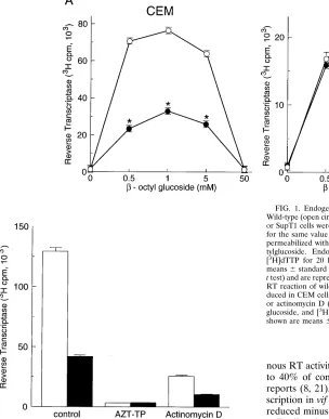
highest level of endogenous RT activity was obtained when virions were permeabilized with 1 mMb-octylglucoside (Fig. 1A and data not shown). In the absence of detergent or in the presence of 50 mMb-octylglucoside, endogenous RT activity was low or undetectable (Fig. 1A). Thus, subsequent
experi-8702 GONCALVES ET AL. J. VIROL.
on November 9, 2019 by guest
http://jvi.asm.org/
ments were performed with 1 mMb-octylglucoside unless oth-erwise noted.
The endogenous RT activity of wild-type and vif mutant virions produced in nonpermissive CEM cells and permissive SupT1 cells was examined (Fig. 1A). vif mutant virions pro-duced in this CEM cell line cannot initiate a second round of infection in any cell type, whereas those produced in SupT1 cells exhibit the same phenotype as the wild type (14). Pelleted virions were normalized for the same exogenous RT activity. The corresponding p24 values determined by p24 enzyme-linked immunosorbent assay were similar. Virions were per-meabilized with different concentrations of b-octylglucoside, and endogenous RT activity was determined. In the presence of 0.5 to 5 mMb-octylglucoside, vif mutant virions produced in CEM cells showed endogenous RT activity reduced to 30 to 40% of the wild-type level (P,0.01) (Fig. 1A). The difference between wild-type and vif mutant virions was less apparent when stronger detergent conditions were used. In contrast, endogenous RT activity was not affected by the vif mutation when virions were produced in SupT1 cells. Zidovudine (AZT)-triphosphate inhibited endogenous RT activity by
.95% (Fig. 1B), thus excluding possible contamination by cellular DNA polymerases. Actinomycin D, which predomi-nantly inhibits plus-strand synthesis (2, 8), inhibited
endoge-nous RT activity in both wild-type and vif mutant virions to 30 to 40% of control levels (Fig. 1B), consistent with previous reports (8, 21). Thus, the defect in endogenous reverse tran-scription in vif mutant virions may result at least in part from reduced minus-strand synthesis.
To directly analyze the viral DNAs synthesized in wild-type and vif mutant virions in vitro, the endogenous RT reaction products were labeled with [32P]dCTP, denatured by alkaline
hydrolysis, and analyzed on agarose gels (Fig. 2). Wild-type virions produced in CEM cells synthesized DNA products which ranged in size from 0.5 to 9.7 kb. Most of the DNA products were subgenomic species of less than approximately 7 kb, consistent with a previous report (4). vif mutant virions produced in CEM cells demonstrated a significant reduction in viral DNA synthesis (Fig. 2A). The synthesis of short DNA products was reduced compared with the wild-type level. In addition, the synthesis of viral DNA products of .6 kb was markedly diminished or undetectable even when the blots were overexposed. In contrast, the levels of viral DNA synthesis in wild-type and vif mutant virions produced in SupT1 cells were similar (Fig. 2A). Analysis of the viral DNAs synthesized at different times demonstrated a progressive increase in the quantity and length of DNA products over a 20-h time course, indicating that the reaction products are formed in an orderly and sequential way (Fig. 2B). However, the quantity of viral DNA synthesized at each time point was significantly reduced in vif mutant virions. Together, these results demonstrate that
vif mutant virions produced in nonpermissive cells show a
significant reduction in viral DNA synthesis in vitro.
Analysis of minus- and plus-strand viral DNA synthesis.To
[image:3.612.70.374.77.464.2]determine whether Vif affects specific steps during minus- or
FIG. 1. Endogenous RT activity in wild-type and vif mutant virions. (A) Wild-type (open circles) and vif mutant (closed circles) virions produced in CEM or SupT1 cells were used for the endogenous RT reaction. Virions normalized for the same value of exogenous RT activity (0.53106
cpm per sample) were permeabilized with reaction buffer containing different concentrations ofb -oc-tylglucoside. Endogenous RT activity was measured by incorporation of [3
H]dTTP for 20 h at 378C and binding to DE81 filters. Values shown are means6standard errors of the means (n53;p, P,0.01 by paired Student’s
t test) and are representative of three independent experiments. (B) Endogenous
RT reaction of wild-type (white bars) and vif mutant (black bars) virions pro-duced in CEM cells performed in the presence of AZT-triphosphate (100mM) or actinomycin D (20mg/ml). Virions were permeabilized with 1 mMb -octyl-glucoside, and [3
H]dTTP incorporation was measured as for panel A. Values shown are means6standard errors of the means (n53).
on November 9, 2019 by guest
http://jvi.asm.org/
plus-strand synthesis, the endogenous RT reaction products were analyzed by Southern blotting with strand-specific probes (Fig. 3). The endogenous RT reaction was performed in the absence of radioisotopes for 20 h, and the DNA products were extracted, ethanol precipitated, treated with RNase, denatured by alkaline hydrolysis, and resolved on agarose gels. The DNA was then transferred to nylon membranes and hybridized to
FIG. 2. Viral DNA products synthesized in wild-type and vif mutant virions in vitro. (A) Viral DNA synthesized in the endogenous RT reaction of wild-type (lane 1) and vif mutant (lane 2) virions produced in CEM or SupT1 cells. The reaction was performed in the presence of [32
P]dCTP for 20 h at 378C. (B) Viral DNA products synthesized in the endogenous RT reaction of wild-type and vif mutant virions over a 20-h time course. The reaction products were labeled by continuous incorporation of [32
P]dCTP. For both panels, the endogenous RT reaction was performed following permeabilization of virions with 1 mMb -oc-tylglucoside. The reaction products were treated with RNase and proteinase K-SDS, extracted with phenol-chloroform, ethanol precipitated, and denatured with NaOH. The denatured DNA products were resolved by agarose gel elec-trophoresis in TBE running buffer and visualized by autoradiography. The size markers are HindIII fragments of lambda DNA. The single strands of the denatured 6.6-kb marker fragment appear as a doublet (4). Similar results were observed in three independent experiments.
FIG. 3. Analysis of minus- and plus-strand viral DNA synthesis in wild-type and vif mutant virions. The endogenous RT reaction of wild-type (lanes 1) and
vif mutant (lanes 2) virions produced in CEM cells was performed for 20 h at
378C in the absence of radionucleotides following permeabilization with 1 mM
b-octylglucoside. The DNA products were denatured and resolved on agarose gels as described for Fig. 2, transferred to nylon membranes, and hybridized to 32P-labeled strand-specific riboprobes. (A) Synthesis of minus-strand HIV-1
DNA. HIV-1 U3(2) and gag/pol(2) riboprobes were used to specifically detect minus-strand HIV-1 DNA. (B) Synthesis of plus-strand HIV-1 DNA. The U3(1) and gag/pol(1) riboprobes were used to specifically detect plus-strand HIV-1 DNA. Parallel reactions performed in the presence of the plus-strand inhibitor actinomycin D (Act D) (20mg/ml) are shown on the right. The size markers are as described for Fig. 2. Similar results were observed in three independent experiments.
8704 GONCALVES ET AL. J. VIROL.
on November 9, 2019 by guest
http://jvi.asm.org/
32P-labeled riboprobes complementary to minus- or
plus-strand HIV-1 DNA. Minus-plus-strand DNA synthesized in wild-type and vif mutant virions in vitro was detected by Southern blotting with riboprobes of plus-strand polarity encompassing the U3 or gag-pol regions (Fig. 3A). The U3(2) probe detects minus-strand products which have minimally completed the first template switching event. In wild-type virions, Southern blotting with the U3(2) probe detected minus-strand DNA products which ranged in size from 0.5 to 9.7 kb (Fig. 3A). More than 95% of the minus-strand DNA products were,7 kb long, with the most abundant products,2 kb long. A small amount of full-length minus-strand DNA, representing ,1% of the total, was detected. In vif mutant virions, the synthesis of minus-strand DNA products ,5 kb in length was reduced compared with that of the wild type. In addition, the synthesis of minus-strand DNA products of.6 kb was markedly dimin-ished or undetectable. Full-genome-length minus-strand DNA was not detectable in vif mutant virions even when the blots were overexposed to visualize the short DNA products at an intensity similar to that of the wild type. The blots were rehy-bridized with the gag/pol(2) probe, which detects minus-strand DNA which has been reverse transcribed through the left half of the genome. Hybridization with the gag/pol(2) probe gave results similar to those obtained with the U3(2) probe, indi-cating that minus-strand synthesis was discontinuous with ini-tiation at multiple sites.
The preceding experiment indicates that vif mutant virions are defective in their ability to synthesize minus-strand DNA. To determine the effect of Vif on synthesis of the plus strand, the blots from the preceding experiment were reprobed with U3 and gag/pol riboprobes of minus-strand polarity. The U3(1) probe detects initial plus-strand DNA products imme-diately adjacent to the 39 PPT. The gag/pol(1) probe detects plus-strand DNA products synthesized after the second tem-plate switch or initiated at the central PPT. In wild-type virions, the U3(1) and gag/pol(1) probes detected plus-strand DNA products of 0.5 to 9.7 kb (Fig. 3B). In contrast, plus-strand DNA products longer than 4 kb could not be detected in vif mutant virions even when the blots were overexposed to visu-alize the short DNA products at an intensity similar to that of the wild type. The most abundant plus-strand DNA products were,2 kb, most likely representing discontinuous synthesis with initiation at multiple sites (2). Similar results were ob-served when the blots were reprobed with the gag/pol(1) probe (Fig. 3B). In the presence of actinomycin D, only low-molec-ular-weight plus-strand DNA was detected in wild-type and vif mutant virions by hybridization with the U3(1) probe. How-ever, actinomycin D almost completely abolished the synthesis of plus-strand DNA detected with the gag/pol(1) probe (Fig. 3B). Thus, actinomycin D blocked plus-strand elongation and the initiation of plus-strand synthesis at some, but not all, sites. Together, these results indicate that vif mutant virions show reduced synthesis of short minus- and plus-strand DNA prod-ucts in addition to those of full genomic length.
Quantitative PCR analysis of endogenous reverse
transcrip-tion. Quantitative PCR was used to further analyze the
syn-thesis of early and late viral DNA products in the endogenous RT reaction. Wild-type and vif mutant virions were normalized for the same quantity of exogenous RT activity prior to per-formance of the endogenous RT reaction for 20 h. The endog-enous RT reaction products were amplified by PCR using different primer pairs to detect HIV-1 DNA sequences within initial, intermediate, and completed or nearly completed re-verse transcripts and quantitated by comparison with HIV-1 DNA standards of known copy number. The R/U5 primer pair is specific for the R/U5 sequence in the LTR within the first
region of viral DNA synthesized. This primer pair can detect viral DNA synthesis in nascent virions (44) and detects virtu-ally all HIV-1 DNA synthesized during reverse transcription (43). The SK38/SK39 and LA8/LA9 gag primer pairs detect HIV-1 DNA within partially completed reverse transcripts syn-thesized through the gag region. The M667/M661 and M667/ LA9 LTR/gag primer pairs flank the primer binding site and detect HIV-1 DNA within completed or nearly completed re-verse transcripts.
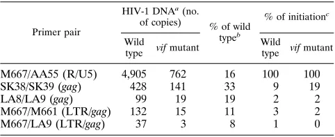
Quantitative PCR analysis showed that initial and late re-verse transcripts were both significantly reduced in the endog-enous RT reaction of vif mutant virions (Fig. 4 and Table 1). The formation of initial reverse transcripts detected by the R/U5 primer pair was reduced to 16% of the wild-type level. Completed or nearly completed reverse transcripts detected by the M667/M661 and M667/LA9 primer pairs were reduced to 8 to 11% of the wild-type level. Consistent with these findings, the levels of intermediate reverse transcripts corresponding to DNA synthesized through the gag region detected by the SK38/ SK39 and LA8/LA9 primer pairs were also significantly
re-FIG. 4. Quantitative PCR analysis of viral DNA synthesis in the endogenous RT reaction of wild-type and vif mutant virions produced in CEM cells, per-formed for 20 h at 378C in the absence of radionucleotides following normaliza-tion of the samples for the same value of exogenous RT activity. The endogenous RT reaction samples were diluted 1:1,000, and 1ml per reaction was used for 25 cycles of PCR amplification with the different primer pairs. The negative control is the endogenous RT reaction buffer. The positive control is 1 ng of pHXB2 plasmid DNA. The cloned HIV-1JR-CSFDNA standards (50 to 5,000 copies) were amplified in parallel. Quantitation of the PCR products is shown in Table 1. Similar results with each primer pair were obtained in two or three indepen-dent experiments.
on November 9, 2019 by guest
http://jvi.asm.org/
duced. In both wild-type and vif mutant virions, the quantity of HIV-1 DNA amplified with the LTR/gag primer pairs was 1 to 3% of the copy number amplified with the R/U5 primer pair, indicating that the majority of reverse transcripts did not pro-ceed to completion. These results suggest that the formation of initial reverse transcripts is significantly reduced in vif mutant virions. However, a significant fraction of the DNA synthesized can be detected by primers designed to detect regions of the genome associated with completed or nearly completed re-verse transcripts.
Sucrose gradient analysis of detergent-treated virions.To
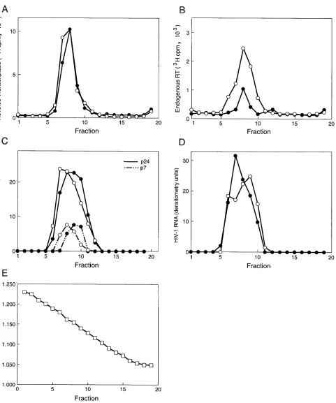
determine the effect of 1 mMb-octylglucoside on virion integ-rity, wild-type and vif mutant virions were analyzed on 10 to 60% sucrose gradients following treatment with the detergent (Fig. 5). The gradient distribution of exogenous and endoge-nous RT activity, p24 Gag, p17 Gag, NC protein, and viral RNA was examined. The gradient distribution of endogenous RT activity paralleled that of exogenous RT activity (Fig. 5A and B). The gradient distribution and quantity of exogenous RT activity were similar in wild-type and vif mutant virions, while endogenous RT activity in vif mutant virions was reduced to 30 to 40% of the wild-type level (Fig. 5A and B). The quantities of p24 Gag, NC protein, and viral RNA in gradient fractions of wild-type and vif mutant virions were similar (Fig. 5C and D). A minor shift in the gradient distribution of p24 Gag and NC protein by one fraction (Fig. 5C) was observed in
vif mutant virions in two independent experiments. The p17
Gag protein was detected only in fractions 6 to 9 and cosedi-mented with p24 Gag (not shown). The fractions which con-tained endogenous and exogenous RT activity, p24 Gag, p17 Gag, NC protein, and viral RNA corresponded to a density of 1.15 to 1.18 g/ml (Fig. 5E). This density corresponds to that of intact HIV-1 virions rather than virion cores, which have a density of 1.25 g/ml (26). These results suggest that 1 mM
b-octylglucoside does not grossly disrupt virion integrity but do not exclude the possibility of subtle structural alterations.
Dimerization of wild-type and vif mutant genomic viral
RNA.The packaged retroviral RNA genome is a dimer which consists of two genomic RNA plus strands linked together near their 59 ends (12, 13). The viral RNA dimer undergoes a maturation process to a more compact and stable form after virus release (12, 13). Altered viral RNA dimerization would be likely to adversely affect reverse transcription, since dimer-ization is necessary for template switching, while
monomeriza-tion of the dimerized RNA may be necessary for the comple-tion of viral DNA synthesis (29, 41). To determine whether Vif affects the dimerization of the viral RNA genome or the sta-bility of the dimer linkage, viral RNA was extracted from wild-type and vif mutant virions and analyzed by nondenatur-ing Northern blot analysis. Aliquots of viral RNA were incu-bated for 10 min at different temperatures ranging from 25 to 658C, resolved by electrophoresis on nondenaturing agarose gels, transferred to nylon membranes, and hybridized with the
gag/pol(1) riboprobe. Northern blotting of the viral RNA demonstrated that viral RNA dimerization and electrophoretic mobility were similar in wild-type and vif mutant virions (Fig. 6). In addition, the thermal stabilities of the viral RNA dimers were similar in wild-type and vif mutant virions in the absence or presence of 5 mM Mg21(Fig. 6). The observed increase in
the melting temperature of HIV-1 RNA dimers from 55 to 608C in the presence of 5 mM Mg21is consistent with previous
reports (12, 13). Sedimentation analysis of the HIV-1 RNA on 5 to 25% sucrose gradients (13) confirmed the conversion of dimeric HIV-1 RNA to its monomeric form following 10 min of incubation at 658C by a shift in the gradient density from fractions 2 to 8 to fractions 12 to 17, respectively, of 19 gradient fractions (not shown). These results indicate that Vif does not affect the dimerization of genomic viral RNA or the stability of the RNA dimer linkage.
DISCUSSION
This study demonstrates that vif mutant virions are defective in their ability to synthesize viral DNA in vitro. vif mutant virions show a significant reduction in endogenous RT activity despite similar levels of exogenous RT activity. Our studies demonstrate that this reflects reduced synthesis of both early and late viral DNA products. Analysis of the viral DNA prod-ucts on agarose gels showed reduced formation of short minus-and plus-strminus-and DNA products in addition to those of full genomic length. The defect in plus-strand synthesis may be secondary to the defect in minus-strand synthesis, since the minus strand serves as the template for plus-strand synthesis. Quantitative PCR analysis of endogenous reverse transcription provided further evidence for reduced formation of both initial and completed reverse transcripts. These results are consistent with previous studies which demonstrate that vif mutant virions show reduced (37, 40) or undetectable (3, 7) proviral DNA synthesis after acute infection in vivo. Together, these findings suggest that Vif is important for an early event after virus entry but preceding or during the early stages of viral DNA synthe-sis. This may be due to an intrinsic effect on reverse transcrip-tion or a preceding postentry event, such as virion uncoating or disassembly of the virion core.
[image:6.612.58.299.99.198.2]Although proviral DNA synthesis is undetectable after acute infection with vif mutant virions produced in PBMC (7), vif mutant virions produced in some nonpermissive cell lines can initiate reverse transcription but are defective in their ability to complete proviral DNA synthesis in vivo (35, 37, 40). This may reflect the choice of producer or target cells, which can modify the severity of the vif mutant defect. In view of these observa-tions, it is noteworthy that our analysis of endogenous reverse transcripts on agarose gels (Fig. 3) provides preliminary evi-dence that Vif may be important for the elongation of minus-and plus-strminus-and DNA, in addition to affecting the early stages of reverse transcription. Together, these findings raise the pos-sibility that Vif may be important for efficient RT processivity once reverse transcription has been initiated. An alternative possibility supported by a recent study (35) is that elongation might be defective because of premature disassembly of the TABLE 1. Quantitative PCR amplification of HIV-1 DNA
synthesized in the endogenous RT reaction of wild-type and vif mutant virions
Primer pair
HIV-1 DNAa(no.
of copies) % of wild
typeb
% of initiationc
Wild
type vif mutant
Wild
type vif mutant
M667/AA55 (R/U5) 4,905 762 16 100 100
SK38/SK39 (gag) 428 141 33 9 19
LA8/LA9 (gag) 99 19 19 2 2
M667/M661 (LTR/gag) 132 15 11 3 2
M667/LA9 (LTR/gag) 37 3 8 1 0
a
The PCR products amplified by the different primer pairs listed in Fig. 4 were quantitated by radioanalytic image analysis using an AMBIS imager by compar-ison with HIV-1 DNA standards of known copy number which were amplified in parallel.
b
Percentage of the wild-type HIV-1 DNA copy number amplified with the same primer pair.
c
Percentage of the HIV-1 DNA copy number amplified with the M667/AA55 primer pair.
8706 GONCALVES ET AL. J. VIROL.
on November 9, 2019 by guest
http://jvi.asm.org/
FIG. 5. Sucrose gradient analysis of wild-type and vif mutant virions treated with 1 mMb-octylglucoside. Wild-type (open circles) and vif mutant (closed circles) virions produced in CEM cells were treated with 1 mMb-octylglucoside and loaded onto 10 to 60% sucrose gradients prepared in the same concentration of detergent. (A) Exogenous RT activity of fractions. (B) Endogenous RT activity of fractions. (C) HIV-1 p24 and p7 detected by trichloroacetic acid precipitation and SDS-PAGE analysis of fractions. (D) HIV-1 viral RNA detected by Northern dot blot analysis of fractions using the gag/pol(1) riboprobe. Data in panels A and B are from the same gradients (100- and 10-ml aliquots, respectively), and those in panels C and D are from parallel gradients. For panel C, cultures were labeled with [35
S]methionine and [35
S]cysteine for 12 h before pelleting of virions. The data in panels C and D were quantitated by densitometry of the autoradiograms. (E) Density of fractions determined with a refractometer.
on November 9, 2019 by guest
http://jvi.asm.org/
nucleoprotein complex, with degradation of the viral RNA by nucleases. Our quantitative PCR analysis did not detect a significant defect in the relative completion of endogenous reverse transcription in vif mutant virions. However, it is pos-sible that our ability to detect a defect in the completion of viral DNA synthesis may have been obscured by the amplifi-cation of short DNA products arising from discontinuous syn-thesis due to aberrant initiation at multiple sites (2, 4) in addition to full-length products. A very low level of completed reverse transcripts was detected in vif mutant virions by PCR, raising the possibility that there may be a small fraction of vif mutant particles which are as active as wild-type particles and can proceed to completion of viral DNA synthesis. Further studies are required to determine whether reverse transcrip-tion in vif mutant virions is intrinsically defective or blocked at a discrete stopping point(s) (24).
Our finding that vif mutant virions are defective for viral DNA synthesis in vitro together with the previous demonstra-tion that vif mutant virions exhibit an abnormal virion core structure (19) suggests that a component of the core is likely to be a target for Vif function. The virion core contains the RT, viral RNA genome, the NC protein, integrase, protease, and cellular tRNALysencased by a shell of p24 Gag. The initiation
of reverse transcription in vivo occurs within intact virions or in the cytoplasm after virus entry and uncoating of the virion core (43, 44). The efficiency of viral DNA synthesis is determined by
several factors, including those which directly affect the activity of the RT enzyme (i.e., divalent cations), the NC protein, the configuration of the RNA or DNA template, and the spatial organization of these components within the nucleoprotein complex. A small quantity of Vif may be incorporated into the virion core (22, 26), raising the possibility that Vif itself might associate with a core component(s) and thereby might directly stimulate reverse transcription or preceding early postentry events. However, whether virion incorporation of Vif is specific or biologically important remains to be determined (5). The possibility that Vif directly affects the RT enzyme seems un-likely, since exogenous RT activity and the quantity of virion-associated RT protein do not appear to be altered by vif mu-tations (17, 35, 40). Our results (Fig. 5 and 6) and previous studies suggest that Vif does not affect viral RNA encapsida-tion (3, 40), dimerizaencapsida-tion, or the stability of the RNA dimer linkage. Altered Gag processing has been demonstrated in vif mutant virions (3, 34), raising the possibility that Vif might affect the NC protein, p24 Gag, or the spatial organization of Gag proteins within the nucleoprotein complex and thereby influence reverse transcription. For example, an effect on the NC protein might affect reverse transcription, since in vitro studies have shown that the NC protein decreases the proba-bility of termination of reverse transcription at certain pause sites, enhances strand transfers, promotes selective DNA an-nealing, and accelerates the renaturation of complementary DNA sequences (20, 25, 39). Further studies are required to identify the target(s) for Vif function in the virion core.
To our knowledge, this study is the first to use the nonionic detergent b-octylglucoside for virion permeabilization in the endogenous RT reaction. We found that permeabilization with 1 mM b-octylglucoside gave higher levels of endogenous re-verse transcription than equivalent concentrations of Nonidet P-40 or Triton X-100. Our studies also showed that the effect of Vif on endogenous reverse transcription was less apparent under stronger detergent conditions (17). Sucrose gradient analysis indicates that 1 mMb-octylglucoside does not grossly disrupt virion integrity, although the possibility of subtle struc-tural alterations cannot be excluded. Consistent with previous reports (4, 8), we found that HIV-1 DNA synthesized in vitro was predominantly subgenomic length. A minor amount of full-genome-length viral DNA was synthesized, but the major-ity of DNA products were less than approximately 7 kb. The most abundant products were,2 kb, representing minus- and plus-strand products most likely arising from discontinuous synthesis with initiation at multiple sites (2, 4). The data sug-gest that it is likely that discontinuous priming occurred, pos-sibly because of aberrant initiation from residual genomic RNA fragments (2, 4). Alternatively, short reverse transcripts containing sequences normally found only in intermediate or full-length products may have arisen from aberrant template switching to internal sites (4). Specific subgenomic bands de-scribed in a previous study (4) were seen in only some exper-iments and were not reproducibly observed (17). Most of the viral DNA detected in the cytoplasm early after infection in vivo consists of a full-length minus strand and a discontinuous plus strand (27). Thus, HIV-1 viral DNA synthesis in vitro differs in several respects from viral DNA synthesis in vivo.
[image:8.612.58.298.73.378.2]We propose the following model for the role of Vif in viral DNA synthesis. Vif acts on a component of the nucleoprotein complex which contains the reverse transcription machinery either during virus assembly or via direct contact within the virion core. This permits efficient viral DNA synthesis because of an intrinsic effect on reverse transcription or a preceding postentry event(s), such as virion uncoating, disassembly of the virion core, or formation of a stable nucleoprotein complex. In
FIG. 6. Northern blot analysis of HIV-1 RNA dimers in wild-type and vif mutant virions. RNA isolated from pelleted wild-type and vif mutant virions was dissolved in a buffer containing 10 mM Tris-HCl (pH 7.5), 1 mM EDTA, and 1% SDS without magnesium (A) or with 5 mM MgCl2(B). Virions were normalized for the same value of exogenous RT activity. Aliquots of RNA were heated for 10 min at the indicated temperatures prior to electrophoresis on nondenaturing agarose gels and Northern blot analysis using the gag/pol(1) riboprobe.
8708 GONCALVES ET AL. J. VIROL.
on November 9, 2019 by guest
http://jvi.asm.org/
permissive cells, Vif is not required because of the presence of a complementing cellular factor or the absence of an inhibitory factor. This model does not exclude the possibility that Vif might also affect other early events, such as virus entry (3) or the nuclear transport of proviral DNA. These observations raise the possibility that drugs targeted to Vif function may provide a new therapeutic approach to inhibiting HIV-1 re-verse transcription.
ACKNOWLEDGMENTS
We thank Heinrich Gottlinger for providing the pGEMgagpol plas-mid and Joseph Sodroski, Heinrich Gottlinger, Alan Engelman, and Xiaoyu Yang for critical reading of the manuscript. AZT-triphosphate was obtained from the NIH AIDS Research and Reference Reagent Program.
This work was supported by grants from the National Institutes of Health (AI 33837 and AI36186 to D.G. and AI33259 to J.Z.), a Clau-dia A. Barr Investigator award, and the G. Harold and Leila Mathers Charitable Foundation. J.Z. is an Elizabeth Glaser Scientist supported by the Pediatric AIDS Foundation. We also acknowledge Center for AIDS Research (AI28691) and Center for Cancer Research (AO6516) grants for supporting necessary core facilities. J.G. is the recipient of a doctoral fellowship from Junta Nacional De Investigacao Cientifica e Tecnologica, Portugal.
REFERENCES
1. Benz, E. W., Jr., and D. Dina. 1979. Moloney murine sarcoma virions syn-thesize full-genome-length double-stranded DNA in vitro. Proc. Natl. Acad. Sci. USA 76:3294–3298.
2. Boone, L. R., and A. M. Skalka. 1981. Viral DNA synthesized in vitro by avian retrovirus particles permeabilized with melittin. J. Virol. 37:109–116. 3. Borman, A. M., C. Quillet, P. Charneau, C. Dauguet, and F. Clavel. 1995. Human immunodeficiency virus type 1 Vif2mutant particles from restrictive cells: role of Vif in correct particle assembly and infectivity. J. Virol. 69: 2058–2067.
4. Borroto-Esoda, K., and L. R. Boone. 1991. Equine infectious anemia virus and human immunodeficiency virus DNA synthesis in vitro: characterization of the endogenous reverse transcriptase reaction. J. Virol. 65:1952–1959. 5. Camaur, D., and D. Trono. 1996. Characterization of human
immunodefi-ciency virus type 1 Vif particle incorporation. J. Virol. 70:6106–6111. 6. Charneau, P., and F. Clavel. 1991. A single-stranded gap in human
immu-nodeficiency virus unintegrated linear DNA defined by a central copy of the polypurine tract. J. Virol. 65:2415–2421.
7. Courcoul, M., C. Patience, F. Rey, D. Blanc, A. Harmache, J. Sire, R. Vigne, and
B. Spire.1995. Peripheral blood mononuclear cells produce normal amounts of defective Vif2human immunodeficiency virus type 1 particles which are re-stricted for the preretrotranscription steps. J. Virol. 69:2068–2074.
8. Debyser, Z., A.-M. Vandamme, R. Pauwels, M. Baba, J. Desmyter, and E. De
Clerq.1992. Kinetics of inhibition of endogenous human immunodeficiency virus type 1 reverse transcriptase by 29,39-dideoxynucleoside 59-triphosphate, tetrahydroimidazo-[4,5,1-jk][1,4]-benzodiazepin-2(1H)-thione, and 1-[(2-hy-droxyethoxy)methyl]-6-(phenylthio)thymine derivatives. J. Biol. Chem. 267: 11769–11776.
9. Fan, L., and K. Peden. 1992. Cell-free transmission of Vif mutants of HIV-1. Virology 190:19–29.
10. Fisher, A. G., B. Ensoli, L. Ivanoff, M. Chamberlain, S. Petteway, L. Ratner,
R. C. Gallo, and F. Wong-Staal.1987. The sor gene of HIV-1 is required for efficient virus transmission in vitro. Science 237:888–893.
11. Foley, G. E., H. Lazarus, J. Dredd, S. Faber, B. G. Uzn, B. Boone, and R. E.
McCarthy.1965. Continuous culture of human lymphoblasts from peripheral blood of a child with acute leukemia. Cancer 18:522–529.
12. Fu, W., R. J. Gorelick, and A. Rein. 1994. Characterization of human im-munodeficiency virus type 1 dimeric RNA from wild-type and protease-defective virions. J. Virol. 68:5013–5018.
13. Fu, W., and A. Rein. 1993. Maturation of dimeric viral RNA of Moloney murine leukemia virus. J. Virol. 67:5443–5449.
14. Gabuzda, D. H., K. Lawrence, E. Langhoff, E. Terwilliger, T. Dorfman, W. A.
Haseltine, and J. Sodroski.1992. Role of vif in replication of human immu-nodeficiency virus type 1 in CD41T lymphocytes. J. Virol. 66:6489–6495. 15. Gabuzda, D. H., H. Li, K. Lawrence, B. S. Vasir, K. Crawford, and E.
Langhoff.1994. Essential role of vif in establishing productive HIV-1 infec-tion in peripheral blood T lymphocytes and monocyte/macrophages. J. Ac-quired Immune Defic. Syndr. 7:908–915.
16. Gilboa, E., S. Goff, A. Shields, F. Yoshimura, S. Mitra, and D. Baltimore. 1979. In vitro synthesis of a 9 kbp terminally redundant DNA carrying the infectivity of Moloney murine leukemia virus. Cell 16:863–874.
17. Goncalves, J., and D. Gabuzda. Unpublished data.
18. Goncalves, J., P. Jallepalli, and D. H. Gabuzda. 1994. Subcellular localiza-tion of the Vif protein of human immunodeficiency virus type 1. J. Virol.
68:704–712.
19. Ho¨glund, S., A. Ohagen, K. Lawrence, and D. Gabuzda.1994. Role of vif during packing of the core of HIV-1. Virology 201:349–355.
20. Ji, X., G. J. Klarmann, and B. D. Preston. 1996. Effect of human immuno-deficiency virus type 1 (HIV-1) nucleocapsid protein on HIV-1 reverse transcriptase activity in vitro. Biochemistry 35:132–143.
21. Junghans, R. P., P. H. Duesberg, and C. A. Knight. 1975. In vitro synthesis of full-length DNA transcripts of Rous sarcoma virus RNA by viral DNA polymerase. Proc. Natl. Acad. Sci. USA 72:1895–1900.
22. Karczewski, M. K., and K. Strebel. 1996. Cytoskeleton association and virion incorporation of the human immunodeficiency virus type 1 Vif protein. J. Virol. 70:494–507.
23. Khandjian, E. W., and C. Meric. 1986. A procedure for Northern blot analysis of native RNA. Anal. Biochem. 159:227–232.
24. Klarmann, G. J., C. A. Schauber, and B. D. Preston. 1993. Template-di-rected pausing of DNA synthesis by HIV-1 reverse transcriptase during polymerization of HIV-1 sequence in vitro. J. Biol. Chem. 268:9793–9802. 25. Lapadat-Tapolsky, M., H. De Rocquigny, D. V. Gent, B. Roques, R. Plasterk,
and J.-L. Darlix.1993. Interactions between HIV-1 nucleocapsid protein and viral DNA may have important functions in the viral life cycle. Nucleic Acids Res. 21:831–839.
26. Liu, H., X. Wu, M. Newman, G. M. Shaw, B. H. Hahn, and J. C. Kappes. 1995. The Vif protein of human and simian immunodeficiency viruses is packaged into virions and associates with viral core structures. J. Virol.
69:7630–7638.
27. Miller, M. D., B. Wang, and F. D. Bushman. 1995. Human immunodefi-ciency virus type 1 preintegration complexes containing discontinuous plus strands are competent to integrate in vitro. J. Virol. 69:3938–3944. 28. Myers, G., S. F. Josephs, A. B. Rabson, T. I. Smith, and F. Wong-Staal (ed.).
1988. Human retroviruses and AIDS. Los Alamos National Laboratory, Los Alamos, N.Mex.
29. Peliska, J. A., and S. J. Benkovic. 1992. Mechanism of DNA strand transfer reactions catalyzed by HIV-1 reverse transcriptase. Science 258:1112–1118. 30. Ratner, L., W. Haseltine, R. Patarca, K. J. Livak, B. Starcich, S. F. Josephs,
E. R. Doran, J. A. Rafalski, E. A. Whitehorn, K. Baumeister, L. Ivanoff, S. R. Petteway, M. L. Pearson, J. A. Lautenberger, T. S. Papas, J. Ghrayeh, N. T. Chang, R. C. Gallo, and F. Wong-Staal.1985. Complete nucleotide sequence of the AIDS virus, HTLV-III. Nature (London) 313:277–284.
31. Rho, H. M., B. Poiesz, W. Ruscetti, and R. C. Gallo. 1981. Characterization of the reverse transcriptase from a new retrovirus (HTLV) produced by a human cutaneous T-cell lymphoma cell line. Virology 112:355–360. 32. Rothenberg, E., D. Smotkin, D. Baltimore, and R. A. Weinberg. 1977. In vitro
synthesis of infectious DNA of murine leukemia virus. Nature (London)
269:122–126.
33. Sakai, H., R. Shibata, J.-I. Sakuragi, S. Sakuragi, M. Kawamura, and A. Adachi. 1993. Cell-dependent requirement of human immunodeficiency virus type 1 Vif protein for maturation of virus particles. J. Virol. 67:1663–1666.
34. Simm, M., M. Shahbuddin, W. Chao, and D. J. Volsky. 1995. Aberrant Gag protein composition of human immunodeficiency virus type 1 (HIV-1) vif mutant produced in lymphocytes. J. Virol. 69:4582–4586.
35. Simon, J. H. M., and M. H. Malim. 1996. The human immunodeficiency virus type 1 Vif protein modulates the postpenetration stability of viral nucleoprotein complexes. J. Virol. 70:5297–5305.
36. Sodroski, J., W. C. Goh, C. Rosen, A. Tartar, D. Portetelle, A. Burny, and W.
Haseltine.1986. Replicative and cytopathic potential of HTLV-III/LAV with
sor gene deletions. Science 231:1549–1551.
37. Sova, P., and D. Volsky. 1993. Efficiency of viral DNA synthesis during infection of permissive and nonpermissive cells with vif-negative human immunodeficiency virus type 1. J. Virol. 67:6322–6326.
38. Strebel, K., D. Daugherty, K. Clouse, D. Cohen, T. Folks, and M. A. Martin. 1987. The HIV “A“ (sor) gene product is essential for virus infectivity. Nature (London) 328:728–730.
39. Tsuchihashi, Z., and P. O. Brown. 1994. DNA strand exchange and selective DNA annealing promoted by the human immunodeficiency virus type 1 nucleocapsid protein. J. Virol. 68:5863–5870.
40. von Schwedler, U., J. Song, C. Aiken, and D. Trono. 1993. vif is crucial for human immunodeficiency virus type 1 proviral DNA synthesis in infected cells. J. Virol. 67:4945–4955.
41. Weiss, S., G. Ha¨usl, M. Famulok, and B. Ko¨nig.1993. The multimerization state of retroviral RNA is modulated by ammonium ions and affects HIV-1 full-length cDNA synthesis in vitro. Nucleic Acids Res. 21:4879–4885. 42. Yong, W., H. S. Wyman, and J. A. Levy. 1990. Optimal conditions for
synthesizing complementary DNA in the HIV-1 endogenous reverse tran-scriptase reaction. AIDS 4:199–206.
43. Zack, J. A., S. J. Arrigo, S. R. Weitsman, A. S. Go, A. Haislip, and I. S. Y.
Chen.1990. HIV-1 entry into quiescent primary lymphocytes: molecular analysis reveals a labile, latent viral structure. Cell 61:213–222.
44. Zhang, H., Y. Zhang, T. P. Spicer, L. Z. Abbott, M. Abbott, and B. J. Poiesz. 1993. Reverse transcription takes place within extracellular HIV-1 virions: potential biological significance. AIDS Res. Hum. Retroviruses 9:1287–1296.