A new species of Microcharon from marine interstitial waters, Shizuoka, Japan... 13
A new species of Microcharon from marine interstitial
waters, Shizuoka, Japan (Isopoda, Lepidocharontidae)
Jeongho Kim1, Wonchoel Lee1, Ivana Karanovic1,2
1 Department of Life Science, Hanyang University, Seoul, Korea 2 Institute for Marine and Antarctic Studies, University of Tasmania, Hobart, Tasmania, 7001, Australia
Corresponding author: Ivana Karanovic (ivana.karanovic@utas.edu.au)
Academic editor: S. Brix | Received 1 February 2017 | Accepted 18 April 2017 | Published 14 June 2017
http://zoobank.org/3E41270A-A8CA-40FE-AD10-210200232F5D
Citation: Kim J, Lee W, Karanovic I (2017) A new species of Microcharon from marine interstitial waters, Shizuoka, Japan (Isopoda, Lepidocharontidae). ZooKeys 680: 13–31. https://doi.org/10.3897/zookeys.680.12048
Abstract
A new species of Microcharon Karaman, 1934 (Asellota: Lepidocharontidae) is described from Miho-Uchihama beach, Shizuoka, Japan. Microcharon tanakai sp. n. differs from its congeners by having nine simple, five penicillate setae on antennal article 6; one simple distal seta on article 1 of the mandibular palp and having the apical lobe of male pleopod 1 convex, rounded, armed with seven setae. A key to Asian species of the genus and 16S rRNA of the new species are provided.
Keywords
interstitial, isopoda,Japan, morphology, taxonomy
Introduction
Members of the genus Microcharon Karaman, 1934, are free-living interstitial isopods. Being highly adapted to the narrow spaces of the interstitial environment, the species possesses an elongated body without visual organs or pigmentation (Coineau 1994, 2000; Wilson and Wägele 1994). When Karaman (1934) described Microcharon, it was included in the family Microparasellidae Karaman, 1934. However, following a recent revision (Galassi et al. 2016), the genus is currently a member of the family Lepidocha-rontidae. Besides Microcharon, Lepidocharontidae also includes Lepidocharon Galassi & Bruce, 2016 and Janinella Albuquerque, Boulanouar & Coineau, 2014. The most ZooKeys 680: 13–31 (2017)
doi: 10.3897/zookeys.680.12048 http://zookeys.pensoft.net
Copyright Jeongho Kim et al. This is an open access article distributed under the terms of the Creative Commons Attribution License (CC BY 4.0), which permits unrestricted use, distribution, and reproduction in any medium, provided the original author and source are credited.
RESEARCH ARTICLE
prominent diagnostic character of Microcharon is the shapes of pereonites 1-7. Pere-onites are cylindrical and each is rectangularly shaped in dorsal view whereas,
Lepidocha-ron and Janinella have trapezoidal pereonites (Galassi et al. 2016).
The genus Microcharon is one of the best studied groups of the family Lepidocha-rontidae and 69 species have been described from all over the world (Boyko et al. 2008). The majority of species are known from Europe, especially from the Mediter-ranean region (Coineau 1994; Boyko et al. 2008; Galassi et al. 2016). On the other hand, only the following three Asian species have been recorded so far: M. halophilus Birstein & Ljovuschkin, 1965; M. kirghisicus Jankowskaya, 1964; and M. raffaellae Pesce, 1979. They are known from Turkmenistan, Kyrgyzstan, and Iran respectively (Coineau 1994; Boyko et al. 2008; Galassi et al. 2016). Diversity of Microcharon in East Asia remains particularly unknown. Only Shimomura et al. (2006) briefly noted a single species of Microcharon collected together with an ingolfiellidean amphipod from marine interstitial of Okinawa archipelago.
During a survey of the interstitial fauna of Miho-Uchihama beach (Shizuoka, Ja-pan), a small number of psammolittoral isopods were collected together with other marine interstitial fauna such as, harpacticoid copepods, nematodes and ostracods. The isopod specimens had a typical Microcharon body plan, but a unique combination of morphological characters which lends support to the establishment of the new species described herein. Beside its description an identification key to the four Asian species of this genus currently recorded. In addition, a partial sequence of 16S rRNA gene was obtained and this may be useful for the future phylogenetic study studies of
Microcha-ron and the family LepidochaMicrocha-rontidae.
Materials and methods
Specimen collection and identification
A new species of Microcharon from marine interstitial waters, Shizuoka, Japan... 15
Figure 1. Sampling site. The arrow indicating the type locality of Microcharon tanakai sp. n. (Miho-Uchihama beach, Shizuoka, Japan).
(Seoul). Measurements were done following the method of Riehl and Brandt (2010). All measurements were taken from the dorsal view of line drawings using the distance measurement tools of Adobe Acrobat Professional. The ratios of appendages were given in distal to proximal order, excluding setae. The body ratios were given in anteromedial to posteromedial point order excluding appendages. Whole body length was measured from biggest female specimen. Terminology is largely based on Galassi et al. (2016).
DNA extraction and amplification
The amplification protocol consisted of the initial denaturation at 94°C for 2 min and 35 cycles each consisting of denaturation at 94°C for 50 sec, annealing at 50°C for 50 sec, extension at 72°C for 1 min 20 sec; the final extension was at 72°C for 7 min. Successful amplifications were confirmed by electrophoresis on 1% agarose gel. The PCR products were purified for sequencing reactions, using the Labopass PCR Purification Kit (COSMO Co. Ltd., Korea) following the guidelines provided with the kit. DNA was sequenced on an ABI automatic capillary sequencer (Macrogen, Seoul, Korea) using the same set of primers. All obtained sequences were visualized us-ing Finch TV version1.4.0 (http://www.geospiza.com/Products/finchtv.shtml). Each sequence was checked for the quality of signal and sites with possible low resolution, and corrected by comparing forward and reverse strands.
Systematics
Suborder Asellota Latreille, 1802
Family Lepidocharontidae Galassi & Bruce, 2016
Genus Microcharon Karaman, 1934
Type species. Microcharon stygius (Karaman, 1933)
Genus diagnosis (modified from Galassi et al. 2016). Body cylindrical; rostrum weakly developed or absent; antennula 5 or 6 articles; antennal flagellum longer than podomere, pereonites rectangular in dorsal view, with subparallel lateral margins; free pleonite as wide as preonite 7; pereopodal coxal plate hardly discernible, incorporated to sternite body wall; distolateral lobe of male pleopod 1 with folded hyaline lamella running parallel to lateral margin; female operculum as long as pleotelson, with two or four apical setae; well-developed uropods with slender endopod and exopod.
Microcharon tanakai sp. n.
http://zoobank.org/638F2AF3-DF77-4293-8077-8E3ECC810F82
Figures 2–10
Type locality. Interstitial water of coarse sand, Miho-Uchihama beach, Shizuoka city, Shizuoka Prefecture, Japan, 35°01'83"N, 138°51'71"E (Fig. 1).
Material examined. Holotype: adult female, (NIBRIV0000787789) com-pletely dissected and mounted in lactophenol on four slides; paratype 1 female (NI-BRIV0000787790) dissected on three slides, paratype 2 female (NIBRIV0000787791) dissected on one slide; adult male pleotelson dissected on three slides; 2 females and 1 male used for SEM.
A new species of Microcharon from marine interstitial waters, Shizuoka, Japan... 17
lateral lobe of maxillula with eleven robust setae; distal apex of male pleopod 1 convex, round, with three apical, four subapical setae; protopod of male pleopod 2.9 times longer than wide; protopod of uropod 3.1 times longer than wide, with fifteen setae.
Description of the female holotype. Body (Fig. 2A): elongate, slender in whole
appearance, total length, 1.95 mm, measured from anteromedial point of cephalon to posteromedial point of pleotelson, body approximately 8.5 times longer than wide, maximum body width in pereonite 3, 0.92 times of maximum width of pleotelson; color of preserved specimens transparent, whole surface of body with many ornamen-tation looking like lines.
Cephalon (Fig. 2B): 1.12 times longer than wide and 0.12 times of whole body;
anterior margin with weak rostrum; lateral margins straight, with four pairs of simple setae, three pairs of simple setae along dorsomedial surface.
Pereon (Fig. 2A, C): 0.68 times of whole body length, medial margin of tergite
convex, pereonites with lateral margin, straight, pereonite 1, 0.62 times longer than wide, four simple setae along lateral margin, two setae on dorsal surface; pereonite 2, 1.12 times longer than wide, with four pairs of setae along lateral margin, two setae on anteromedial edge; pereonite 3, 1.06 times longer than wide, with four pairs of setae along lateral margin, two pairs of setae along dorsal surface; pereonite 4, 1.02 times longer than wide, with nine setae on both lateral and dorsal surface; pereonite 5, 0.99 times longer than wide, with six setae on both lateral and dorsal margin; pereonite 6, 1.13 times longer than wide, with eight simple setae on dorsal margin; pereonite 7, 1.02 times longer than wide, with two setae on both anterolateral corner and three pairs of setae along dorsal surface.
Pleonite 1 (Fig. 2A): as wide as pereonite 7, 0.38 times longer than wide, with two
simple setae on dorsomedial margin.
Pleotelson (Fig. 2D): 1.33 times longer than wide, wider than preceding pereonites,
with several setae, becoming slightly narrow from basal part to distal end, weak cleft on middle of posterior rim.
Antennula (Fig. 2E): 6 articles; article 1 robust, 1.5 times longer than wide, with
three setae: two distomedial simple, one penicillate short setae distolaterally; article 2, smaller than 1, distoventral projection, 1.8 times longer than wide, with two simple, three penicillate setae, one of them elongate, stout, reaching distal tip of antennula; article 3, naked, 1.5 times longer than wide; article 4 with one proximolateral simple seta, two penicillate setae distolaterally; article 5, 1.2 times longer than wide with one simple seta, one aesthetasc distomedially; article 6, smallest 0.6 times of article 5, with one aesthetasc, one penicillate, three simple setae on distal end.
Antenna (Fig. 3A, B): six podomeres, twelve flagellar articles; article 1 globular in
A new species of Microcharon from marine interstitial waters, Shizuoka, Japan... 19
Figure 3. Microcharon tanakai sp. n., holotype, female. A antennal articles 4-6 with flagellum, dorsal
distal margin; flagellar articles from 2 to 8 subequal in length, armature of each articles as follows: article 1 with two simple setae distally, article 2 with four simple setae dis-tally, article 3 with three simple setae disdis-tally, article 4 with three simple setae disdis-tally, article 5 with four simple setae distally, article 6 with six simple setae distally, article 7 with three simple setae distally, article 8 with four simple setae distally, article 9 with four simple setae distally, article 10 with four simple setae distally, article 11 with three simple setae distally, article 12 with four simple setae distally
Mandible (Figs 3E–G, 8B): body robust, curved inwardly; pars incisiva of both
mandibles with four cusps; right mandible (Fig. 3F), lacinia mobilis, with five cusps, tapering proximally three pinnate, three simple setae located below lacinia mobilis; pars
molaris of both mandibles with one simple, one pinnate distal setae, slightly longer
than pars molaris, almost same length as seta on palp; left mandible (Fig. 3E), pars
inci-siva with four cusps, lacinia mobilis missing, with three pinnate and one naked robust
setae located below pars incisiva; palp (Fig. 3G) with three articles, inserted on cuticu-lar projection; article 1 with one simple seta distally (Fig. 8B), article 2 longest, with two pinnate and several hair-like setae on distal margin, article 3 curved outwardly, with five pinnate and with several hair-like setae on lateral margin.
Paragnaths (Fig. 3D): deeply incised, with two lobes, each distal part of lobes
cov-ered with numerous setae.
Maxillula (Fig. 4D): lateral lobe with eleven strong robust setae on distal edge,
some denticulate, some setulose, two spinular rows along outer margin; mesial lobe nearly half as long and wide as outer endite, with three short simple and several hair-like setae on distal end.
Maxilla (Fig. 4C): lateral rami slightly shorter than median one, with four
pecti-nate setae on distal end respectively; mesial ramus coalescent with basis, much thicker than others with eight strong setae distally and one of them pectinate.
Maxilliped (Fig. 3C): epipodite wide, apically pointed, distal tip reaching distal
part of first palp article; basis 2.1 times longer than wide; palp with 5 articles; article 1, 0.5 times longer than wide, with two simple setae on both distal corners; article 2, 1.7 times longer than article 1, with two simple setae on distomedial corner; article 3 tapering distally, with three simple setae along medial margin, one seta on distolateral corner; article 4 curved inwardly, with four simple setae distally; article 5 with five sim-ple setae, 2 claws distally; endite 2.4 times longer than wide, with five apical pinnate setae, numerous spinules on distomedial corner.
Pereopods 1–4 (Figs 4A, B, 5A, B): inserted on pereon anterolaterally (Fig. 8C);
A new species of Microcharon from marine interstitial waters, Shizuoka, Japan... 21
Figure 4. Microcharon tanakai sp. n., holotype, female. A pereopod 1, lateral B pereopod 2, lateral
Figure 5. Microcharon tanakai sp. n., holotype, female. A pereopod 3, lateral B pereopod 4, lateral
A new species of Microcharon from marine interstitial waters, Shizuoka, Japan... 23
Figure 6. Microcharon tanakai sp. n., holotype, female. A pereopod 6, lateral B pereopod 7, lateral
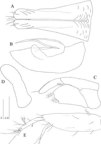
Figure 7. Microcharon tanakai sp. n., male. A pleopod 1, dorsal B pleopod 2, dorsal C pleopod 3, dorsal
A new species of Microcharon from marine interstitial waters, Shizuoka, Japan... 25
Figure 8. Scanning electron microscope images of Microcharon tanakai sp. n., A female antennal scale, lateral, B female mandibular palp, lateral C female pereopod 1-3, lateral, D female coxal plate of perepod 2, ventral E female bifurcate seta of pereopod 4, lateral, F female articular plate of pereopod 5, lateral. Scale bar unit µm.
Figure 9. Scanning electron microscope images of Microcharon tanakai sp. n., G pereonite 7 and free ple-onite, ventral, paratype male H penial papillae, ventral, I pleopod 1, dorsal, J distal part of male pleopod 1, dorsal K proximal part of male pleopod 1, dorsal L distolateral view of male pleopod 1. Scale bar unit µm.
two claws (Fig. 8E), two pairs of setae on both dorsal and ventral margin (vs pereopod 1 with three simple, two simple setae on both dorsoventral margin respectively).
Pereopods 5–7 (Figs 5C, 6A, B): inserted on pereon posterolaterally, subequal in
A new species of Microcharon from marine interstitial waters, Shizuoka, Japan... 27
Figure 10. Scanning electron microscope images of Microcharon tanakai sp. n., M lateral view of male pleopod 1 N protopod of male pleopod 2, ventral O pleopod 3, dorsal, female J distal tip of pleopod 3 exopod. Scale bar unit µm.
margin; merus, with four simple setae on both side of distal corner; carpus, with one bifid, two simple, one penicillate setae (vs pereopod 7 with one bifid, one simple, one penicillate setae); propodus, pereopod 5 with two bifid, two simple, one penicillate setae (vs pereopod 6 with two bifid, thre simple, one penicillate setae; pereopod 7 with two bifid, two simple setae), with sclerite on distal margin, covering proximal part of dacty-lus (Fig. 8F); dactydacty-lus with two claws, two pairs of setae on both dorsal, ventral margins.
Female operculum (Fig. 6C): 1.2 times longer than wide, with no ornamentation
on dorsal surface, with four setae on distal margin.
Uropods (Fig. 7E): protopod robust, slightly longer than pleotelson, length 3.1 times
longer than wide, with thirteen simple and two robust setae proximomedially; endopod 0.5 times longer than protopod, 1.7 times longer than exopod, with nine simple and four penicillate setae; exopod 0.6 times longer than endopod, with four simple setae distally.
Description of male. Penial papillae (Fig. 9G, H): located at posteromedial
mar-gin of pereonite 7 in ventral view, coalescent, proximal marmar-gin round, tapering dis-tally, but distal margin straight, with proximomedial opening channel.
Pleopod 1 (Figs 7A, 9I–L, 10M): elongate, total length almost reaching
wide (measured at widest section of proximal part); separated in half by medial sperm tube running from triangular opening on proximal part of medial groove, distolateral edge of hyaline lamella straight, without ornamentations; parallel to lateral margin of pleopod, in lateral view distal end of lobes convex, rounded at distal apex, each with three simple setae distally, four subapical setae on ventral margin.
Pleopod 2 (Figs 7B, 10N): protopod elongate, robust, 2.9 times longer than wide;
appendix masculina, curved, tapering distally, tip nearly reaching to protopod apex, with no armature; exopod rounded apically, broad.
Pleopod 3 (Figs 7C, 10O, P): no sexual dimorphism, endopod two-articulated,
second article suboval, with ornamentation like turtle shell shape, and with one api-cal, one mesial, one lateral plumose setae; exopod two-articulated, clearly longer than endopod, reaching far beyond tip of endopod, with one simple seta distally.
Pleopod 4 (Fig. 7D): rudimentary, uniramous, no ornamentation, distal margin
rounded.
Etymology. The species is named in honor of the collector, Dr. Hayato Tanaka, to express our appreciation for his support in this study.
Key to Asian species of Microcharon
1 Endopod of pleopod 3 with three plumose setae; article 1 of mandibular palp with one single seta distally ...M. tanakai sp. n.
– Endopod of pleopod 3 naked; article 1 of mandibular palp naked ...2
2 Antennula consist of 6 articles ...M. halophilus – Antennula consist of 5 articles ...3
3 Female operculum with 4 distal simple setae; distal part of male pleopod 1, rounded ...M. raffaellae – Female operculum with 2 distal simple setae; distal part of male pleopod 1,
straight ...M. kirghisicus
DNA amplification
A new species of Microcharon from marine interstitial waters, Shizuoka, Japan... 29
Discussion
Microcharon tanakai sp. n. is identified as a member of the genus Microcharon based on
the combination of the following characters: 1) body slender, elongate and all somites subequal in width, 2) pereopods 1-4 inserted anterolaterally, pereopods 5-7 inserted pos-terolaterally 3) coxal plates indiscernible in dorsal view, 4) pleotelson longer than wide, longer than any pereonite, 5) antennal article three with scale laterally 6) antennal flagel-lum longer than podomeres 7) maxillipedal palps composed of five articles, which are broader than the endite, 8) pereopod 1 leg-like, 9) uropods with long, broad protopod, both endopod and expod shorter than protopod in all other marine species of
Micro-charon and exopod inserted subapically (Wilson and Wägele 1994; Albuquerque et al.
2014; Galassi et al. 2016). The new species can be clearly distinguished from the three Asian species. Like M. halophilus described from the Kaptar-Khana cave, Turkmenistan (Birstein and Ljovuschkin 1965), it shows six articles of antennula. However, their dif-ferences include: a weak rostrum on the anterior margin of cephalon in the new species; larger length/width ratio of article 6 of the antennula (1.7 vs 1); setal formula of the an-tennula (article 1 with two simple, one penicillate setae vs one simple seta); the presence of one simple distal seta on the article 1 of the mandibular palp; only a simple setae along the medial margin (vs with combination of bifid, simple setae) on the carpus and propo-dus of pereopod; the female operculum with four setae distally (vs two distal setae in M.
halophilus); and the endopod of the pleopod 3 with three penicillate setae (vs naked in M. halophilus). The other Asian species, M. kirghisicus and M. raffaellae described from
Kyr-gyzstan (Jankowskaya 1964) and Iran (Pesce 1979) can be easily distinguished from M.
tanakai by the number of articles on the antennula (5 vs 6); both the carpus and
propo-dus of the pereopod with only one simple seta along the medial margin and a difference in the setal formula of the endopod of pleopod 3 (naked vs with three penicillate setae).
We were able to amplify 16S rRNA partial sequence of M. tanakai. To date, 228 16S rRNA sequences of the suborder Janiroidea G.O. Sars, 1897 have been deposited on GenBank belonging to the following 7 families: Desmosomatidae G.O. Sars, 1897, Macrostylidae Hansen, 1916, Joeropsididae Nordenstam, 1933, Janiridae G.O. Sars, 1897, Munnopsidae Lilljeborg, 1864, Haploniscidae Hansen, 1916 and Acanthaspi-diidae Menzies, 1962. Although this molecular marker has not been commonly used for barcoding (Hebert et al. 2003; Costa et al. 2007), the 16S rDNA has been proven a valid marker for distinguishing morphologically similar species of families Serolidae Dana, 1852, Chaetiliidae Dana, 1849 and Munnopsidae Lilljeborg, 1864 (Held 2000; Held and Wägele 2005; Raupach et al. 2007).
Acknowledgements
Foundation of Korea (NRF) and National Institute of Biological Resource (NIBR) funded by the Ministry of Environment (MOE), Republic of Korea. We would like to express our great appreciation to Dr. Nicole Coineau for her kind help and invaluable comments for the improvement of this paper.
References
Albuquerque EF, Boulanouar M, Coineau N (2014) First record of Janinella nom. n. (Crusta-cea: Isopoda: Microparasellidae) in the South Atlantic: revision of the genus and descrip-tion of a new Brazilian species. Journal of Natural History 48(29–39): 1817–1823. https:// doi.org/10.1080/00222933.2013.877996
Altschul SF, Gish W, Miller W, Myers EW, Lipman DJ (1990) Basic local alignment search tool. Journal of Molecular Biology 215: 403–410. https://doi.org/10.1016/S0022-2836(05)80360-2
Birstein J A, Ljovuschkin SI (1965) Subterranean Paraselloidea (Crustacea, Isopoda) in USSR. Zoologicheskii Zhurnal 44: 997–1013.
Boyko CB, Bruce NL, Merrin KL, Ota Y, Poore GCB, Taiti S, Schotte M, Wilson GDF (2008 onwards) (Eds) World marine, Freshwater and Terrestrial isopod crustaceans database. http://www.marinespecies.org/isopoda [on 2017-03-28]
Coineau N (1994) Evolutionary biogeography of the Microparasellid isopod
Microcha-ron (Crustacea) in the Mediterranean Basin. Hydrobiologia 287: 77–93. https://doi.
org/10.1007/BF00006898
Coineau N (2000) Adaptations to interstitial groundwater life. In: Wilkens H, Culver DC, Humphreys WF (Eds) Subterranean ecosystems, ecosystems of the world 30. Elsevier, 189–210.
Costa FO, deWaard JR, Bouthillier J, Ratnasingham S, Dooh RT, Hajibabaei M, Hebert PDN (2007) Biological identification through DNA barcodes: the case of the Crustacea. Canadian Journal of Fisheries and Aquatic Sciences, 64: 272–295. https://doi.org/10.1139/f07-008 Galassi DMP, Bruce NL, Fiasca B, Dole-Oliver MJ (2016) A new family Lepidocharontidae
with description of Lepidocharon gen. n., from the Great Barrier Reef, Australia, and re-definition of the Microparasellidae (Isopoda, Asellota). Zookeys 594: 11–50. https://doi. org/10.3897/zookeys.504.8049
Hebert PDN, Cywinska A, Ball SL, deWaard JR (2003) Biological identifications through DNA barcodes. Proceedings of the Royal Society of London B 270: 313–321. https://doi. org/10.1098/rspb.2002.2218
Held C (2000) Phylogeny and Biogeography of Serolid Isopods (Crustacea, Isopoda, Seroli-dae) and the Use of Ribosomal Expansion Segments in Molecular Systematics. Molecular Phylogenetics and Evolution 15(2): 165–178. https://doi.org/10.1006/mpev.1999.0739 Held C, Wägele JW (2005) Cryptic speciation in the giant Antarctic isopod Glyptonotus
ant-arcticus (Isopoda: Valvifera: Chaetiliidae). Scientia Marina: 69(2) 175–181. https://doi.
A new species of Microcharon from marine interstitial waters, Shizuoka, Japan... 31
Jankowskaya AJ (1964) Relict Crustacean of coastal bottom waters of the lake Issyk Kul (North Tien-Shan). Zoologicheskii Zhurnal 43(7): 975–986.
Karaman S (1934) Beitrage zur Kenntnis des Isopoden-Familie Microparasellidae. Mitteilungen über Hohlen- und Karstforschung 1934: 42–44.
Palumbi SR, Martin A, McMillan WO, Stice L, Grabowski G (1991) The simple fool’s guide to PCR, Version 2.0. http://palumbi.stanford.edu/SimpleFoolsMaster.pdf
Pesce GL (1979) The first Microparasellid from subterranean water of Iran, Microcharon
raf-faellae n. sp. (Crustaca, Isopoda). Vie et Milieu, 28–29, fasc. 2 série C 237–245.
Riehl T, Brandt A (2010) Descriptions of two new species in the genus Macrostylis Sars, 1864 (Isopoda, Asellota, Macrostylidae) from the Weddell Sea (Southern Ocean), with a synonymi-sation of the genus Desmostylis Brandt, 1992 with Macrostylis. ZooKeys 57: 9–49. https://doi. org/10.3897/zookeys.57.310
Raupach MJ, Malyutina M, Brandt A, Wägele JW (2007) Molecular data reveal a highly diverse species flock within the munnopsoid deep-sea isopod Betamorpha fusiformis (Barnard, 1920) (Crustacea: Isopoda: Asellota) in the Southern Ocean. Deep-Sea Research II 54: 1820–1830. https://doi.org/10.1016/j.dsr2.2007.07.009
Shimomura M, Ohtsuka S, Tomikawa K (2006) Ingolfiella inermis n. sp., a New Interstitial Ingolfiellid Amphipod from Okinawa, Southern Japan (Peracarida, Amphipoda). Crusta-ceana 79(9): 1097–1105. https://doi.org/10.1163/156854006778859614