RESEARCH
Suction assisted liposuction does not
impair the regenerative potential of adipose
derived stem cells
Dominik Duscher
1,2,4*, Anna Luan
1, Robert C. Rennert
1, David Atashroo
1, Zeshaan N. Maan
1, Elizabeth A. Brett
1,
Alexander J. Whittam
1, Natalie Ho
1, Michelle Lin
1, Michael S. Hu
1, Graham G. Walmsley
1,3, Raphael Wenny
2,
Manfred Schmidt
2, Arndt F. Schilling
4, Hans‑Günther Machens
4, Georg M. Huemer
2, Derrick C. Wan
1,
Michael T. Longaker
1,3and Geoffrey C. Gurtner
1*Abstract
Background: Adipose‑derived stem cells (ASCs) have been identified as a population of multipotent cells with promising applications in tissue engineering and regenerative medicine. ASCs are abundant in fat tissue, which can be safely harvested through the minimally invasive procedure of liposuction. However, there exist a variety of dif‑ ferent harvesting methods, with unclear impact on ASC regenerative potential. The aim of this study was thus to compare the functionality of ASCs derived from the common technique of suction‑assisted lipoaspiration (SAL) versus resection.
Methods: Human adipose tissue was obtained from paired abdominoplasty and SAL samples from three female donors, and was processed to isolate the stromal vascular fraction. Fluorescence‑activated cell sorting was used to determine ASC yield, and cell viability was assayed. Adipogenic and osteogenic differentiation capacity were assessed in vitro using phenotypic staining and quantification of gene expression. Finally, ASCs were applied in an in vivo model of tissue repair to evaluate their regenerative potential.
Results: SAL specimens provided significantly fewer ASCs when compared to excised fat tissue, however, with equivalent viability. SAL‑derived ASCs demonstrated greater expression of the adipogenic markers FABP‑4 and LPL, although this did not result in a difference in adipogenic differentiation. There were no differences detected in osteo‑ genic differentiation capacity as measured by alkaline phosphatase, mineralization or osteogenic gene expression. Both SAL‑ and resection‑derived ASCs enhanced significantly cutaneous healing and vascularization in vivo, with no significant difference between the two groups.
Conclusion: SAL provides viable ASCs with full capacity for multi‑lineage differentiation and tissue regeneration, and is an effective method of obtaining ASCs for cell‑based therapies.
© 2016 Duscher et al. This article is distributed under the terms of the Creative Commons Attribution 4.0 International License (http://creativecommons.org/licenses/by/4.0/), which permits unrestricted use, distribution, and reproduction in any medium, provided you give appropriate credit to the original author(s) and the source, provide a link to the Creative Commons license, and indicate if changes were made. The Creative Commons Public Domain Dedication waiver (http://creativecommons.org/ publicdomain/zero/1.0/) applies to the data made available in this article, unless otherwise stated.
Background
Adipose tissue has recently been identified as a promis-ing source of multipotent cells for use in regenerative
medicine. Adipose-derived stem cells (ASCs) are cells of mesenchymal origin with a capacity to differentiate through adipogenic, osteogenic, and chrondrogenic lin-eages, among others [1, 2]. Notably, in contrast to bone marrow-derived mesenchymal stem cells (BM-MSCs), ASCs derived from adipose tissue are abundant [3] and relatively easily obtainable [1, 2, 4]. Due to their high yield in adipose tissue, ASCs additionally have the poten-tial to be used in clinical therapy without the need for expansion in culture.
Open Access
*Correspondence: dominikduscher@me.com; ggurtner@stanford.edu 1 Hagey Laboratory for Pediatric Regenerative Medicine, Division of Plastic Surgery, Department of Surgery, Stanford University School of Medicine, Stanford, CA, USA
2 Section of Plastic, Aesthetic and Reconstructive Surgery, Johannes Kepler University, Linz, Austria
The potential utility of ASCs in tissue engineering and cell-based regenerative therapies has been confirmed in a variety of pre-clinical and clinical applications. For exam-ple, pullulan-collagen hydrogel scaffolds seeded with ASCs have been demonstrated to increase vascularity and improve wound healing [5, 6]. With regard to skeletal regenerative potential, implantation of an ASC-seeded hydroxyapatite-coated poly (lactic-co-glycolic acid) scaf-fold into a critical-sized calvarial defect resulted in signif-icant healing of the defect within 8 weeks [7, 8]. Finally, the adipogenic and angiogenic capabilities of ASCs have been utilized in the technique of cell-assisted lipotransfer (CAL), in which fat grafts are enriched with their native ASCs to improve retention and variability [9–12].
However, there exist a variety of different methods to obtain adipose tissue in clinical practice, with unclear impact on the viability and regenerative potential of ASCs. The current standard method for fat harvest for regenerative medicine purposes is liposuction. Specifi-cally, suction-assisted lipoaspiration (SAL) [13], which uses manual movement of a small suction cannula to mechanically disrupt the adipose tissue, is most widely used [14, 15]. Previous work from our laboratory has demonstrated that relative to SAL, laser-assisted lipo-suction (LAL) leads to reduced ASC viability and in vivo regenerative potential [16], while ultrasound-assisted liposuction (UAL) does not affect ASC yield, prolifera-tion, differentiation or capacity for tissue regeneration [17]. However, it remains to be determined what effects SAL itself has on key ASC characteristics. Therefore, the aim of this study was to determine the effects of SAL on ASC yield, viability, in vitro adipogenic and osteogenic differentiation capabilities, as well as in vivo regenerative potential by comparing ASCs derived from SAL lipoaspi-rates and those from resected adipose tissue.
Methods
Human adipose tissue collection and stromal vascular fraction isolation
Human adipose tissue was obtained from three healthy female donors after informed consent under approval of the Stanford University Institutional Review Board (Protocol no. 2188). Both abdominoplasty and suction-assisted lipoaspiration specimens were collected from each patient. Patients were female, 36–54 years of age, and had no known comorbidities. SAL was performed at a negative pressure of 760 mmHg using a 5 mm rounded, blunt cannula.
Lipoaspirate was processed to obtain the stromal vascu-lar fraction as described previously [2]. Briefly, lipoaspi-rate was washed with sterile phosphate-buffered saline, followed by removal of the oil and blood/saline layers. The remaining fat layer was digested with Type II collagenase
(Sigma-Aldrich; St. Louis, MO) in Medium 199 (Cellgro; Manassas, VA, USA) in a 37 °C water bath at 180 rpm for 30 min. The mixture was centrifuged at 1500g for 20 min at 4 °C and the supernatant was discarded. The cellular pellet was re-suspended in Dulbecco’s Modified Eagle’s Medium (DMEM) (Invitrogen; Carlsbad, CA, USA) with 10 % fetal bovine serum (FBS), filtered through a 100 µm pore size cell strainer (Corning; Corning, NY, USA), cen-trifuged at 300g for 15 min, and the supernatant was dis-carded once again. The cell pellet was then re-suspended in red cell lysis buffer and centrifuged once more time before re-suspending the stromal vascular fraction (SVF) in complete medium. Excised abdominoplasty specimens were de-epithelialized, mechanically minced into small pieces, and then digested and processed in the same man-ner as the lipoaspirate samples.
Fluorescence‑assisted cell sorting analysis
Our group recently demonstrated significant differences in the transcriptional profiles of primary ASCs when compared to cultured cells stressing the importance of using primary or very early passage cells in in all transla-tional studies [18]. Therefore we utilized freshly isolated SVF and stained it for immediate fluorescence-activated cell sorting (FACS) to identify the ASC fraction. ASCs were defined by the established surface marker profile CD45-/CD31-/CD34+ [16, 19, 20]. Mouse anti-human monoclonal antibodies CD31-PE, CD45-PeCy7, and CD34-APC (BD Biosciences; San Jose, CA, USA) were used and propidium iodide staining was employed to exclude dead cells. Analysis was performed using a BD FACSAria machine (BD Biosciences).
In vitro viability assay
Freshly extracted ASCs from SAL and excisional fat were seeded into a 96-well plate for determination of viability by MTT assay (Vybrant MTT Cell Proliferation Assay Kit, Invitrogen; Carlsbad, CA, USA).
In vitro osteogenic differentiation
ASCs derived from SAL lipoaspirates and excised adipose tissue at passage two were cultured in osteogenic differ-entiation media (ODM), containing 10 % FBS, 1 % peni-cillin/streptomycin, 100 μg/mL ascorbic acid, and 10 mM β-glycerol 2-phosphate [21]. An alkaline phosphatase assay (Sigma-Aldrich) was performed after 7 days in cul-ture with ODM, and mineralization was assessed using Alizarin Red staining at day 14. Alizarin Red staining was extracted with 20 % methanol and 10 % acetic acid in dis-tilled water, and quantified using a spectrophotometer at 450 nm.
again at day 7 and day 14 in osteogenic culture, and processed using the RNeasy Mini Kit (Qiagen; Hilden, Germany). Reverse transcription was performed using TaqMan Reverse Transcription Reagents (Invitrogen). An ABI Prism 7900HT Sequence Detection System (Applied Biosystems; Foster City, CA, USA) was used to perform quantitative real-time polymerase chain reaction (qRT-PCR) with Power SYBR Green PCR Master Mix (Applied Biosystems) as the reporter. qRT-PCR analysis was con-ducted to detect gene expression levels of the early osteogenic marker Runt-related transcription factor-2 (RUNX-2) as well as the late osteogenic marker osteocal-cin (OCN). Expression levels of RUNX-2 and OCN were normalized to beta-actin expression values.
In vitro adipogenic differentiation
Cells from both groups were passaged twice and seeded in standard 6-well plates in triplicate at equal density. After reaching 70 % confluence, ASCs were cultured in adipogenic differentiation medium (ADM), consisting of DMEM, 10 % FBS, 1 % penicillin/streptomycin, 10 μg/mL insulin, 1 μM dexamethasone, 0.5 mM methylxanthine, and 200 μM indomethacin. Lipid accumulation was determined using Oil Red O (ORO) staining after 7 days in culture with ADM. Staining was imaged using a Leica DC300 camera and Leica DM IL inverted contrasting microscope at 10× magnification, then extracted with isopropanol, and quantified by absorbance spectropho-tometry at 520 nm.
Total RNA was harvested at day 0 and day 7 of adi-pogenic induction culture. Expression levels of the adipogenic differentiation markers peroxisome prolif-erator-activated receptor γ (PPAR-γ), fatty acid bind-ing protein 4 (FABP4/AP2), and lipoprotein lipase (LPL) were determined at two time points during adipogenic differentiation. Gene expression values were normalized to beta-actin.
Animals
All mice were housed in the Stanford University Veteri-nary Service Center in accordance with NIH and insti-tution-approved animal care guidelines. All procedures were approved by the Stanford Administrative Panel on Laboratory Animal Care.
In vivo excisional wound model
Nude male Crl:CD-1-Foxn1nu mice (Charles River
Labo-ratories, Wilmington, MA, USA http://www.criver.com) between 8 and 12 weeks of age were randomized to three treatment groups: unseeded hydrogel control or hydrogel seeded with human ASC isolated from SAL lipoaspirates or resected adipose tissue. Pullulan-collagen hydrogel was produced as and seeded as described previously [5,
22]. Briefly, 2.5 × 10⁵ human ASCs suspended in 15 μL of
PBS solution were pipetted onto hydrophobic wax paper and the hydrogel absorbed the cells actively by capil-lary, hydrophobic and entropic forces [5]. As previously described [23], two 6 mm full thickness wounds were cre-ated at the dorsum of each mouse. Each wound was held open by donut shaped silicone rings sutured on with 6-0 nylon sutures to prevent wound contraction and allow for healing by granulation. Wounds were covered with an occlusive dressing (Tegaderm, 3 M, St. Paul, MN, http:// www.3m.com). Photographs were taken on days 0, 3, 5, 7, 9, 11, 13 and 15 and wound area was measured using ImageJ software (National Institute of Health, Bethesda, MD, http://www.nih.gov) (n = 8 wounds/group).
Assessment of wound vascularity
To evaluate wound vascularity, immunohistochemical staining for the endothelial cell marker CD31 was per-formed as described previously (n = 8 wounds/condi-tion) [22]. Briefly, wounds were harvested upon closure and processed for paraffin sectioning. Seven micron thick paraffin sections were stained with primary anti-body (1:100 Rb α CD31, Ab28364, Abcam, Cambridge, UK, http://www.abcam.com) overnight at 4 °C, fol-lowed by secondary antibody staining (1:400 AF547 Gt α Rb, Life Technologies, Grand Island, NY, USA http:// www.lifetechnologies.com). Cell nuclei were visualized with the nuclear stain DAPI. ImageJ (National Institute of Health, Bethesda, MD, USA http://www.nih.gov) was used to binarize immunofluorescent images taken with the same gain, exposure, and excitation settings as previ-ously described [22]. Intensity threshold values were set automatically and quantification of CD31 staining was determined by pixel-positive area per high power field.
Statistical analysis
Data are shown as mean ± SEM. Statistical analyses were performed using GraphPad Prism software (GraphPad Software, Inc.). Statistical comparisons were made using Student’s t-tests and ANOVAs, with Bonferroni correc-tions for multiple comparisons where appropriate. A *p value of < 0.05 was considered statistically significant.
Results
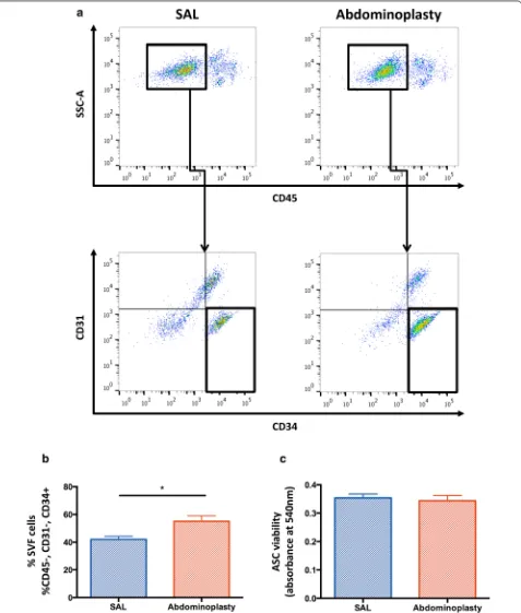
SAL yields a decreased frequency of ASCs
SAL does not compromise ASC viability
A reduction of ASC yield in SAL lipoaspirates, did not result in an impaired ASC viability. An MTT assay was performed to assess the impact of SAL on ASC viability when compared to excision. Cell viability was not signifi-cantly different between ASCs harvested by SAL or exci-sion (p = 0.53) (Fig. 1c).
Osteogenic differentiation potential
In order to determine the osteogenic differentiation potential of ASCs isolated from either SAL or excisional fat samples, ASCs were cultured in ODM for 14 days. There were no significant differences detected in alka-line phosphatase activity after 7 days in ODM (p = 0.44)
(Fig. 2a) or in mineralization of the extracellular matrix at day 14, as measured by Alizarin Red assay (p = 0.06)
(Fig. 2b). Similarly, there were no significant differences in expression of the osteogenic differentiation markers
RUNX-2 or OCN between the SAL- and excision-derived ASCs, at any time points assessed (Fig. 2c).
Adipogenic differentiation potential
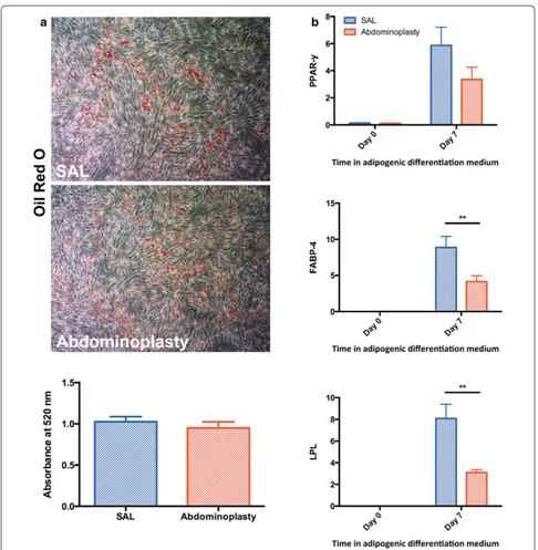
Cells were cultured in ADM for 7 days to determine adi-pogenic differentiation potential of ASCs derived from SAL or excisional fat. Lipid accumulation was confirmed by ORO staining after 7 days of culture in ADM, indi-cating appropriate adipogenic differentiation of ASCs. Quantification of Oil Red O staining showed no sig-nificant differences between the two groups (p = 0.47)
(Fig. 3a). Additionally, RNA was harvested prior to induction of differentiation at day 0 and again after 7 days of culture in ADM to correlate adipogenic marker tran-script expression levels with observed in vitro adipo-genic differentiation. Interestingly, gene expression of the intermediate and late adipogenic differentiation mark-ers FABP-4/AP2 and LPL were significantly enhanced in ASCs harvested by SAL compared to those from exci-sional fat tissue (**p < 0.01) (Fig. 3b). This difference was seen after 7 days of adipogenic induction, but not at day 0 before induction of differentiation in ADM. Similarly, gene expression of the early adipogenic marker PPAR-γ
showed a trend toward greater expression at day 7 in cells derived from SAL, however this did not reach statistical significance (p = 0.06).
SAL and excisional fat derived ASCs equally enhance wound healing
To evaluate the therapeutic functionality of SAL derived ASCs versus ASCs isolated from excisional fat in vivo, cell-seeded hydrogels [5] were applied to a previously established model of murine cutaneous healing [5, 24]. Consistent with their unimpaired in vitro functional-ity, ACSs obtained via SAL demonstrated comparable
therapeutic efficacy for cutaneous regeneration versus cells isolated from abdominoplasty samples (Fig. 4a). Both ASC treatment groups displayed significantly improved healing kinetics as early as day three compared to unseeded hydrogel controls (Fig. 4b). The acceler-ated healing rates directly resulted in significantly faster wound closure times in the ASCs groups (11.4 and 10.8 vs. 13.8 days, **p < 0.01) (Fig. 4c). These data indicate that ASCs derived from either SAL or excision both have a positive effect on in vivo regeneration.
SAL and excisional fat derived ASCs both enhance wound vascularity
Improvement of wound healing by ASCs is widely attrib-uted to enhanced vascularization of the wound bed [17,
25–28]. Indeed, both ASC treatment groups showed sig-nificantly enhanced neovascularization compared with acellular scaffold controls (*p < 0.05), confirming our results regarding in vivo regenerative potential (Fig. 5). Similar to the wound healing outcomes, no signifi-cant differences between the two ASC groups could be detected. This further corroborates that in vivo regenera-tive potential is preserved in SAL-derived ASCs.
Discussion
enable an exact assessment potentially superior to clini-cal comparisons utilizing one-step cell isolation protocols in the operating room.
In this study, we found that ASCs obtained from both SAL and excised abdominoplasty tissue occurred at high frequencies and viability, although excised adipose tissue
provided greater yields of ASCs when compared to SAL. Factors influencing ASC yield have been discussed con-troversially in the literature. In addition to harvesting technique, patient demographics can affect ASC fre-quency in adipose tissue. Generally, there are no detect-able differences in ASC yield or proliferation with age
[image:6.595.56.541.86.572.2][19, 34]. However, high donor age and comorbidities such as diabetes significantly impair ASC functionality [19,
35] and donor gender affects ASC properties, with more robust osteogenic differentiation in ASCs from male patients [36]. Furthermore, previous studies from our group have demonstrated depot-specific differences in
ASCs, with ASCs isolated from the flank and thigh show-ing greater osteogenic potential but ASCs from the flank having lesser adipogenic capabilities when compared to the arm and abdomen [37].
The capability of ASCs to differentiate down multi-ple lineages is of critical importance in their utility in
Fig. 3 SAL derived ASCs have similar adipogenic lineage differentiation capacities. a Representative images and quantification of Oil Red O staining following adipogenic differentiation of SAL and abdominoplasty derived ASCs. b RT‑PCR quantifying the expression of adipogenic markers in vitro.
[image:7.595.56.542.84.581.2]tissue engineering and cell-based regenerative therapies. Previous studies from our group have found no signifi-cant difference in osteogenic differentiation potential between suction-assisted lipoaspiration and third-gen-eration ultrasound-genthird-gen-eration lipoaspiration, despite the mechanical disruption delivered during ultrasound application [17]. In contrast, ASCs derived from LAL have been shown to suffer from decreased osteogenic dif-ferentiation capacity relative to those from SAL [16]. The results from this study demonstrate that SAL does not impair the osteogenic differentiation potential of ASCs. This is not surprising, since SAL delivers a mechanical effect rather than heat, and is thus an approach more similar to UAL than LAL.
Interestingly, we found that expression of adipogenic differentiation markers FABP-4 and LPL were signifi-cantly higher in SAL derived ASCs when compared to those harvested from excisional abdominoplasty fat
tissue. Expression of the early adipogenic marker PPAR-γ was also greater, although not significantly so. Our find-ings corroborate those in a recent study by Keck et al. [38], who determined expression of adiponectin, PPAR-γ, and GLUT4 to be significantly increased in ASCs from PAL, a technique analogous to SAL, when compared to manual aspiration. A potential explanation for these findings may be found in the cellular effects of mecha-notransduction, the conversion of mechanical forces to biochemical signals [39]. It is becoming increasingly probable that ASCs are subject to significant mecha-notransductive effects [40, 41], much as are other progen-itor cell types. However, the literature is still developing in this area and results thus far have been inconsistent and largely focused on either adipocytes or adipose tissue as a whole [42, 43]. Due to the conflicting data regarding mechanotransduction in ASCs, one may turn to the BM-MSC literature for potential clues. Importantly, shear
[image:8.595.59.540.87.453.2]stress has been previously shown to cause changes in cytoskeletal distribution in MSCs, ultimately leading to alterations in differentiation potential. Specifically, Chang et al. [44] found that shear stress led to increased sion of the early adipokine PPAR-γ and decreased expres-sion of the early osteogenic gene RUNX-2 [44]. Here, we see similar effects in ASCs, with trends toward increased PPAR-γ and decreased RUNX-2 expression in ASCs isolated from lipoaspirates when compared to those obtained from excised adipose tissue. Therefore we may conclude that mechanical forces exerted during lipoaspi-ration alters ASC biology, at least at the transcriptional level. However, further work is needed to clarify these potential effects.
ASCs harbor great promise for tissue regenera-tion applicaregenera-tions [5]. In this study we demonstrate that SAL-harvested ASCs have an identical potential for the enhancement of cutaneous healing when compared to ASCs derived from excisional fat. Furthermore, wounds treated with either ASC population displayed signifi-cantly greater vascularity compared to an unseeded
scaffold control group. These promising findings distin-guish SAL as a reliable method for obtaining ASCs suit-able and effective for regenerative medicine approaches, and an equivalent source of ASCs when compared to those derived from three-dimensionally intact adipose tissue.
Conclusion
ASCs represent a promising source of multipotent cells for tissue engineering and regenerative medicine. Suc-tion-assisted lipoaspiration offers a possibility for rela-tive ease of harvest of ASCs with minimal donor site morbidity. Here we show that SAL lipoaspirates provide a slightly decreased yield of viable ASCs when com-pared to resected adipose tissue. ASCs derived from SAL retain full multipotency and regenerative capabili-ties. Overall, these findings suggest that SAL is a reliable and effective method of obtaining ASCs for tissue engi-neering approaches and cell-based therapies when com-pared to the gold standard of minimally-manipulated excisional adipose tissue, and does not damage ASCs in
[image:9.595.58.543.86.423.2]terms of viability, osteogenic and adipogenic differen-tiation capacity, wound regenerative potential, or wound neovascularization.
Authors’ contributions
All listed authors contributed to the idea generation, design, and completion of this work. DD contributed to the idea generation, experimental work and manuscript preparation. AL, RCR, DA, ZNM, EAB, AJW, NH, ML, MSH and GGW contributed to the experimental work. MS, AFS, HGM, GMH, DCW, MTL and GCG guided the idea generation, experimental work and manuscript prepara‑ tion. All authors read and approved the final manuscript.
Author details
1 Hagey Laboratory for Pediatric Regenerative Medicine, Division of Plastic Sur‑ gery, Department of Surgery, Stanford University School of Medicine, Stanford, CA, USA. 2 Section of Plastic, Aesthetic and Reconstructive Surgery, Johannes Kepler University, Linz, Austria. 3 Institute for Stem Cell Biology and Regenera‑ tive Medicine, Stanford University School of Medicine, Stanford, CA, USA. 4 Department of Plastic Surgery and Hand Surgery, Technical University Munich, Munich, Germany.
Acknowledgements
Funding for the stem cell research conducted in our laboratory has been provided by the National Institutes of Health (R01‑DK074095, R01‑AG025016, R01‑DE021683, R21‑DE024230), the Hagey Family Endowed Fund in Stem Cell Research and Regenerative Medicine, and The Oak Foundation. The authors would like to thank Dr. Dean Vistnes at the Kaplan Cosmetic Surgery Center for lipoaspirate sample collection. Cell sorting was completed at the Stanford Shared FACS Facility.
Competing interests
DD and GCG are listed on the patent “Efficient Stem Cell Delivery Into Bioma‑ terials Using a Novel Capillary Driven Encapsulation Technique” and GCG is listed on the patent “Intelligent Biodegradable Pullulan Regenerative Matrix for Tissue Engineering” assigned to Stanford University. AL, RCR, DA, ZNM, EAB, AJW, NH, ML, MSH, GGW, RW, MS, AFS, HGM, GMH, DCW and MTL have no potential conflicts of interest, affiliations or financial involvement with any organization or entity with a financial interest in or financial conflict with the subject matter or materials discussed herein.
Received: 9 February 2016 Accepted: 27 April 2016
References
1. Zuk PA, et al. Multilineage cells from human adipose tissue: implications for cell‑based therapies. Tissue Eng. 2001;7(2):211–28.
2. Zuk PA, et al. Human adipose tissue is a source of multipotent stem cells. Mol Biol Cell. 2002;13(12):4279–95.
3. Aust L, et al. Yield of human adipose‑derived adult stem cells from lipo‑ suction aspirates. Cytotherapy. 2004;6(1):7–14.
4. De Ugarte DA, et al. Comparison of multi‑lineage cells from human adipose tissue and bone marrow. Cells Tissues Organs. 2003;174(3):101–9. 5. Garg RK, et al. Capillary force seeding of hydrogels for adipose‑derived
stem cell delivery in wounds. Stem Cells Transl Med. 2014;3(9):1079–89. 6. Kosaraju R, et al. Adipose‑derived stem cell‑seeded hydrogels increase
endogenous progenitor cell recruitment and neovascularization in wounds. Tissue Eng Part A. 2016;22(3–4):295–305.
7. Cowan CM, et al. Adipose‑derived adult stromal cells heal critical‑size mouse calvarial defects. Nat Biotechnol. 2004;22(5):560–7.
8. Walmsley GG, et al. Surveillance of stem cell fate and function: a system for assessing cell survival and collagen expression in situ. Tissue Eng Part A. 2016;22(1–2):31–40.
9. Garza RM, et al. Studies in fat grafting: part IV. Adipose‑derived stromal cell gene expression in cell‑assisted lipotransfer. Plast Reconstr Surg. 2015;135(4):1045–55.
10. Luan A, et al. Cell‑assisted lipotransfer improves volume retention in irra‑ diated recipient sites and rescues radiation‑induced skin changes. Stem Cells. 2016;34(3):668–73.
11. Paik KJ, et al. Studies in fat grafting: part v. cell‑assisted lipotransfer to enhance fat graft retention is dose dependent. Plast Reconstr Surg. 2015;136(1):67–75.
12. Yoshimura K, et al. Cell‑assisted lipotransfer for cosmetic breast augmen‑ tation: supportive use of adipose‑derived stem/stromal cells. Aesthet Plast Surg. 2008;32(1):48–55 (discussion 56–7).
13. Scuderi N, et al. Power‑assisted lipoplasty versus traditional suction‑ assisted lipoplasty: comparative evaluation and analysis of output. Aesthet Plast Surg. 2005;29(1):49–52.
14. Grazer FM. Suction‑assisted lipectomy, suction lipectomy, lipolysis, and lipexeresis. Plast Reconstr Surg. 1983;72(5):620–3.
15. Collins PC, Field LM, Narins RS. Liposuction surgery and autologous fat transplantation. Clin Dermatol. 1992;10(3):365–72.
16. Chung MT, et al. Isolation of human adipose‑derived stromal cells using laser‑assisted liposuction and their therapeutic potential in regenerative medicine. Stem Cells Transl Med. 2013;2(10):808–17.
17. Duscher D, et al. Ultrasound‑assisted liposuction does not compromise the regenerative potential of adipose‑derived stem cells. Stem Cells Transl Med. 2016;5(2):248–57.
18. Januszyk M, et al. Evaluating the effect of cell culture on gene expression in primary tissue samples using microfluidic‑based single cell transcrip‑ tional analysis. Microarrays. 2015;4(4):540–50.
19. Duscher D, et al. Aging disrupts cell subpopulation dynamics and dimin‑ ishes the function of mesenchymal stem cells. Sci Rep. 2014;4:7144. 20. Suga H, et al. Functional implications of CD34 expression in human adi‑
pose‑derived stem/progenitor cells. Stem Cells Dev. 2009;18(8):1201–10. 21. Levi B, Longaker MT. Concise review: adipose‑derived stromal cells for
skeletal regenerative medicine. Stem Cells. 2011;29(4):576–82. 22. Rustad KC, et al. Enhancement of mesenchymal stem cell angiogenic
capacity and stemness by a biomimetic hydrogel scaffold. Biomaterials. 2012;33(1):80–90.
23. Wong VW, et al. Pullulan hydrogels improve mesenchymal stem cell delivery into high‑oxidative‑stress wounds. Macromol Biosci. 2011;11(11):1458–66.
24. Galiano RD, et al. Quantitative and reproducible murine model of exci‑ sional wound healing. Wound Repair Regen. 2004;12(4):485–92. 25. Duscher D, et al. Stem cells in wound healing: the future of regenerative
medicine? A Mini‑Review. Gerontology. 2016;62(2):216–25.
26. Hu MS, et al. Stem cell‑based therapeutics to improve wound healing. Plast Surg Int. 2015;2015:383581.
27. Bhang SH, et al. Angiogenesis in ischemic tissue produced by sphe‑ roid grafting of human adipose‑derived stromal cells. Biomaterials. 2011;32(11):2734–47.
28. Hadjipanayi E, Schilling AF. Hypoxia‑based strategies for angiogenic induction: the dawn of a new era for ischemia therapy and tissue regen‑ eration. Organogenesis. 2013;9(4):261–72.
29. Aronowitz JA, Lockhart RA, Hakakian CS. Mechanical versus enzymatic isolation of stromal vascular fraction cells from adipose tissue. Springer‑ plus. 2015;4:713.
30. Strong AL, et al. The current state of fat grafting: a review of har‑ vesting, processing, and injection techniques. Plast Reconstr Surg. 2015;136(4):897–912.
31. Domenis R, et al. Adipose tissue derived stem cells: in vitro and in vivo analysis of a standard and three commercially available cell‑assisted lipotransfer techniques. Stem Cell Res Ther. 2015;6:2.
32. Shridharani SM, Broyles JM, Matarasso A. Liposuction devices: technology update. Med Devices (Auckl). 2014;7:241–51.
33. Nagy MW, Vanek PF Jr. A multicenter, prospective, randomized, single‑ blind, controlled clinical trial comparing VASER‑assisted Lipoplasty and suction‑assisted Lipoplasty. Plast Reconstr Surg. 2012;129(4):681e–9e. 34. Mojallal A, et al. Influence of age and body mass index on the yield and
proliferation capacity of adipose‑derived stem cells. Aesthet Plast Surg. 2011;35(6):1097–105.
• We accept pre-submission inquiries
• Our selector tool helps you to find the most relevant journal
• We provide round the clock customer support
• Convenient online submission
• Thorough peer review
• Inclusion in PubMed and all major indexing services
• Maximum visibility for your research
Submit your manuscript at www.biomedcentral.com/submit
Submit your next manuscript to BioMed Central
and we will help you at every step:
36. Aksu AE, et al. Role of gender and anatomical region on induction of osteogenic differentiation of human adipose‑derived stem cells. Ann Plast Surg. 2008;60(3):306–22.
37. Levi B, et al. Depot‑specific variation in the osteogenic and adipogenic potential of human adipose‑derived stromal cells. Plast Reconstr Surg. 2010;126(3):822–34.
38. Keck M, et al. Power assisted liposuction to obtain adipose‑derived stem cells: impact on viability and differentiation to adipocytes in comparison to manual aspiration. J Plast Reconstr Aesthet Surg. 2014;67(1):e1–8. 39. Duscher D, et al. Mechanotransduction and fibrosis. J Biomech.
2014;47(9):1997–2005.
40. Yilgor Huri P, et al. Biophysical cues enhance myogenesis of human adipose derived stem/stromal cells. Biochem Biophys Res Commun. 2013;438(1):180–5.
41. Bodle JC, Hanson AD, Loboa EG. Adipose‑derived stem cells in functional bone tissue engineering: lessons from bone mechanobiology. Tissue Eng Part B Rev. 2011;17(3):195–211.
42. Shoham N, Gefen A. Mechanotransduction in adipocytes. J Biomech. 2012;45(1):1–8.
43. Atashroo D, et al. Studies in fat grafting: part II. Effects of injec‑ tion mechanics on material properties of fat. Plast Reconstr Surg. 2014;134(1):39–46.