RESEARCH
A proof-of-concept study with the
tyrosine kinase inhibitor nilotinib
in spondyloarthritis
Jacqueline E. Paramarta
1*, Maureen C. Turina
1, Troy Noordenbos
1,2, Tanja F. Heijda
1, Iris C. Blijdorp
1,2,
Nataliya Yeremenko
1,2and Dominique Baeten
1,2Abstract
Background: To evaluate the immunomodulating and clinical effects of nilotinib, a tyrosine kinase inhibitor, in a proof-of-concept study in spondyloarthritis (SpA) assessing the mast cell as potential novel therapeutic target in this disease.
Methods: Twenty eight patients with active peripheral (pSpA) and/or axial SpA (axSpA) were included in a rand-omized, double-blind, placebo-controlled clinical trial (Trial registration: Trialregister.nl NTR2834). Patients were treated 1:1 with nilotinib or placebo for 12 weeks, followed by an open label extension for another 12 weeks. Paired synovial tissue biopsies, serum sampling and assessment of clinical symptoms were performed serially.
Results: In pSpA (n = 13) synovial inflammation appeared to diminish after 12 weeks of nilotinib treatment as evidenced by histopathology (decrease in number of infiltrating CD68+ and CD163+ macrophages and mast cells). Compared to placebo mRNA expression of c-Kit as mast cell marker (p = 0.037) and of pro-inflammatory cytokines such as IL-6 (p = 0.024) were reduced. The reduction of synovial inflammation was paralleled by a decrease in serum biomarkers of inflammation such as C-reactive protein (p = 0.024) and calprotectin (p = 0.055). Also clinical param-eters such as patient’s global assessment of disease activity (p = 0.031) and ankylosing spondylitis disease activity score (p = 0.031) showed improvement upon 12 weeks of nilotinib but not placebo treatment. This improvement was further augmented at week 24. In contrast to pSpA, neither serum biomarkers of inflammation nor clinical parameters improved upon nilotinib treatment in axSpA. During the trial one serious adverse event occurred, which was consid-ered unrelated to the study drug.
Conclusions: This small proof-of-concept study suggests that nilotinib treatment modulates inflammation and clini-cal symptoms in pSpA. A similar effect was not seen in axSpA.
Trial registration: trialregister.nl registration code NTR2834 registered 31 March 2011
Keywords: Spondyloarthritis, Nilotinib, Tyrosine kinase inhibitor, Randomized controlled trial, Mast cells
© 2016 The Author(s). This article is distributed under the terms of the Creative Commons Attribution 4.0 International License (http://creativecommons.org/licenses/by/4.0/), which permits unrestricted use, distribution, and reproduction in any medium, provided you give appropriate credit to the original author(s) and the source, provide a link to the Creative Commons license, and indicate if changes were made. The Creative Commons Public Domain Dedication waiver (http://creativecommons.org/ publicdomain/zero/1.0/) applies to the data made available in this article, unless otherwise stated.
Background
The introduction of tumor necrosis factor (TNF) inhibi-tors has improved the management of
spondyloarthri-tis (SpA) tremendously [1]. However, some patients do
not respond sufficiently to TNF inhibitors or experience side effects. Also, treatment discontinuation leads to
fast relapse of disease [2, 3], and pathological
osteopro-liferation continues under anti-TNF treatment [4–8].
Accordingly, there is still an unmet medical need for new therapies.
Immunopathological studies on peripheral SpA (pSpA) synovitis recently identified the mast cell as potential therapeutic target. Mast cells are key players of the innate immune system and produce and secrete a variety of
Open Access
*Correspondence: j.e.paramarta@amc.uva.nl
1 Department of Clinical Immunology and Rheumatology,
Academic Medical Center/University of Amsterdam, Meibergdreef 9, 1105 AZ Amsterdam, The Netherlands
cytokines [9, 10]. Besides their well-established role in allergies, these cells also play an important role in
rheu-matoid synovitis [9–13]. We recently proposed that mast
cells might be more important in SpA than in
rheuma-toid arthritis (RA) based on the following: [13] Firstly,
the infiltration with c-Kit + mast cells in SpA synovitis
is markedly higher compared to RA. Secondly, this infil-tration is already observed in early disease and is not affected by effective anti-TNF treatment. Thirdly, mast cells are the major interleukin (IL)-17 expressing cells in pSpA and the proportion of mast cells expressing IL-17 is significantly higher in SpA than RA synovitis. Fourthly, proof-of-concept studies demonstrated that IL-17A blockade effectively down-modulates inflammation and clinical symptoms in the ankylosing spondylitis (AS) and
psoriatic arthritis (PsA) subtypes of SpA [14–16]. Finally,
sulfasalazine, the only disease-modifying anti-rheumatic
drug (DMARD) with proven efficacy in pSpA [17], has
shown to inhibit degranulation and TNF secretion by
mast cells [18, 19].
Mast cells can be targeted in vivo by tyrosine kinase inhibitors such as imatinib and nilotinib, which are reg-istered for the treatment of chronic myeloid leukemia
(CML) [20, 21]. Originally developed to inhibit c-Abl
on malignant leucocytes, these drugs also appeared to inhibit c-Kit, the receptor for stem cell factor, thereby inducing apoptosis of mast cells, including synovial
mast cells [22]. Accordingly, we recently demonstrated
in ex vivo biopsy tissue cultures that imatinib strongly reduced spontaneous production and secretion of pro-inflammatory cytokines including IL-6, IL-8 and IL-17 by
SpA synovium [13]. In line with this data, a small open
label trial with imatinib in six SpA patients showed a decrease in clinical and serum markers of disease activity
upon 3 months of treatment [23].
The objective of the present study was to evaluate mast cells as potential therapeutic target in SpA by con-ducting a proof-of-concept randomized controlled trial with nilotinib. Nilotinib is a second-generation tyros-ine kinase inhibitor which is more effective in the treat-ment of CML and has a better safety profile compared to
imatinib [24, 25]. As the rational for tyrosine kinase
inhi-bition is mainly based on synovial studies, we explored the immunomodulating effects on synovial histopathol-ogy, systemic inflammation, and symptoms of pSpA. Additionally, we evaluated clinical efficacy in axial SpA (axSpA) in this exploratory study.
Methods
Patients and study design
Twenty-eight patients diagnosed with SpA by their treat-ing rheumatologist and fulfilltreat-ing the European
Spon-dyloarthropathy Study Group (ESSG) criteria [26] were
included in a single centre, double-blind, investigator ini-tiated clinical trial and randomized (1:1) to receive nilo-tinib (Tasigna; Novartis Pharmaceuticals) 400 mg twice daily or matching placebo capsules (Tiofarma B.V.) for 12 weeks, followed by an open label extension with nilo-tinib for another 12 weeks. The Assessment of Spondy-loarthritis International Society (ASAS) criteria were not yet published when this clinical trial was designed, there-for the ESSG criteria were used in this study. Patients were considered to have pSpA when they had an arthri-tis, to have axSpA when they had inflammatory back pain, and combined disease if they had both an arthritis and inflammatory back pain. Patients were between 18 and 65 years old and had active disease despite treatment with non-steroidal anti-inflammatory drugs (NSAIDs). Active disease was defined as: patient’s and physician’s
global assessment of disease activity of ≥40 mm, as
well as ≥1 swollen and ≥1 tender joint in case of pSpA
(n = 11), Bath ankylosing spondylitis disease activity
index (BASDAI) of ≥4 in case of axSpA (n = 15), or both
in case of combined disease (n = 2). The cohort included
ten AS, ten PsA, and eight undifferentiated SpA patients.
Stable doses of NSAIDs, corticosteroids (≤10 mg/day
prednisone equivalent), methotrexate, sulfasalazine and leflunomide were allowed during the trial, but not intra-articular corticosteroids. Prior anti-TNF therapy (in case the reason for discontinuation was not primary
failure) was permitted after a washout period (≥4 weeks
in case of etanercept, ≥8 weeks in case of infliximab and
≥10 weeks for the other TNF inhibitors). All patients
gave written informed consent to participate in the study as approved by the Ethics Committee of the Academic Medical Center/University of Amsterdam (Trialregister. nl registration code NTR2834). Clinical characteristics at
baseline are summarized in Table 1.
Synovial immunopathology
Synovial biopsies were obtained by mini-arthroscopy at baseline, weeks 12 and 24 in pSpA patients with active
knee or ankle arthritis (n = 8) as described previously
[27, 28]. Samples (6–8 per patient) were either
snap-frozen in Tissue-Tek® O.C.T.™ (Sakura) for histological
evaluation or immediately stored in liquid nitrogen for subsequent RNA extraction and gene expression analysis.
peroxidise link, aminoethylcarbazole substrate as chro-mogen (all Dako), and hematoxylin as counterstain. Parallel sections were incubated with isotype and con-centration-matched monoclonal antibodies as nega-tive controls. Samples were stained in a single run to minimize technical biases. Stained sections were scored semiquantitatively for cellular infiltration by three inde-pendent observers (NY, ICB and DB) who were blinded to the patient’s treatment allocation and treatment
dura-tion, as described previously [29–31].
For gene expression analysis, mRNA was extracted using RNA Stat-60 (Tel-Test), then treated with DNase I (Invitrogen) and reverse-transcribed using a Rever-tAid H Minus First Strand complementary DNA Syn-thesis Kit (Fermentas). The RNA concentration was determined with a NanoDrop spectrophotometer. Analy-sis of mRNA by qPCR was performed using a StepOne-Plus Real-Time PCR System (Applied Biosystems) using GAPDH as housekeeping gene. Predesigned TaqMan probe and primer sets for IL-6 (Hs00174131_m1), IL-8 (Hs00174103_m1), TNF (Hs00174128_m1), IL-17A (Hs00174383_m1), IL-17F (Hs00369400_m1), IL-23 (Hs00372324_m1), c-Kit (Hs00174029_m1), and GAPDH
(4310884E) were assayed according to the manufacturer’s protocol (Applied Biosystems).
Serum biomarkers
Serum analysis included safety measurements (liver function, renal function, blood cell counts) as well as C-reactive protein (CRP) and erythrocyte sedimentation rate (ESR). Serum levels of matrix metalloproteinase-3 (MMP-3, Biotrak, Amersham Pharmacia Biotech) and calprotectin (Hycult Biotech), two potential
biomark-ers of inflammation in SpA [32, 33], were measured by
enzyme-linked immunosorbent assays (ELISAs) accord-ing to the manufacturer’s instructions.
Clinical assessments
Clinical assessments consisted of safety evaluation (con-sisting of the patient’s history, physical examination, laboratory tests, urinalysis and electrocardiograms), patient’s and physician’s global assessment of disease activity on a visual analogue scale, ankylosing spondylitis disease activity score (ASDAS) and ASDAS improvement
criteria [34, 35] in all patients. This was complemented by
swollen and tender joint count (SJC66 and TJC68) in case
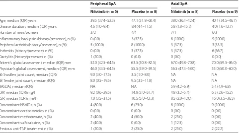
Table 1 Baseline characteristics of the study population by treatment group
Significance of the comparisons is determined by Mann–Whitney U test for continuous variables and Fisher’s exact test for categorical variables. There were no significant differences between the nilotinib and placebo groups
SpA spondyloarthritis; IQR interquartile range; BASDAI bath ankylosing spondylitis disease activity Index; CRP C-reactive protein; ESR erythrocyte sedimentation rate;
NSAIDs non-steroidal anti-inflammatory drugs; TNF tumor necrosis factor; NA not applicable
Peripheral SpA Axial SpA
Nilotinib (n = 5) Placebo (n = 8) Nilotinib (n = 8) Placebo (n = 9)
Age, median (IQR) years 39.5 (37.4–52.3) 47.1 (31.8–60.4) 38.0 (36.1–42.4) 40.1 (34.5–46.7) Disease duration, median (IQR) years 4.6 (1.0–9.4) 8.4 (4.4–11.5) 5.8 (1.8–15.3) 4.0 (1.6–12.7)
Number of men/women 3/2 4/4 7/1 6/3
Inflammatory back pain (history/presence), n (%) 0 (0.0) 3 (37.5) 8 (100.0) 9 (100.0) Peripheral arthritis (history/presence), n (%) 5 (100.0) 8 (100.0) 3 (37.5) 3 (33.3) Enthesitis (history/presence), n (%) 0 (0.0) 3 (37.5) 3 (37.5) 6 (66.7)
Dactylitis (history/presence), n (%) 1 (20.0) 0 (0.0) 0 (0.0) 0 (0.0)
Patient’s global assessment, median (IQR) mm 52.0 (42.5–64.5) 63.5 (50.8–82.5) 67.0 (49.8–70.8) 70.0 (59.5–86.0) Physician’s global assessment, median (IQR) mm 46.0 (43.5–64.5) 55.5 (49.0–59.5) 56.5 (47.3–58.0) 55.0 (50.0–60.0) 66 Swollen joint count, median (IQR) 9.0 (3.0–17.5) 3.5 (1.0–8.0) NA NA
68 Tender joint count, median (IQR) 8.0 (0.5–19.5) 9.5 (3.5–11.8) NA NA
BASDAI, median (IQR) NA NA 5.9 (4.2–6.9) 5.4 (4.9–6.8)
CRP, median (IQR) mg/l 9.2 (0.6–29.5) 14.8 (3.0–51.7) 4.8 (3.2–5.4) 6.3 (2.6–15.2) ESR, median (IQR) mm/h 7.0 (3.5–31.5) 10.5 (5.0–42.3) 8.5 (2.0–12.0) 16.0 (3.5–36.5)
Concomitant NSAIDs, n (%) 4 (80.0) 6 (75.0) 8 (100.0) 9 (100.0)
Concomitant corticosteroids, n (%) 0 (0.0) 0 (0.0) 0 (0.0) 0 (0.0)
Concomitant methotrexate, n (%) 2 (40.0) 4 (50.0) 2 (25.0) 0 (0.0)
Concomitant sulfasalazine, n (%) 2 (40.0) 0 (0.0) 1 (12.5) 0 (0.0)
[image:3.595.59.540.101.369.2]of pSpA, and BASDAI and BASDAI50 response in case
of axSpA [36, 37].
Statistical analysis
Data are presented as the median and interquartile range (IQR). pSpA and axSpA were analyzed separately as the rational for tyrosine kinase inhibition is mainly based on synovial studies in pSpA and the assessment in axSpA was more exploratory. In case of combined SpA the patient’s data were included in both groups. The nilo-tinib and placebo group were compared to each other by Mann–Whitney U tests for continuous variables and Fisher’s exact test for categorical variables. Differences between various time points were assessed by Wilcoxon matched pairs tests by treatment group. For biological data (synovial immunopathology and systemic biomark-ers) the weeks 0–12 data of the original nilotinib group were pooled with the weeks 12–24 data of the original placebo group (which was treated with nilotinib from week 12 onwards) to increase the power of these proof-of-concept analyses. The clinical data, which are more sensitive to placebo effects, were not pooled but analyzed
as observed with the last observation carried forward. P
values of <0.05 were considered statistically significant,
and p values <0.1 and ≥0.05 were considered to represent
a trend.
Results
Immunomodulation of synovial inflammation by nilotinib treatment
To assess the modulation of synovial inflammation by nilotinib, synovial biopsies were obtained before and after treatment in pSpA patients with knee or ankle arthritis. Since the number of snap-frozen biopsies which passed quality control for immunohistological
analy-sis was small (n = 3 in the nilotinib group and n = 4 in
the placebo group) we only used descriptive analyses for
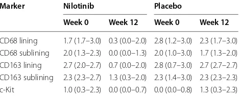
this part of the analysis. As shown in Table 2, the
num-ber of infiltrating CD68+ and CD163+ macrophages,
which are markers for synovial inflammation in SpA [29,
30, 38], numerically decreased in both the lining layer
and the synovial sublining upon nilotinib treatment. In contrast, there was no consistent modulation of synovial macrophage numbers between baseline and week 12 of placebo treatment. Nilotinib treatment also decreased
the number of c-Kit + synovial mast cells, while there
was a numerical increase after placebo treatment. This was confirmed by qPCR analysis of mRNA expression, as c-Kit expression showed a significant decrease upon nilo-tinib treatment but augmented upon placebo treatment
over 12 weeks (p = 0.037) (Fig. 1a). Additionally,
nilo-tinib treatment induced a decrease of the synovial mRNA expression of the pro-inflammatory cytokines IL-6
(p = 0.024) and IL-23 (p = 0.024) compared to placebo,
but not of IL-8 (p = 0.378) and TNF (p = 0.500) (Fig. 1b–
e). Expression levels of IL-17A and IL-17F were too low (even in the baseline biopsies) to allow reliable detection.
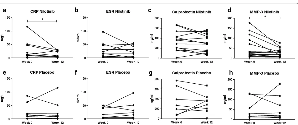
Modulation of systemic inflammation by nilotinib treatment in pSpA
As the tissue analysis indicated that nilotinib down-reg-ulates synovial inflammation, we next examined whether systemic biomarkers of inflammation are also
modu-lated by nilotinib in pSpA (Fig. 2). CRP remained stable
in the placebo group but decreased significantly from 9.2
(IQR 1.7–33.1) to 5.2 (IQR 1.7–25.1) mg/l (p = 0.024)
upon 12 weeks of nilotinib treatment. This was particu-larly marked in patients with high levels at baseline. CRP levels decreased even further after 24 weeks of nilotinib
treatment to 4.0 (IQR 0.4–25.5) mg/l (p = 0.031
com-pared to baseline). The effect on ESR (p = 0.141) was less
clear. Calprotectin showed a trend towards improvement after 12 weeks treatment with nilotinib decreasing from 359.9 (IQR 183.3–484.9) to 287.9 (IQR 116.7–457.1) ng/
ml (p = 0.055), but stayed stable in the placebo group.
MMP-3 also mainly decreased in patients with high lev-els at baseline, but overall the median showed a slight increase after 12 weeks of nilotinib from 28.5 (IQR 11.6–
104.8) to 29.3 (IQR 12.3–46.0) ng/ml (p = 0.034).
Improvement of clinical symptoms after nilotinib treatment in pSpA
In line with the synovial tissue and serum biomarker analyses nilotinib treatment induced a significant
reduc-tion in patient’s global assessment at week 12 (p = 0.031)
as well as after an additional 12 weeks of open label
treat-ment with nilotinib (p = 0.031) (Fig. 3a). Moreover, the
placebo treated patients did not show any changes in patient’s global assessment during the first phase of the study but showed significant improvement after entering
the open label phase with nilotinib treatment (p = 0.012)
(Fig. 3a). The physician’s global assessment showed a
Table 2 Immunomodulatory effect of nilotinib versus pla-cebo treatment on synovial histopathology
Values are the median (IQR) assessed on a semiquantitative scale
Marker Nilotinib Placebo
Week 0 Week 12 Week 0 Week 12
[image:4.595.305.540.114.210.2]trend towards improvement at week 12 (p = 0.063) and
improved significantly at week 24 (p = 0.031) (Fig. 3b).
The SJC66 and TJC68 decreased numerically between week 0 and 12 of nilotinib but not placebo treatment and decreased further at week 24, which was
signifi-cant for the SJC66 (p = 0.049). After entering the open
label phase with nilotinib the originally placebo treated patients also showed significant improvement in SJC66
(p = 0.031) (Fig. 3c, d). The ASDAS, a composite
meas-ure originally developed for axSpA but which was also
shown to be useful in pSpA [39], improved after 12 weeks
of nilotinib treatment (p = 0.031), which was not the case
for placebo (p = 0.371) (Fig. 3e). ASDAS clinically
impor-tant improvement was reached by 40.0 % of the nilotinib
group at week 12 (Fig. 3f) and by 53.8 % of the total study
population at week 24.
c-kit Nilotinib
0 2 4 6
Week 0 Week 12
mR NA re la tive expr ess io n c-kit Placebo 0 2 4 6
Week 0 Week 12
mR NA re la tive expr ess io n IL-6 Nilotinib 0 2 4 6
Week 0 Week 12
mR NA re la tive expr essi on IL-6 Placebo 0 2 4 6
Week 0 Week 12
mR NA re la tive expr essi on IL-8 Nilotinib 0 2 4 6 8
Week 0 Week 12
mR NA re la tive exp re ssi on IL-8 Placebo 0 2 4 6 8
Week 0 Week 12
mR NA re la tive expr essi on TNF Placebo 0 1 2 3
Week 0 Week 12
mR NA re la tive expr essi on TNF Nilotinib 0 1 2 3
Week 0 Week 12
mR NA re la tive expr essi on IL-23 Placebo 0.0 0.5 1.0 1.5 2.0
Week 0 Week 12
mR NA re la tive expr essi on IL-23 Nilotinib 0.0 0.5 1.0 1.5 2.0
Week 0 Week 12
mR NA re la tive expr ess io n
a b c d e
f g h i j
Fig. 1 Synovial tissue mRNA expression in peripheral spondyloarthritis. Effect of nilotinib and placebo treatment on in vivo synovial tissue mRNA expression in peripheral spondyloarthritis as assessed by quantitative polymerase chain reaction. The panel represents the transcription of c-Kit, interleukin-6 (IL-6), IL-8, tumor necrosis factor (TNF), and IL-23 before and after treatment with nilotinib (a–e) or placebo (f–j). The lines connect the data points for each patient between weeks (week) 0 and 12
CRP Nilotinib
Week 0 Week 12
0 50 100 150 * mg /l ESR Nilotinib
Week 0 Week 12
0 50 100 150 mm/ h CRP Placebo
Week 0 Week 12
0 50 100 150 mg /l ESR Placebo
Week 0 Week 12
0 50 100 150 mm/ h MMP-3 Nilotinib
Week 0 Week 12 0 50 100 150 200 * ng/ ml MMP-3 Placebo
Week 0 Week 12 0 50 100 150 200 ng /m l Calprotectin Nilotinib
Week 0 Week 12 0 200 400 600 800 ng/ ml Calprotectin Placebo
Week 0 Week 12 0 200 400 600 800 ng /m l
a b c d
e f g h
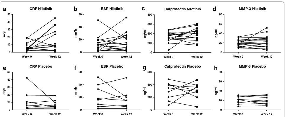
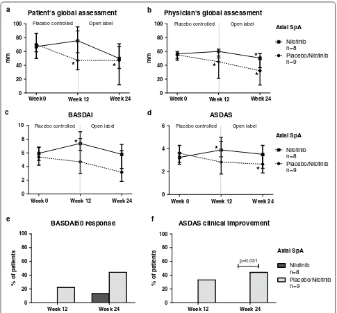
[image:5.595.61.539.86.261.2] [image:5.595.62.538.334.535.2]Lack of effect of nilotinib treatment in axSpA
Although the rational to study nilotinib treatment in SpA was based on synovial tissue findings in pSpA, we also explored the effect of nilotinib in axSpA since the published open label trial with imatinib also showed
improvement in axial symptoms [23]. In contrast with
pSpA, however, serum CRP (baseline 4.2 (IQR 2.9–8.0)
mg/l versus week 12 7.8 (IQR 2.4–25.5) mg/l, p = 0.054)
as well as the other tested systemic biomarkers of inflam-mation did not show a reduction but even an increase
after nilotinib treatment (Fig. 4). More importantly,
the clinical parameters did not improve upon nilotinib treatment, while the placebo response was
unexpect-edly high for this patient group (Fig. 5). Patient’s and
Swollen joint count
Week 0 Week 12 Week 24 0
5 10 15
20 Placebo controlled Open label
*
*
nu
mb
er
of
jo
in
ts
Tender joint count
Week 0 Week 12 Week 24 0
5 10 15 20
nu
mb
er
of
jo
in
ts
Placebo controlled Open label Patient's global assessment
0 20 40 60 80
100 Placebo controlled Open label
*
*
*
Week 0 Week 12 Week 24
mm
Physician's global assessment
0 20 40 60 80
100 Placebo controlled Open label
Week 0 Week 12 Week 24
*
*
mm
ASDAS clinical improvement
Week 12 Week 24
0 20 40 60 80 100
%
of
pa
ti
en
ts
a b
c
n=5
n=8 Nilotinib
Placebo/Nilotinib
Peripheral SpA ASDAS
Week 0 Week 12 Week 24
0 2 4 6
*
Placebo controlled Open label
e f
Nilotinib n=5
Placebo/Nilotinib n=8
Peripheral SpA
Nilotinib n=5
Placebo/Nilotinib n=8
Peripheral SpA d
[image:6.595.58.540.85.553.2]physician’s global assessment were unchanged at week 12 for the nilotinib arm but, surprisingly, was significantly decreased in the placebo group (from 70 (IQR 60–86)
to 47 (IQR 34–96) mm, p = 0.046, and from 55 (IQR
50–60) to 45 (IQR 21–54) mm, p = 0.010, respectively).
BASDAI (from 5.9 (IQR 4.2–6.9) to 7.4 (IQR 6.3–8.1),
p = 0.039) and ASDAS (from 3.2 (IQR 2.6–3.6) to 3.9
(IQR 3.3–5.0), p = 0.020) were even increased at week
12 in the nilotinib treated patients. ASDAS clinically important improvement at week 12 was reached more often in the placebo group (33.3 %) than in the nilotinib
group (0 %) (p = 0.072), with a similar pattern for
BAS-DAI50 response (22.2 versus 0 % respectively, p = 0.156).
This was even more striking at week 24 in which ASDAS clinically important improvement was reached in 44.4 % of the placebo treated patients compared to 0 % in the
nilotinib group (p = 0.031), with again a similar pattern
for the BASDAI50 response (44.4 versus 12.5 %
respec-tively, p = 0.149). Collectively, these data indicate a lack
of effect of nilotinib on axSpA.
Safety analysis
Although the present study is too small in size and dura-tion to come to stringent safety conclusions, there were no unexpected safety signals in comparison with already
available large scale data in CML [24, 25]. The overall
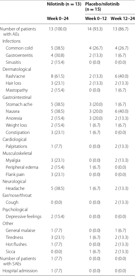
number of adverse events (AEs) was high in both the
nilotinib and placebo group (Table 3). The most
com-mon AEs were headache, dermatologic conditions (rash or acne), and gastrointestinal complaints (stomach ache or nausea). In contrast to the CML trials, haematological
and biochemical AEs occurred rarely (anemia, throm-bocytopenia, hypomagnesemia, and lipase elevation occurred in one patient each). Significant prolongation of the QT-interval on the electrocardiogram did not occur. All AEs were transient but dose reduction (from 400 mg twice daily to 400 mg once daily) was needed in seven patients (mostly temporarily), after which the AEs resolved. One patient developed an acute cholecystitis at week 21 for which he was hospitalized; this was consid-ered a serious adverse event unrelated to the study drug.
Discussion
We recently proposed that mast cells may contribute to SpA synovitis by indicating that the number of infiltrat-ing mast cells was specifically increased in SpA versus RA synovitis, SpA synovial mast cells contained IL-17A as assessed by immunostaining, and ex vivo targeting of mast cells with imatinib reduced inflammation in
syno-vial biopsy cultures [13]. The present study supports
the role of mast cells in synovial inflammation by dem-onstrating histological, biological and clinical effects of nilotinib treatment in pSpA. Tyrosine kinase inhibi-tors such as nilotinib and imatinib target c-Kit, which is crucial for the survival of mast cells. In agreement with previous ex vivo studies demonstrating that imatinib
induces apoptosis of synovial mast cells [22], in vivo
treatment with nilotinib induced a decrease in the num-ber of synovial mast cells and in c-Kit mRNA expression in pSpA. This was associated with a decrease in infiltrat-ing macrophages, synovial expression of pro-inflamma-tory cytokines such as IL-6, systemic CRP levels, and
CRP Nilotinib
Week 0 Week 12 0
10 20 30 40 50
mg
/L
CRP Placebo
Week 0 Week 12
0 10 20 30 40 50
mg
/L
ESR Nilotinib
Week 0 Week 12
0
20 40 60
mm
/h
ESR Placebo
Week 0 Week 12 0
20 40 60
mm
/h
MMP-3 Nilotinib
Week 0 Week 12 0
20 40 60 80
ng
/m
l
MMP-3 Placebo
Week 0 Week 12
0 20 40 60 80
ng
/m
l
Calprotectin Nilotinib
Week 0 Week 12
0 200 400 600 800
ng
/m
l
Calprotectin Placebo
Week 0 Week 12 0
200 400 600
ng
/m
l
a b c d
e f g h
Fig. 4 Serum biomarkers in axial spondyloarthritis. Effect of nilotinib and placebo on serum biomarkers of patients with axial spondyloarthritis.
The panel represents the C-reactive protein (CRP), erythrocyte sedimentation rate (ESR), calprotectin and matrix metalloproteinase-3 (MMP-3)
levels before (week 0) and after treatment (week 12) with nilotinib (a–d) or placebo (e–h). The lines connect the data points for each patient. *P
[image:7.595.58.540.86.284.2]clinical disease activity parameters. Importantly, the immunomodulatory effect of nilotinib was consistent across biological and clinical measurements and was not observed in the placebo group. Together with the
pub-lished small open label study with imatinib [23], these
data indicate that tyrosine kinase inhibitors targeting mast cells appear to be able to suppress inflammation in pSpA and thereby support the potential role of mast cells in SpA pathogenesis.
Due to the proof-of-concept design, this study has limitations which should be considered when interpret-ing the data. Firstly, the number of pSpA patients in the double-blind phase was small, particularly in the syno-vial evaluation as the quality of the synosyno-vial biopsies was insufficient in a number of patients. Therefore the data for the biological parameters were pooled (weeks 0–12 of the originally nilotinib treated patients with weeks 12–24 of the patients receiving placebo originally and BASDAI50 response
Week 12 Week 24
0 20 40 60 80 100
%
of
pa
tien
ts
BASDAI
Week 0 Week 12 Week 24
0 2 4 6 8
10 Placebo controlled Open label
*
ASDAS
Week 0 Week 12 Week 24
0 2 4
6 Placebo controlled Open label
*
*
Patient's global assessment
0 20 40 60 80
100 Placebo controlled Open label
Week 0 Week 12 Week 24
*
*
mm
Physician's global assessment
0 20 40 60 80 100
Week 0 Week 12 Week 24
Placebo controlled Open label
*
*
*
mm
ASDAS clinical improvement
Week 12 Week 24
0 20 40 60 80 100
p=0.031
%
of
pa
ti
en
ts
c
a b
n=8
n=9 Nilotinib
Placebo/Nilotinib
Axial SpA
Nilotinib
Placebo/Nilotinib n=8
n=9
Axial SpA
Nilotinib
Placebo/Nilotinib n=8
n=9
Axial SpA
f e
d
[image:8.595.56.541.86.537.2]subsequently nilotinib). Secondly, the week 12 primary endpoint was perhaps too short. Indeed, both CRP and clinical parameters tended to decrease even further after an additional 12 weeks of treatment. Thirdly, the study did not allow to define mechanistically how nilotinib
exerted its immunomodulatory effects. Even though our analysis suggested an effect on synovial IL-23 but not TNF expression, the number of good quality syno-vial biopsies was too small to determine reliably which inflammatory pathways are or are not modulated by nilo-tinib. For example, mRNA expression of key cytokines such as IL-17A and IL-17F was too low for reliable analysis. Furthermore, c-Kit is not only expressed on mast cells but on other immune cells as well, including innate lymphoid cells, which can express and produce
pro-inflammatory mediators [40]. Moreover, nilotinib
can also target other tyrosine kinases such as c-Fms and platelet derived growth factor receptor (PDGF-R).
C-Fms is expressed on CD163+ macrophages, which
are significantly increased in SpA synovitis [29, 41, 42],
and PDGF-R is a key molecule on myofibroblasts, which were recently shown to be specifically increased in SpA
versus RA synovitis [43]. Since targeting mast cells,
innate lymphoid cells, CD163+ macrophages, as well as
myofibroblasts all might be beneficial in pSpA, it remains unknown how nilotinib actually reduces synovial and systemic inflammation. Finally, the potency of nilotinib compared to other treatments for pSpA such as
sulfasala-zine [17] or TNF blockade [39] is unknown, as we did not
include an active comparator group.
Strikingly, the biological and clinical effects observed in pSpA were completely absent in axSpA. This is in agree-ment with the concept that peripheral and axSpA might be driven by slightly distinct cellular and molecular
mecha-nisms [44]. For example, the major cellular source of IL-17
in pSpA are mast cells and to a lesser degree neutrophils
[13], while in axSpA neutrophils and myeloperoxidase
(MPO)+ cells are the major IL-17 expressing cells [45]. The
discrepant response to nilotinib between pSpA and axSpA is also in line with previous data with sulfasalazine, which
also targets mast cells [18, 19] and has proven clinical
effi-cacy in peripheral but not axial disease [17]. Taken together,
the histopathology and the studies with sulfasalazine and nilotinib point towards partially distinct inflammatory pathways in peripheral versus axial disease. However when interpreting these data, it must be considered that it was not the intention of the current trial to compare the efficacy of nilotinib between pSpA and axSpA, but between nilo-tinib and placebo in an exploratory fashion, hence pSpA and axSpA were not compared to each other. The effect of nilotinib on enthesitis, dactylitis and extra-articular mani-festations (psoriasis, inflammatory bowel disease, uveitis) of SpA remains to be investigated.
A final observation was the pronounced placebo response in axSpA, which was unusually high compared
to what is commonly reported [46–48]. Although we do
not have a clear explanation for this finding, this observa-tion indicates that open label trials in axSpA should be
Table 3 AEs possibly related to nilotinib, occurring in more than one patient and all SAEs
Values are the number (percentage) of patients. The SAE concerned one case of hospital admission following laparoscopic surgery because of acute cholecystitis. The placebo/nilotinib treated patients received placebo from week 0 until week 12, and nilotinib from week 12 until week 24
AE adverse event; SAE serious adverse event
Nilotinib (n = 13) Placebo/nilotinib (n = 15)
Week 0–24 Week 0–12 Week 12–24
Number of patients
with AEs 13 (100.0) 14 (93.3) 13 (86.7) Infections
Common cold 5 (38.5) 4 (26.7) 4 (26.7) Gastroenteritis 4 (30.8) 2 (13.3) 1 (6.7) Sinusitis 2 (15.4) 0 (0.0) 0 (0.0) Dermatological
Rash/acne 8 (61.5) 2 (13.3) 6 (40.0) Hair loss 3 (23.1) 2 (13.3) 2 (13.3) Mastopathy 2 (15.4) 0 (0.0) 1 (6.7) Gastrointestinal
Stomach ache 5 (38.5) 3 (20.0) 1 (6.7) Nausea 5 (38.5) 3 (20.0) 6 (40.0) Anorexia 2 (15.4) 3 (20.0) 2 (13.3) Weight loss 2 (15.4) 1 (6.7) 1 (6.7) Constipation 3 (23.1) 1 (6.7) 0 (0.0) Cardiological
Palpitations 1 (7.7) 0 (0.0) 2 (13.3) Musculoskeletal
Myalgia 3 (23.1) 0 (0.0) 2 (13.3) Peripheral edema 2 (15.4) 1 (6.7) 0 (0.0) Flank pain 3 (23.1) 0 (0.0) 0 (0.0) Neurological
Headache 5 (38.5) 1 (6.7) 2 (13.3) Ear/nose/throat
Cough 0 (0.0) 0 (0.0) 2 (13.3)
Psychological
Depressive feelings 2 (15.4) 0 (0.0) 0 (0.0) Other
General malaise 1 (7.7) 0 (0.0) 1 (6.7) Tiredness 3 (23.1) 1 (6.7) 2 (13.3) Hot flushes 1 (7.7) 0 (0.0) 2 (13.3)
Sicca 0 (0.0) 1 (6.7) 2 (13.3)
Number of patients
with SAEs 1 (7.7) 0 (0.0) 0 (0.0)
[image:9.595.57.290.113.589.2]interpreted with caution and pleas for a double-blind pla-cebo-controlled arm not only in large phase III trials but also in proof-of-concept trials.
Conclusions
This proof-of-concept study supports the concept that mast cells can contribute to synovial inflammation in SpA and that tyrosine kinase inhibition targeting these cells has a biological and clinical immunomodulatory effect in pSpA. A similar response was not observed in axSpA in this small exploratory trial. These results sup-port further clinical evaluation of nilotinib in larger clini-cal trials in pSpA as well as evaluation of other drugs targeting mast cells in SpA.
Abbreviations
AE: adverse event; AS: ankylosing spondylitis; ASAS: Assessment of Spondy-loarthritis International Society; ASDAS: ankylosing spondylitis disease activity score; axSpA: axial spondyloarthritis; BASDAI: bath ankylosing spondylitis disease activity index; CML: chronic myeloid leukemia; CRP: C-reactive protein; IL: interleukin; IQR: interquartile range; DMARD: disease modifying anti-rheu-matic drug; ELISA: enzyme-linked immunosorbent assays; ESR: erythrocyte sedimentation rate; ESSG: European Spondyloarthropathy Study Group; MMP: matrix metalloproteinase; MPO: myeloperoxidase; NSAID: non-steroidal anti-inflammatory drug; PDGF-R: platelet derived growth factor receptor; PsA: psoriatic arthritis; pSpA: peripheral spondyloarthritis; RA: rheumatoid arthritis; SJC: swollen joint count; SpA: spondyloarthritis; TJC: tender joint count; TNF: tumor necrosis factor.
Authors’ contributions
All authors were involved in drafting the article or revising it critically for important intellectual content, and all authors approved the final version to be published. Study conception and design: JEP, DB. Data acquisition: JEP, NY, TFH, ICB, MCT, TN. Analysis and interpretation of the data: JEP, NY and DB. All authors read and approved the final manuscript.
Author details
1 Department of Clinical Immunology and Rheumatology, Academic Medical
Center/University of Amsterdam, Meibergdreef 9, 1105 AZ Amsterdam, The Netherlands. 2 Laboratory of Experimental Immunology, Academic Medical
Center/University of Amsterdam, Meibergdreef 9, 1105 AZ Amsterdam, The Netherlands.
Acknowledgements
We thank Novartis Pharmaceuticals and Tiofarma B.V. for the supply of the study medication for this investigator initiated and independent study. Also we would like to thank the following rheumatologists for referring patients: Drs. M. Kortekaas, Dr. C.M. Verhoef, Dr. K. Vos, Dr. H.J. Dinant, Drs. M.N. Nabibux, Drs. C.L. Jonckheere, Dr. A.A.M. Blaauw, Drs. L.T. Burgemeister, Prof. Dr. M.H. van Rijswijk, Dr. W.H. van der Laan, and Drs. A. van Sijl. We thank A. van Tillo, Drs. M.W. Tang, D. Pots, Dr. D.M. Gerlag, and Prof. Dr. R. Landewé for their help in this study. Prof. Dr. D. Baeten was supported by a VIDI grant from The Netherlands Organization for Scientific Research (NWO) and by a grant from the Dutch Arthritis Foundation (Reumafonds).
Competing interests
The authors declare that they have no competing interests.
Availability of data and material Please contact author for data requests.
Ethics approval and consent to participate
All patients gave written informed consent to participate in the study as approved by the Ethics Committee of the Academic Medical Center/University of Amsterdam.
Received: 22 January 2016 Accepted: 5 October 2016
References
1. Dougados M, Baeten D. Spondyloarthritis. Lancet. 2011;377:2127–37. 2. Baraliakos X, Listing J, Brandt J, Zink A, Alten R, Burmester G, et al. Clinical
response to discontinuation of anti-TNF therapy in patients with ankylos-ing spondylitis after 3 years of continuous treatment with infliximab. Arthritis Res Ther. 2005;7:R439–44.
3. Paramarta JE, Heijda TF, Baeten DL. Fast relapse upon discontinuation of tumour necrosis factor blocking therapy in patients with peripheral spon-dyloarthritis. Ann Rheum Dis. 2013;72:1581–2.
4. Braun J, Baraliakos X, Hermann KG, Deodhar A, van der Heijde D, Inman R, et al. The effect of two golimumab doses on radiographic progression in ankylosing spondylitis: results through 4 years of the GO-RAISE trial. Ann Rheum Dis. 2014;73:1107–13.
5. van der Heijde D, Landewe R, Einstein S, Ory P, Vosse D, Ni L, et al. Radiographic progression of ankylosing spondylitis after up to 2 years of treatment with etanercept. Arthritis Rheum. 2008;58:1324–31. 6. van der Heijde D, Landewe R, Baraliakos X, Houben H, Tubergen AV,
Williamson P, et al. Radiographic findings following 2 years of inflixi-mab therapy in patients with ankylosing spondylitis. Arthritis Rheum. 2008;58:3063–70.
7. van der Heijde D, Salonen D, Weissman BN, Landewe R, Maksymowych WP, Kupper H, et al. Assessment of radiographic progression in the spines of patients with ankylosing spondylitis treated with adalimumab for up to 2 years. Arthritis Res Ther. 2009;11:R127.
8. Finzel S, Kraus S, Schmidt S, Hueber A, Rech J, Engelke K, et al. Bone ana-bolic changes progress in psoriatic arthritis patients despite treatment with methotrexate or tumour necrosis factor inhibitors. Ann Rheum Dis. 2013;72:1176–81.
9. Sandler C, Lindstedt KA, Joutsiniemi S, Lappalainen J, Juutilainen T, Kolah J, et al. Selective activation of mast cells in rheumatoid synovial tissue results in production of TNF-alpha, IL-1beta and IL-1Ra. Inflamm Res. 2007;56:230–9.
10. Woolley DE, Tetlow LC. Mast cell activation and its relation to proinflam-matory cytokine production in the rheumatoid lesion. Arthritis Res. 2000;2:65–74.
11. Hueber AJ, Asquith DL, Miller AM, Reilly J, Kerr S, Leipe J, et al. Mast cells express IL-17A in rheumatoid arthritis synovium. J Immunol. 2010;184:3336–40.
12. Eklund KK. Mast cells in the pathogenesis of rheumatic diseases and as potential targets for anti-rheumatic therapy. Immunol Rev. 2007;217:38–52.
13. Noordenbos T, Yeremenko N, Gofita I, van de Sande M, Tak PP, Canete JD, et al. Interleukin-17-positive mast cells contribute to synovial inflamma-tion in spondylarthritis. Arthritis Rheum. 2012;64:99–109.
14. Baeten D, Baraliakos X, Braun J, Sieper J, Emery P, van der Heijde D, et al. Anti-interleukin-17A monoclonal antibody secukinumab in treatment of ankylosing spondylitis: a randomised, double-blind, placebo-controlled trial. Lancet. 2013;382:1705–13.
15. McInnes IB, Sieper J, Braun J, Emery P, van der Heijde D, Isaacs JD, et al. Efficacy and safety of secukinumab, a fully human anti-interleukin-17A monoclonal antibody, in patients with moderate-to-severe psoriatic arthritis: a 24-week, randomised, double-blind, placebo-controlled, phase II proof-of-concept trial. Ann Rheum Dis. 2014;73:349–56.
16. Baeten DL, Kuchroo VK. How cytokine networks fuel inflammation: interleukin-17 and a tale of two autoimmune diseases. Nat Med. 2013;19:824–5.
17. Clegg DO, Reda DJ, Abdellatif M. Comparison of sulfasalazine and placebo for the treatment of axial and peripheral articular manifestations of the seronegative spondyloarthropathies: a Department of Veterans Affairs Cooperative Study. Arthritis Rheum. 1999;42:2325–9. 18. Barrett KE, Tashof TL, Metcalfe DD. Inhibition of IgE-mediated mast cell
degranulation by sulphasalazine. Eur J Pharmacol. 1985;107:279–81. 19. Bissonnette EY, Enciso JA, Befus AD. Inhibitory effects of sulfasalazine and
• We accept pre-submission inquiries
• Our selector tool helps you to find the most relevant journal
• We provide round the clock customer support
• Convenient online submission
• Thorough peer review
• Inclusion in PubMed and all major indexing services
• Maximum visibility for your research
Submit your manuscript at www.biomedcentral.com/submit
Submit your next manuscript to BioMed Central
and we will help you at every step:
20. Giles FJ, O’Dwyer M, Swords R. Class effects of tyrosine kinase inhibitors in the treatment of chronic myeloid leukemia. Leukemia. 2009;23:1698–707. 21. Weisberg E, Manley PW, Breitenstein W, Bruggen J, Cowan-Jacob SW, Ray A, et al. Characterization of AMN107, a selective inhibitor of native and mutant Bcr-Abl. Cancer Cell. 2005;7:129–41.
22. Juurikivi A, Sandler C, Lindstedt KA, Kovanen PT, Juutilainen T, Leskinen MJ, et al. Inhibition of c-kit tyrosine kinase by imatinib mesylate induces apoptosis in mast cells in rheumatoid synovia: a potential approach to the treatment of arthritis. Ann Rheum Dis. 2005;64:1126–31. 23. Eklund KK, Remitz A, Kautiainen H, Reitamo S, Leirisalo-Repo M. Three
months treatment of active spondyloarthritis with imatinib mesylate: an open-label pilot study with six patients. Rheumatology (Oxford). 2006;45:1573–5.
24. Saglio G, Kim DW, Issaragrisil S, le Coutre P, Etienne G, Lobo C, et al. Nilotinib versus imatinib for newly diagnosed chronic myeloid leukemia. N Engl J Med. 2010;362:2251–9.
25. Kantarjian HM, Giles F, Gattermann N, Bhalla K, Alimena G, Palandri F, et al. Nilotinib (formerly AMN107), a highly selective BCR-ABL tyrosine kinase inhibitor, is effective in patients with Philadelphia chromosome-positive chronic myelogenous leukemia in chronic phase following imatinib resistance and intolerance. Blood. 2007;110:3540–6.
26. Dougados M, van der Linden S, Juhlin R, Huitfeldt B, Amor B, Calin A, et al. The European Spondyloarthropathy Study Group preliminary criteria for the classification of spondyloarthropathy. Arthritis Rheum. 1991;34:1218–27.
27. Baeten D, Van den Bosch F, Elewaut D, Stuer A, Veys EM, De Keyser F. Needle arthroscopy of the knee with synovial biopsy sampling: technical experience in 150 patients. Clin Rheumatol. 1999;18:434–41.
28. Gerlag DM, Tak PP. How to perform and analyse synovial biopsies. Best Pract Res Clin Rheumatol. 2009;23:221–32.
29. Baeten D, Moller HJ, Delanghe J, Veys EM, Moestrup SK, De Keyser F. Asso-ciation of CD163+ macrophages and local production of soluble CD163 with decreased lymphocyte activation in spondyloarthropathy synovitis. Arthritis Rheum. 2004;50:1611–23.
30. Baeten D, Kruithof E, De Rycke L, Boots AM, Mielants H, Veys EM, et al. Infiltration of the synovial membrane with macrophage subsets and polymorphonuclear cells reflects global disease activity in spondyloar-thropathy. Arthritis Res Ther. 2005;7:R359–69.
31. Kruithof E, Baeten D, De Rycke L, Vandooren B, Foell D, Roth J, et al. Syno-vial histopathology of psoriatic arthritis, both oligo- and polyarticular, resembles spondyloarthropathy more than it does rheumatoid arthritis. Arthritis Res Ther. 2005;7:R569–80.
32. Vandooren B, Kruithof E, Yu DT, Rihl M, Gu J, De Rycke L, et al. Involvement of matrix metalloproteinases and their inhibitors in peripheral synovitis and down-regulation by tumor necrosis factor alpha blockade in spondy-loarthropathy. Arthritis Rheum. 2004;50:2942–53.
33. De Rycke L, Baeten D, Foell D, Kruithof E, Veys EM, Roth J, et al. Differential expression and response to anti-TNFalpha treatment of infiltrating versus resident tissue macrophage subsets in autoimmune arthritis. J Pathol. 2005;206:17–27.
34. Lukas C, Landewe R, Sieper J, Dougados M, Davis J, Braun J, et al. Devel-opment of an ASAS-endorsed disease activity score (ASDAS) in patients with ankylosing spondylitis. Ann Rheum Dis. 2009;68:18–24.
35. Machado P, Landewe R, Lie E, Kvien TK, Braun J, Baker D, et al. Ankylos-ing spondylitis disease activity score (ASDAS): definAnkylos-ing cut-off values for disease activity states and improvement scores. Ann Rheum Dis. 2011;70:47–53.
36. Garrett S, Jenkinson T, Kennedy LG, Whitelock H, Gaisford P, Calin A. A new approach to defining disease status in ankylosing spondylitis: the bath ankylosing spondylitis disease activity index. J Rheumatol. 1994;21:2286–91.
37. Braun J, Davis J, Dougados M, Sieper J, van der Linden S, van der Heijde D. First update of the international ASAS consensus statement for the use of anti-TNF agents in patients with ankylosing spondylitis. Ann Rheum Dis. 2006;65:316–20.
38. Kruithof E, De Rycke L, Vandooren B, De Keyser F, FitzGerald O, McInnes I, et al. Identification of synovial biomarkers of response to experimental treatment in early-phase clinical trials in spondylarthritis. Arthritis Rheum. 2006;54:1795–804.
39. Paramarta JE, De Rycke L, Heijda TF, Ambarus CA, Vos K, Dinant HJ, et al. Efficacy and safety of adalimumab for the treatment of peripheral arthritis in spondyloarthritis patients without ankylosing spondylitis or psoriatic arthritis. Ann Rheum Dis. 2013;72:1793–9.
40. Bernink JH, Peters CP, Munneke M, te Velde AA, Meijer SL, Weijer K, et al. Human type 1 innate lymphoid cells accumulate in inflamed mucosal tissues. Nat Immunol. 2013;14:221–9.
41. Baeten D, Demetter P, Cuvelier CA, Kruithof E, Van Damme N, De Vos M, et al. Macrophages expressing the scavenger receptor CD163: a link between immune alterations of the gut and synovial inflammation in spondyloarthropathy. J Pathol. 2002;196:343–50.
42. Vandooren B, Noordenbos T, Ambarus C, Krausz S, Cantaert T, Yeremenko N, et al. Absence of a classically activated macrophage cytokine signature in peripheral spondylarthritis, including psoriatic arthritis. Arthritis Rheum. 2009;60:966–75.
43. Yeremenko N, Noordenbos T, Cantaert T, van Tok M, van de Sande M, Canete JD, et al. Disease-specific and inflammation-independent stromal alterations in spondylarthritis synovitis. Arthritis Rheum. 2013;65:174–85. 44. Baeten D, Breban M, Lories R, Schett G, Sieper J. Are spondyloarthritides
related but distinct conditions or a single disease with a heterogeneous phenotype? Arthritis Rheum. 2013;65:12–20.
45. Appel H, Maier R, Wu P, Scheer R, Hempfing A, Kayser R, et al. Analysis of IL-17(+) cells in facet joints of patients with spondyloarthritis sug-gests that the innate immune pathway might be of greater relevance than the Th17-mediated adaptive immune response. Arthritis Res Ther. 2011;13:R95.
46. Van den Bosch F, Kruithof E, Baeten D, Herssens A, De Keyser F, Mielants H, et al. Randomized double-blind comparison of chimeric monoclonal antibody to tumor necrosis factor alpha (infliximab) versus placebo in active spondyloarthropathy. Arthritis Rheum. 2002;46:755–65. 47. van der Heijde D, Kivitz A, Schiff MH, Sieper J, Dijkmans BA, Braun J, et al.
Efficacy and safety of adalimumab in patients with ankylosing spondylitis: results of a multicenter, randomized, double-blind, placebo-controlled trial. Arthritis Rheum. 2006;54:2136–46.