RESEARCH
Treatment of rats with
Jiangzhi
Capsule
improves liquid fructose-induced fatty liver:
modulation of hepatic expression of SREBP-1c
and DGAT-2
Yuanyang Zhao
1, Yongquan Pan
2, Yifan Yang
3, Robert Batey
4, Jianwei Wang
5*and Yuhao Li
3*Abstract
Background: Jiangzhi Capsule is an Australian listed patented traditional Chinese medicine and has been used for management of lipid abnormalities over the past 10 years. To obtain a better understanding regarding Jiangzhi Capsule, the present study investigated the effects and underlying mechanisms of Jiangzhi Capsule on chronic fructose overconsumption-induced lipid abnormalities.
Methods: Male rats were treated with liquid fructose in their drinking water over 14 weeks. Jiangzhi Capsule was co-administered (once daily, by oral gavage) during the last 7 weeks. Indexes of lipid and glucose homeostasis were determined enzymatically, by ELISA and/or histologically. Gene expression was analyzed by real-time PCR, Western blot and/or immunohistochemistry.
Results: Treatment with Jiangzhi Capsule (100 mg/kg) attenuated fructose-induced excessive triglyceride accumulation and Oil Red O-stained area in the liver. This effect was accompanied by amelioration of
hyperinsulinemia. There was no significant difference in intakes of fructose and chow, and body weight between fructose control and fructose Jiangzhi Capsule-treated groups. Mechanistically, Jiangzhi Capsule downregulated fructose-stimulated hepatic overexpression of sterol regulatory element binding protein (SREBP)-1/1c at the mRNA and protein levels. Accordingly, the SREBP-1c downstream genes, acetyl-CoA carboxylase-1 and stearoyl-CoA desaturase-1, were also inhibited. In addition, acyl-coenzyme A:diacylglycerol acyltransferase (DGAT)-2 expression at the mRNA and protein levels in the liver was also inhibited after Jiangzhi Capsule treatment. In contrast, Jiangzhi Capsule affected neither carbohydrate response element binding protein, peroxisome proliferator-activated receptor (PPAR)-gamma and DGAT-1, nor PPAR-alpha and its target genes.
Conclusions: These findings demonstrate the anti-steatotic action of Jiangzhi Capsule in fructose-fed rats, and modulation of hepatic SREBP-1c and DGAT-2 involved in hepatic de novo synthesis of fatty acids and triglyceride, respectively. Our findings provide an evidence-based and mechanistic understanding of Jiangzhi Capsule supporting its application for the prevention and/or treatment of fatty liver and its associated disorders in clinical practice. Keywords: Acyl-coenzyme A:diacylglycerol acyltransferase, Lipid, Jiangzhi Capsule, Liver, Sterol regulatory element-binding protein-1c, Triglyceride
© 2015 Zhao et al. This article is distributed under the terms of the Creative Commons Attribution 4.0 International License (http://creativecommons.org/licenses/by/4.0/), which permits unrestricted use, distribution, and reproduction in any medium, provided you give appropriate credit to the original author(s) and the source, provide a link to the Creative Commons license, and indicate if changes were made. The Creative Commons Public Domain Dedication waiver (http://creativecommons.org/ publicdomain/zero/1.0/) applies to the data made available in this article, unless otherwise stated.
Open Access
*Correspondence: wjwcq68@163.com; yuhao@sitcm.edu.au 3 Endocrinology and Metabolism Group, Sydney Institute of Health Sciences/Sydney Institute of Traditional Chinese Medicine, Sydney, NSW, Australia
5 Laboratory of Traditional Chinese Medicine, Chongqing Medical University, Chongqing, China
Background
Nonalcoholic fatty liver disease has become an important public health problem due to its high prevalence, poten-tial progression to severe liver disease, and association with cardiometabolic abnormalities [1, 2]. Fatty liver, the hallmark of nonalcoholic fatty liver disease, is linked to obesity, insulin resistance and type 2 diabetes [2]. How-ever, there is no effective therapy currently approved by The Food and Drug Administration of the United States of America for treatment of this common disorder. Tra-ditional Chinese medicine (TCM) has been used to treat liver disease in China since ancient times. Numerous Chinese herbs and active components have been tested for treatment of nonalcoholic fatty liver disease. Evi-dence from randomized controlled trials has suggested the efficacy and safety of TCM therapies in the treatment of nonalcoholic fatty liver disease [3]. TCM herbs have predominately been used clinically in the form of for-mulas containing an average of ten herbs [3]. However, relatively few evidence-based investigations have been undertaken to examine the therapeutic activities and the underlying mechanisms of action associated with TCM formulas for nonalcoholic fatty liver disease.
Excessive fat accumulation in the liver can occur as a result of numerous factors among which increased fat synthesis plays a pivotal role [2]. Hepatic de novo fatty acid synthesis may contribute to excessive lipid accumu-lation in the liver with the enzymes responsible for fatty acid synthesis being transcriptionally regulated [2]. Sterol regulatory element-binding protein (SREBP)-1c is the principal inducer of de novo hepatic lipogenesis by modu-lating lipogenic enzymes, such as acetyl-CoA carboxylase (ACC) and stearoyl-CoA desaturase (SCD)-1 [2, 4]. In contrast, acyl-coenzyme A:diacylglycerol acyltransferase (DGAT)s are the enzymes those catalyze the final step and rate-limiting reaction in triglyceride synthesis. DGAT-1 likely plays a role in intestinal repackaging of free fatty acids, whereas DGAT-2 is predominately expressed in the liver and catalyzes the final step of triglyceride
biosynthe-sis [2]. Recent studies have demonstrated that DGAT-2
plays an important role in hepatocyte triglyceride synthe-sis, thereby contributing to hepatic steatosis [5, 6]. Reduc-tion of DGAT-2 expression by antisense oligonucleotide attenuates hepatic steatosis in high fat diet-induced obese mice and ob/ob mice [7], and in high fat diet-fed rats [8].
Jiangzhi Capsule is an Australian listed patented TCM formula (AUST L 134445) and has been used for man-agement of lipid abnormalities over the past 10 years. It is composed of 13 herbs: Radix Astragali, Poria, Folium Nelumbinis, Rhizoma Alisma, Fructus Crataegi, Fructus Chaenomelis, Radix et Rhizoma Salviae Miltiorrhizae,
Radix et Rhizoma Notoginseng, Pollen Typhae, Rhizoma et Radix Polygoni cuspidati, Herba Taraxaci, Radix
Polygoni multiflori and Fructus Ligustri Lucidi. Many of individual herbs in this formula, such as Rhizoma Alisma
[9], Radix Salviae Miltiorrhizae [10, 11], Radix Notogin-seng [12–14] and Fructus Ligustri Lucidi [15] have been reported to regulate glucose and lipid metabolism and/or to protect the liver against injuries. Kwon et al. [16] found
that the formula consisting of Astragalus
membrana-ceus, Crataegus pinnatiida, Alisma orientale, Salvia miltiorrhiza, Morus alba and Pueraria lobata attenu-ated alcohol-induced fatty liver and liver damage in rats.
Treatment with Fructus Crataegi decreased hepatic
SREBP-1c mRNA expression in apolipoprotein E-defi-cient mice [17]. Emodin, an active component contained in both Radix Polygoni multiflori and Rhizoma et Radix Polygoni cuspidati ameliorated high fat diet-induced excessive hepatic triglyceride accumulation, accompa-nied by a downregulation of hepatic SREBP-1c protein expression in rats [18]. We have recently demonstrated that oleanolic acid, one of the prominent active com-ponents contained in Fructus Ligustri Lucidi, improves fructose-induced fatty liver via the hepatic SREBP-1c
pathway [19]. On the other hand, emodin has also been
noted to decrease DGAT-1 content within in vitro mod-els of steatosis hepatic L02 cell [20] in addition to reports highlighting water extracts of Radix Polygoni multiflori
can decrease hepatic DGAT activity in high fat diet-fed rats [21]. Oleanolic acid has also been noted to inhibit DGAT activity in rat liver microsomes [22]. The tanshi-nones (cryptotanshinone, 15,16-dihydrotanshinone I,
tanshinone IIA and tanshinone I) isolated from Radix
Salviae Miltiorrhiza also showed inhibitory effect on DGAT activity in rat liver [23]. Whilst studies on the indi-vidual herbal components provide preliminary evidence, there is, however, a lack of evidence-based knowledge in the metabolic effects and the underlying mechanisms of
Jiangzhi Capsule.
Fructose has now become a major constituent of our modern diet with chronic overconsumption increas-ing developmental risk of fatty liver, dyslipidemia,
insu-lin resistance and obesity in animals and humans [4,
24]. Research has shown that sugar-sweetened
nonalco-holic beverages, such as soft drinks, appear as the major source of fructose for all classes of age considered with the exception of children younger than 6 years and adults older than 50 years [4]. In the present study, we tested the effects of Jiangzhi Capsule on liquid fructose-induced lipid abnormalities and further investigated the underly-ing mechanisms in rats.
Methods
Preparation and identification of Jiangzhi Capsule
identified by the botanist Dr. Dawen Zhao. The voucher specimens were deposited in Guangdong Yifang
Pharma-ceutical Co., Ltd, China. Radix et Rhizoma Notoginseng
(Panax notoginseng (Burk.) F. H. Chen, voucher speci-men no. S0020GZYJ106, 5%) was ground into find pow-der. Radix Astragali (Astragalus membranaceus (Fisch.)
Bge. Var. mongholicus (Bge.) Hsiao, voucher specimen
no. H0300/GZYJ118, 8%), Poria (Poria cocos (Schw.)
Wolf, voucher specimen no. F0070/BJZY100, 8%), Folium
Nelumbinis (Nelumbo nucifera Gaertn., voucher
speci-men no. H0100/GZYJ170, 3%), Rhizoma Alisma (Alisma
orientalis (Sam.) Juzep., voucher specimen no. Z0030/
BJZY083, 8%), Fructus Crataegi (Crataegus pinnatiida
Bge. var. major N. E. Br., voucher specimen no. S0150/
GZYJ173, 10%), Fructus Chaenomelis (Chaenomeles
speciosa (Sweet) Nakai, voucher specimen no. M0160/
GZYJ022, 6%), Radix et Rhizoma Salviae miltiorrhizae
(Salvia miltiorrhiza Bge., voucher specimen no. D0100/ BJZY032, 10%), Pollen Typhae (Typha angustifolia L.,
voucher specimen no. P0050/GZYJ182, 6%), Rhizoma et
Radix Polygoni cuspidati (Polygonum Cuspidatum Sieb. et Zucc., voucher specimen no. H0113/BJZY135, 10%),
Herba Taraxaci (Taraxacum mongolicum Hand.-Mazz.,
voucher specimen no. P0040/GZYJ142, 10%), Radix
Polygoni multiflori (Polygortum multiflorum Thunb.,
voucher specimen no. H0360/BJZY063, 8%) and Fructus
Ligustri lucidi (Ligustrum lucidum Ait., voucher speci-men no. N0080/GZYJ138, 8%) were ground into crude powder and extracted with water for two times (10 vol-umes of water for 2 h boiling and 7 volvol-umes of water for 1 h boiling). The combined filtrate was evaporated under reduced pressure below 50°C. The yield of the extract was
28%. The powdered Radix et Rhizoma Notoginseng and
the extract were completely mixed to produce Jiangzhi
Capsule used in the present study. For quality control,
Jiangzhi Capsule was identified by HPLC process similar to that described in the Chinese Pharmacopoeia (Ver-sion 1, 2010). Briefly, HPLC profiles were performed on an Agilent 1100 ZG-0090 HPLC instrument with Agi-lent Chimstation System. The chromatography was car-ried out on an Agilent XDB-C18 5 μm 250 × 4.6 mm
(for determination of ginsenoside Rg1, ginsenoside
Rb1 and notoginsenoside R1) or Purospher-star 5 μm
150 × 4.6 mm (for salvianolic acid B determination) col-umn. The sample injection volume was 10 µl. The mobile phase for salvianolic acid B determination was consist of methanol, acetonitrile, formic acid and water (ratio of 30:10:1:59 respectively), while determination of gin-senoside Rg1, gingin-senoside Rb1 and notogingin-senoside R1 was a gradient consisting of a mixture of water and ace-tonitrile (0–40 min, 80:20; 40–50 min, 80 → 70:20 → 30;
50–74 min, 70:30; 74–84 min, 70 → 20:30 → 80;
84–100 min, 20:80). Pure salvianolic acid B, ginsenoside
Rg1, ginsenoside Rb1 and notoginsenoside R1 (pur-chased from National Institutes for Food and Drug Con-trol, Beijing, China) were used as external standards. Peak areas were quantified at 286 nm for salvianolic acid B, and at 203 nm for ginsenoside Rg1, ginsenoside Rb1 and notoginsenoside R1.
Animals and treatment protocols
All animal procedures were conducted according to international, national and institutional rules regarding animal experimentation, and approved by the Animal Ethics Committee, Chongqing Medical University, China.
Male Sprague–Dawley rats weighing 210–230 g and the standard chow were supplied by the Laboratory Animal Center, Chongqing Medical University, China. Rats were housed in a temperature controlled facility (21 ± 1°C, 55 ± 5% relative humidity) with a 12-h light/ dark cycle. Animals were allowed free access to water and the standard chow for at least 1 week prior to starting the experiments.
Fructose in drinking water used for the present study has been described previously [19, 25–28]. Thirty-three rats were divided initially into two groups: water con-trol free access to water (n = 6), and fructose group free access to 10% fructose solution (w/v, preparation every day) (n = 27). This fructose group had continued free access to 10% fructose solution for the duration of the study but was further divided into the following three groups (n = 9) 7 weeks after study commencement:
fruc-tose control, frucfruc-tose Jiangzhi Capsule 25 mg/kg and
fructose Jiangzhi Capsule 100 mg/kg. Animals in
Jiang-zhi Capsule-treated groups were administered Jiangzhi
prompt dislocation of the neck vertebra. The liver was collected and weighed, and the ratio of liver weight to body weight calculated. Segments of liver were snap frozen in liquid nitrogen and stored at −80°C for subse-quent determination of gene/protein expression, and tri-glyceride and total cholesterol contents.
Determination of triglyceride and total cholesterol contents in liver
Triglyceride and total cholesterol contents in liver were determined as described previously [29]. Briefly, 100 mg of tissue was homogenized and extracted with 2 ml of isopropanol. After centrifugation (3,000 rpm), the triglyc-eride and total cholesterol contents in supernatants were determined enzymatically (Wako, Osaka, Japan).
Histological examination
A portion of liver was fixed with 10% formalin and embedded in paraffin. Three-micron sections were cut and stained with hematoxylin and eosin for examina-tion of liver histology (BX-51, Olympus Corporaexamina-tion, Tokyo, Japan). To further confirm lipid droplet accumu-lation, 6-μm frozen sections were stained with Oil Red O. Forty fields in three individual sections were randomly selected, and the Oil Red O-stained area and the total tissue area were measured using an ImageJ 1.43 analyz-ing system. The ratio of the Oil Red O-stained area to the total tissue area was calculated (%).
Real‑time PCR
Real time PCR was performed as described previously [25, 26]. Total RNA was isolated from the livers of indi-vidual rats using TRIzol (Takara, Dalian, China). cDNA was synthesized using M-MLV RTase cDNA Synthesis Kit (Takara, Dalian, China) according to the manufacturer’s instructions. Real time PCR was performed with the CFX 96 Real Time PCR Detection System (Biorad Laboratories Inc, Hercules, CA, USA) using the SYBR® Premix Ex Taq™ II (Takara, Dalian, China). The sequences of primers are shown in Additional file 1: Table S1. The gene expression from each sample was analysed in duplicates and normal-ized against the internal control gene β-actin. Levels in water control rats were arbitrarily assigned a value of 1.
Western blot
Western blot was performed as described previously [25]. Total and nuclear proteins were prepared individu-ally from livers using the kits for tissue and nuclear pro-tein extraction (Pierce Biotechnology, Rockford, IL, USA), according to the manufacturer’s instructions. Pro-tein concentration was determined using the Bradford method (Bio Rad Laboratories, Hercules, CA, USA) using bovine serum albumin as a standard. Protein (30 μg) was
subjected to SDS-PAGE analysis on a 10% gel, and then electrotransferred onto polyvinylidene fluoride mem-brane (Amersham, Buckinghamshire, UK). SREBP-1 and DGAT-2 (dilution 1:200, Santa Cruz Biotechnology, Santa Cruz, CA, USA) were detected with a goat polyclonal anti-body and rabbit polyclonal antianti-body, respectively. Detec-tion of signals was performed using the ECL Western blot detection kit (Pierce Biotechnology, Rockford, IL, USA) with anti-goat and anti-rabbit horseradish peroxidase-con-jugated IgG (dilution 1:5,000, Santa Cruz Biotechnology, Santa Cruz, CA, USA) as second antibody, respectively. Polyclonal rabbit Lamin A/C antibody (dilution 1:1,000, Cell Signaling Technologies, Beverly, MA, USA) was used as loading control to normalize the signal obtained for nuclear SREBP-1 protein. Mouse monoclonal β-actin anti-body (dilution 1:1,000, Santa Cruz Biotechnology, Santa Cruz, CA, USA) was used as loading control to normalize the signal obtained for DGAT-2 protein. The immunore-active bands were visualized by autoradiography and the density was evaluated using ImageJ 1.43. Levels in water control rats were arbitrarily assigned a value of 1.
Immunohistochemistry
A portion of liver was fixed with 4% paraformaldehyde,
dehydrated and embedded in paraffin. Sections (3 μm)
were dewaxed in xylene, rehydrated in ethanol and treated with 3% H2O2 in absolute methanol for 30 min. Next,
sec-tions were immersed in citrate buffer (pH = 6.0), boiled for 10 min and cooled down at room temperature. Slides were blocked with normal goat serum for 30 min and then incubated with rabbit polyclonal DGAT2 anti-bodies (dilution 1:200, Santa Cruz Biotechnology, Santa Cruz, CA, USA) at 4°C overnight. Next, the samples were submitted to ABC (kit from Zhongshan Golden Bridge Biotechnology, Beijing, China) (biotin: 1 h; streptavidin: 30 min, 37°C), followed by incubation with DAB (kit from Zhongshan Golden Bridge Biotechnology, Beijing, China) for 1 min. Counterstaining was performed with May-er’s hematoxylin. Omission of the primary antibody was served as the negative control.
Data analysis
All results are expressed as means ± SEM. Data were
analyzed by ANOVA using the StatView software, and followed by The Student–Newman–Keuls test to locate the differences between groups. P < 0.05 was considered to be statistically significant.
Results
Identification of the contents of some active components in Jiangzhi Capsule
It has been reported that Radix Salviae miltiorrhizae [10,
pharmacological activities. In the present study, some
typical active components contained in Radix Salviae
miltiorrhizae and Radix Notoginseng of Jiangzhi Cap-sule were identified and quantified by HPLC as follows: salvianolic acid B, 0.533% (retention time 10.755 min), ginsenoside Rg1, 0.787% (retention time 37.289 min), ginsenoside Rb1, 0.640% (retention time 71.332 min) and notoginsenoside R1, 0.199% (retention time 24.869 min).
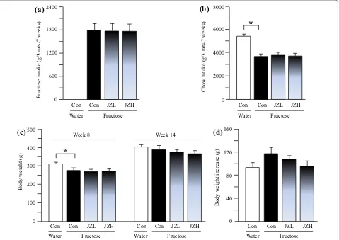
Intakes of fructose and chow, and body weight in rats
The results showed that fructose control rats ate less
chow, compared to water controls (Figure 1b). There
was no significant difference in intakes of fructose (Fig-ure 1a) and chow (Figure 1b) between fructose control
and fructose Jiangzhi Capsule-treated groups. Water
control rats were heavier than fructose controls; there was no difference in body weight between fructose con-trol and fructose Jiangzhi Capsule groups before
treat-ments commenced (Figure 1c). There was no difference
in body weights between groups at the endpoint of the experiment (Figure 1c); the body weight gain in fructose
control intended to increase compared to water control, which tended to decrease after Jiangzhi Capsule treat-ment (Figure 1d).
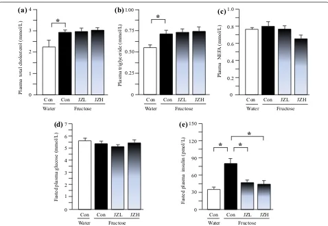
Blood biochemical parameters in rats
Compared to water control rats, fructose controls showed higher plasma concentrations of total choles-terol (Figure 2a), triglyceride (Figure 2b) and insulin (Figure 2e), whereas there was no significant difference in plasma NEFA (Figure 2c) and glucose (Figure 2d) con-centrations between water control and fructose control.
Jiangzhi Capsule at both 25 and 100 mg/kg significantly
suppressed the insulin increase (Figure 2e). However,
Jiangzhi Capsule showed minimal effect on plasma con-centrations of total cholesterol, triglyceride, NEFA and glucose (Figure 2a–d).
Liver‑associated parameters in rats
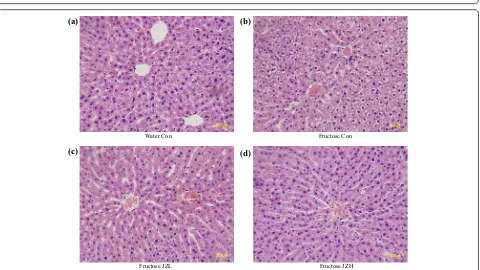
Although fructose feeding did not significantly affect liver weight (Figure 3a) and the ratio of liver weight to body weight (Figure 3b), hepatic total cholesterol (Figure 3c)
(a)
Fructose
intake
(g/3
rats/7
weeks)
Chow intake (g
/3
rats/7
weeks)
(b)
*
Body
weight
(g
)
(c)
Con JZL JZH Con
Week 8 Week 14
Body
weight
increase
(g
)
0 40 80 120 160
(d)
Con JZL JZH
0 100 200 300 400 500
JZL JZH 0
600 1200 1800 2400
Con
Fructose
JZL JZH Con JZL JZH
*
0 2000 4000 8000
6000
Water Con
Fructose Water
Con
Fructose Water
Fructose Water
Fructose Water
[image:5.595.61.540.360.699.2]Con Con Con
Figure 1 Intakes of fructose (a) and laboratory chow (b), body weight (c) and body weight gain (d) in water control, fructose control and fructose
and triglyceride (Figure 3d) contents were increased after fructose feeding. Accordingly, fructose feeding increased
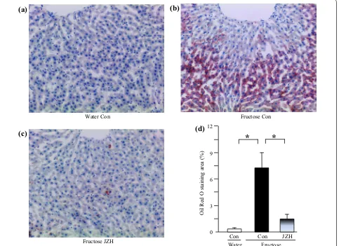
vacuolization (Figure 4b) and Oil Red O staining area
(Figure 5b), indicative of fructose-induced excess hepatic lipid droplet accumulation. Jiangzhi Capsule treatment (both dosages) did not alter liver weight (Figure 3a), ratio of liver weight to body weight (Figure 3b) or hepatic total cholesterol content (Figure 3c). However, Jiangzhi Cap-sule substantially decreased hepatic triglyceride content (Figure 3d). This coincided with vacuolization (Figure 4c, d) and Oil Red O staining area (Figure 5c, d) in the liver being also significantly reduced.
Hepatic gene/protein expression in rats
As the treatment with Jiangzhi Capsule at 100 mg/kg
showed more pronounced effects on hepatic triglyceride accumulation, comparisons in gene/protein expression are restricted to water control, fructose control, and fruc-tose Jiangzhi Capsule 100 mg/kg groups.
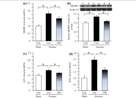
By real-time PCR 14-week fructose feeding increased hepatic expression of mRNAs encoding SREBP-1c
(Figure 6a), ACC-1 (Figure 6c), SCD-1 (Figure 6d) and
DGAT-2 (Figure 7b). The increased contents of nuclear
SREBP-1 protein (Figure 6b) and DGAT-2 protein
(Fig-ure 7c) were further demonstrated by Western blot anal-ysis. Immunohistochemical staining results also showed upregulated DGAT-2 protein expression in fructose con-trol compared to water concon-trol (Figure 7d). Seven-week
Jiangzhi Capsule treatment downregulated mRNA lev-els of SREBP-1c (Figure 6a), ACC-1 (Figure 6c), SCD-1 (Figure 6d) and DGAT-2 (Figure 7b). The results of pro-tein expression further confirmed the suppression of SREBP-1 (Figure 6b) and DGAT-2 (Figure 7c, d) by Jiang-zhi Capsule treatment.
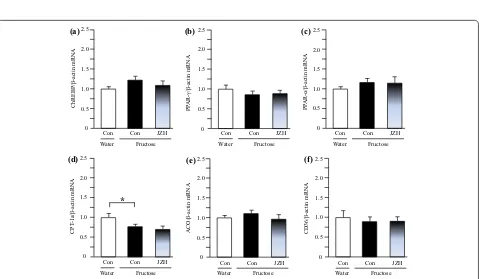
Also with regards to the liver, fructose feeding did not significantly alter mRNA levels of DGAT-1 (Figure 7a), ChREBP (Figure 8a), PPAR-γ (Figure 8b), PPAR-α (Fig-ure 8c), acyl-CoA oxidase (ACO) (Figure 8e) and CD36 (Figure 8f), but downregulated carnitine palmitoyltrans-ferase (CPT)-1a expression (Figure 8d). Jiangzhi Capsule treatment showed minimal effect on expression of these genes (Figure 8a–f).
Fasted
plasma
gl
ucose
(mmol
/L
)
Fasted
plasma
insulin (pmol/L
)
(e)
(d)
*
*
Con JZL JZH Con JZL JZH
*
Plasma total cholesterol
(mmol/
L)
Plasma trig
ly
ceride
(mmol/
L)
0
(a)
(b)
0 0.25 0.50 0.75 1.00
Plasma
NE
FA
(m
mo
l/L
)
(c)
0 0.2 0.4 0.6 0.8 1.0
1 2 4
3
*
Con JZL JZH
*
Con JZL JZH
Con JZL JZH
Fructose
Water Water Fructose Water Fructose
Con Con
Con
Fructose
Water Water Fructose
Con Con
0 1 2 3 4 5 6 7
[image:6.595.59.537.87.417.2]0 30 60 90 120 150
Li
ver
weight
(g
)
0
(a)
4 8 12
Li
ver
weight/body
weight
(mg/g)
(b)
0 10 20 30 40
Li
ver
trig
ly
ceride
(mg/g
tissue)
*
*
*
Con JZL JZH Con JZL JZH
Con JZL JZH
Li
ver
total cholesterol
(mg/g
tissue
)
(c)
*
(d)
0 2 4 6 8 10
Con JZL JZH 0
20 40 60 Fructose
Water Water Fructose
Fructose
Water Water Fructose
Con Con
[image:7.595.59.538.89.394.2]Con Con
Figure 3 Liver weight (a), the ratio of liver weight to body weight (b), liver total cholesterol content (c) and liver triglyceride content (d) in water control, fructose control and fructose Jiangzhi Capsule (JZ)-treated rats. Data are means ± SEM (n = 6–9 each group). *P < 0.05. Con control, JZL JZ 25 mg/kg, JZH JZ 100 mg/kg.
Water Con Fructose Con
Fructose JZL Fructose JZH
(a) (b)
(c) (d)
[image:7.595.60.540.429.699.2]Discussion
The present results clearly demonstrated that treatment of rats with Jiangzhi Capsule decreased fructose feeding-induced excess hepatic triglyceride accumulation and increased vacuolization and Oil Red O staining area in the livers. However, Jiangzhi Capsule did not affect chow and fructose intakes and body weight, and had minimal effect on plasma concentrations of total cholesterol, tri-glyceride, NEFA and glucose. Therefore, these findings suggest a specific anti-steatotic effect of Jiangzhi Capsule in rats.
Fructose, by providing large amounts of hepatic triose-phosphate as precursors for fatty acid synthesis, is highly lipogenic [4]. Recent findings suggest that the increase in hepatic de novo lipogenesis is one of the major pro-viders of lipids in fructose-induced fatty liver [4, 31]. A high-fructose diet has been shown to induce the expres-sion of the transcription factor SREBP-1c [4]. In addition,
fructose consumption may also activate another hepatic transcription factor ChREBP, which upregulates the expression of hepatic lipogenic genes responsible for fatty acid synthesis [4]. In the present study, Jiangzhi Capsule treatment substantially suppressed fructose-stimulated hepatic overexpression of both SREBP-1c mRNA and nuclear SREBP-1 protein. Accordingly, the overexpres-sion of SREBP-1c downstream genes ACC-1 and SCD-1 was also downregulated, however, hepatic ChREBP
expression remained unchanged after Jiangzhi Capsule
treatment. Thus, these results suggest that modulation of hepatic SREBP-1c-mediated expression of the genes responsible for hepatic de novo fatty acid synthesis con-tributes to the anti-steatotic effect of Jiangzhi Capsule in rats.
Recently, we have demonstrated that mangiferin, a prominent component contained in many anti-obese and anti-diabetic herbs, ameliorates fructose-induced
Water Con Fructose Con
Fructose JZH
*
*
(a)
Oil Red
O staining
area
(%
)
(d)
Con Con JZH
3 6 9 12
0
(b)
(c)
[image:8.595.59.542.91.442.2]Fructose Water
fatty liver by suppressing hepatic DGAT-2 expression in spontaneously hypertensive rats [28]. In the present study, treatment with Jiangzhi Capsule also significantly inhibited hepatic DGAT-2 expression at the mRNA and protein levels, but was without effect on DGAT-1 expres-sion in fructose-fed rats. Thus, these findings suggest that inhibition of hepatic DGAT-2 is also responsible for
Jiangzhi Capsule-elicited attenuation of fructose-induced excessive hepatic triglyceride accumulation.
Reduction in hepatic fatty acid oxidation and increased fatty acid uptake into liver appear to have only minor roles in hepatic triglyceride deposition [2]. PPAR-α, pre-dominantly expressed in the liver and, to a lesser extent, in the heart and muscle, has a crucial role in control-ling fatty acid oxidation and uptake through direct tran-scriptional control of the genes, such as CPT1a, ACO and CD36 [32]. The induction of fatty acid oxidation by PPAR-α activation improves plasma lipid profiles. In a variety of mouse models, PPAR-α agonists lower plasma
triglycerides, reduce adiposity and improve hepatic and muscular steatosis [32]. In contrast, PPAR-γ is predomi-nantly expressed in adipose tissue and normally at low level in liver [32]. PPAR-γ is associated with regulation of the genes encoding molecules that promote a com-bination of lipid storage and lipogenesis [32]. In mice, activation of PPAR-γ in liver appears to contribute to the development of hepatic steatosis [33, 34]. It has been reported that the contribution of de novo lipogenesis to fructose-induced hypertriglyceridemia is small [35]. Fur-thermore, research findings suggest a DGAT-2-induced disconnection between liver and circulating triglyceride levels. In transgenic mice overexpressing hepatic DGAT-2, there were increased liver triglyceride content and reduced circulating triglyceride level [36, 37]. In the pre-sent study, treatment with Jiangzhi Capsule did not alter
hepatic expression of PPAR-α, CPT-1a, ACO, CD36 and
PPAR-γ in fructose-fed rats. Thus, our findings in gene expression do not support the involvement of the hepatic
(a)
Con JZH
SREBP-1c/β
-actin
m
RN
A
Nuclear
SREBP-1/lamin
A/
C
protei
n
(b)
0 0.5 1.0 1.5 SREBP-1 Lamin A/C
Con JZH
ACC-1/
β-actin
m
RN
A
SCD
-1/β
-actin
mRN
A
0 0.5 1.0 1.5 2.0 2.5
(c)
Con JZH
0 0.5 1.0 1.5 2.0
2.5
(d)
Con JZH
0 0.5 1.0 1.5 2.0 2.5 3.0
*
*
*
*
*
*
*
*
Fructose Water
Fructose Water
Fructose Water
Fructose Water
Con Con
Con Con
[image:9.595.61.537.90.431.2]1 2 3 4 5 6
DG AT -1/β-actin m RN A DG AT -2/β-actin m RN A (b) 0 0.5 1.0 1.5 2.0 2.5 (a) 0 0.5 1.0 1.5 2.0 2.5 DG AT -2/β-actin p rotein (c) 0 DGAT-2 β-actin * * (d)
Water Con Fructose Con Fructose JZH
0.5 1.0 1.5 * * Con JZH Fructose Water
Con Con JZH
Fructose Water
Con Con JZH
Fructose Water
[image:10.595.57.539.88.321.2]Con 1 2 3 4 5 6
Figure 7 Hepatic expression of mRNAs encoding acyl-coenzyme A:diacylglycerol acyltransferase (DGAT)-1 (a) and DGAT-2 (b), and protein of DGAT-2 by Western blot (c lanes 1, 2 water control; lanes 3, 4 fructose control; lanes 5, 6 fructose JZ 100 mg/kg) and immunohistochemical staining (d) in water control, fructose control and fructose Jiangzhi Capsule (JZ)-treated rats. Data are means ± SEM (n = 6–9 each group). *P < 0.05. Con control, JZH JZ 100 mg/kg.
PP AR-α/β-actin mRN A ACO/β-actin m RN A CD36/β-actin mRNA CP T-1a /β-actin m RN A (c) 0 0.5 1.0 1.5 2.0 2.5 (d) 0 0.5 1.0 1.5 2.0 2.5 (e) 0 0.5 1.0 1.5 2.0 2.5 (f) 0 0.5 1.0 1.5 2.0 2.5 * ChREBP/β-actin m RN A PP AR-γ/β -actin mRN A (a) 0 0.5 1.0 1.5 2.0 2.5 (b) 0 0.5 1.0 1.5 2.0 2.5 Con JZH Fructose Water
Con Con JZH
Fructose Water
Con Con JZH
Fructose Water Con Con JZH Fructose Water
Con Con JZH
Fructose Water
Con Con JZH
Fructose Water
Con
[image:10.595.60.539.370.649.2]PPAR-α and PPAR-γ pathways in the anti-steatotic effect of Jiangzhi Capsule in rats. A consideration may be that modulation of hepatic de novo lipogenesis via the SREBP-1c and DGAT-2 pathways by Jiangzhi Capsule is insuffi-cient to improve fructose-induced hypertriglyceridemia.
Although hepatic steatosis is strongly associated with the development of insulin resistance, it remains unclear whether insulin resistance causes the excessive accumu-lation of triglyceride in the liver, or whether the increase in triglyceride itself or of metabolite intermediates may play a causal role in the development of insulin resistance
[2]. Some studies have shown that the accumulation of
intrahepatic lipids precedes the state of insulin resistance [2]. In the present study, the anti-steatotic effect of
Jiang-zhi Capsule treatment was accompanied by pronounced
amelioration of fructose-induced hyperinsulinemia. Further investigations are needed to determine whether
Jiangzhi Capsule improves insulin resistance through attenuation of excessive hepatic triglyceride accumula-tion or whether the improvement of fatty liver is partially secondary to the amelioration of insulin resistance.
Conclusions
Our present results demonstrate the anti-steatotic action of Jiangzhi Capsule in fructose-fed rats and modulation of hepatic SREBP-1c and DGAT-2 that are involved in hepatic de novo synthesis of fatty acids and triglyceride, respectively. Our findings provide an evidence-based and
mechanistic understanding of Jiangzhi Capsule for the
prevention and/or treatment of fatty liver and its associ-ated disorders in clinic.
Abbreviations
ACC: acetyl-CoA carboxylase; ACO: acyl-CoA oxidase; ChREBP: carbohydrate response element binding protein; CPT: carnitine palmitoyltransferase; DGAT: acyl-coenzyme A:diacylglycerol acyltransferase; NEFA: non-esterified fatty acids; PPAR: peroxisome proliferator-activated receptor; SCD: stearoyl-CoA desaturase; SREBP: sterol regulatory element-binding protein; TCM: traditional Chinese medicine
Authors’ contributions
YZ performed the experiments, analyzed/interpreted data and drafted the manuscript. YP, YY, and RB analyzed/interpreted data. JW and YL contributed to the concept, designed experiments, analyzed/interpreted data and finalized the manuscript. All authors read and approved the final manuscript.
Author details
1 Faculty of Basic Medical Sciences, Chongqing Medical University, Chong-qing, China. 2 The Laboratory Animal Center, Chongqing Medical University, Chongqing, China. 3 Endocrinology and Metabolism Group, Sydney Institute of Health Sciences/Sydney Institute of Traditional Chinese Medicine, Sydney, NSW, Australia. 4 Central Clinical School, Royal Prince Alfred Hospital, The Uni-versity of Sydney, Sydney, NSW, Australia. 5 Laboratory of Traditional Chinese Medicine, Chongqing Medical University, Chongqing, China.
Additional files
Additional file 1: Table S1. Primer sequences for real time PCR assays.
Acknowledgements
We thank Mr. Alan Yeung for his excellent assistance in the preparation of this manuscript. This work was financially supported by National Natural Science Foundation of China (Grant 81374033), China, and Sydney Institute of Health Sciences/Sydney Institute of Traditional Chinese Medicine, Australia.
Compliance with ethical guidelines
Competing interests
The authors declare that they have no competing interests.
Received: 1 October 2014 Accepted: 12 May 2015
References
1. Marchesini G, Bugianesi E, Forlani G, Cerrelli F, Lenzi M, Manini R et al (2003) Nonalcoholic fatty liver, steatohepatitis, and the metabolic syn-drome. Hepatology 37:917–923
2. Postic C, Girard J (2008) Contribution of de novo fatty acid synthesis to hepatic steatosis and insulin resistance: lessons from genetically engi-neered mice. J Clin Invest 118:829–838
3. Shi KQ, Fan YC, Liu WY, Li LF, Chen YP, Zheng MH (2012) Traditional Chi-nese medicines benefit to nonalcoholic fatty liver disease: a systematic review and meta-analysis. Mol Biol Rep 39:9715–9722
4. Tappy L, Lê KA (2010) Metabolic effects of fructose and the worldwide increase in obesity. Physiol Rev 90:23–46
5. Cases S, Stone SJ, Zhou P, Yen E, Tow B, Lardizabal KD et al (2001) Clon-ing of DGAT2, a second mammalian diacylglycerol acyltransferase, and related family members. J Biol Chem 276:38870–38876
6. Yamazaki T, Sasaki E, Kakinuma C, Yano T, Miura S, Ezaki O (2005) Increased very low density lipoprotein secretion and gonadal fat mass in mice overexpressing liver DGAT1. J Biol Chem 280:21506–21514
7. Yu XX, Murray SF, Pandey SK, Booten SL, Bao D, Song XZ et al (2005) Anti-sense oligonucleotide reduction of DGAT2 expression improves hepatic steatosis and hyperlipidemia in obese mice. Hepatology 42:362–371 8. Choi CS, Savage DB, Kulkarni A, Yu XX, Liu ZX, Morino K et al (2007)
Sup-pression of diacylglycerol acyltransferase-2 (DGAT2), but not DGAT1, with antisense oligonucleotides reverses diet-induced hepatic steatosis and insulin resistance. J Biol Chem 282:22678–22688
9. Hong X, Tang H, Wu L, Li L (2006) Protective effects of the Alisma orienta-lis extract on the experimental nonalcoholic fatty liver disease. J Pharm Pharmacol 58:1391–1398
10. Ji W, Gong BQ (2008) Hypolipidemic activity and mechanism of purified herbal extract of Salvia miltiorrhiza in hyperlipidemic rats. J Ethnopharma-col 119:291–298
11. Chen J, Deng J, Zhang Y, Yang J, He Y, Fu W et al (2014) Lipid-lowering effects of Danhong injection on hyperlipidemia rats. J Ethnopharmacol 154:437–442
12. Yoshikawa M, Murakami T, Ueno T, Yashiro K, Hirokawa N, Murakami N et al (1997) Bioactive saponins and glycosides. VIII. Notoginseng (1): new dammarane-type triterpene oligoglycosides, notoginsenosides-A, -B, -C, and -D, from the dried root of Panax notoginseng (Burk.) F.H. Chen. Chem Pharm Bull 45:1039–1045
13. Ng TB (2006) Pharmacological activity of sanchi ginseng (Panax notogin-seng). J Pharm Pharmacol 58:1007–1019
14. Ji W, Gong BQ (2007) Hypolipidemic effects and mechanisms of Panax notoginseng on lipid profile in hyperlipidemic rats. J Ethnopharmacol 113:318–324
15. Yim TK, Wu WK, Pak WF, Ko KM (2001) Hepatoprotective action of an oleanolic acid-enriched extract of Ligustrum lucidum fruits is mediated through an enhancement on hepatic glutathione regeneration capacity in mice. Phytother Res 15:589–592
16. Kwon HJ, Kim YY, Choung SY (2005) Amelioration effects of traditional Chinese medicine on alcohol-induced fatty liver. World J Gastroenterol 11:5512–5516
18. Tzeng TF, Lu HJ, Liou SS, Chang CJ, Liu IM (2012) Emodin, a naturally occurring anthraquinone derivative, ameliorates dyslipidemia by activat-ing AMP-activated protein kinase in high-fat-diet-fed rats. Evid Based Complement Altern Med 2012:781812
19. Liu C, Li Y, Zuo G, Xu W, Gao H, Yang Y et al (2013) Oleanolic acid dimin-ishes liquid fructose-induced fatty liver in rats: role of modulation of hepatic sterol regulatory element-binding protein 1c-mediated expres-sion of genes responsible for de novo fatty acid synthesis. Evid Based Complement Altern Med 2013:534084
20. Wang W, He Y, Lin P, Li Y, Sun R, Gu W et al (2014) In vitro effects of active components of Polygonum Multiflorum Radix on enzymes involved in the lipid metabolism. J Ethnopharmacol 153:763–770
21. Lin P, He YR, Lu JM, Li N, Wang WG, Gu W et al (2014) In vivo lipid regula-tion mechanism of Polygoni Multiflori Radix in high-fat dietfed rats. Evid Based Complement Altern Med 2014:642058
22. Dat NT, Cai XF, Rho MC, Lee HS, Bae K, Kim YH (2005) The inhibition of diacylglycerol acyltransferase by terpenoids from Youngia koidzumiana. Arch Pharm Res 28:164–168
23. Ko JS, Ryu SY, Kim YS, Chung MY, Kang JS, Rho MC et al (2002) Inhibitory activity of diacylglycerol acyltransferase by tanshinones from the root of
Salvia miltiorrhiza. Arch Pharm Res 25:446–448
24. Johnson RJ, Perez-Pozo SE, Sautin YY, Perez-Pozo SE, Sautin YY, Manitius J et al (2009) Hypothesis: could excessive fructose intake and uric acid cause type 2 diabetes? Endocr Res 30:96–116
25. Gao H, Guan T, Li C, Zuo G, Yamahara J, Wang J et al (2012) Treatment with ginger ameliorates fructose-induced fatty liver and hypertriglyceri-demia in rats: modulation of the hepatic carbohydrate response element binding protein-mediated pathway. Evid Based Complement Altern Med 2012:570948
26. Wang J, Gao H, Ke D, Zuo G, Yang Y, Yamahara J et al (2013) Improvement of liquid fructose-induced adipose tissue insulin resistance by ginger treatment in rats is associated with suppression of adipose macrophage-related pro-inflammatory cytokines. Evid Based Complement Altern Med 2013:590376
27. Li Y, Wang J, Gu T, Yamahara J, Li Y (2014) Oleanolic acid supplement attenuates liquid fructose-induced adipose tissue insulin resistance through the insulin receptor substrate-1/phosphatidylinositol 3-kinase/ Akt signaling pathway in rats. Toxicol Appl Pharmacol 277:155–163
28. Xing X, Li D, Chen D, Zhou L, Chonan R, Yamahara J et al (2014) Mangiferin treatment inhibits hepatic expression of acyl-coenzyme A:diacylglycerol acyltransferase-2 in fructose-fed spontaneously hyper-tensive rats: a link to amelioration of fatty liver. Toxicol Appl Pharmacol 280:207–215
29. Rong X, Li Y, Ebihara K, Zhao M, Naowaboot J, Kusakabe T et al (2010) Angiotensin II type 1 receptor-independent beneficial effects of telmisar-tan on dietary-induced obesity, insulin resistelmisar-tance and fatty liver in mice. Diabetologia 53:1727–1731
30. Wang BQ (2010) Salvia miltiorrhiza: chemical and pharmacological review of a medicinal plant. J Med Plants Res 4:2813–2820
31. Stanhope KL, Schwarz JM, Keim NL, Griffen SC, Bremer AA, Graham JL et al (2009) Consuming fructose-sweetened, not glucose-sweetened, beverages increases visceral adiposity and lipids and decreases insulin sensitivity in overweight/obese humans. J Clin Invest 119:1322–1334 32. Evans RM, Barish GD, Wang YX (2004) PPARs and the complex journey to
obesity. Nat Med 10:355–361
33. Chao L, Marcus-Samuels B, Mason MM, Moitra J, Vinson C, Arioglu E et al (2000) Adipose tissue is required for the antidiabetic, but not for the hypolipidemic, effect of thiazolidinediones. J Clin Invest 106:1221–1228 34. Gavrilova O, Haluzik M, Matsusue K, Cutson JJ, Johnson L, Dietz KR et al (2003) Liver peroxisome proliferator-activated receptor gamma contrib-utes to hepatic steatosis, triglyceride clearance, and regulation of body fat mass. J Biol Chem 278:34268–34276
35. Chong MF, Fielding BA, Frayn KN (2007) Mechanisms for the acute effect of fructose on postprandial lipemia. Asia Pac J Clin Nutr 85:1511–1520 36. Monetti M, Levin MC, Watt MJ, Sajan MP, Marmor S, Hubbard BK et al
(2007) Dissociation of hepatic steatosis and insulin resistance in mice overexpressing DGAT in the liver. Cell Metab 6:69–78
37. Jornayvaz FR, Birkenfeld AL, Jurczak MJ, Kanda S, Guigni BA, Jiang DC et al (2011) Hepatic insulin resistance in mice with hepatic overexpression of diacylglycerol acyltransferase 2. Proc Natl Acad Sci USA 108:5748–5752
Submit your next manuscript to BioMed Central and take full advantage of:
• Convenient online submission
• Thorough peer review
• No space constraints or color figure charges
• Immediate publication on acceptance
• Inclusion in PubMed, CAS, Scopus and Google Scholar
• Research which is freely available for redistribution