JOURNAL OF VIROLOGY, Dec. 1973,p.1568-1578 Copyright0 1973 AmericanSocietyforMicrobiology
Vol.12,No.6 PrintedinU.S.A.
Gamma
Ray-Induced Small Plaque
Mutants of
Western
Equine Encephalitis Virus
B. SIMIZU, S. YAMAZAKI, K. SUZUKI, AND T. TERASIMA
Department of Virology and Rickettsiology, Central VirusDiagnostic Laboratory, Departmentof Chemistry, National Institute of Health,Shinagawa, Tokyo141, andDepartment ofPhysiologyandPathology,National
Institute of Radiological Sciences, Chiba 280, Japan
Received for publication 18July 1973
SmallplaquemutantsofWesternequine encephalitisviruswereobtained from thesurviving fractionsofwild-type virus whichwasirradiated withgammarays.
The frequency with which small plaque mutants appeared in the surviving fraction increased with the radiation dose. These mutants were not more resistant to radiation than wild-type virus.Thegrowthrate ofamutant, S127, waslower than that ofwild-type.Clonally purifiedmutantvirionspresentedtwo
peaks inavelocity sedimentation profile; peak1correspondedtothepeakofwild
typeandpeak 2 moved faster than peak 1. Virions of both peakswereinfectious andconsistently formed small plaques in chicken embryo cells. Virions reisolated fromeither peak and grown inchicken embryo cells also revealedtwopeaks in sedimentationanalysis.Inthe electron microscope examination peak2provedto
consist ofgiant form particles, eachofwhich containedmorethanonenucleoid
surrounded with a common envelope. Despite this remarkable morphological difference, densities of thewild-type and S127 mutant virions were similar in
cesium chloridegradients. The RNAsandproteinsof mutantvirionscould not be
distinguished from thoseofwild typeonthe basis of sizeorcharge.
Mutations can be induced in animal viruses
by treatment with chemical mutagens (7,
33),
However, induction of mutants from
single-strandedRNAvirusesby ionizing radiation has
been relatively limited thus far (33) although
radio-induced mutants of Rous sarcoma virus
have been reported recently
by
Golde (11). Inthe course of isolation oftemperature-sensitive
mutants (28) we attempted to use gamma
radiation for induction of the mutants. As a
result, some small plaque mutants emerged
among the survivors of the radiation at
rela-tivelyhighrate,
although
temperature-sensitivemutants were not detected inthe examination
of a few hundred plaques. The virions of the mutants sedimented faster than those of the
wild-type when analyzed in sucrose gradients,
and electron microscope examination showed
the morphological variations of the mutant
virions tobein sizeand shape.
Most investigations dealingwith plaque
mu-tants in RNA viruses have been concerned
primarily with theirbiological properties.
Evi-dence obtained by physico-chemical and
sero-logical studies indicate that the surface
struc-ture of some ofthese mutants may also differ. This isreflectedindifferences inadsorption and
elution pattern on some chromatographic col-umns(9, 16), in coatproteins testedin
serologi-cal studies (4, 35), and inamino acid composi-tion of mutant proteins (22). Studieson
mor-phological differencesinmutant virionsrelating
plaque type variations have been limitedthus
far.
In this investigation, we report the isolation
and preliminary characterization of small
plaque mutants ofWestern equineencephalitis
(WEE) virusobtainedfrom thesurviving
frac-tions aftergamma rayirradiation.Relationship
betweenmorphologicalvariations ofthemutant
virionsand smallplaque formation is discussed.
MATERIALS AND METHODS
Virus and cell culture. The McMillan strain of
WEE virus was used. Before this experiment, it was
purifiedby the plaque cloning technique.The proce-dures for the preparation of WEE virus stocks and chicken embryo (CE) monolayer cultures have been described (27, 28). Eagle minimal essential medium (MEM)supplementedwith 5% calf serum was used as the cell culturemedium.
Gammarayirradiation.Virus was suspendedin cell culture medium and put into glass ampoules. Those ampoules were exposed to gamma radiation
from acobalt-60source at adoserate of1.4 x 104 rads
1568
on November 10, 2019 by guest
http://jvi.asm.org/
GAMMA RAY-INDUCEDWEE VIRUS MUTANT
per min. They were kept cold in ice water during irradiation to minimize heat inactivation and then storedat -80C untiluse.
Procedures for labeling virus. CE monolayers were infected with WEEvirusatamultiplicityof 10
plaque-forming units (PFU) per cell after a 60-min incubation with MEMcontaining1 ;tgofactinomycin Dperml. Viral RNAwaslabeled by incubating cells
from 1 hpostinoculationwithfreshMEM containing
3H-uridine (1 uCi/ml) for 12 h (28). Viral proteins werelabeled by incubating cellsfrom 3 h
postinocula-tion withfreshMEMcontaining 3H-tyrosine (5
;iCi/
ml)for 9h. The culturefluids containing thelabeled
viruseswereharvested and purifiedasdescribedlater.
Concentration and purification ofviruses. Cul-turefluids containing viruseswereclarified by centrif-ugationat3,000rpmfor15minandthenat10,000xg
for 15minat4C. Thesupernatantfluidwas
centrifu-gated to pellet virus in the no. 30 rotor (Spinco,
Beckman, L2-65B)at 74,000 x gfor120minat4 C.
Theviruspelletswereresuspendedinasmall volume
ofTNEbuffer(30) (0.05MTris,0.1MNaCl,0.001M ethylenediaminetetraacetate, pH 7.4) and pipetted vigorouslyto breakupaggregates.The virus suspen-sionwasthencentrifuged at10,000x gfor 15minto removeaggregates.Thesupernatantfluidwaslayered
over a preformed 0 to 40% (wt/wt) linear sucrose gradientprepared inTNEbuffer andcentrifuged in
the SW 27 rotor at 52,000 x g for 2 h (rate zonal
centrifugation). The visible band of virus near the
middleofthetubewasremoved anddialyzed
exten-sively againstTNEbufferand, ifnecessary,rebanded
inasecondsucrosegradientforfurtherpurification.
Extraction of RNA. RNA was extracted from
labeledvirusbyphenol-sodium dodecylsulfate(SDS)
at 60 C. Samples were diluted with phosphate
buf-feredsaline containing 0.15% SDS (final
concentra-tion).Anequalvolume ofwater-saturatedphenolwas
added and the mixturewasshakenfor3minat60 C.
Two phaseswereseparated by low-speed centrifuga-tion; the upper aqueous phase was removed and
reextractedonce more with freshphenol. RNAfrom
the finalaqueousphasewasprecipitatedtwicewith2
volumes ofice-cold ethanol. The RNAprecipitatewas
collected by centrifugation in the cold and was
resuspended in TNEbuffer solution with orwithout
1%SDS.
Polyacrylamide gel electrophoresis.
Polyacryl-amidegels, 10cminlength,werepolymerizedinglass
tubes 0.4 cm in diameter. The gelscontained 7.5%
(wt/vol) acrylamide, 0.2%N,N'-bismethylene acryl-amide, 0.05% ammonium persulphate, and 0.03%
N,N,N',N'-tetramethylenediamine, in 0.1 M
so-diumphosphate buffer, pH 7.2,with0.1% SDS (31).
Virus samples in phosphate buffer solution were
acidified with 0.1 volume ofglacial acetic acid and
then made 0.5Minureaand 1%inSDS. Afterheating
in a boiling water bath for 3 min, samples were dialyzed atroomtemperature overnight against0.01
M ofphosphate buffer solution with 0.5 M ofurea,
0.1% of 2-mercaptoethanol and 0.1% of SDS. After
dialysis Iwovolume of 60%sucroseandofbromophenol
bluewereadded, and200-,litersampleswerelayered
onto gels. Electrophoresis was carried out at room
temperature at 10 mA/column for 3 h. Gels were extruded from the tubes and frozenondry ice. Frozen gels were sliced into 2-mmsegments. Each slicewas dissolved in 0.5 ml of30%H202 at 80C for 2 hand mixed with5ml ofscintillation fluid (toluene, 1liter; TritonX 100,500ml;PPO,10g;POPOP,0.3g).The radioactivity was counted in a Beckman LS-200B liquidscintillation spectrometer.
Electron microscopy. Virus samples purified by sucrose gradients were mixed with 2% solution of uranyl acetate and incubated for 1 min at room temperature. Afterwashingoncewithsaline,samples werestained withan aqueous2% solution of sodium phosphotungstate at pH 7.0 for 30 s (T. Kitano, personal communication). The preparationswere ap-plied to carbon films on copper grids; they were examinedin aHitachiHU-11B electron microscopeat anoperatingvoltage of75 kVandat amagnification ofx40,000.
Hemagglutination (HA) test. The test was per-formed according to the procedure previously de-scribed (29) using goose erythrocytes. Hemagglutinin units (HAU) were expressed as reciprocals of the highest dilution of viruses.
Chemicals. Actinomycin D was provided by
Merck, Sharp & Dohme, West Point, Pa. 'H-uridine (20 Ci/mmol) and 3H-tyrosine (36 Ci/mmol) were obtained from theRadiochemical Centre, Amersham, England. Ribonuclease-free sucrose was purchased fromNakaraiChemicals Ltd., Japan. Acrylamide and N,N'-bis methylene acrylamide were bought from the Wako PureChemical Ind. Ltd., Japan.
RESULTS
Effect of gamma ray irradiation on WEE
virus. The lethal and mutagenic effects of
gamma rays on WEE virus were examined.
When
avirussuspensionincell culture mediumwasexposed togammarays, therewas alinear
relation between the dose of radiation andvirus
inactivationasseen inTable 1 (also Fig. 2). In
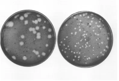
assaying survivors, we sometimes noted small
plaquesamong plaquesofwild-type virus(Fig.
1).Thefrequency ofsmallplaqueformers inthe
survivingfraction wasincreased with increasing
radiation dose (Table 1). Small plaques were
isolated and clonally purified by three
succes-sive passages from plaque to plaque. These
small plaque formers were found to be true
mutants because the characters described
be-low, especiallyin plaque size, weremaintained
TABLE 1. Effect of gamma ray irradiationon WEE virus
Gamma ray Survival (PFU) Frequencyofsmall (rad) plaquesinsurvivors
0 107.8 3/2042 (0.14%)
500 106.1 18/1782(1.01%)
1,000 104.4 24/1384(1.73%) 1,500 102.4 26/ 788(3.29%)
1569
VOL.12,1973
on November 10, 2019 by guest
http://jvi.asm.org/
SIMIZU ET AL.
afterseveral passages in CE cells and BHK-21
cellsoradultmousebrains.
Inordertodetermine whether thesemutants
were induced or selected by radiation, the
wild type and three mutants were
exposed
togamma rayirradiation and the survivingcurves
obtained were compared. Figure 2 indicates
that the wildtype and the mutants showed a
similar dose-response curve, suggesting that
these mutants were not obtained
by
selectionforrelativeresistance tothe radiation.
Further-more,the factthatfrequency of mutants among survivors wasincreased with the radiation dose
favors the hypothesis that the mutants were
induced by radiation.
Biological properties of small plaque
mutants. Six
small-plaque
mutants obtainedfrom the irradiated virus and onespontaneous
mutant werecompared with wildtypein
regard
tothebiological
markers which had beeninves-tigated on WEE virus in our
laboratory,
i.e.,temperature sensitivity, heat
stability,
plaqu-ing efficiency under dextran sulfate agar, and
virulence for mice (26-28,
32).
As summarizedin Table 2, the results obtained with these
mutants vary from one mutant to another but
marked differences in characteristics of wild
typeandthesemutants are not seen.
In a one-step growth experiment, growth patterns of the wild type and a small plaque mutant, S127, were compared in CE monolayer
cells (Fig. 3). Mutant virus grew more slowly
than wildtypeand the yieldof the mutant was
lower thanthat of wild type for the test period.
Even at 24 h after inoculation, the yield of the mutant didnotreachthelevel of the wild type.
The total (released and cell associated) virus
yieldofthemutant was also lower than that of
wildtype. The limited growth in CE cells is a prominentcharacteristic of this mutant.
Sedimentation behaviors of S127 mutant.
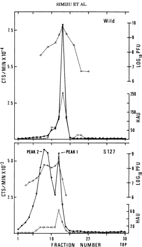
When the partially purified S127 mutant virus
was examined by rate zonal centrifugation, we
noted that it showed two visible bands in sucrose gradients. Therefore, mutant viruses
were labeled with 3H-uridine and analyzed by
rate-zonal centrifugationto seethedistribution
oftheirradioactivity, infectivity, and HA
activ-ity. Asshown in Fig. 4, tworadioactivepeaks of
infectivity and HA activity are seen in the
velocity sedimentation profile of the mutant
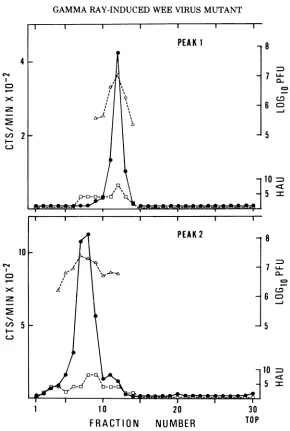
S127. Toseparatethesetwo
peaks,
fraction 14ofpeak 1and fractions6 to 9ofpeak2inFig. 4
were collected and resedimented by rate zonal
centrifugation. As seen in
Fig.
5, two peakssedimented separately corresponding to each
I
FIG. 1. Plaquesproduced with the wild type (left) and a smallplaquemutant, S092(right) in CE cells.
1570 J. VIROL.
on November 10, 2019 by guest
http://jvi.asm.org/
[image:3.497.54.449.376.643.2]GAMMA RAY-INDUCEDWEEVIRUSMUTANT
e 0.01 0
C-\-0.001
-I
0 250 500 750 1000
[image:4.497.51.244.59.350.2]GAMMA-RAY(KILORADS)
FIG. 2. Survivingfractionsof wild type and small plaque mutants irradiated with gamma rays.
Sym-bols:0,wild-type;0, S113 mutant; *, S118 mutant;
[image:4.497.257.449.238.533.2]0,S127mutant.
TABLE 2. Biological propertiesof wild-type and small plaquemutantsof WEE virus
EOPa EOP5 Heatingc
Virus at42/ under at 50 C, LD,O/PFUd
37C DS-agar 30min
(%)
(%)
(%)
Wild-type 69 100 3.0 6.7/7.9(2)
S025 29 109 3.1 6.5/7.6(4)
S092 32 92 2.2 6.9/8.9(3)
S113 19 120 6.8 7.1/8.0(3)
S118 39 114 2.7 6.9/8.0(3)
S127 66 93 0.8 6.3/7.7(3)
S132 32 38 10.7 7.1/7.9(2.5)
Spontaneous 90 Not done 4.4 6.8/8.6(4)
aEfficiencyofplaquingat37/42C (28).
'Efficiency of plaquing under agar containing dextran
sulfate (32).
cSurvivingfraction afterheatingat50C for 30 min.
dLD,.inadultmice/PFUinCE cells. Parentheses indicate themeansurvival time(days)of 10miceinoculatedwith 100
LD,, ofeachvirus.
peakfractionshown in thebottom
panel
ofFig.
4;peak2movesfaster, andisbroader thanpeak
1 inboth radioactivity andinfectivity.
Virions in both peaks in Fig. 5 formed small
plaques in CE cells and their plaques were
reducedbyantiserum raised ina rabbit
immu-nized with wild type virus to the same degree
asthose of wildtype (data not shown).
In thenext experiment, density of virions was
examined by equilibrium centrifugation in a
cesium chloride gradient. Mutant virions from fractions 6 to 14 and wild type virions from
fractions 14 and 15 in Fig. 4 were collected and
dialyzed to remove sucrose and then mixed in
the solution of cesium chloride(startingspecific
gravity 1.243g/ml). As seen in Fig. 6, densities of wild type and mutant virions were 1.234
g/cm3
and 1.226g/cm3,
respectively. In a 20 to60% linear sucrose density gradient, both
vi-ruses showed 1.19 g/cm3 in density, although
the peak of mutant was broader than that of
10
9
-.
J
'" 8
Q-co
7
7.:
6
5
O 1 2 4 6 8 10 " 24
HOURS AFTER INOCULATION
FIG. 3. One-step growth ofthewild-typeand S127 mutant of WEE virus in CE cells. CE cells were
inoculated with the virusat aninput multiplicity of
10PFUpercell.Aftera60-min adsorption period,the
inoculated cultures were washed twice with MEM
and then incubated at 37C; duplicate plates were
sampledatselected intervals.A partofculturefluid
wasstoredafter clarifying by low-speed centrifugation
for released virus assay.Infected cellswere collected
into culturefluidwitha rubberpolicemanand then
frozen and thawed three cycles toobtain totalvirus.
Symbols: 0, wild-type released virus; 0, wild-type
totalvirus;*,mutantreleasedvirus;0,mutanttotal
virus.
i
0
o ,
I I
W
I I I
II
I I I
II II
I I I I I I
II II
I I
-P
"W,"
I I I I I IAl
VOL.12,1973 1571'
-t I I I I --j--l[-O'I-]
on November 10, 2019 by guest
http://jvi.asm.org/
[image:4.497.53.246.392.552.2]SIMIZU ETAL.
7.;.
If\
~~~~~~~~~9
CLL
T T
Cfl
C5-,
6
250
2.5
150~
50
PEAK
2-
-.-PEAK
IS
127
9C-,)5.o0
A C
1
10
23
30
1-C,,~~~~ ~ ~ ~ ~ ~~~~~~~~C
2s5
- 616
0120
1 1~~~I0 21 30
[image:5.497.108.388.53.539.2]FRACTION
NUMBER
TOPFIG. 4. Sucrosegradient analysis ofthewild-typeandS127mutantvirionssynthesizedin CE cells. Twenty
plates ofCE cellswereinfectedwith eitherwild-typeorS127mutantin thepresenceof1.5,ugof actinomycinD perml and 1 UCi of 3H-uridine per ml.At 12h afterinoculation, the culturefluids wereharvested and the labeled virionswereconcentrated. Concentrated virionswerelayeredover apreformed linear0 to40%osucrose gradientand then centrifuged in theSW27rotor at52,000 x gfor2h. Fractions(1 ml) were collectedand sampleswereassayed foracid-precipitable radioactivity(0), infectivity(A),and HAactivity(0).
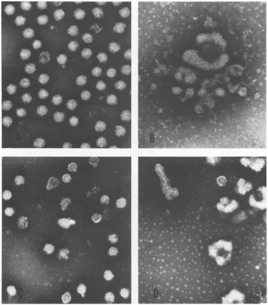
wild type (data not shown). These data taken to confirm these observations, the virions of
together reveal that the wild type and mutant wildtype andS127 mutant were examined in an
virions are similar in density, but different in electron microscope (Fig. 7). The wild type
shape orsize. virions showed quiteuniform enveloped
spheri-Electron microscope observation. In order cal particles about 60 nm in diameters, as
i.579 J.VIROL.
on November 10, 2019 by guest
http://jvi.asm.org/
GAMMARAY-INDUCED WEE VIRUSMUTANT
4
CD
10
CDJ
- 2-c 2 c-3
PEAK 1
PEAK2
8
7
7LL.
c, OC0~
6 °=
5
10
D
5 =
8
7Lh.
7fi 60~
5
10 =
5 =
1
1
0
20
30
FRACTION
NUMBER
TOPFIG. 5. Separation oftwopeaksof S127mutantinsucrosegradients. Peak1from fraction14andpeak2from fractions 6to 9inFig.4werecollected and resedimented byrate zonalcentrifugation. Fractions (I ml) were collected and samples wereassayed foracid-precipitable radioactivity (0), infectivity (A), and HA activity (0).
reported by Morgan et al. (21) and Grimley et
al. (12), whereas the mutant virions
evidently
showedtwosizetypes,i.e., the
regular
spherical
particle (peak 1) and the giant particle(peak 2),
which consistedof two tofournucleoids. These giant form particles already appeared in the
concentrated viruspreparation before
purifica-tion insucrose(Fig.7B).Therefore,theyare not
likely to be an artefact due to exposure to
sucrose duringpurification.
Viral components of S127 mutant. The
mutantviruswasfurther examined for its RNA
and protein content to compare with those of
wild type virus. Viral RNAwhich was extracted
from the purified mutant virions was
sedi-mentedinalinear15to30% sucrosegradientat
the same rate as the 41SRNA extracted from wild type (Fig. 8). Viral proteins extracted from
similarlypurified mutant virions were analyzed
by polyacrylamidegelelectrophoresis.Asshown
in Fig. 9, viral core and envelope polypeptides migrated to positions identical with those of wild type. The molecularweight ofeach
poly-peptide was 6.0 x 10⁴ and 3.8 x 10',
respec-tively, as estimated by electrophoresis with
standard marker proteins. These patterns of
VOL.12,1973 15"73
on November 10, 2019 by guest
http://jvi.asm.org/
[image:6.497.102.392.49.480.2]6
0.l
|
|
l
l135
0 * 1
250
10~~~~~
CL
L
2b
0 0
@0
2
100
80
Wild
a
a'~~~~~
60~
-40
0. - 20
-J
- 0i
C,
,1.35
00
C-,
*
-41.30
00
5
10
c-10
0
*
P*0C
4
1.1 5
20
S127
Ijis
aa
~~~~~~~10=
1
5
10
15
20T
PFRACTIONS
FIG. 6. BuoyantdensitiesofpurifiedWEEvirions in cesium chloride
density
gradients.
A 1.5-mlamountof
3H-uridine-labeledvirionspurifiedtwiceinsucrosegradientswasmixedwith3.5ml
of
TNEsolutioncontaining
1.6 g of cesium chloride. Centrifugation wasperformed in SW50L rotor at 100,000 x g
for
46h at 4 C. Fifteen-drop fractionswerecollectedfromthebottomofthetube,andsampleswereassayedfor
acid-precipita-bleradioactivity (filledcircles), and HAactivity (opencircles).Measurementofrefractiveindexwasmadeon
allfractions, and densitiesat25Cwerecalculatedfromanempiricallydetermined relationbetweenrefractive
index anddensity.
1574
on November 10, 2019 by guest
http://jvi.asm.org/
[image:7.497.117.398.26.626.2]GAMMARAY-INDUCEDWEE VIRUS MUTANT
[image:8.497.61.443.71.505.2].inw _ - - * w.
FIG. 7. Electronmicrographs ofWEEvirions. x100,000.A,wild-type; B,concentrated crude S127 mutant;
C, peak 1(Fig. 5) particles ofS127 mutant; D,peak2(Fig. 5)particles ofS127 mutant.
RNA andproteinsaresimilartothosedescribed
forgroup Aarbovirusesby Sreevalsanetal.(29)
and Acheson and Tamm
(2).
Geneticstability of small
plaque
mutants.To determine whether these altered properties
of small plaque mutants resulted from a true
genotypic alteration and, if so, whether this
property was stable, the following experiments
weredone.Viruses labeled with3H-uridinewere
sedimentedin sucrose
gradients
under thesameconditions stated
above,
and 30 fractionscol-lectedfrom eachtubewereassayed for
radioac-tivity and HA activity. As a result, wild type
and its large
plaque
mutant, L49(unpublished
data),
showed asingle peak,
whereas S118mutant aswell as S127 mutant showed 2peaks.
A spontaneous small plaque mutant derived
from wild type alsoshowed2peaks. However,a revertantpresentedasingle peak corresponding
tothat of wild type. The revertantwasobtained
from a wild type sized plaque that rarely
appearedafterseveralpassages ofS127mutant
inCE cells.
Two clones were isolated from peak 1
(frac-15"I5
VOL.12,1973
on November 10, 2019 by guest
http://jvi.asm.org/
l l killing effect of radiation which is presumably
due to irreparable chain breaks.Therefore, the
observed dose-dependent occurrence of
small
plaque mutants in the wild typeWEE
viruses
3 mightbe explained by radiation-induced
muta-tion. However, we should note that the wild type used here is relatively mutable in its
ild
plaque
type
character. After being extensively
purified by plaquing, the wild type virus still
- S 121 contained
spontaneous
smallplaque
mutants at2- the rate of
0.1%
(Table
1).This is not aspecial
l-
lcase
inplaque-type mutationsofanimalviruses28S 18S (7, 20, 24, 33). In view of such highmutability, it still remains a possibility that the mutants
wereselectedbythe relative resistance to
radia-tion, although a significant difference in
sensi-tivity to radiation
between
wild type andmu-tants was not demonstrated. A
significant
difference in sensitivity to gamma radiation
might
not bereadily
detectable when thema-1
10O
20O
30
jority
of the infectiousparticles
are"mono-FRACTION NUMBER TOP ploid" in a mutant
population.
We wouldex-FIG. 8. Sedimentation analysis of WEE viral
RNA.
pectthatpreexisting
mutants
could be selectedRNA was extractedfrompurified 3H-uridine labeled
virions and was sedimented in a 15 to 30% (w/w) Wild
sucrose densitygradient. Centrifugation wasfor12h at 24,000 rpm in theSW27rotorand 1-ml fractions were collected. Arrows indicate the positions of co- 2_
sedimented 28S RNA and 18S RNA markers
ex-tractedfrom CE cells.
tion 12) and peak2 (fraction4) in Fig. 5.They
were grown once in CE cells for labeling virus
with 3H-uridine, and then applied to sucrose
gradients.
These two clonespresented
two2 peaks again, indicating that the regular spheri- ccal particle of peak 1 is not a revertant but a SS127
mutant which is stable in theability to
repro-duceparticles oftwo types.
DISCUSSION
Since ionizingradiation causes breaks in the
polynucleotide chains, arboviruses containing 2
single-stranded RNA polynucleotides (3) are
highly
sensitive
to radiation compared withviruses
which
contain
double-stranded
polynu-cleotides
(10).
Inactivationof theWEEvirusby I0 ,5 2'0 25 3'0 35 4 45gamma ray followed a one-hit curve, and the E FRACTION NUMBER
37% survivaldoseofthis virusdetermined from FIG. 9. SDS-polyacrylamide gel electrophoresis of
Fig. 2 was 150krads.This value is inagreement proteins extractedfrompurified WEE virions. Wild-with those reported for single-stranded RNA type and S127 mutant viruses were labeled with
viruses (10, 12). 3H-tyrosine. One hour before infection CE cells were
Ionizing radiation yields highly reactive and incubated with1.5pgofactinomycin D perml. Three
short-lived radicals inthe medium surrounding hours after infection the medium was removed, and
orwithinviruses(10, 14) and ultimately leads to MEMwith 5
'Ci
of3H-tyrosine
permlwasadded. Atalterationsofviralgenometroughthprod 12 h, culture fluids were harvested and virions were
alterations ofvlralgenomethroughthe produc- purifiedasdescribedinthetext. Thevirions
dis-tion ofreparable chain breaks orexcitations in rupted in 1% SDS, 0.5% ureaand0.1%
2-mercapto-DNA or RNA molecules. Accordingly, viruses ethanol andelectrophoresedina7.5%gelat10mAfor could receive a mutagenic effect other than a 3h.
1576
SIMIZUETAL. J.VIROL.on November 10, 2019 by guest
http://jvi.asm.org/
[image:9.497.56.246.53.296.2] [image:9.497.258.449.70.544.2] [image:9.497.260.450.312.553.2]GAMMARAY-INDUCED WEE VIRUS MUTANT
by
gamma raysif each genome within a"multi-ploid" particle is intrinsically infectious and
the multiploid particles are significantly more
resistant to irradiation. The fact that a spon-taneous mutant closely resembled the induced mutants in biological and physical characters mayalsosuggest this possibility.
We demonstrated two types of
morphologi-cally different particles in mutant virions by
sedimentation analysis and electron
micro-graphs. Both were infectious and formedsmall
plaques inCE cells, and contained the complete
RNAs andproteins
comparable
tothose of wildtype.Therefore, thesemutants arequite
differ-ent from the incomplete or defective particles
recently reported forSindbis virus (23, 25)
and
for other animal viruses (17). Despite this
morphological difference the densityof
mutants-wassimilartothatofwildtype.Variantstrains
of some animal viruses are known to have
different densitiesin cesiumchloridefrom those
of their parental strains (6, 19).
As shown in electron micrographs, our
mu-tants basically consisted of aroughly spherical
orsomewhat polygonal nucleoid havinga
well-defined envelope with fine projections.
How-ever, the virions collected from peak 2 in a sucrose gradient (Fig. 5) contained morethan
onenucleoid surrounded byacommonenvelope
(giantform). Itisunlikely that thesegiant form
particles are aggregates made up during the
process of purification, since an attempt to
break up these particles by sonication failed
and, in addition, these particles were found
even inthe crude viral material.
Envelopment of nucleocapsids to form
ma-turevirions occurseitheratthe cellsurface (1)
or along cytoplasmic membranes (12), and
ac-cumulation of nucleocapsids around
membra-nous vacuoles is characteristic in group A
ar-bovirus morphogenesis (5, 8, 12, 21). Judging
from our electron micrographs, we concluded
thatafewnucleoidswere
presumably
envelopedby a common membrane at the final step of maturation. Similar giant forms have been
noted
by
Klimenko et al. (18) for Venezuelanequine
encephalitis
virus,Higashi
etal. (15) forChikungunya
virus, and Tan(34)
for SemlikiForest virus in arboviruses, although these
au-thors did not report physical properties, and
plaque morphologyofthe viruses. The
produc-tion of the two type particles of S127 mutant
wasconstantly observed at the same rate after
recloning of the mutant in CE cells or BHK
cells. Furthermore, the particles which resem-bled those of wild type were not revertants of
the mutants, because
they
formed smallplaques in CE cells. Therefore, we concluded
that the production oftwo
particle
typeswas afunction
of themutantgenomerather than thatofthe host. The mechanism of the production of
both types of particles is not known at the
present time.
Apreliminary experiment showed that
infec-tivity of S127 mutant was about seven times
lower than that of wildtype whencomparedon
the basis of RNA content. This result shows
that most of the lower PFU to RNA ratio is
ascribed tothe multiploidnature ofthe
parti-cles. Aparticle with a few pieces ofviral RNA
can only makeone plaque. Furthermore, these
particles may have some disadvantages in the
replicating process.Thus limitedgrowth of the
virusmay result in developingasmall plaque.
ACKNOWLEDGMENTS
We aremostgratefultoT. Kitano,M.Kobayashi, and K. Hashimotointhislaboratory for theirtechnical advices. We are also indebted toMasako Wagatsuma for her excellent technical assistance.
A part ofthis investigationwas done in Department of Microbiology SchoolofMedicine ChibaUniversitywhile the authors(B. Simizu and S. Yamazaki) stayed there.
LITERATURE CITED
1. Acheson, N. H., and I. Tamm. 1967. Replication of Semliki Forest virus: an electron microscopic study. Virology 32:128-143.
2. Acheson, N. H., and I. Tamm.1970.Structuralproteins ofSemliki Forest virus and itsnucleocapsid. Virology 41:321-329.
3. Arif, B. M., and P. Faulkner.1972.Genome ofSindbis virus.J. Virol. 9:102-109.
4. Barron, A. L., and D. T. Karzon. 1965. Studies of mutantsof ECHO6 virus. 1. Biologic and serologic characteristics. Amer. J. Epidemiol.81:323-331. 5. Brown, D. T., M. R. F. Waite, and E. R. Pfefferkorn.
1972.Morphology andmorphogenesis ofSindbis virus as seen with freeze-etching techniques. J. Virol. 10:524-536.
6. Crawford, L.V. 1960. AstudyoftheRous sarcomavirus by density gradient centrifugation. Virology 12:143-153.
7. Fenner, F., and J. F. Sambrook. 1964.The genetics of animal viruses. Annu. Rev. Microbiol. 18:199-226. 8. Friedman, R. M., J. G. Levin, P. M. Grimley, and I.K.
Berezesky. 1972. Membrane-associated replication compex inarbovirus infection. J. Virol. 10:504-515.
9. Germanov, A. B., M. I. Sokolov, and N. A. Parasiuk.
1972.TheapplicationofDEAE-cellulose column chro-matography to the selection of S+ revertants and determination of reversionfrequencyin populationof S- mutants of fowl plague virus. J. Gen. Virol. 17:49-54.
10. Ginoza, W. 1970. The action ofradiation onviruses,p. 174-194.In H.Dertinger andH.Jung(ed.),Molecular radiationbiology.Springer-Verlag,New York. 11. Golde,A. 1970. Radio-induced mutants ofthe
Schmidt-Ruppin strain of Rous sarcoma virus. Virology 40:1022-1029.
12. Grimley, P.M.,J.G.Levin,I. K. Berezesky, andR.M. Friedman.1972.Specificmembranousstructures
asso-ciated withthereplicationof group Aarboviruses. J. Virol.10:492-503.
13. Gruber,J. 1971.ImmunogenicityofpurifiedVenezuelan equineencephalitisvirusinactivatedby ionizing radia-tion.Infect.Immunity3:574-579.
14. Heine, J. W., and G. J. Galasso. 1968. Effects of
X-1577 VOL.12,1973
on November 10, 2019 by guest
http://jvi.asm.org/
SIMIZUET AL.
irradiated aqueous solutions on vesicular stomatitis
virus. J.Virol.2:1147-1153.
15. Higashi,N., A. Matsumoto, K.Tabata, andY.
Nagato-mo.1967.Electronmicroscopestudy of development of
Chikungunya virus in green monkey kidney stable
(VERO) cells. Virology 33:55-69.
16. Hodes, H. L., H. D. Zepp, and E. Ainbender. 1960. A physical property as a virus marker. Difference in avidity of cellulose resinforvirulent(Mahoney) and attenuated (LSc, 2ab) strain of type 1 poliovirus. Virology11:306-308.
17. Huang, A. S., and D. Baltimore. 1970. Defectiveviral particles andviraldiseaseprocesses.Nature(London)
226:325-327.
18. Klimenko, S. M., F.I. Yershov, V. P. Gofman, A. P.
Nabatnikov, and V. M. Zhdanov.1965.Architecture of Venezuelan equine encephalomyelitis virus. Virology 27:125-128.
19. Martin, S. A., and H. J.Zweerink. 1972. Isolationand characterization of two types of bluetongue virus particles. Virology 50:495-506.
20. Marshall, L. D., R. P. Scivani, and W. C. Reeves. 1962. Variation in the size of plaques produced in tissue cultureby strainofwestern equine encephalitisvirus. Amer. J. Hyg.76:216-224.
21. Morgan, C., C. Howe, andH.M. Rose. 1961.Structure anddevelopmentofvirusesasobserved inthe electron microscopeV. Western equine encephalomyelitis virus. J.Exp. Med.113:219-234.
22. Moscarello, M. A., andM. E.Kaighn.1964.Theamino acid composition of three plaque-type mutants of encephalomyocarditis virus. Biochim. Biophys. Acta 90:161-163.
23. Mussgay, M.,and R. Rott. 1964.Studiesonthestructure
ofahemagglutinating component ofa groupA arbo
virus(Sindbis). Virology23:573-581.
24. Quersin-Thiry,L. 1961. Nutritiverequirementsofasmall
plaque mutant ofwestern equine encephalitis virus. Brit.J.Exp.Pathol. 42:511-522.
25. Schlesinger, S., M. Schlesinger, and B. W. Burge.1972. DefectivevirusparticlesfromSindbisvirus.Virology 48:615-617.
26. Simizu, B., and N. Takayama. 1969. Isolation oftwo
plaque mutantsofwestern equine encephalitis virus differinginvirulencefor mice. J.Virol.4:799-800. 27. Simizu, B., and N. Takayama. 1971. Relationship
be-tweenneurovirulence andtemperaturesensitivity ofan
attenuated western equine encephalitis virus. Arch. GesamteVirusforsch.35:242-250.
28. Simizu, B., and N. Takayama. 1972. Virulence of a
temperature-sensitive mutant ofwestern equine
en-cephalitis virus. Arch. Gesamte Virusforsch. 38:328-337.
29. Sreevalsan, T., and P. T. Allen. 1968. Replication of
western equine encephalomyelitis virus.II.
Cytoplas-micstructure involved in thesynthesis and
develop-mentofthevirions.J. Virol.2:1038-1046.
30. Strauss, J. H., Jr.,B. W.Burge,and J. E. Darnell. 1969. Sindbis virus infection of chick and hamster cells: synthesis of virus-specific proteins. Virology 37:367-376.
31. Summers, D. F., J. V. Maizel, Jr., J. E. Damell,Jr. 1965. Evidence forvirus-specificnoncapsid proteinsin poli-ovirus-infected HeLa cells. Proc. Nat. Acad. Sci. U.S.A.54:505-513.
32. Takayama, N. 1972. Further characterizationofan
at-tenuatedwesternequineencephalitis virus: searchfor in vitro markers. Arch. Gesamte Virusforsch. 36:363-371.
33. Takemoto, K. K.1966.Plaquemutantsof animal viruses. Progr.Med.Virol.8:314-348.
34. Tan, K. B. 1970. Electron microscopy of cells infected with Semliki Forest virus temperature-sensitive
mu-tants: correlation ofultrastructural and physiological observation. J. Virol.5:632-638.
35. Thiry,L. 1964. Someproperties of chemically induced small-plaque mutantsofNewcastledisease virus. Vi-rology24:146-154.
1578 J.VIROL.