Extensive counter-ion interactions seen at the
surface of subtilisin in an aqueous medium
†
Michele Cianci,*aJacopo Negroni,aJohn R. Helliwellband Peter J. Hallingc
The extent of protein and counter-ion interactions in solution is still far from being fully described and understood. In low dielectric media
there is documented evidence that counter-ions do bind and affect
enzymatic activity. However, published crystal structures of macro-molecules of biological interest in aqueous solution often do not report the presence of any counter-ions on the surface. The extent of counter-ion interactions within subtilisin in an aqueous medium has been investigated crystallographically using CsCl soak and X-ray
wavelength optimised anomalous diffraction at the Cs K-edge. Ten
Cs+, as well as six Clsites, have been clearly identified, revealing that
in aqueous salt solutions ions can bind at defined points around the
protein surface. The counter-ions do not generally interact with formal charges on the protein; formally neutral oxygens, mostly
backbone carbonyls, mostly coordinate the Cs+ions. The Clion sites
are also found likely to be near positive charges on the protein surface. The presence of counter-ions substantially changes the protein surface electrical charge. The surface charge distribution on a protein is commonly discussed in relation to enzyme function. The correct
identification of counter-ions associated with a protein surface is
necessary for a proper understanding of an enzyme's function.
Introduction
Ions can interact at many places on a protein molecule. The mechanism of interaction between ions and a protein is far from being fully understood. It is also a matter of debate whether the nature of such interactions is site-specic, or nonspecici.e.depending on interaction with nearby groups.1,2
Some interactions take place at relatively strong and specic binding sites, and have been well studied.3 At higher salt concentrations, more ions become associated, with important and signicant effects on function associated with Hofmeister effects.4–6 Properties following the Hofmeister series include enzyme activity, protein stability, protein–protein interactions, protein crystallization, optical rotation of sugars and amino acids and bacterial growth, as reviewed by Zhang and Cremer.7 Further elucidation of the sites of ion interactions on protein surfaces is warranted so as to better rationalize ion-specic, Hofmeister, effects.
Molecular dynamics simulations have been used to elucidate the affinity of cations like K+and Na+on protein surfaces in aqueous solutions. These cations show a strong preference for aspartic and glutamic acid side chains, with minor contribu-tions from other side chains or from carbonyl oxygens.8
The “law of matching water affinity”9 suggests that K+, a chaotrope, clusters water molecules only weakly, and, as such, it is mismatched in its water affinity to the strongly hydrated phosphates and carboxylates. A K+ion would form weak, labile interactions with phosphate and carboxylate.10
Molecular dynamics simulations have also shown that large soanions such as SCNand Iinteract with the backbone of a folded protein via a hybrid binding site that consists of the amide nitrogen and an alpha-carbon, with a Clion binding far more weakly to the site.11
Protein X-ray crystallography represents a source of infor-mation on these interactions with ions. If their identication is specically targeted, condence in ion assignments can be improved. The most common cations used in crystallisation, Na+ and NH4+, are iso-electronic with water and hence will usually be falsely modelled in a protein X-ray crystal structure as waters, unless distinctive features of their coordination envi-ronment are clearly seen. Replacing these common counter-ions with ones based on heavier elements, but as similar as possible in their size and their chemistry, combined with anomalous diffraction measurements with soer X-rays12,13can be used to unambiguously identify their binding sites.14,15We
aEuropean Molecular Biology Laboratory c/o DESY, 22603 Hamburg, Germany. E-mail:
[email protected]; Fax: +49 40 89902149; Tel: +49 40 89902118
bSchool of Chemistry, University of Manchester, Oxford road, Manchester, M13 9PL,
UK. E-mail: [email protected]
cWestCHEM, Department of P & A Chemistry, University of Strathclyde, Glasgow G1
1XL, UK. E-mail: [email protected]; Fax: +44 (0)141 548 4822; Tel: +44 (0) 141 548 2683
†Electronic supplementary information (ESI) available. See DOI: 10.1039/c4ra06448h
Cite this:RSC Adv., 2014,4, 36771
Received 30th June 2014 Accepted 17th July 2014
DOI: 10.1039/c4ra06448h
www.rsc.org/advances
COMMUNICATION
Open Access Article. Published on 18 July 2014. Downloaded on 18/11/2015 10:25:40.
This article is licensed under a
Creative Commons Attribution 3.0 Unported Licence.
View Article Online
have been exploring such methods to give denitive informa-tion about such counter-ion interacinforma-tions when investigating proteins that had been transferred to a predominantly non-aqueous environment (acetonitrile) and where interactions with counter-ions might be expected to become stronger. The enzyme subtilisin Carlsberg, in acetonitrile, was shown to have no fewer than eleven dened binding sites for Cs+cations and eight for Clanions, with clear evidence from an enzyme assay that their binding affected function.14
We report here that the same enzyme, shown to retain activity in 3.5 M aqueous NaCl,16has a substantial number of clear binding sites for these counter-ions. Using the same approach of CsCl soak and optimised anomalous diffraction measurements with soer X-rays, ten Cs+and six Clsites are unambiguously identied. Thisnding is rather unexpected, because the improved solvation of ions in water might be expected to reduce their binding to the protein in comparison with their behaviour in non-aqueous media. The electrical potential surface of the protein will be substantially affected by the presence of these counter-ions.
Experimental section
Crystallization, cryo-protection and Cs soaking of crystals
Subtilisin Carlsberg was purchased from Sigma (product code: P5380) and used for crystallization without further purication. The protein powder was dissolved in 330 mM Na cacodylate buffer at pH 5.6 to a concentration of 10 mg mL1and crys-tallized by the batch method from a buffer solution saturated with Na2SO413% (w/v) as precipitant.14Needle morphology crystals grew over a period of two weeks to typical dimensions 5050400mm3. These‘native’crystals were cryo-protected using a solution of 25% glycerol and 75% buffer reservoir. The caesium derivative was prepared by soaking a single crystal with the above cryoprotectant containing 2 M CsCl for 1 minute.
X-ray diffraction data collection and processing and subsequent protein structure renement
[image:2.595.41.548.325.715.2]Data collection was performed on SRS BL10017 Daresbury Laboratory. Diffraction data were collected using soer X-rays13
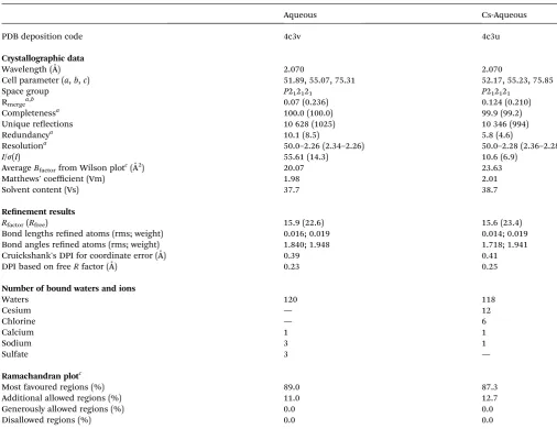
Table 1 Data collection, data processing and refinement statistics for aqueous subtilisin, with and without Cs soak
Aqueous Cs-Aqueous
PDB deposition code 4c3v 4c3u
Crystallographic data
Wavelength (˚A) 2.070 2.070
Cell parameter (a,b,c) 51.89, 55.07, 75.31 52.17, 55.23, 75.85
Space group P212121 P212121
Rmergea,b 0.07 (0.236) 0.124 (0.210)
Completenessa 100.0 (100.0) 99.9 (99.2)
Unique reections 10 628 (1025) 10 346 (994)
Redundancya 10.1 (8.5) 5.8 (4.6)
Resolutiona 50.0–2.26 (2.34–2.26) 50.0–2.28 (2.36–2.28)
I/s(I) 55.61 (14.3) 10.6 (6.9)
AverageBfactorfrom Wilson plotc(˚A2) 20.07 23.63
Matthews' coefficient (Vm) 1.98 2.01
Solvent content (Vs) 37.7 38.7
Renement results
Rfactor(Rfree) 15.9 (22.6) 15.6 (23.4)
Bond lengths rened atoms (rms; weight) 0.016; 0.019 0.014; 0.019
Bond angles rened atoms (rms; weight) 1.840; 1.948 1.718; 1.941
Cruickshank's DPI for coordinate error (A)˚ 0.39 0.41
DPI based on freeRfactor (A)˚ 0.23 0.25
Number of bound waters and ions
Waters 120 118
Cesium — 12
Chlorine — 6
Calcium 1 1
Sodium 3 1
Sulfate 3 —
Ramachandran plotc
Most favoured regions (%) 89.0 87.3
Additional allowed regions (%) 11.0 12.7
Generously allowed regions (%) 0.0 0.0
Disallowed regions (%) 0.0 0.0
aThe values in parentheses are for the highest-resolution shell.bR
merge¼ShklSi|Ii(hkl) hI(hkl)i|/ShklSi|Ii(hkl)|.cData taken from PROCHECK.25
Open Access Article. Published on 18 July 2014. Downloaded on 18/11/2015 10:25:40.
This article is licensed under a
at an X-ray wavelength of 2.070 ˚A and these data processed using the HKL2000 soware package.18Initial crystallographic phases were calculated from the PDB model code 2WUW14for the aqueous subtilisin model and 2WUV14for the Cs derivative subtilisin model. These protein molecular models were rened with the CCP4 Refmac 5.019and the protein structure regions displaying different conformations were rebuilt with COOT.20 Anomalous difference Fourier electron density maps were calculated and those peaks contoured at a 4slevel or higher were considered signicant. Ion assignments were made on the basis of coordination geometry at a peak, the map peak height, andnally theB-factor and occupancy of each site. The indi-vidual Cland Cs+ion occupancies were rened with aB-factor set equal to the average of their surrounding protein atoms within a radius of 7 ˚A of each ion. This adjustment was per-formed using the soware program ION_GRINDER, a Python script which uses the Python interfaces of the Computational Crystallography Toolbox (CCTBX)21 to work on PDB models. Final Rfactor (Rfree) were 15.9 (22.6) and 15.6 (23.4) for the aqueous subtilisin model and for the Cs derivative subtilisin model respectively. Overall X-ray diffraction data and protein model renement statistics for both are reported in Table 1. The gures presented in this paper were made using CCP4MG.22
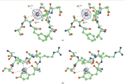
Fig. 1 Crystallographically detected counter-ion sites for subtilisin in
[image:3.595.49.285.51.266.2]water: Cs in grey, Cl in green. Anomalous difference electron density Fourier map contour level is at 4s.
Table 2 Ion sites in the aqueous Cs structure. Ion site numbering follows the series reported in Cianciet al.(2010): Occ¼ion site occupancy;B
-fac¼ionB-factor;scontour level¼maximum peak height of the anomalous difference electron density Fourier mapa
Atom Occ. B-fac. s Potentially charged groups within 0.8 nm Coordinating atoms within 0.4 nm
Cs-1 0.73 30.31 29 His39 ND (0.68), Cl3**(0.76) Leu42 O (0.30), His39 O (0.30), Ala37 O (0.30), W40 (0.34)
Cs-2 0.79 31.37 29 Cl8 (0.49), Glu195 OE (0.71) Ala194 O (0.30), Leu196 O (0.31), Ser260 O (0.31), Gly193 O (0.33), Ser260 OG (0.39), W34 (0.35), W152 (0.37)
Cs-3 0.26 31.86 11 Cl7 (0.72), Asp60 OD (0.79) Ser98 O (0.33), Gly61 O (0.33), W98 (0.25)
Cs-5 0.36 33.76 11 Glu54**OE (0.39) Tyr256 ring C's (0.34–0.39), W127**(0.31), Glu54** OE (0.39), Ala52**O (0.29)
Cs-6 0.24 31.37 6 Arg247 NH (0.72) Tyr143 O (0.33), Ser244 OG (0.37), Tyr143 ring C's (0.37–0.39), W113 (0.23)
Cs-7 0.24 35.68 7 Asp120 OD (0.57), Cl3 (0.71) Leu241 O (0.28), Pro239 O (0.32), W46 (0.27), W62 (0.28), W119 (0.32)
Cs-9 0.22 29.49 4 Ala1 N (0.50), Asp41 OD (0.77) Thr3 OG (0.28), Ala1 O (0.30), Gly80 O (0.33), Thr3 N (0.38), W53 (0.27), W107**(0.23)
Cs-10 0.12 34.61 5 Cl5 (0.42), Arg186 NH (0.71) Ser156 O (0.33), Asn158 O (0.34)
Cs-11 0.12 29.94 4 None Gly47 O (0.35), Tyr57 OH (0.40)
Cs-12 0.24 45.47 5 None Ser159 O (0.31), Thr162 OG (0.31), Ser159 OG (0.37) Cl-1 1.00 16.00 22 Asp181 OD (0.76) Asn185 ND (0.30), Ser184 O (0.33), Tyr263 OH (0.38),
Leu257 O (0.38), W123 (0.34), Gln2**O (0.28), W54**(0.36)
Cl-3 0.74 39.36 6 Cs7 (0.71), Cs1**(0.76) Asn240 O (0.30), Gln245 NE (0.32) Cl-4 0.64 31.88 5 Glu195 OE (0.41), Asp172 OD (0.51), Na2
(0.63), Lys170 NZ (0.67*), Lys136 NZ (0.73*), Tyr167 OH (0.61)
Lys170 O (0.30), W3 (0.35), W87 (0.40)
Cl-5 0.48 44.62 5 Lys15**NZ (0.38), Cs10 (0.42), Arg186 NH (0.66)
Asn158 O (0.29), Asn158 ND (0.29)
Cl-7 0.39 32.75 5 Lys265**NZ (0.69), Cs3 (0.72) Ser99 O (0.30), W147 (0.33), Asn248**ND (0.31) Cl-8 0.27 25.81 4 Cs2 (0.49), Glu195 OE (0.66), Glu197 OE
(0.76)
Ala194 O (0.30), W22 (0.32)
aAtoms are identied by the usual PDB codes, with O, being the alpha-carbonyl oxygen;*–the amino groups of Lys136 and Lys170 appear to be in
salt bridges with the carboxylates of Asp140 and Glu195 respectively;**–these groups are in an adjacent protein molecule in the crystal.
Open Access Article. Published on 18 July 2014. Downloaded on 18/11/2015 10:25:40.
This article is licensed under a
[image:3.595.45.554.387.705.2]Results and discussion
The clear anomalous signal shows very well dened sites (>4s
level Fig. 1 and Table 2). All the caesium sites, and most of the chloride ones, had partial occupancies. The total occupancies for Cs+and Clsum to +3.3 and3.5 respectively, indicating almost zero (0.2) net charge from the counter-ions. In contrast, the caesium and chloride counter-ions in the previ-ously determined subtilisin crystal structure in acetonitrile had a net charge of1.7.
In protein crystallography ion's partial occupancies and ion's Bfactorare a measure of ion mobility likelihood. It is reasonable to think that the protein molecule has only some of the sites fully occupied, and perhaps some are mutually exclusive.
The distribution of cesium ions on the subtilisin surface in an aqueous medium was predicted by molecular dynamics simulations in a preliminary study and showed good agreement with the cation sites observed here crystallographically.23The electrical potential surface of subtilisin is altered by the protein–counter-ion interactions at high salt concentrations (Fig. 2). Patches of positive electric potential appear to increase
Fig. 2 Change of electrostatic potential of the protein surface. Red areas have a negative potential, blue areas a positive potential, white areas are
neutral. (a) Subtilisin soaked in a CsCl salt aqueous solution; (b) in aqueous solution; (c and d) views at 180. Electrostatic potential of the protein surface were computed using CCP4MG.22
Fig. 3 Stereo view of two representative Cs+ion binding sites. Atoms colored according to atom type: blue¼N; red¼O; green¼C, grey¼Cs.
Anomalous difference Fourier map contour level at 4s. (a) Cs site 2 coordination by carbonyls; (b) Cs site 6 interacting with tyrosine ring.
Open Access Article. Published on 18 July 2014. Downloaded on 18/11/2015 10:25:40.
This article is licensed under a
[image:4.595.67.552.197.308.2] [image:4.595.44.550.362.700.2]in area on the protein surface overall and in close proximity of the catalytic triad (Fig. 2). Molecular dynamics simulations show a poor agreement of the distribution of chloride ions on the subtilisin surface in an aqueous medium compared with the anion sites observed crystallographically.23 It was calculated that the surface potential of subtilisin is more negative in aqueous solution than in the crystal, limiting the binding of anions.23In fact, patches of negative electric potential do not show an extension of the areas in a high salt condition (Fig. 2). Protein electrical potential surfaces are commonly calculated and discussed in structure–function relationships. However, such calculations normally do not consider any counter-ions, as well as the intrinsic difficulty of predicting ionisable amino acid side chain pKas,24and electric potentials are very different and even opposite in sign if counter-ions are included.14,23Clearly, an experimental determination of the cation and anion substructures should be sought for any of those systems where electrical potential surfaces are studied, especially at high salt concentrations.
Previous suggestions are that cations are expected to be coordinated mainly by hydrated protein side-chain carboxyl-ates, with a minor contribution from carbonyl oxygens and other side chains.8,10In contrast, we observe that, most of the Cs+ ions are predominantly coordinated by formally neutral oxygens, mostly backbone carbonyls: Fig. 3a shows Cs site 2 as an example. Two of the Cs+ions appear to show coordination by the aromatic rings of Tyr residues, presumably in a charge–p
interaction (Fig. 3b). Only Cs site 5 is closely associated with a charged protein group, and since it interacts with two different protein molecules, the site is probably an artefact of the crystal packing. The “law of matching water affinity”9would suggest relatively weak association of large cations like Cs+with protein carboxylates.
Most of the Clion sites have relatively few of the potential H-bond donors and they are also not near likely protein +ve charges. All the sites show coordinating interactions with an atom in an adjacent protein molecule in the crystal. Cl site 5, for instance, is placed between a Lys amino group and a Cs+ ion associated with the symmetry related molecule and it might be a crystal packing site. In these cases we have to accept the possibility that Cl-sites are a result of crystal packing, as conrmed by the results of the molecular dynamics simula-tions,23and would not have particular affinity for counter-ions when the enzyme is in solution. This possibility is also in agreement with the observation that Clbinds far more weakly to the protein surface.11
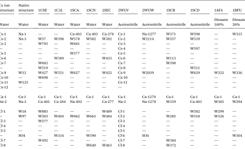
We can also compare the counter-ion sites found in this study with those in previously reported studies, and also with one for our aqueous crystals not soaked with CsCl (Table 2). The tightly bound Ca-1 is conserved across all reported subtilisin structures.
[image:5.595.45.550.415.722.2]In the Cs-1 site, three of the previously determined crystal structures reported a Ca2+(Table 3). In our aqueous structure that has not had a CsCl soak, we conrm a sodium atom (Na-1)
Table 3 Equivalent ion sites in related PDB structures of subtilisin Carlsberg
Cs ion structure
Native
structure 1CSE 1C3L 1SCA 1SCN 2SEC 2WUV 2WUW 1SCB 1SCD 1AF4 1BFU
Water Water Water Water Water Water Water Acetonitrile Acetonitrile Acetonitrile Acetonitrile
Dioxane 100%
Dioxane 20%
Cs-1 Na-1 — — Ca-403 Ca-403 Ca-278 Cs-1 Na-1277 W371 W598 — W315
Cs-2 Na-3 W57 W398 W578 W582 W382 Cs-2 W2114 W357 W539 — —
Cs-3 — W795 — W681 — — Cs-3 — — — — —
— — — — — — — Cs-4 — — W597 — —
Cs-5 — — — W577 — — Cs-5 — — — — —
Cs-6 — — W389 — — W433 Cs-6 — W113 — — —
Cs-7 — W602 — — — — Cs-7 — W398 — — —
— — W519 — — — — Cs-8 — — W511 — —
Cs-9 W12 W427 W351 W627 — W422 Cs-9 W2039 — W629 W322 W336
Cs-10 — W698 — — — — Cs-10 — — — — —
Cs-11 W125 — — — — — Cs-11 — — — — —
Cs-12 — — — — — — — — — — — —
Ca-1 Ca-1 Ca-1 Ca-1 Ca-1 Ca-1 Ca-1 Ca-1 Ca-1279 Ca-1 Ca-1 Ca-1 Ca-1
Na-2 Na-2 Ca-401 Ca-284 Na-402 — Ca-277 Na-2 Na-1278 W359 Ca-403 W305 W294
Cl-1 W18 W885 — — — W489 Cl-1 — — W582 W299 —
— W97 W503 W404 W662 W665 W484 Cl-2 — W285 W518 W326 —
Cl-3 — W577 — — — — Cl-3 — — — — —
Cl-4 — — — — — — Cl-4 — — — — —
Cl-5 — — — — — — Cl-5 — — — — —
— SO4 — W334 — W590 — Cl-6 SO4 — — — W304
Cl-7 — W492 — — — — Cl-7 — W384 — — —
Cl-8 — — — — W640 W463 Cl-8 — W372 — — —
Open Access Article. Published on 18 July 2014. Downloaded on 18/11/2015 10:25:40.
This article is licensed under a
in this position distinguishing it from a water or from bivalent cation site. In site Cs-2, a water molecule is modelled in allve previous structures. Four carbonyl oxygens, a side chain hydroxyl and a water molecule coordinate the site. This appears more like octahedral coordination rather than a tetrahedron of waters. For all but one of the other Cs+sites in our structure, a water molecule has been modelled in at least one of the other aqueous structures. In many of these sites the coordination geometry does not obviously indicate a counter-ion rather than a water molecule, so without the optimized anomalous diff er-ence electron density data it would not be easily recognized. Our structure also contains a site clearly assigned to a Na+(Na-2). The same site has been reported as containing Na+ in some other structures, but as a second Ca2+in others (Table 3). This site showed no anomalous difference Fourier map peak at 2.070 ˚
A wavelength, which excluded a Ca2+(Ca2+f”at this wavelength is 2.95 e). The assignment as a calcium is not supported by our ndings.
Turning to the Clsites we identify, four of the six sites have been modelled as containing waters in one or more of the previously reported crystal structures.
We can also compare the counter-ion sites with those found in our previous subtilisin crystal structure soaked with CsCl and acetonitrile (2WUV).14All but one Cs+site, and all the Clsites,
were also found in this acetonitrile structure (Table 2). In addition, the acetonitrile based crystal structure had further counter-ion sites, 2 Cs+and 2 Cl, which are not found in the aqueous crystal structure. The favoured sites for ion interac-tions are not very different in acetonitrile and aqueous media in subtilisin.
Conclusions
The present study extends the understanding association of counter-ions with the surface of subtilisin and of proteins in general. Subtilisin shows signicant numbers of dened counter-ion sites even in aqueous conditionsi.e.with its large dielectric constant. Most counter-ion sites did not involve close interaction with formal protein charges. The presence of these counter-ions substantially changes the protein electrical potential surface, and which is an effect commonly discussed in relation to function.
The interactions between proteins and counter-ions may have important inuences on enzyme function, especially in systems where quite high salt concentrations are encountered. To understand these effects, it is important to have information about the sites of interaction, and which we provide in this case study.
Acknowledgements
We are grateful to STFC Daresbury Laboratory for the provision of beamtime. Dr D. Lousa and Prof. C. M. Soares (Universida de Nova de Lisboa) are thanked for the helpful discussions. Structures with and without Cs have been deposited to Protein Data Bank codes: 4c3u and 4c3v.
References
1 R. L. Baldwin,Biophys. J., 1996,71, 2056–2063.
2 D. J. Tobias and J. C. Hemminger,Science, 2008,319, 1197– 1198.
3 T. Dudev and C. Lim,Chem. Rev., 2014,114, 538–556. 4 P. Jungwirth and B. Winter,Annu. Rev. Phys. Chem., 2008,59,
343–366.
5 W. Kunz,Curr. Opin. Colloid Interface Sci., 2010,15, 34–39. 6 P. Lo Nostro and B. W. Ninham,Chem. Rev., 2012,112, 2286–
2322.
7 Y. Zhang and P. S. Cremer,Curr. Opin. Chem. Biol., 2006,10, 658–663.
8 L. Vrbka, J. Vondr´aˇsek, B. Jagoda-Cwiklik, R. V´acha and P. Jungwirth, Proc. Natl. Acad. Sci. U. S. A., 2006, 103, 15440–15444.
9 K. D. Collins,Biophys. Chem., 2012,167, 43–59. 10 K. D. Collins,Biophys. Chem., 2006,119, 271–281.
11 K. B. Rembert, J. Paterov´a, J. Heyda, C. Hilty, P. Jungwirth and P. S. Cremer,J. Am. Chem. Soc., 2012,134, 10039–10046. 12 M. Cianci, P. J. Rizkallah, A. Olczak, J. Raery, N. E. Chayen, P. F. Zagalsky and J. R. Helliwell, Acta Crystallogr., Sect. D: Biol. Crystallogr., 2001,57, 1219–1229.
13 K. Djinovic-Carugo, J. R. Helliwell, H. Stuhrmann and M. S. Weiss,J. Synchrotron Radiat., 2005,12, 410–419. 14 M. Cianci, B. Tomaszewski, J. R. Helliwell and P. J. Halling,J.
Am. Chem. Soc., 2010,132, 2293–2300.
15 C. Mueller-Dieckmann, S. Panjikar, A. Schmidt, S. Mueller, J. Kuper, A. Geerlof, M. Wilmanns, R. K. Singh, P. A. Tucker and M. S. Weiss,Acta Crystallogr., Sect. D: Biol. Crystallogr., 2007,63, 366–380.
16 D. N. Okamoto, M. Y. Kondo, J. A. N. Santos, S. Nakajima, K. Hiraga, K. Oda, M. A. Juliano, L. Juliano and I. E. Gouvea,Biochim. Biophys. Acta, 2009,1794, 367–373. 17 M. Cianci, S. Antonyuk, N. Bliss, M. W. Bailey, S. G. Buffey,
K. C. Cheung, J. A. Clarke, G. E. Derbyshire, M. J. Ellis, M. J. Enderby, A. F. Grant, M. P. Holbourn, D. Laundy, C. Nave, R. Ryder, P. Stephenson, J. R. Helliwell and S. S. Hasnain,J. Synchrotron Radiat., 2005,12, 455–466. 18 Z. Otwinowski and W. Minor,Methods Enzymol., 1997,276,
307–326.
19 G. N. Murshudov, A. A. Vagin and E. J. Dodson, Acta Crystallogr., Sect. D: Biol. Crystallogr., 1996,53, 240–255. 20 P. Emsley, B. Lohkamp, W. G. Scott and K. Cowtan, Acta
Crystallogr., Sect. D: Biol. Crystallogr., 2010,66, 486–501. 21 R. W. Grosse-Kunstleve, N. K. Sauter, N. W. Moriarty and
P. D. Adams,J. Appl. Crystallogr., 2002,35, 126–136. 22 S. McNicholas, E. Potterton, K. S. Wilson and M. E. M. Noble,
Acta Crystallogr., Sect. D: Biol. Crystallogr., 2011,67, 386–394. 23 D. Lousa, M. Cianci, J. R. Helliwell, P. J. Halling, A. M. Baptista and C. M. Soares, J. Phys. Chem. B, 2012, 116, 5838–5848.
24 S. J. Fisher, J. Wilkinson, R. H. Henchman and J. R. Helliwell, Crystallogr. Rev., 2009,15, 231–259.
25 R. A. Laskowski, M. W. MacArthur, D. S. Moss and J. M. Thornton,J. Appl. Crystallogr., 1993,26, 283–291.
Open Access Article. Published on 18 July 2014. Downloaded on 18/11/2015 10:25:40.
This article is licensed under a