Copyright© 1970 AmericanSociety forMicrobiology PrintedinU.S.A.
Evidence
for
a
Single-Stranded
Adenovirus-Associated
Virus
Genome:
Isolation and
Separation of
Complementary
Single Strands
K. 1. BERNS AND J. A. ROSE
LaboratoryofBiology ofViruises, National Institute ofAllergyanid InifectioutsDiseases, Bethesda, Marylanld 20014 Received for publication25February 1970
Single-strandedadenovirus-associated virus type 2deoxyribonucleicacid (AAV-2
DNA) has been isolated from the virion after enzymatic pretreatment of the particles
byheatingat 53Cfor 1 hr in 0.015 M NaCl plus 0.0015 M sodium citrate in the
presence of 1 %sodium dodecyl sulfate. Double-stranded AAV-2 DNA present as a marker is not denatured by this treatment. AAV-2 single-stranded DNA is composed of twocomplementary species which can be separated in neutral CsClwhen 5-bro-modeoxyuridine has been substituted for thymidine in the DNA. The present re-portis the first documented instance of the separation of complementary strands of ananimalvirus DNA.
The adenovirus-associated viruses (AAV) are sinall, defective deoxyribonucleic acid (DNA) viruses which require an adenovirus as a helper
for production infection (1, 7). Purified AAV
DNA was found to be double-stranded with a molecular weightof 3.0 x 10⁶ to 3.6 x 10⁶ (10,
12). Crawford et al. (5), however, suggested that AAV DNA was single-stranded in situ and only
formed a double helix during extraction. Rose
etal. (11) andMayor etal. (9) have now
demon-stratedthat AAV DNA isindeed single-stranded
in situ, and that the double-stranded form of
purified AAV DNA results from the annealing
of single strands from different virions during
extraction (11).
Inthis paper we report the isolation of single-stranded AAV DNA from virions and the
physi-calseparation ofthe single strands into two
com-plementary species.
MATERIALS AND METHODS
Materials. Preparation and assay of virus stocks has been described (7, 13). KBcellswerefromaline
originallyprovided byM. Green. Optical grade CsCl and 5-bromodeoxyuridine (BUDR)-6-'H (12.7 c/ mmole) were obtained from Schwarz BioResearch Inc., Orangeburg, N.Y.
Thymidine-2-'4C
(52.8 mc/mmole) and thymidine-methyl-3H (>15 c/mmole)
were purchased from New England Nuclear Corp.,
Boston, Mass. 32p was obtained from Tracer Lab,
Waltham, Mass. Crystallized-lyophilized trypsin and 2X crystallized papain were fromWorthington Bio-chemicalCorp., Freehold, N.J.; 5-fluorodeoxyuridine (FUDR) from Hoffman-La Roche, Inc., Nutley,
N.J.; BUDR from Calbiochem, LosAngeles, Calif.;
and Sarkosyl (NL 97) from Geigy Industrial Chem-icals, Ardsley, N.Y.
Virus growth andpurification. Thegrowthand
puri-fication of 3H-BUDR adenovirus type 2 (adenoid 6)
has been described (l1). AAV-2 (AAV-2H) (7) was
producedin KBcellsinsuspension culture [in Eagle's
medium (6) supplementedwith5% horseserum] with adenovirustype 2ashelper.Forproductionof AAV-2 containing either 3H- or '4C-thymidine cells were
co-infected withadenovirus type 2 [10tissueculture in-fectious doses(TCID)50/cell]andAAV-2 (10 TCID5o/ cell), and 6 hr later 3H-or 14C-thymidine was added to a final concentration of 1.0 or 0.1 ,c/ml, respec-tively. The cells were harvested after 48 hr at 37 C. To obtain 3H-BUDR AAV-2, cells were similarly
infected. FUDR (0.5 ,ug/ml) wasadded 13.5 hr after infection, and 3H-BUDR (0.5
,pc/ml)
and BUDR (10,g/ml) 14 hr after infection. The infection wasterminated at 48 hr. For production of 3P-BUDR AAV-2,cells were infected asabove, and i3 hrafter infection the cells were collected, washed, and
re-suspended in Eagle's medium containing reduced
P04 (10-1 M) supplemented with '% dialyzed horse
serum. FUDR (0.5 ,ug/ml) was then added, and 30 minlater 32p (2
Ac/ml)
and BUDR (10,ug/ml) wereadded. The infected cells were harvested at 48 hr. To purify AAV, cell harvests from 1 liter cultures
were resuspended in 27 ml of phosphate-buffered
saline and sonically treated. The preparations were treated with
2%7o
deoxycholate and 0.02c% trypsin at37 C for 30 min. After debris had been removed by
low-speed centrifugation, virus was pelleted by cen-trifugation in the Spinco model L ultracentrifuge at 22,000rev/min for3 hrat4 C in the SW 25.1 rotor.
Sedimented virus was resuspended in 4 ml
tris(hy-693
on November 11, 2019 by guest
http://jvi.asm.org/
BERNS AND ROSE
droxymethyl)aminomethane (Tris), pH 7.9, by sonic treatment and the AAV-2 waspurified by CsCl cen-trifugation as described previously (13, 11).
DNA extraction. The extraction ofadenovirus DNA and the double-stranded form of AAV DNA have beendescribed(11).
DNAsedimentation. DNA was sedimented through preformed, linear, neutral sucrose gradients contain-ing 1 M NaCl (2). CsCl solutions for isopycnic cen-trifugation were 0.05 M Tris (pH 7.9) and 0.001 M (ethylenedinitrilo) tetraacetic acid and contained 0.15%Sarkosyl in afinal volume of 4.5 ml. The spe-cific conditionsfor each gradientaredescribed in the figures.
Denaturation and annealing ofDNA. Denaturation of DNA in alkali and the neutralization of alkali-denatured DNA have been described (2). DNAwas annealed in 0.15 MNaCl plus 0.015Msodium citrate
(1X SSC) fort hrat70C.
RESULTS
Isolation of single-stranded DNA from AAV. AAV DNAis single-strandedinsitu (9, 11), but
thepurified form is double-stranded (10, 13). We
were interested in
isolating single-stranded
DNAunder conditions which would (i) not denature
the double-stranded form of the DNA, and
(ii)
not permit annealing of single strands into the
double-stranded form. To do this experiment,
it is necessary to be able to distinguish single-stranded AAV-2 DNA from thedouble-stranded
form. The two forms of AAV-2 DNA may be separated by isopycnic CsCl centrifugation (12)
and by zonal sedimentation through a neutral sucrose gradient (Fig. 1). AAV DNA which had
been denatured in alkali and neutralized sedi-mented morerapidly than double-stranded DNA inaneutral sucrosegradientcontaining 1 MNaCl (Fig. 1A and B). The ratio ofdistances traveled
was 1.57 (Fig. 1C). AAV single-stranded DNA
rapidly anneals to form double-stranded DNA (9, 11),and it is assumed that thetrailingmaterial
observedin
Fig.
1BrepresentsAAVDNAwhichhas annealed at room temperature between the
time of neutralization and the
completion
of thesucrose run (5
hr).
Thereported
method for the purification ofAAV DNA (11,13) releasesDNAfrom the particles after enzymatic pretreatment
by heating the virus at 50C in 1 X SSC in the
presence of 1 % sodium dodecyl sulfate (SDS).
These conditions of salt and temperature allow annealing of AAVsingle strands (11). Alteration of conditionstoprevent
annealing
(e.g.,decreasedionicstrengthorincreased temperature, orboth)
would result in the releaseofsingle strands into solution (9). Enzymatically pretreated AAV particles containing 3H-DNA were heated
to-gether with purified double-stranded 14C-AAV
DNAat53C for1 hrin 0.1XSSCin the presence
A
E 400 -Q_ B
200_
500
300
100
5 10 15 20 25 30 35
[image:2.487.263.450.54.372.2]FRACTION NUMBERS
FIG. 1. Sedimentation of14C-AAV-2 DNA through
linear5 to20% neutralsucrosegradients.
Cenitrifuga-tioczwasfor4hrat40,000rev/min and20 C inSpinco model SW 50 rotor. (A) Double-stranded AA V-2 DNA. (B) Single-stranded AAV-2 DNA. Double-strandedAAV-2 DNA was alkali-denatured and then neutralized. (C) A mixture of single- and double-stranded AAV-2 DNA. The single-stranded AA V-2 DNAsediments more rapidly thani thedouble-stranided
form.
of 1% SDS. The DNA was then sedimented
through neutral sucrose (Fig. 2). Although the
14C-AAV DNA still sediments as
double-strandedDNA, thenewly released 3H-AAVDNA
sedimentsassingle strands. The ratio of distances traveled was 1.58. There istrailing 3H-DNA
(ap-proximately 25%) but no more than seen in the
case of purified AAV DNA which has been
alkali-denatured (Fig. IB). Thus, under
condi-tions which do not denature purified double-stranded AAV DNA, single-stranded DNA is recovered from the particles. The same prepara-tionwasbanded in CsCl(Fig.3). The
"4C-double-stranded DNAbandsat adensity of 1.714g/cm3. The 3H-DNA peak (fraction 17) is 12 mg/cm3 more dense. This is the density difference
ob-694 J. VIROL.
on November 11, 2019 by guest
http://jvi.asm.org/
SINGLE-STRANDED AAV-2
A-4C J 200L
20 25 30
FRACTIONNUMRERS
a-~~~~~~~~~~~~~~~~~~~~~~~~~~~~~~~~~~-100 J
200-5 10 15 20 25
FRACTION NUMBER
FIG. 2. Sucrose sedimentation of newly released 3H-AAV-2 DNA and double-stranded '4C-AAV-2 marker DNA. After enzymatic pretreatment, AAV-2 particles containing3H-DNA weremixed with double-stranded 14C-AA V-2 DNA and heatedat 53 CI hr in
0.JX SSC in the presence of 1% SDS. Conditions of centrifugationwere those described in thelegendto
Fig1.
175
-174C
.1| 7 3z
,'O-1724o
[image:3.487.43.235.49.212.2]171
T J 70 ' 69FIG. 3. Isopycnic CsCIcentrifugation for 72 hr at
33,000 rev/min at 20 C in Spinco model 40 rotor.
Newly released 3H-AAV-2 DNA and double-stranded 14C-AAV-2 marker DNA. The conditionsofDNA
re-leaseweretlhose describedinthelegendtoFig.2.
served between native and denatured AAV-2 DNAin theanalytical ultracentrifuge (12).There
isskewingof the 3Hbandtowardlighter density.
This result has beenrepeatedly obtained,and we
believeit represents
annealing
which hasoccurredduring the course of
centrifugation
(72 hr at20C; reference
3).
Thesharp
peak of 3H near thetopofthegradient probably
representsDNA stillassociated with capsid protein.Isopycnic centrifugation of denatured BUDR AAV DNA. Roseetal. (11) had noted that
den-sity hybridAAVdouble-stranded DNA molecules
(i.e., molecules composed of BUDR- and
non-BUDR-substituted single
strands)
banded in abimodal distribution in CsCl. It was sug-gested that the components mightrepresent two
complementary species which contain different
amounts of thymidine. An extensive random
substitution of thymidine by BUDR would
re-sult, therefore, in two single-stranded species
with substantially different densities in CsCl and thus account for the bimodal hybrid. (Non-BUDR-substituted, denatured AAV DNA bands in CsCl as asingle uniform band) (12).
Double-stranded 3H-BUDR AAV DNA banded in CsCl
as a uniform component and was 75 mg/cm3
more dense than unsubstituted
'4C-double-stranded AAV DNA (Fig. 4). When 3H-BUDR
DNA was denatured in alkali, neutralized, and
banded in CsCl, two bands were observed (Fig.
5A). If these bands represented two single-stranded
species,
theheavier band would be ex-pected to have moreradioactivity,
because it would contain more 3H-BUDR. Under ourconditions this is not the case. The light band
contains more radioactivity and, in addition, is
skewed toward light density. These results could beattributedtothefactthat thepeak of the light band isclosetothedensity oftheoriginal
double-stranded 3H-BUDR DNA so that the light band
of single polynucleotide chains would bedifficult
todistinguish from double-stranded DNA which
had formed during the experiment (i.e., the two
bands would overlap). To test this possibility,
double-stranded 3H-BUDR DNA was added as
amarker tothedenatured, neutralized 3H-BUDR
single strands (Fig. 5B). Theheavy bandis again
present, andaportion ofthelightbandisseen as a shoulder on the dense side of the double-stranded marker DNA. An estimate of the counts/min representing light strands can be made from the skew of the double-stranded
DNA band in Fig. 5B. The light strandswould
contain 40% fewer counts/min than the heavy
strands. The heavy band was approximately 40
mg/cm3, and the light band-shoulder was about
301Nr
*
_1 II
~~~~I
<i
O 5 IX
- T
0&?6-I0 15 20 25 FRACTION NUMBER
30
11.86
-I
C'-S.,_
to
r.
-0
z
1.78B?
1701
I162
FIG. 4. Isopycnic CsCI centrifugation for 48 hrat
40,000rev/minat25 CinSpincomodel SW 50rotorof double-stranded 14C-thymidine AAV-2 DNA, double-stranded3H-BUDR AAV-2DNA,anddouble-stranded
32P-BUDRAAV-2 DNA.
695
VOL. 5, 1970
on November 11, 2019 by guest
http://jvi.asm.org/
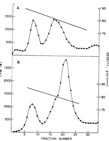
[image:3.487.41.234.304.407.2] [image:3.487.249.439.462.585.2]BERNS AND ROSE
A. -1.85 thesucrose
sedimentation
dataillustratedinFig.
7.
Annealing
conditions were the same as for1500
_
Fig.
- 1
6. DNA from theheavy
band inFig.
5AA
X0
\° does not form double-stranded DNA whenself-ooo-
Xl.75
\ / \.annealed (Fig. 7A). DNA from the dense side oft1 75 the light band remains predominantly
single-500 j \by \
stranded,
but there is asignificant
amount oftrailing materialwhich we assume to be
double-stranded DNA
resulting
from contamination ofz the light single strands with double-stranded
B. -\
*500
;-2000 1851
400L-T \ ~~~~~~1.85_ .
1500-.80
1000- 0
-1.75
500-5 10 15 20 25 30 B ,
FRACTION NUMBER
FIG. 5. Isopycnic CsCI centrifugation for 48 hr at 400-40,000 rev/min at 25C in Spinco model 65 rotor of E 3H-BUDRAAV-2DNA. (A) 3H-BUDRAAV-2DNA wasalkali-denatured andthen neutralized. (B)
Double-stranded 3H-BUDR AAV-2 DNA was added to the | 200-solutiondescribedinpartA as adensity marker.
8 mg/cm' more dense than the double-stranded o r-L
form of the DNA.
Complementarityoftheheavy andlight compo- .
nents. The sedimentation characteristics of both I C
single- and double-stranded
'H-BUDR
DNA inneutral sucrose were determined. Double- 400 stranded 'H-BUDR AAV DNA sediments 1.16
times as fast as double-stranded 14C-thymidine
AAV DNA (Fig. 6A). 'H-BUDR AAV DNA
which has been alkali-denatured and neutralized sediments 1.85 times as fast as the double-stranded
"4C-AAV
DNA (Fig. 6B). When the denatured 3H-BUDR AAV DNA is annealed at70 Cin 1x SSCfor 1
hr,
it sediments asdouble- -__stranded
'H-BUDR
DNA (Fig. 6C). Thus, cj lo 15 20 25 30'H-BUDR
AAV single polynucleotide chains FRACTION NUMBERwhich have been annealed into double-stranded FIG. 6. Sucrose sedimentation of3H-BUDR AAV-2
DNA canbereadily distinguished from the origi- DNA and '4C-thymidine AAV-2 DNA. Conditions of
nal single strands in a neutral sucrose gradient. centrifugationwerethosedescribed inthelegendto Fig.
If thetwobandsseeninFig. 5Aprimarilyrepre- 1.
(A)
Double-stranded 'H-BUDR AAV-2 DNA andsentthe separation of two species ofAAV single double-stranded 14C-thymidine AAV-2 DNA. (B) strandswhicharecomplementary, neither popula- Single-stranded 3H-BUDR AA V-2 DNA derived from tion should form double-stranded DNA when double-stranded DNA by alkali-denaturation and exposedtoannealing
cosubsequent
neutralization and double-stranded14C-exposed to annealig conditions, but double- thymidineAAV-2 DNA. (C) The single-stranded
3H-stranded DNA should be formed when the two BUDR AA V-2 DNA from part B was annealed at speciesaremixed under thesameannealingcondi- 70 C forIhr in IX SSC. Double-stranded '4C-thymi-tions. The results of this experimentareshown in dineAAV-2 DNA present as a marker.
696 J. VIROL.
on November 11, 2019 by guest
http://jvi.asm.org/
[image:4.487.59.250.54.299.2] [image:4.487.263.450.180.529.2]A.r,
___'
200k
300
1-)
100
600
300 C.
I
L=3
5 10 5 20 25 30
'FRACTION NUMBER
200 tions (Fig. 8B). The light 3H-BUDR band was
slightly more dense than double-stranded
3H-BUDR DNA and was broad, again indicating
that the preparation is not completely free of
100 double-stranded DNA (Fig. 8C). A mixture of
the heavy and light bands does reform double-stranded DNA under annealing conditions (Fig.
8D). The annealed DNA appears as a
homo-geneouspeakatadensity about 4 mg/cm3 lighter
I than the
32P-BUDR marker,
exactly
as observed.__in Fig.
8A.0 50
200
100
FIG.7. Sucrose sedimentation of 3H-BUDRAAV-2 single polynucleotide chains after attempting
anneal-ing. Conditions of annealing were those described in
Fig. 6C. Conditions of centrifugation were those
de-scribed in thelegendtoFig. 1. Double-stranded
14C-thymidine AAV-2 DNA waspresent in allruns as a
marker. (A) 3H-BUDR AAV-2 heavy strandsfrom Fig.5Awereexposedtoannealingconditions. (B)
IH-BUDR AAV-2 light strandsfrom theheavier side of
the light-density peak in Fig. 5A were exposed to annealing conditions. (C) The single strands from partsAand Bweremixed and then annealed.
DNA thatformed during the CsClrun(Fig. 7B).
Whenequivalent amountsof theheavy and light strands are annealed together, a pattern similar
to that obtained with the original 3H-BUDR double-stranded DNA is seen (Fig. 7C). There
isnopeak of single-stranded DNA.Weconclude
that we have physically separated the
comple-mentary single polynucleotide chains of AAV
DNA, although the light strands are
contami-nated with double-stranded DNA. Confirmation ofthis conclusion was obtained byrepeating the
preceding experiment and measuring the extent
offormation of double-stranded DNA in CsCl density gradients. 32P-BUDR double-stranded AAV DNA was usedas a density marker. This
DNA preparation banded at a slightly greater
density than the double-stranded 3H-BUDR AAV DNA (-4to 5 mg/cm3), indicating 4% greater
BUDR incorporation (Fig. 4 and 8A; reference
11). The heaby 3H-BUDR band formed no
double-stranded DNA under annealing
condi-DISCUSSION
That AAV DNA is single-stranded in situhas
nowbeen demonstrated in this and previous
com-munications (9, 11). Roseetal. (11) showedthat double-stranded AAVDNAisformedby the
an-nealing of single strands released from different particles during the DNA extraction procedure. In this paper we demonstrated that extracted
AAV DNA iscomposed oftwospecies of single polynucleotide chains which do not self-anneal but do form double-stranded DNAwhen mixed under annealing conditions, i.e., they are
com-plementary. The heavy BUDR single strands
canbeseparated from the light BUDR strands in one operation. However, the light strands are
contaminated with double-strandedDNA; there-fore, an additional purification step isnecessary
to obtain a unique preparation of light strands.
These data are compatible with the suggestion
that an AAV particle contains one single
poly-nucleotide chain (+ or -) whichhasa
molecu-larweight of approximately 1.5 x 10⁶ (5, 9, 11).
Inlight ofourpresentknowledgeof the struc-ture and composition of AAV DNA, it is not
surprising that the resolution of the structure of AAV DNAinsituhasprovendifficult.The
exist-ence ofcomplementary strands, the small size of
theDNA, therelatively highoverallguanine plus
cytosine content, the extraction procedure, and the experimental conditions required to study theDNAwouldallcontributetorapid formation of double-strandedDNA(18, 19).Themagnitude oftheproblemisillustrated in thispaper. In the
relatively short experiments involving sucrose
gradients, about 25% of the single strands have
annealed (Fig. 1,2, 6); however,over50%,7 ofthe samesingle-stranded DNAcentrifuged for 72 hr
in CsClat25 Chasannealed(Fig. 3).Evenunder
conditions where the complementary single strands have different densities in CsCl (i.e., BUDR labeling), approximately 40%0 of the
DNA has annealed (Fig. 5).
We were able to separate the complementary AAV single strands because, apparently, theydo not contain equal amounts ofthymidine. Thus,
B.rl~~~~~~~~~~~~~~~~~~~~~~~~~~~~~~~~~~
B. ,
;p
:Sr
r:
697
on November 11, 2019 by guest
http://jvi.asm.org/
[image:5.487.41.231.54.300.2]BERNS AND ROSE
-r
I)
1.823 1.792 1.788
[image:6.487.57.246.58.478.2]DENSITY, (g/cc)
FIG. 8. Isopycnic CsCIcentrifugation of3H-BUDR
AAV-2 single polynucleotide chains after attempted
annealing. Conditions of annealing werethosedescribed
in Fig. 6C. Conditions ofcentrifugation were those described in the legend to Fig. 5. 52P-BUDR AAV-2
double-strandedDNA wasusedas adensitymarker in
all gradients. (A) 3H-BUDR AAV-2 double-stranded
DNA. (B) 3H-BUDR AAV-2 heavy strands from
Fig. SA were exposed to annealing conditions. (C)
3H-BUDR AAV-2 light strandsfrom the heavier side
ofthe light density peak in Fig. 5A were exposed to
annealing conditions. (D) The single strands from
partsB andCweremixedand thenannealed.
when BUDR is substituted for thymidine, there isa difference in density between the two-strand species. Assuming a 75% substitution of
thymi-dine byBUDR (11), the difference in thymidine
content of the two strands would be about40% (i.e., the heavy strand would be 26% T and the
light strand 18% T), if the expected density for
single strands with equal thymidine content
would be theaverageof thetwodensitiesobserved. Differences in thymidine content of
comple-mentary, single polynucleotide chains equal to
those assumed here have been reported for the
replicative form of
OX
174DNA (15, 16) andforHeLacelllightsatellite DNA(14).Weattempted
the separation of the complementary strands of
BUDRadenovirusDNA, but therewasonly one peak of denatured DNA in neutral CsCl.
BUDR-substituted double-stranded AAV
DNA sediments more rapidly in neutral sucrose than non-BUDR-substituted DNA. The dif-ference observed is too great to be accounted for
simply by the expected increase in mass caused
byBUDR (4).Interestingly,theratioof distances
sedimented by single- and double-stranded
BUDR labeled AAV DNA in neutral sucrose is thesame asthatobserved for the unlabeled DNA (1.58). This is in reasonable agreement with the data ofStudier (17) whichwould predit a ratio of 1.66 foradouble-stranded DNA of molecular
weight3 x 106 which iscomposed oftwointact
complementary singlestrands.
The present report is the first documented
in-stance of the separation of complementary
strands ofananimal virus DNA. Thesefindings
willpermita more detailed study of AAV DNA
infectivity (8) andallow analysisof strand-specific
transcription in vivo. Information derived from
such studies mayhelp reveal the molecular basis
for AAVdefectiveness.
ACKNOWLEDGMENT WethankC.Silvermanfor technicalassistance.
LITERATURECITED
1.Atchison,R.W.,B. C. Casto, and W. M. Hammon. 1965. Adenovirus-associateddefectivevirus particles. Science 149: 754-756.
2. Berns,K. I. and C.Silverman. 1970. Thenatural occurrence of cross-linkedvacciniadeoxyribonucleicacid. J. Virol. 5: 299-304.
3. Berns, K.I.,and C. A.Thomas.1961.Astudyof single poly-nucleotidechainsderivedfrom T2 and T4bacteriophage. J.Mol. Biol.3:289-300.
4. Burgi, S.,and A.D.Hershey. 1963. Sedimentation rate as a measureofmolecularweightof DNA. Biophys. J. 3:309-321.
5.Crawford,L. V.,E. A.C. Follett,M. G.Burdon, and D. J. McGeoch. 1969.The DNA of a minute virus of mice. J. Gen. Virol. 4:37-46.
6. Eagle,H. 1959.Aminoacid metabolismin mammalian cell cultures.Science 130:432-437.
7. Hoggan, M.D.,H.Blacklow,and W. P. Rowe.1966. Studies of small DNAvirusesfound in variousadenovirus prepara-tions: physical, biological,and immunological character-istics.Proc. Nat.Acad.Sci.U.S.A. 55:1467-1474. 8. Hoggan,M.D.,A. J.Shatkin,N. R. Blacklow, F. Koczot,
andJ. A. Rose. 1968.Helper-dependent infectious
deoxy-698 J. VIROL.
on November 11, 2019 by guest
http://jvi.asm.org/
ribonucleic acid from adenovirus-associated virus. J. Virol. 2:850-851.
9. Mayor, H. D., K. Torikai, J. L. Melnick, andM. Mandel. 1969. Plus and minus single-stranded DNA separately encapsidated in adeno-associated satellite virions. Science
166:1280-1282.
10. Parks, W. P., M. Green, M. Pifia, and J. L. Melnick. 1967. Physico-chemical characterization of adeno-associated satellite virustype4and its nucleic acid. J. Virol.1:980-987.
11. Rose, J. A., K. I.Berns, M. D. Hoggan, and F. J. Koczot.
1969. Evidence forasingle-stranded adenovirus-associated virusgenome:formation ofa DNAdensity-hybridon
re-leaseof viral DNA. Proc. Nat. Acad. Sci. U.S.A.
64:863-869.
12. Rose, J. A., M. D. Hoggan, and A. J. Shatkin. 1968. Genetic relatedness studies with adenovirus-associated viruses. J.
Virol.2:999-1005.
13. Rose, J. A., M.D. Hoggan, and A. J. Shatkin. 1966. Nucleic
acid fromanadeno-associatedvirus: chemical and physical studies. Proc. Nat. Acad. Sci. U.S.A.56:86-92.
14. Schildkraut, C. L., and J. J. Maio. 1969. Fractions ofHeLa
DNA differingintheircontentof guanine +cYtosine.J.
Mol.Biol. 46:305-312.
15. Sinsheimer, R. L. 1959. A single-stranded deoxyribonucleic acid frombacteriophageOX-174.J. Mol. Biol. 1:43-53. 16. Sinsheimer, R. L., B. Starman, C. Nagler, and S. Guthrie.
1962. Theprocessofinfection with bacteriophageOX-174.1.
Evidence fora"replicative form." J. Mol. Biol.4:142-160.
17. Studier, F. W. 1965. Sedimentation studies of the size and shape of DNA. J. Mol.Biol. 11:373-390.
18. Studier, F. W. 1969. Effects of the conformation of single-stranded DNAon renaturation andaggregation. J. Mol. Biol. 41:199-209.
19. Wetmur, J. G., and N. Davidson. 1968. Kinetics of renatura-tionof DNA. J. Mol. Biol. 31:349-370.