Complete Reversible Refolding of a G-Protein
Coupled Receptor on a Solid Support
Natalie Di Bartolo1*, Emma L. R. Compton2, Tony Warne3, Patricia C. Edwards3, Christopher G. Tate3, Gebhard F. X. Schertler4, Paula J. Booth1¤
1School of Biochemistry, University of Bristol, Bristol, United Kingdom,2Organisational and Staff Development Unit, University of Strathclyde, Glasgow, United Kingdom,3MRC Laboratory of Molecular Biology, Cambridge Biomedical Campus, Cambridge, United Kingdom,4Laboratory of Biomolecular Research, Paul Scherrer Institute, Villigen, Switzerland
¤ Current address: Department of Chemistry, King’s College London, London, United Kingdom
Abstract
The factors defining the correct folding and stability of integral membrane proteins are poorly understood. Folding of only a few select membrane proteins has been scrutinised, leaving considerable deficiencies in knowledge for large protein families, such as G protein coupled receptors (GPCRs). Complete reversible folding, which is problematic for any membrane protein, has eluded this dominant receptor family. Moreover, attempts to recover receptors from denatured states are inefficient, yielding at best 40–70% functional protein. We present a method for the reversible unfolding of an archetypal family member, theβ1 -adrenergic receptor, and attain 100% recovery of the folded, functional state, in terms of ligand binding, compared to receptor which has not been subject to any unfolding and retains its original, folded structure. We exploit refolding on a solid support, which could avoid unwanted interactions and aggregation that occur in bulk solution. We determine the changes in structure and function upon unfolding and refolding. Additionally, we employ a method that is relatively new to membrane protein folding; pulse proteolysis. Complete refolding ofβ1-adrenergic receptor occurs in n-decyl-β-D-maltoside (DM) micelles from a urea-denatured state, as shown by regain of its original helical structure, ligand binding and protein fluorescence. The successful refolding strategy on a solid support offers a defined method for the controlled refolding and recovery of functional GPCRs and other membrane proteins that suffer from instability and irreversible denaturation once isolated from their native membranes.
Introduction
A central feature of biological activity is the correct folding of proteins into their functional states. Integral membrane proteins have been largely overlooked in the extensive investigations levelled at understanding this folding process. Biophysical studiesin vitroare currently the method of choice for elucidating molecular level mechanistic detail [1–6] and these are being
OPEN ACCESS
Citation:Di Bartolo N, Compton ELR, Warne T, Edwards PC, Tate CG, Schertler GFX, et al. (2016) Complete Reversible Refolding of a G-Protein Coupled Receptor on a Solid Support. PLoS ONE 11 (3): e0151582. doi:10.1371/journal.pone.0151582
Editor:Jose M. Sanchez-Ruiz, Universidad de Granada, SPAIN
Received:May 26, 2015
Accepted:March 1, 2016
Published:March 16, 2016
Copyright:© 2016 Di Bartolo et al. This is an open access article distributed under the terms of the
Creative Commons Attribution License, which permits unrestricted use, distribution, and reproduction in any medium, provided the original author and source are credited.
Data Availability Statement:All relevant data are within the paper and its Supporting Information files.
Funding:The authors acknowledge funding from BBSRC grant BB/G002037/1. PJB also thanks the Wellcome Trust (grant 72075) as well as the Royal Society for a Woflson Research Merit Award and Leverhulme Trust for a Research Fellowship. Research in CGT’s laboratory was funded by a core grant from the Medical Research Council [MRC U10519215].
complemented by studies using cellular membrane extracts to capture the additional cellular machineries involved in correct membrane protein foldingin vivo[7–10]. One of the largest knowledge gaps lies with biophysical studies of transmembrane helical proteins and there is a dearth of information on many large membrane protein families. A case in point are GPCRs. These constitute the largest family of membrane receptors, with over 800 human GPCRs iden-tified, and are the focus of considerable research and industrial interest [11,12]. The primary function of GPCRs is to respond to extracellular stimuli and activate an array of intracellular signalling pathways through their interaction with numerous G protein subtypes as well as through G-protein independent pathways, often in a ligand-specific manner [13,14]. The lack ofin vitrofolding information on this particular protein class has significant ramifications. It impacts directly upon achieving and maintaining a correctly folded, functional structure for the plethora of biophysical, biochemical and structural work aimed at ascertaining functional mechanisms for GPCRs. Obtaining sufficient quantities of properly folded GPCRs forin vitro structural and functional studies has proven tremendously difficult, not least due to the poor long-term stability of the receptors in detergents and associated loss of structure and function. These practical issues, together with the inherent importance of understanding folding, have highlighted the significance of investigating GCPR refolding and stability. One of the most promising advances in this area has been the use of columns as solid supports for recovering functional GPCR from inclusion bodies, using SDS to solubilise the inclusion bodies generally with amphipols or bicelles to recover folded protein [15–19]. The most successful cases result in ~40–70% recovery of functional protein. Solubilisation of inclusion bodies in urea gives much lower yields of 25%. The extent of denaturation of the SDS or urea-solubilised state has not been assessed in detail—these denaturants could in fact be primarily solubilising protein that is already largely folded from the inclusion bodies, so little or no refolding is required. Nor have there been any reports on the controlled, reversible unfolding and refolding a receptor. Reversible unfolding has however, been demonstrated for several otherα-helical membrane proteins from different protein classes including diacylglycerol kinase (DGK) [20], the potas-sium channel, KscA [21,22], the galactose transporter, GalP [3] and the small multidrug resis-tance transporter, EmrE [1].
Here, we present a systematic folding study of the thermostabilised turkeyβ1-adrenergic receptor (β1AR-m23) [23,24], using urea and SDS denaturants. This mutant offers an exciting opportunity for folding studies as the increased stability makes refolding a viable prospect. We compare refolding in detergent micelles composed of DM in bulk solution with a solid support. The latter approach gives unprecedented reversible refolding of the GPCR, from a urea-dena-tured state, with a 100% yield. Although interactions with other lipids or proteins in native membranes may be important for the functional folded state of GPCRsin vivo, here we study the receptor in isolation and address function with respect to ligand binding only. It is vital to attain complete recovery of such function as membrane proteins are notorious for perturba-tions of ligand binding constants and reduced stability when solubilised in detergents, even without any refolding.
Materials and Methods
Materials
DM was purchased from Anatrace Inc. (Maumee, OH, USA). DMPC was from Avanti Polar Lipids Inc (Alabaster, Al). CHAPS was from Calbiochem. [3H] (-) dihydroalprenolol (DHA) was from Amersham. Ni-NTA agarose beads were from Qiagen. All other chemicals were of the highest grade and purchased from Sigma-Aldrich, unless stated otherwise.
Abbreviations:β1AR-m23, thermostabilisedβ1
-adrenergic receptor; CD, circular dichroism; CHAPS, 3-[(3-Cholamidopropyl)dimethylammonio]-1-propanesulfonate; CHAPSO, 3-[(3-Cholamidopropyl) dimethylammonio]-2-hydroxy-1-propanesulfonate; CHS, cholesteryl hemisuccinate; CMC, critical micelle concentration; DHA, [3H] (-) dihydroalprenolol; DM,
n-decyl-β-D-maltoside; DMPC, L-α
-1,2-dimyristoylphosphatidylcholine; GPCRs, G-protein coupled receptors; LTB4, leukotriene B4receptor;
Protein expression and purification
Purifiedβ1AR-m23 was kindly provided by the laboratory of Chris Tate (MRC, Laboratory of Molecular Biology). Theβ1AR-m23 construct is truncated at both the N- (residues 3–32) and C-terminus (after residue Leu367) and contains a C-terminal tag of six histidine residues for purification. A segment, comprising residues 244–271, is also deleted. The construct contains eight point mutations: C116L improves expression; C358A at the C-terminus of Helix 8 removes a palmitoylation site; R68S, M90V, Y227A, A282L, F327A and F338A improve ther-mal stability. The receptor was expressed using the baculovirus system in High 5 cells. Mem-brane preparation, solubilisation, immobisilied metal ion affinity chromatography (IMAC) and alprenolol ligand-affinity chromatography were all performed as previously described [23]. The receptor was purified in buffer containing 20 mM Tris pH 7.8, 350 mM NaCl, 0.2 mM EDTA, 100 nM alprenolol and 0.1% decylmaltoside (DM).
Urea unfolding
Fluorescence and CD unfolding experiments were carried out at room temperature at a final β1AR-m23 concentration of 0.45μM (0.0162 mg/ml) and 4.5μM (0.162 mg/ml). For urea unfolding experiments in DM,β1AR-m23 was diluted into buffer containing 25 mM Tris pH 7.5, 150 mM NaCl, 0.1 mM EDTA, 0.5% DM and varying concentrations of urea (0–9 M) and incubated for 30 min with gentle mixing before spectral analysis was carried out. A higher con-centration of DM was required in unfolding experiments carried out in urea, than SDS, due to the effects of urea on increasing the cmc of the detergent. Where possible averaged results are from independent experiments performed using different samples but the same protein prepa-ration. However, in those experiments requiring large amounts of sample, such as CD, aver-aged results are sometimes from independent experiments using samples from different protein preparations. Variations in the CD signal and fluorescence were observed between dif-ferent preparations of protein, most likely due to errors in determining protein concentration.
Refolding from a urea-denatured state
Fluorescence spectroscopy
Intrinsic protein fluorescence emission spectra were recorded on a Fluoromax-2 (Jobin Yvon) at room temperature and in a 10 mm pathlength cell. Samples were excited at 280 nm and emission spectra collected between 295–450 nm with 1 nm excitation and 5 nm emission band-widths. Unfolding curves were generated by plotting the red-shift in the fluorescence emission maximum against denaturant concentration.
Circular dichroism spectroscopy
CD spectra were measured at room temperature between 200–260 nm in 1 nm intervals with a 1 s integration time and a band width of 1 nm. Cells with pathlengths of 5 and 0.5 mm were used for protein concentrations of 0.45 and 4.5μM, respectively. Data was analysed using CDtools software [26] and unfolding curves were generated by plotting the reduction in heli-city, calculated from changes in the CD signal at the 222 nm band which is characteristic ofα -helical proteins, against denaturant concentration. The 222 nm band was used to monitor fold-ing due to the absorbance of urea at low wavelengths.
Radioligand binding assays
Saturation binding assays were carried out with [3H](-) dihydroalpenolol essentially as previ-ously described [27]. Briefly, assays were performed in 25 mM Tris pH 7.5, 150 mM NaCl and 0.5% DM with 60 nM [3H](-) DHA in a final volume of 120μl which were incubated on ice for 1 h. Bound and free radioligand were then separated by centrifugal gel filtration using columns packed with 4.4 ml sephadex G-25M (GE Healthcare) pre-equilibrated with DM buffer as above. Tritiated antagonist was determined by liquid scintillation counting. Non-specific bind-ing was determined in the presence of 10μM s-propranolol to folded and unfolded protein samples and also incubated on ice for 1 h. In urea unfolding experiments measuring the ligand binding activity of refoldedβ1AR-m23, binding assays were performed after the refolding pro-cedure on the Ni2+affinity resin (see theMaterials and Methodsabove). The activity of the refolded receptor was compared to that determined of the originally purified receptor that had not been denatured by urea. In these folded control experiments the receptor was bound to the column but not treated with urea and instead the DM-buffer remained unchanged. Unfolded control experiments were also performed in whichβ1AR-m23 was eluted in 8 M urea. Follow-ing elution from the column and determination of protein concentration by the Markwell-Lowry assay, the receptor was diluted to 0.1μM in the appropriate DM-containing buffer before addition of 5μl (~18 ng) to each radioligand binding assay (120μl total reaction vol-ume) giving a final receptor concentration of 4.2 nM (with the radioligand being 60 nM).
Pulse proteolysis
The use of pulse proteolysis to followβ1AR-m23 unfolding was initially tested in bulk solution using a 1-min pulse as follows;β1AR-m23 was diluted into buffer containing 25 mM Tris pH 7.5, 150 mM NaCl, 0.5% DM, 10 mM CaCl2and urea (2–8 M) at a final concentration of 0.45μM and allowed to equilibrate for 30 min at room temperature. Proteolysis was initiated by adding a 50x stock solution of thermolysin in 2.5 M NaCl and 10 mM CaCl2to a final con-centration of 0.2 mg/ml. After 1 min incubation, the reactions were quenched by the addition of 13.3μM phosphoramidon and vortexed. Quenched reactions were analysed on 12% (w/v) SDS-PAGE stained with SYPRO Red fluorescent dye (Molecular Probes). Band intensities were quantified with the image analysis software AlphaErase FC. Pulse proteolysis was also used to monitorβ1AR-m23 unfolding on a Ni2+-NTA column and compared to that in bulk solution. In these experiments, 0.45 or 4.5μMβ1AR-m23 in either bulk solution or bound to Ni2+-NTA agarose beads was incubated in buffer as above containing 3 or 8 M urea for 2 or 27 min (for a 5 or 30 min total unfolding time). Proteolysis was initiated by the addition of thermolysin and quenched by the addition of phosphoramidon, as above.β1AR-m23 was eluted from Ni2+ -NTA agarose with 300 mM imidazole and all samples analysed by SDS-PAGE. Also in these experiments, the length of digestion, both on the column and in bulk solution, was increased to 3 min to compensate for the diminished activity of thermolysin in high concentrations of urea.
Results
Urea unfolding of
β
1AR-m23 in DM in bulk solution and on a solidsupport
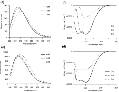
Intrinsic protein fluorescence and far-UV circular dichroism (CD) were used to monitor changes inβ1AR-m23 structure in bulk solution in DM and in the presence of the chemical denaturant urea. Unfolding was assessed by a reduction in helical secondary structure (as shown by a decrease in the 222 nm CD band characteristic ofαhelical structure). The change in tryptophan fluorescence was also monitored during denaturation, as an increase in exposure of tryptophan residues to water is accompanied by a red-shift in the fluorescence emission maximum to longer wavelengths. Although the latter fluorescence change cannot necessarily be linked directly to changes in folded and unfolded state populations.
Dilution of 0.45μMβ1AR-m23 in DM into buffer containing 8 M urea resulted in a protein fluorescence band that had red-shifted by ~ 9 nm (from 329.9 nm in DM to 339.3 nm in urea) (Fig 1a). Under the same conditions a decrease in intensity of the 222 nm CD band, from ~ -26,070 deg.cm2.dmol-1to -10,480 deg.cm2.dmol-1, was observed corresponding to a loss of ~60% of its startingα-helix (Fig 1b). Plots of the red-shift in the fluorescence emission maxi-mum correlated with those for reduction in helicity against urea concentration. These fluores-cence and CD data exhibit similar midpoints of unfolding,Cm, centred around 4.0 ± 0.1 M and 4.5 ± 0.1 M, respectively, and similar slopes at theCmof 0.90 ± 0.07 nmM-1and 0.82 ± 0.06% reduction in helicityM-1, respectively (S1a, S1bandS2Figs). Unfolding was also carried out at a 10-fold higher protein concentration (4.5μM) (Fig 1c and 1d). Under these conditions the receptor loses ~47% of its startingα-helix (Fig 1d).
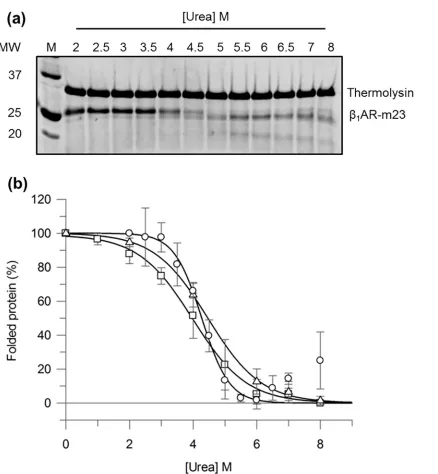
digestion of unfolded protein while keeping folded protein intact. The fraction of folded pro-tein can then be quantified by SDS-PAGE and plotted against denaturant concentration to determineCm. The validity of this method for monitoringβ1AR-m23 unfolding in urea was ini-tially tested in bulk solution using a 1 min pulse. The SDS-PAGE inFig 2ashows the amount of folded receptor remaining in varying urea concentrations following 1-min pulse proteolysis with thermolysin.β1AR-m23 remains largely intact in 2–3 M urea (folding conditions) with band intensities comparable to that of undigested receptor. In higher urea concentrations (3–7 M) the fraction of folded receptor decreases, as demonstrated by the disappearance of the intact protein band and appearance of lower molecular weight cleavage products. In 7 M urea and above the intact protein band begins to reappear, probably due to the reduced activity of ther-molysin in high urea [28,30]. TheCmvalue obtained by pulse proteolysis (4.3 ± 0.1 M with an associated slope of 0.4 ± 0.1% folded proteinM-1) is in excellent agreement with that deter-mined by fluorescence and far-UV CD (Fig 2bandTable 1). It should be noted, that in the complete absence of urea some foldedβ1AR-m23 was digested. Although this could be over-come by adding less protease this also reduced the sensitivity of the assay.
Fig 1.β1AR-m23 unfolding in DM and in urea.Fluorescence and far UV CD spectra (a and b, respectively) of 0.45μM and (c and d, respectively) 4.5μM
β1AR-m23 in DM in the original folded state in the presence of 0 M urea (solid lines) and 3 M urea (dashed lines), and unfolded in 8 M urea (dotted lines). All
buffers contained 25 mM Tris pH 7.5, 150 mM NaCl, 0.1 mM EDTA and 0.5% DM. Spectra are averages from a minimum of three independent experiments on different samples. For folded protein (in 0 M urea) the band intensity at 222 nm was -26070±2400 deg.cm2.dmol-1and -27780±5400 deg.cm2.dmol-1at a
protein concentration of 0.45μM and 4.5μM, respectively. The wavelength at the fluorescence emission maximum for folded protein was 329.9±0.8 nm and 329.8±0.3 nm at a protein concentration of 0.45μM and 4.5μM, respectively. The intensity at the fluorescence emission maximum for folded protein was 474000±118000 and 2404000±399000 at a protein concentration of 0.45μM and 4.5μM, respectively.
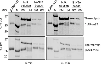
[image:6.612.40.458.77.399.2]Pulse proteolysis was also used to monitorβ1AR-m23 unfolding on a Ni2+-NTA column and compared to that in bulk solution. The length of digestion was increased to 3 min to com-pensate for the diminished activity of the protease in 8 M urea (used for unfolding). Following equilibration of 4.5μMβ1AR-m23 on a solid support under folding (3 M urea) or unfolding
Fig 2. Monitoringβ1AR-m23 unfolding in bulk solution by pulse proteolysis.0.2 mg/ml thermolysin was used to digest 0.45μMβ1AR-m23,
pre-equilibrated in buffer containing 25 mM Tris pH 7.5, 150 mM NaCl, 0.1 mM EDTA, 0.5% DM, 10 mM CaCl2and urea (2–8 M) for 30 min, for 1 min. (a) A
representative SDS-PAGE gel ofβ1AR-m23 following 1-min pulse proteolysis. (b) The percentage of foldedβ1AR-m23 remaining as determined by pulse
proteolysis (circles), fluorescence (squares) and far-UV CD (triangles). The percentage of folded protein at each urea concentration was determined by; pulse proteolysis from the amount of undigested protein as measured by SDS-PAGE; fluorescence from the red-shift in the fluorescence emission maximum; and CD from the degree ofα-helical structure as measured by the CD intensity at 222 nm. Resulting values were normalised between 0% and 100% with 100% representing the fully folded protein in DM and the 0% the partly unfolded 8 M urea state that possesses some helical content. Error bars show±SD (standard deviation) and are the result of a minimum of three independent experiments on different samples.
conditions (8 M urea), as judged by bulk solution fluorescence and CD experiments (Fig 1c and 1d), clear differences in proteolytic susceptibility were observed; after a 5 min incubation in urea 60.3 ± 10.7% of the amount of receptor present in 3 M urea remained intact in 8M urea (Fig 3andTable 2). The concentration of thermolysin used in these proteolysis experiments (~ 6μM) was similar to that of the receptor meaning unfolded protein is likely to be digested slower than in conditions of an excess of protease. Experiments performed with higher concen-trations of protease or with longer digestion periods resulted in greater problems with digestion of folded protein (data not shown). However, when the experiments were carried out with 10-fold less protein (0.45μM), ensuring an excess of protease, greater difference in proteolytic susceptibility were observed in 3 and 8 M urea; only 41.5 ± 5.1% of the amount of receptor in 3
Table 1. Cmvalues for 0.45μMβ1AR-m23 unfolded in urea as determined by 1-min pulse proteolysis, fluorescence and circular dichroism.
Cm[M] Slope [% folded protein M-1]
Pulse proteolysis 4.3±0.1 0.4±0.1
Fluorescence 4.0±0.4 0.9±0.2
Circular dichroism 4.5±0.3 0.8±0.1
Cmvalues for 0.45μMβ1AR-m23 unfolded in urea as determined by 1-min pulse proteolysis,fluorescence
and circular dichroism. Unfolding curves inFig 2werefit to determine the denaturant concentration at the midpoint of unfolding (Cm) together with the slope at theCm. Errors are±SD and are the result of a minimum of 3 independent experiments on different samples.
doi:10.1371/journal.pone.0151582.t001
Fig 3. Comparingβ1AR-m23 unfolding in bulk solution and on a solid support by pulse proteolysis.Example SDS-PAGE gels ofβ1AR-m23 following
3-min pulse proteolysis after a 5 or 30 min incubation in urea. Briefly, 0.45 or 4.5μMβ1AR-m23 in either bulk solution or bound to Ni2+-NTA column was
equilibrated with buffer containing 25 mM Tris pH 7.5, 150 mM NaCl, 0.1 mM EDTA, 0.5% DM, 10 mM CaCl2and 3 (folding conditions) or 8 M (unfolding
conditions) urea followed by digestion with 0.2 mg/ml thermolysin for 3 min. The original foldedβ1AR-m23 is shown for comparison (lanesβ1AR-m23) and molecular mass markers are indicated in kDa (lanes M).
[image:8.612.41.473.389.653.2]M urea remained intact in 8 M urea (Fig 3andTable 2).β1AR-m23 unfolding on the column is further supported by its inability to bind [3H] (-) dihydroalprenolol (DHA) following incuba-tion in 8 M urea (S4 Fig).
Reversible folding of
β
1AR-m23 in DM from a urea-denatured state4.5μMβ1AR-m23 in 8 M urea, which has lost a significant amount (~47%) of its startingα -helical structure was used for refolding experiments (Fig 1d). Successful refolding was primar-ily assessed by recovery of ligand binding activity; to that of the purified receptor prior to unfolding. Recovery of the CD and fluorescence spectra to that of the original purified state was also used. Refolding was attempted into DM by two methods: (i) on a solid support using a Ni2+-NTA affinity column and (ii) in bulk solution by rapid dilution.
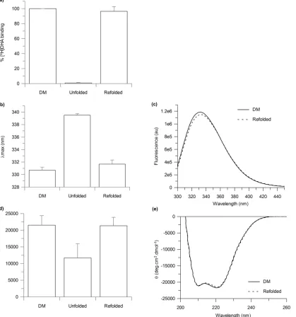
β1AR-m23 refolding on a solid support by removal of urea on a Ni2+-NTA affinity column was determined to be very efficient with recovery of approximately 100% of functional protein, as determined by the recovery of approximately 100% of the original binding activity, to the antagonist [3H]DHA, as compared to that of the originally folded receptor in DM prior to any treatment with urea (Fig 4a). As the predicted molecular weight of our receptor construct is 36 kDa, the theoretical maximum specific binding for pure receptor is 27.8 nmol/mg protein, assuming one binding site per receptor. Ligand binding values of 10.8 ± 1.9 nmol/mg were obtained for the original foldedβ1AR-m23 in DM, which corresponded to ~ 40% of the theo-retical maximum. This is comparable to previously reported ligand binding values obtained for truncatedβ1AR (βAR 34-424/His6) of between 10–12 nmol/mg [27]. The ligand binding activ-ity of the refoldedβ1AR-m23 was 10.5 ± 2.5 nmol/mg. Tryptophan fluorescence and far-UV CD spectra also returned to that of the originally folded receptor in DM (Fig 4b–4e).
Dilution ofβ1AR-m23 into various DM-containing buffers resulted in incomplete refolding as judged by the lack of recovery of the original fluorescence band; the fluorescence emission maximum after dilution of 335.2 nm was only slightly shifted from the urea-unfolded state at 337.8 nm, and did not fully recover to the 329.9 nm maximum of the originally folded receptor in DM (S5 Fig). This difference is too large to be attributed to the small amount (0.8 M) of urea remaining in these samples as demonstrated by unfolding experiments in which urea concen-trations greater than 4 M are required to induce such changes in the fluorescence emission maximum or CD signal at 222 nm (S1a and S1b Fig).
Discussion
[image:9.612.32.578.90.148.2]An in depth knowledge of the structural and molecular basis of GPCR function is lacking, often owing to difficulties in their overexpression, purification and stabilisation. It is therefore
Table 2. The band intensities of intactβ1AR-m23 on SDS-PAGE gels following 3-min pulse proteolysis in urea.
0.45μMβ1AR-m23 4.5μMβ1AR-m23
5 min 30 min 5 min 30 min
Bulk solution 28.8±7.5 31.3±4.1 56.4±11.8 42.1±5.1
Ni-NTA beads 41.5±5.1 40.2±6.9 60.3±10.7 59.8±10.5
The band intensities of intactβ1AR-m23 on SDS-PAGE gels following 3-min pulse proteolysis in 3 M (folding conditions) or 8 M (unfolded conditions) urea
(Fig 3) were calculated using the AlphaErase FC software. Values given are the percentage amounts of intactβ1AR-m23 remaining in 8 M (unfolded
conditions) compared to in 3 M (folded conditions) urea. Each value is the result of an average of between 5 to 9 independent measurements on different samples; errors are±SD.
crucial to develop new and efficient methodologies for obtaining large amounts of native-like, functional and stable GPCRs. Here, we address a fundamental facet of GPCR function, their folding, and report the functional folding (in terms of ligand binding and secondary structure) of the exemplary GPCR,β1AR-m23, from a chemically-denatured state. This has provided important insights into how to obtain and stabilise the correct fold of the receptor. Firstly, the
Fig 4.β1AR-m23 refolding on a Ni2+-NTA column into DM, from urea.4.5μMβ1AR-m23, bound to a Ni2+-NTA column, was unfolded in 25 mM Tris pH
7.5, 150 mM NaCl, 0.5% DM and 8 M urea for 5 mins before refolding into buffer in which the urea had been omitted. The (a) binding of antangonist [3H]DHA to as well as the (b) fluorescence emission maximum and (d) CD signal at 222 nm of the refolded receptor. Results are compared to the originally folded β1AR-m23 in 0.5% DM andβ1AR-m23 unfolded in 8 M urea. Error bars show±SD and are the result of three independent experiments on different samples.
(c) Fluorescence and (e) far UV-CD spectra of the original foldedβ1AR-m23 in 0.5% DM (solid line) and of refoldedβ1AR-m23 in 0.5% DM (dotted line).
Spectra are the average of three independent measurements. The fluorescence emission maximum was 330.7±0.5 nm for the original folded receptor and 331.7±0.7 nm for the receptor refolded in DM. The CD signal at 222 nm was–21520±2840 deg.cm2.dmol-1for the original folded receptor and–
21360±2520 deg.cm2.dmol-1.
[image:10.612.44.463.82.530.2]most stabilising conditions for the isolated receptor, are not necessarily the best for folding; although bicelles containing the lipid DMPC were found to be the most stabilising conditions forβ1AR-m23 (S2andS3Figs), no refolding occurred in any of the bicelle conditions tested but only in DM detergent micelles. Secondly, the extent of helical perturbations in the dena-tured state is not a good indication of the likelihood of successful refolding;β1AR-m23 cannot be fully refolded from an SDS-denatured state even though SDS induces fewer perturbations in helical structure than in urea. At 4.5μMβ1AR-m23 loses 34% of its startingα-helical structure in SDS compared to 47% in urea (S6d Fig) however, only ~ 36% of the original binding activity is restored upon refolding from this SDS denatured state on a Ni2+-NTA column (S7c Fig). We show that the complete refolding ofβ1AR-m23 is achieved into DM micelles, from a urea-denatured state, using an affinity column as a solid support. One possibility is that compared to refolding in bulk solution, a solid support is advantageous in preventing unwanted protein/ protein interactions that favour aggregation. We cannot however, rule out other contributions for example electrostatic effects of the solid surface which may aid in protein folding. Practi-cally, this work provides the groundwork for obtaining stable, functional GPCRS for further structural and functional characterisation.
Although there are only a limited number of examples of refolding studies with GPCRs, the use of columns in recovering functional GPCRs from inclusion bodies solubilised in harsh detergents such as SDS and urea has been reported previously [15–19]. However, no detailed structural information of the denaturant-solubilised state exists, nor has complete refolding and recovery of a native-like state been demonstrated; the leukotriene B4(LTB4) receptor, BLT1, has been functionally folded into detergent-lipid mixed micelles composed of LDAO-asolectin with folding yields of ~ 20–30% (defined as the ratio of the amount of functional pro-tein obtained after dissociation from the column to that initially immobilised, as determined by the affinity constant for LTB4binding) [15,18]; the serotonin receptor, 5-HT4a was folded into the functional conformation in DMPC/CHAPS bicelles with folding yields of ~25% (defined as the ratio of the amount of soluble protein obtained after dissociation from the col-umn to that initially immobilised. Refolding was also assessed at a qualitative level by analysing it’s ligand-binding properties) [17]; the olfactory OR5 receptor was first folded into digitonin detergent micelles with folding yields of ~ 80%, as judged by fluorescence-monitored ligand binding assays, before insertion into lipid vesicles [16]. In all these cases the protein was recov-ered from insoluble inclusion bodies. A similar column-assisted folding approach has also been employed to refold several other membrane proteins from inclusion bodies including the chlo-roplast protein-import channel, Toc 75, and the light harvesting complex II (LHC2). In both cases, the solubilised inclusion bodies were applied to the Ni2+column and refolding initiated by buffer exchange from chaotrope to mild detergent; Triton X-100 (TX) for Toc 75 and n-octyl-β-D-glucopyranoside (OG) for LHC2. In the case of LHC2, this was followed by pigment binding and trimerisation upon transfer to mixed lipid-detergent micelles composed of TX and L-phosphatidyl-D,L-glycerol dipalmitoyl (PG) [31]. Refolding yields of 8% and 3% are
to that of untreated receptor which has not been unfolded, which is vital given the tendency of GPCR stability and ligand binding to be disrupted when solubilised in detergentsin vitro. To our knowledge, this is the first example of such a highly efficient and truly reversible folding system for a GPCR.
The efficiency ofβ1AR-m23 refolding on the beads from urea was found to be dependent on the initial unfolding time in urea; with higher refolding yields obtained following shorter unfolding times in urea (over the range of 5 to 60 min), as judged by the recovery of protein fluorescence and ligand binding activity (S8 Fig). 100% refolding yields with complete recovery of helical structure, protein fluorescence and ligand binding, as compared to the receptor in DM prior to any urea treatment, were only observed following 5 min unfolding in urea. Longer unfolding times of 10, 30 and 60 min caused the refolding yields to drop to 90, 72 and 30%, respectively. We used pulse proteolysis combined with electrophoresis to monitor structural changes inβ1AR-m23 on the column after unfolding in urea for 5 and 30 min. Comparisons of protein band intensities in unfolding (8 M) and folding (3 M) urea concentrations showed the percentage amount of intact receptor remaining in unfolding conditions compared to folding conditions; 60.3 ± 10.7% and 59.8 ± 10.5% of the of the intact protein remained after 5 and 30 min unfolding, respectively (Fig 3andTable 2), thus implying no further unfolding with a lon-ger unfolding time than 5 min.
Interestingly,β1AR-m23 cannot be fully refolded from an SDS-denatured state. Previous reports have found recovery of functional GPCRs from SDS-solubilised inclusion bodies. Much lower yields have been reported for obtaining functional GPCRs from urea-solubilised inclusion bodies Work by Roglet al. reporting on the refolding of Toc75 from bacterial inclu-sion bodies also suggests that incluinclu-sion bodies are more resistant to unfolding compared to their folded counterparts; tryptophan fluorescence spectroscopy was used to show that the unfolded inclusion bodies had an emission peak that was intermediate (346 nm) to that of folded Toc 75 (342 nm) and the same sample unfolded in urea (353 nm) [31]. Our more defined folding system seems to be the key to achieving higher refolding yields and may be applicable to other GPCRs, as well as for membrane proteins in general.
Although our understanding of the various factors affecting the folding yields of GPCRs is still somewhat limited, our results support the idea that preventing aggregation through the formation of intermolecular protein interactions, facilitates folding. This can be achieved by carrying out folding with the receptor immobilised on a solid support, for example on a Ni2+ affinity resin if the protein carries a His-tag and by limiting the length of time that the receptor is in denaturant. Given the large variation in GPCR folding environments reported, it may be that the choice of this environment is specific to each receptor in which case the most straight-forward strategy is to systematically test a range of folding conditions and quantify the associ-ated refolding yields, as demonstrassoci-ated here.
Supporting Information
S1 Appendix. Supporting Methods and Results.
(DOCX)
S1 Fig.β1AR-m23 unfolding in DM-containing buffers.Fluorescence and far-UV CD were
containing 25 mM Tris pH 7.5, 150 mM NaCl, 0.1 mM EDTA, 0.5% DM and varying concen-trations of urea (0–9 M), unless stated otherwise, for 30 min. Plots show unfolding under the following conditions; at a final protein concentration of 0.45μM (open circles, solid line) and at the same protein concentration but in the presence of 350 mM NaCl (open squares, dashed line), 10% (w/v) glycerol (open triangles, dotted line), 0.02% (w/v) CHS (closed circles, solid line) and 1μM alprenolol (solid squares, dashed line) and at a final protein concentration of 4.5μM (solid triangles, dotted line). Error bars show ± SD.
(TIF)
S2 Fig. Fluorescence- and CD-measuredCmvalues forβ1AR-m23 unfolding in urea and
SDS.
(TIF)
S3 Fig.β1AR-m23 unfolding in DMPC/CHAPS.Plots of the red-shift in the fluorescence
emission maximum (λmax)versusconcentration of (a) urea and (b) SDS. Unfolding was per-formed at a final receptor concentration of 0.45μM in bicelles of varyingqvalues; 0.49 (open circles), 0.91 (open squares), 1.68 (open triangles), 2.73 (closed circles) and 3.63 (closed squares), or in DM (closed triangles). All buffers contained 2% (w/v total lipid and detergent) DMPC/CHAPS, 25 mM Tris pH 7.5, 150 mM NaCl and 0.1 mM EDTA. Error bars show ± SD. (TIF)
S4 Fig. Binding activity ofβ1AR-m23 on a Ni2+-NTA column.The amount of radioligand
bound to 4.5μMβ1AR-m23 on a Ni2+-NTA column following 5 min incubation with buffer containing 25 mM Tris pH 8, 150 mM NaCl, 0.1 mM EDTA, 0.5% DM and 8 M urea
(unfolded). Results are compared to control experiments carried out with no protein (no pro-tein) and withβ1AR-m23 but under folding conditions in the absence of urea (native). Error bar show ± SD and are the result of a minimum of four independent experiments on different samples.
(TIF)
S5 Fig. Refoldingβ1AR-m23 by dilution into DM, from urea.Changes in the fluorescence
emission maximum ofβ1AR-m23 initially unfolded at 4.5μM in 8 M urea for 5 min and then diluted 10-fold into various DM-containing buffers all containing 25 mM Tris pH 7.5, 150 mM NaCl and 0.5% DM. Comparisons with that of the original foldedβ1AR-m23 in 0.5% DM and unfoldedβ1AR-m23 in 8 M urea are shown. Error bars show ± SD and are the result of three or four independent experiments on different samples.
(TIF)
S6 Fig.β1AR-m23 unfolding in DM and in SDS.Fluorescence and far-UV CD spectra of (a
and b, respectively) 0.45μM and (c and d, respectively) 4.5μMβ1AR-m23 in 25 mM Tris pH 7.5, 150 mM NaCl, 0.1 mM EDTA and 0.2% DM in the original folded state in the absence of SDS (solid lines) and unfolded in 0.84XSDS(~0.65% SDS) (dotted lines). Fluorescence and CD spectra show the results from a single measurement. For folded protein (in 0XSDS) the CD sig-nal at 222 nm was -18990 deg.cm2.dmol-1and–21530 deg.cm2.dmol-1at a protein concentra-tion of 0.45μM and 4.5μM, respectively. The wavelength at the fluorescence emission was 330.4 nm and 328. 5 nm at a protein concentration of 0.45μM and 4.5μM, respectively. The intensity at the fluorescence emission maximum was 516000 and 3404000 at a protein concen-tration of 0.45μM and 4.5μM, respectively. Folding experiments with SDS were carried out using a different protein preparation to those carried out with urea and were not pursued in great depth due to more successful refolding results achieved with urea.
S7 Fig. Refoldingβ1AR-m23 into DM, from SDS.Refolding of 4.5μMβ1AR-m23 was
per-formed following a 5 min incubation with 25 mM Tris pH 7.5, 150 mM NaCl, 0.1 mM EDTA, 0.2% DM and 0.84XSDS(0.65% SDS) in bulk solution, by rapid dilution or on a Ni2+-NTA col-umn. (a) Fluorescence emission maxima ofβ1AR-m23 diluted 10-fold into various DM-con-taining buffers. (b) Fluorescence emission maxima and (c) binding of antagonist [3H](-)DHA toβ1AR-m23 refolded on a column. Comparisons with that of original foldedβ1AR-m23 in 0.2% DM and unfoldedβ1AR-m23 in 0.84XSDSare shown. Error bars show ± SD and are the result of two or three independent experiments on different samples.
(TIF)
S8 Fig.β1AR-m23 refolded on a Ni2+-NTA column into DM, from urea, over time.Changes
in the intrinsic protein fluorescence and ligand binding activity ofβ1AR-m23 were measured as follows: (a) The fluorescence emission maximum (b) and binding of antagonist [3H]DHA of β1AR-m23 refolded into 25 mM Tris pH 7.5, 150 mM NaCl, 0.5% DM after a 5, 10, 30 and 60 min incubation with 8 M urea at a protein concentration of 4.5μM. Results are compared to original foldedβ1AR-m23 in 0.5% DM and unfoldedβ1AR-m23 denatured in 8 M urea. Error bars show ± SD and are the result of two or three independent experiments on different samples. (TIF)
Author Contributions
Conceived and designed the experiments: NDB PJB ELRC GFXS. Performed the experiments: NDB ELRC. Analyzed the data: NDB. Contributed reagents/materials/analysis tools: TW PCE CGT GFXS. Wrote the paper: NDB PJB.
References
1. Miller D, Charalambous K, Rotem D, Schuldiner S, Curnow P, Booth PJ. In vitro unfolding and refolding of the small multidrug transporter EmrE. Journal of molecular biology. 2009; 393(4):815–32. Epub 2009/08/25. doi:10.1016/j.jmb.2009.08.039PMID:19699749.
2. Harris NJ, Findlay HE, Simms J, Liu X, Booth PJ. Relative Domain Folding and Stability of a Membrane Transport Protein. Journal of molecular biology. 2014; 426(8):1812–25. doi:10.1016/j.jmb.2014.01.012 PMID:24530957
3. Findlay HE, Rutherford NG, Henderson PJ, Booth PJ. Unfolding free energy of a two-domain trans-membrane sugar transport protein. Proceedings of the National Academy of Sciences of the United States of America. 2010; 107(43):18451–6. Epub 2010/10/13. doi:10.1073/pnas.1005729107PMID: 20937906; PubMed Central PMCID: PMCPmc2972933.
4. Bartlett AI, Radford SE. An expanding arsenal of experimental methods yields an explosion of insights into protein folding mechanisms. Nature structural & molecular biology. 2009; 16(6):582–8. Epub 2009/ 06/06. doi:10.1038/nsmb.1592PMID:19491935.
5. Curnow P, Di Bartolo ND, Moreton KM, Ajoje OO, Saggese NP, Booth PJ. Stable folding core in the folding transition state of an alpha-helical integral membrane protein. Proceedings of the National Acad-emy of Sciences of the United States of America. 2011; 108(34):14133–8. Epub 2011/08/13. doi:10. 1073/pnas.1012594108PMID:21831834; PubMed Central PMCID: PMCPmc3161581.
6. Daggett V, Fersht A. The present view of the mechanism of protein folding. Nat Rev Mol Cell Biol. 2003; 4(6):497–502. PMID:12778129
7. Sachse R, Dondapati SK, Fenz SF, Schmidt T, Kubick S. Membrane protein synthesis in cell-free sys-tems: From bio-mimetic systems to bio-membranes. FEBS letters. 2014; 588(17):2774–81. doi:10. 1016/j.febslet.2014.06.007PMID:24931371
8. Ezure T, Nanatani K, Sato Y, Suzuki S, Aizawa K, Souma S, et al. A cell-free translocation system using extracts of cultured insect cells to yield functional membrane proteins. PloS one. 2014; 9(12): e112874. Epub 2014/12/09. doi:10.1371/journal.pone.0112874PMID:25486605; PubMed Central PMCID: PMCPmc4259328.
10. Skach WR. The expanding role of the ER translocon in membrane protein folding. The Journal of cell biology. 2007; 179(7):1333–5. Epub 2008/01/02. doi:10.1083/jcb.200711107PMID:18166647; PubMed Central PMCID: PMCPmc2373491.
11. Venkatakrishnan AJ, Deupi X, Lebon G, Tate CG, Schertler GF, Babu MM. Molecular signatures of G-protein-coupled receptors. Nature. 2013; 494(7436):185–94. Epub 2013/02/15. doi:10.1038/ nature11896PMID:23407534.
12. Mason JS, Bortolato A, Congreve M, Marshall FH. New insights from structural biology into the drugg-ability of G protein-coupled receptors. Trends in pharmacological sciences. 2012; 33(5):249–60. Epub 2012/04/03. doi:10.1016/j.tips.2012.02.005PMID:22465153.
13. Kobilka BK, Deupi X. Conformational complexity of G-protein-coupled receptors. Trends in pharmaco-logical sciences. 2007; 28(8):397–406. Epub 2007/07/17. doi:10.1016/j.tips.2007.06.003PMID: 17629961.
14. Kroeze WK, Sheffler DJ, Roth BL. G-protein-coupled receptors at a glance. Journal of cell science. 2003; 116(Pt 24):4867–9. Epub 2003/11/20. doi:10.1242/jcs.00902PMID:14625380.
15. Baneres JL, Martin A, Hullot P, Girard JP, Rossi JC, Parello J. Structure-based analysis of GPCR func-tion: conformational adaptation of both agonist and receptor upon leukotriene B4 binding to recombi-nant BLT1. Journal of molecular biology. 2003; 329(4):801–14. Epub 2003/06/06. PMID:12787679. 16. Kiefer H, Krieger J, Olszewski JD, Von Heijne G, Prestwich GD, Breer H. Expression of an olfactory
receptor in Escherichia coli: purification, reconstitution, and ligand binding. Biochemistry. 1996; 35 (50):16077–84. Epub 1996/12/17. doi:10.1021/bi9612069PMID:8973178.
17. Baneres JL, Mesnier D, Martin A, Joubert L, Dumuis A, Bockaert J. Molecular characterization of a puri-fied 5-HT4 receptor: a structural basis for drug efficacy. The Journal of biological chemistry. 2005; 280 (21):20253–60. Epub 2005/03/19. PMID:15774473.
18. Baneres JL, Popot JL, Mouillac B. New advances in production and functional folding of G-protein-cou-pled receptors. Trends in biotechnology. 2011; 29(7):314–22. Epub 2011/04/19. doi:10.1016/j.tibtech. 2011.03.002PMID:21497924.
19. Dahmane T, Damian M, Mary S, Popot JL, Baneres JL. Amphipol-assisted in vitro folding of G protein-coupled receptors. Biochemistry. 2009; 48(27):6516–21. Epub 2009/06/19. doi:10.1021/bi801729z PMID:19534448.
20. Lau FW, Bowie JU. A method for assessing the stability of a membrane protein. Biochemistry. 1997; 36 (19):5884–92. Epub 1997/05/13. doi:10.1021/bi963095jPMID:9153430.
21. Barrera FN, Renart ML, Poveda JA, de Kruijff B, Killian JA, Gonzalez-Ros JM. Protein self-assembly and lipid binding in the folding of the potassium channel KcsA. Biochemistry. 2008; 47(7):2123–33. Epub 2008/01/22. doi:10.1021/bi700778cPMID:18205389.
22. Barrera FN, Renart ML, Molina ML, Poveda JA, Encinar JA, Fernandez AM, et al. Unfolding and refold-ing in vitro of a tetrameric, alpha-helical membrane protein: the prokaryotic potassium channel KcsA. Biochemistry. 2005; 44(43):14344–52. Epub 2005/10/26. doi:10.1021/bi050845tPMID:16245951. 23. Warne T, Serrano-Vega MJ, Tate CG, Schertler GF. Development and crystallization of a minimal
ther-mostabilised G protein-coupled receptor. Protein expression and purification. 2009; 65(2):204–13. Epub 2009/03/20. PMID:19297694.
24. Serrano-Vega MJ, Magnani F, Shibata Y, Tate CG. Conformational thermostabilization of the beta1-adrenergic receptor in a detergent-resistant form. Proceedings of the National Academy of Sciences of the United States of America. 2008; 105(3):877–82. Epub 2008/01/15. doi:10.1073/pnas.0711253105 PMID:18192400; PubMed Central PMCID: PMC2242685.
25. Markwell MAK, Haas SM, Bieber LL, Tolbert NE. A modification of the Lowry procedure to simplify pro-tein determination in membrane and lipopropro-tein samples. Analytical biochemistry. 1978; 87(1):206–10. doi:10.1016/0003-2697(78)90586-9PMID:98070
26. Lees JG, Smith BR, Wien F, Miles AJ, Wallace BA. CDtool-an integrated software package for circular dichroism spectroscopic data processing, analysis, and archiving. Analytical biochemistry. 2004; 332 (2):285–9. Epub 2004/08/25. doi:10.1016/j.ab.2004.06.002PMID:15325297.
27. Warne T, Chirnside J, Schertler GF. Expression and purification of truncated, non-glycosylated turkey beta-adrenergic receptors for crystallization. Biochimica et biophysica acta. 2003; 1610(1):133–40. Epub 2003/02/15. PMID:12586387.
28. Park C, Marqusee S. Pulse proteolysis: a simple method for quantitative determination of protein stabil-ity and ligand binding. Nature methods. 2005; 2(3):207–12. Epub 2005/03/23. doi:10.1038/nmeth740 PMID:15782190.
30. Park C, Marqusee S. Probing the high energy states in proteins by proteolysis. Journal of molecular biology. 2004; 343(5):1467–76. Epub 2004/10/20. doi:10.1016/j.jmb.2004.08.085PMID:15491624. 31. Rogl H, Kosemund K, Kuhlbrandt W, Collinson I. Refolding of Escherichia coli produced membrane
protein inclusion bodies immobilised by nickel chelating chromatography. FEBS letters. 1998; 432(1– 2):21–6. Epub 1998/08/26. PMID:9710243.