JOURNAL OFVIROLOGY,Aug. 1975, p.420-433
Copyright 0 1975 AmericanSocietyforMicrobiology PrintedVol.in U.S.A.16,No.2
Structure and
Chemical-Physical
Characteristics of
Lactate
Dehydrogenase-Elevating
Virus
and Its
RNA
MARGOBRINTON-DARNELL* ANDPETER G. W. PLAGEMANN
Department ofMicrobiology,MedicalSchool, University of Minnesota,Minneapolis, Minnesota55455 Received for publication4March1975
Lactate dehydrogenase-elevating virus (LDV) was purified from culture fluid
of infected primary cultures of various mouse tissues (peritoneal macrophage,
bone marrow, spleen, and embryo) and from plasmaofinfected mice. Electron
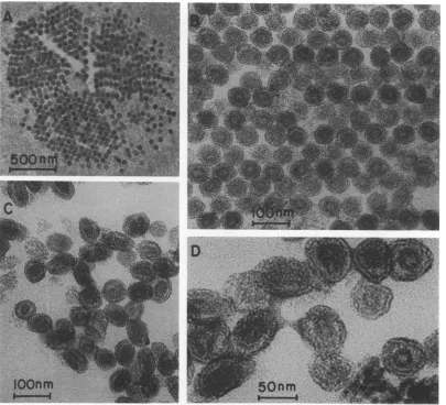
microscopy of negatively stained virus and positively stained sections ofLDV
revealed spherical particles of uniform size with a diameter of about 55 nm,
containinganelectron-densecorewithadiameterofabout30nm.Duringsample
preparation the envelope had a tendencyto slough off anddisintegrateto form
aggregatesof various sizes and small hollowparticles with adiameter of 8to14
nm. Two strains of LDV exhibiteda density of1.13 g/cm3 inisopycnic sucrose
density gradient centrifugation whether propagated in primary cultures ofthe
various mouse tissues or isolated from plasma of infected mice. A brief
incubation of LDV inasolution containing 0.01% Nonidet P-40orTriton Xwas
sufficient to release theviral nucleocapsid, whereas a similartreatmenthad no
effect on Sindbis virus. The nucleocapsid of LDV exhibited a density of 1.17
g/cm3, wasdevoidofphosphatidylcholine, andcontainedonly the smallest of the
viralproteins, VP-1, which hadamolecularweight of about 15,000. The envelope
contained two proteins, VP-2with a molecularweightof18,000 and a
glycopro-tein, VP-3, which migrated heterogenously (24,000 to 44,000 daltons) during
polyacrylamide gelelectrophoresis. When comparedtothesedimentationrateof
29S rRNA, the RNAs of LDV and Sindbis virus sedimented at 48 and 45S,
respectively, whether analyzed by zone sedimentation in sucrose density
gradients containing lowor high salt concentrations or denaturedbytreatment
with formaldehyde. Our results indicate that LDV should be classified as a
togavirus, but that LDV issufficientlydifferent fromalpha andflavivirusestobe
excluded from these groups.
The lactate
dehydrogenase-elevating
virus(LDV) is a
lipoprotein-enveloped
virus ofinter-mediate size (see reference 27) containing a
genome ofsingle-stranded RNA with a mol wt
ofabout 5 x 10⁶ as estimatedbyzone
sedimen-tation in sucrose density gradients (9).
Al-though
LDV resembles the togaviruses incer-tain of its characteristics, it has not yet been
classified because ofvarious uncertainties and
controversies about its structure and
physical-chemical properties. The present studies were
designed to obtain further information about
theseproperties,particularly withrespect to the
structure, stability, and density ofthe virions
and theircores,thenature of theviralstructural
proteins associated with the core and the viral
envelope, andthemolecularweightand
second-ary structure of the viral RNA. A preliminary
report of some of these results has been
pre-sented (M. B. Darnell, J. K. Collins, and P. G.
W. Plagemann, Annu. Meet. Am. Soc. Micro-biol. 1974, V121, p. 220).
MATERIALS AND METHODS
Materials. Materials were purchased as follows:
[5-'H]uridine (26 Ci/mmol) and [3IHIleucine (55 Ci/
mmol) from Schwarz/Mann; D-[3H]mannose (611
mCi/mmol) from Amersham/Searle; D-
['H
Jglucosa-mine (18 Ci/mmol) and [methyl-3H]choline (20 Ci/
mmol) from NewEngland NuclearCorp.;
[carboxyl-'4CIdeoxycholate (3 mCi/mmol) from ICN; Nonidet
P-40(NP-40)fromShellChemical Co.Buffer 12(B12) was composed of 0.1NNaCl,0.05%(vol/vol) mercap-toethanol, 10 mM Tris-chloride (pH 7.4), and 1mM EDTA. Buffer 6 (B6) was composed of 50 mM NaCl, 10 mM EDTA, 10 mM Tris-chloride (pH 7.4), and
0.5%(wt/vol)sodiumdodecyl sulfate (SDS).
Cells and,cell culture.Primarymouse peritoneal macrophage cultureswerepreparedasreported previ-ously (5a). Briefly, macrophages were stimulated in adult Swiss micebyanintraperitoneal injectionof a 1%(wt/vol) starch solution andweregrowninmedium 58B (9,35) in25-cm' plasticFalconflasks.
Bone marrow cultures wereprepared using mouse femurs, which had been dissected freeofmuscleand cut at the joints. The marrow was forced out with medium 58B, and cells were dispersed by pipetting 420
on November 10, 2019 by guest
http://jvi.asm.org/
CHARACTERISTICS 421
and seeded immediately. Spleen and mouse embryo cell cultures were prepared as described previously (35, 36).Spleencells were grown in medium 58B, and mouse embryo cells were propagated in Eagle mini-mal medium supplemented with 10% (vol/vol) fetal calfserumandantibiotics.
Viruses and preparation of unlabeled and ra-dioactively labeled virions. Ourstrainof LDV was obtained in 1971 from the plasma of a C3H mouse carrying a transplantable tumor. Another strain of LDVwas kindly supplied by A. L. Notkins. Pools of LDV wereprepared as follows. Plasma was harvested from 24-h LDV-infected mice and diluted fivefold with medium 58B,filtered through a membrane filter (MA type, 450-nm pore diameter, Millipore Corp.), and stored at -80C. The final suspensions contained approximately 109 median infectious units(ID,,)per ml.Virus titers wereestimated by titration in mice as described previously (33).
West Nile virus (strain E 101) and Sindbis virus (strain SAAR 339) were propagated in brains of newborn hamsters as described previously (8). Brain pools ofWest Nile virus and Sindbis virus contained 108 to 109 and 1010to 1011PFU/ml, respectively, and were stored at -80C. Plaque assays were conducted in LLC-MK2 cells and chicken embryo fibroblasts, respectively (8).
The variousprimary cultures of mouse tissues were infected with 20 to 50 ID,, of LDV or 30 PFU of Sindbis virus per cell2to 24hafterplantinginto 25-cm2Falcon flasksasdescribed previously (5a, 8). LLC-MK2 cell monolayer cultures were infected in the same mannerwith20PFU of West Nile virus per cell. After 1-hperiodsofvirusadsorption,theresidual virus inocula were poured off andreplaced with 5 ml of me-dium 58B in which the concentration of fetal calf serum wasreduced to 2%. The medium was supple-mented with 30
gCi
of ['H]uridine, 25 gCiof ['H]-leucine,30,Ci
of['H]mannose, 10uCi of['H]glucosa-mine, or 10
IsCi
of ['H Icholine per ml. In the caseof choline, mannose, andglucosaminelabeling, the cells wereprelabeledfor 3hbefore infection. The medium containing the isotope was removed during virus adsorption and then added back to the cells. ['H]-uridine and leucine were added to the medium 1 to 3 h after infection. When labeling with leucine, the normal concentration of unlabeled leucine in the medium was reduced 90%. The culture fluids were harvested 24 h after infection, clarified by sequential centrifugation at 500 x g for 5 min and 15,000x g for 20 min, and then immediately layered in 5- to8-ml amounts onto linear 0.5to 1.5 M sucrose gradients made in B12. The gradients were
cen-trifuged in an L-3 Beckman ultracentrifuge in an
SW27rotor at22,000 rpm at4C for14h. One-milli-liter fractions were collected withanISCO gradient fractionator (model 183), which was attached to a continuously recordingspectrophotometer anda frac-tion collector. Aliquots of each fraction or whole fractions were analyzed for radioactivity in acid-insolublematerial(31)and/orinfectivity. Thedensity
of gradient fractions kept at 4C was measured directly by weighing 100-gl aliquots. For additional analyses (seebelow)fractionscontainingvirusorviral
componentswerepooled and mixed with 2.5 volumes of ethanol at -20C and small amounts of total cellular RNA from Novikoff rat hepatoma cells as co-precipitant. The precipitated material was col-lectedbycentrifugationafter24to 48 h at -20C.
LDV was alsolabeled invivo. Mice were injected
intraperitoneally with 500 MCi of [5-3HJuridine 4 h
after anintraperitoneal injection on unlabeled LDV
(10'ID,dmouse),and their blood was collected by the
retro-orbital bleeding technique 24 h after infection.
The plasma from four mice was pooled, diluted
fivefold with B12, and then clarified; LDV was isolated by isopycnic centrifugation on 0.5 to 1.5 M sucrosegradients as describedabove.
Isolation of viral RNAs andanalysisbyvelocity sedimentation. RNA was isolated from virions or viral components by extraction with phenol-SDS-chloroform by the method of Perry et al. (29) orby deproteinization by incubation at 37 C for 5 min with vigorous mixing in B6 containing 2% (wt/vol) SDS.
Samplesof RNAwerecentrifugedinlinear 0.15 to 0.9
Msucrosedensity gradients in an SW27 rotor at 20 C for 10h.The salt composition of the gradientsolutions varied and is indicated in the appropriate experi-ments. One-milliliter fractions were collected from the gradients and analyzed forradioactivity in acid-insoluble material as already described. Sedimenta-tion rates ofRNA wereestimated by the method of Martin and Ames (20) using hotphenol-extracted 29 and 18S rRNA from NlS1-67 cellsasstandards(32).
Isolation of viral proteins and separationby gel
electrophoresis. Samplesofprotein-labeledvirusor
viral components were supplemented with 1% (wt/ vol) SDS, 1% (vol/vol) mercaptoethanol, and0.5 M ureaand incubated at 37 C for 1 h. The sampleswere concentrated to about 0.5 ml by vacuum dialysis againsta solution composed of 10 mM sodium phos-phate (pH 7.4), 0.1% (wt/vol) SDS, and 0.1%(vol/vol) mercaptoethanol. Samples of 0.2 ml of the concen-trated solution were supplemented with 0.04 ml ofa 60% (wt/vol) solution of sucrose and 5
gsl
of a 0.1% (wt/vol) solution ofbromphenol blue. The proteins were separated by electrophoresis in 7.5% (wt/vol) SDS-polyacrylamide gels containing0.2%(wt/vol) N,N'-methylene-bis-acrylamide asdescribedby Maizel
(19). Electrophoresis was in20-cmglass tubes at100 V for 8 h. Thegels were sliced into 1-mmslices, and thesewereallowedtoswell in1ml ofwaterfor1h at room temperature. Then the slices and eluates to-gether wereanalyzed forradioactivity.
Radioactivity determination. Radioactivity was
measuredbyliquid scintillationcounting. All samples were mixed with 8 ml ofa modified Bray solution describedpreviously (34).
Electron microscopy. LDV samples prepared as described in the appropriate experiments were al-lowed toadsorbtocharged copper grids covered with aParlodian film. Theexcess liquidwassyphonedoff withapieceoffilterpaper, andadropof 1%(wt/vol) phosphotungstic acid(pH 7.0) wasputontothegrid. In some instancesvirusadsorbed to gridswas fixed
with2% glutaraldehyde (pH7.2) for5min and then
rinsed twicewith distilledwaterbeforestainingwith phosphotungstic acid.PelletedLDV waspreparedfor
on November 10, 2019 by guest
http://jvi.asm.org/
422 BRINTON-DARNELL AND PLAGEMANN
thin sectioning by sequential fixation with 2% (vol/ vol)glutaraldehyde and1%(wt/vol)osmiumtetroxide in 0.1 M phosphate buffer (pH 7.2), followed by stainingwith 2%(wt/vol) uranylacetate,dehydration inethanol, andembedding inanepoxy resinmixture (Epon 812) according to the method of Luft (18). Sections were cut with adiamond knife in an LKB Ultratome andthen mountedoncoppergridscovered with Parlodian film. The sectionswerestained with uranyl acetate (2%, wt/vol)andReynoldlead citrate. All preparations were examined with a Siemens Elmiskop electron microscope.
RESULTS
Density of LDV virions. Resultsfrom
previ-ousstudies (9) indicatedthat LDVpropagated
in primary mouse macrophage cultures has an
unusually lowdensity for an enveloped animal
virus (1.12 to 1.13g/cm3). Asimilar value (1.14
g/cm
3) has beenreported
for macrophage-propagated LDV by Michaelides andSchlesin-ger (21), whereas a density of 1.17 g/cm3 was
reported forLDVpropagated in mouse embryo
cultures (25) or isolated from the plasma of
infected mice (37). These different results
sug-gested the possibility that thedensity of LDV
mightvarywith the type ofhost cell and reflect differences in thelipid composition ofthe host membrane. Another possibility was that the
differing resultswereduetothe use of different
strains ofLDV. Our results negate both
possi-A.LDV FROM MEC CULTURES B. LDV FROM MA
'~ [LDV
o0 o-A
0. '-~~~~~~~~~B
z
0
U.
bilities. We have propagated LDV in primary
cultures of mouseperitoneal macrophages, and
of mouse bone marrow, spleen, and embryo
cells, and have also isolated LDV from the plasma of infected mice. In each case LDV
bandedat a density ofabout 1.13g/cm3during
isopycniccentrifugation in sucrose density
gra-dients. Virus was located in the gradients by
infectivity titrations or a radioactive label in the
viral RNA or by bothmethods. Representative gradient profiles of LDV propagated in mouse
embryo cultures and peritoneal macrophage
cultures and of virus isolated from mouse
plasma areillustratedinFig. 1. Furthermore, a
second strain of LDV, kindly supplied by A. L.
Notkins, exhibitedthe same density when
prop-agated in peritoneal macrophage cultures and
analyzed inthe same manner (Fig. 1B).
Detergent and proteinase treatment of LDV. Studies by Michaelidesand Schlesinger (21) have shown that treatment of LDV with
0.2% NP-40 at room temperature for 1 h
re-leased two of the viral proteins in a soluble
form. A similar treatment has been used to
isolate the nucleocapsid of alphaviruses (13),
butinthecase of LDV the physical properties of
the particles remaining after NP-40 treatment
had not been studied in detail. The following
results demonstrate that LDV is much more
sensitive to detergent treatment than alpha
BOTTOM T
FRACTION NUMBER TOP
FIG. 1. Isopycnic sucrose density gradient centrifugation of two strains of LDV propagated in primary culturesofmouse embryo cells (MEC [A]),and macrophages (B), and isolated from plasma (C). The viruses werepropagated in cell cultures and inmice in the presence of[5-3H]uridine as described in Materials and Methods. Samples of6mlofculturefluidorfivefold-diluted plasma were centrifuged through linear 0.5 to 1.5 Mgradients ofsucroseinB12inanS W27rotor at22,000 rpm at4C for 14 h.Aliquots of the gradient fractions wereanalyzed for density, infectivity, and radioactivity in acid-insoluble material. Frame B is a composite of thegradient profiles of culture fluid from cells infected withourstrainofLDV(A) or that of Notkins (B).
J. VIROL.
on November 10, 2019 by guest
http://jvi.asm.org/
[image:3.503.67.457.398.592.2]togaviruses. The results in Fig. 2A illustrate
that a 10-min incubation of uridine-labeled
LDV at4 C in asolution containingaslittle as
0.01% (vol/vol)NP-40 completely converted the
virions to particles which equilibrated at a
density ofabout 1.17
g/cm3,
withaconcomitantloss of99.9%oftheinfectivity. Incubation with
0.005%NP-40 hadnoeffectonthe density of the
virions, but treatment with 0.1 or 0.2% NP-40
had the same effect as incubation with 0.01%
(data not shown). Zone sedimentation of the
RNAextracted from NP-40-treated particles by
treatment with SDS demonstrated that this
RNA was intact 48S viral RNA (Fig. 2B). The
RNAwithin these particles, however,was
com-pletely degraded after a brief exposure of the
particles to RNase, whereas the RNA within
mature virions wasresistant tosuch treatment
(Fig. 2D). Incubation of LDV in solutions
con-taining 0.01% (vol/vol) Triton X (Fig. 2E) or
0.05% (wt/vol) deoxycholate (data not shown)
at4 C for 10 min also resultedin itsconversion
to a particle with an approximate density of
1.17 g/cm3. In contrast, incubation of Sindbis
virus in solutionscontaining these low
concen-trations of detergents did not affect its density
(1.175 g/cm3; Fig. 2C). Additional studies have
shown that a concentration of at least 0.1%
NP-40 was required to quantitatively convert
Sindbis virustofreenucleocapsidswhich hada
density of1.23g/cm3 (datanotshown).
Inanother experimentweincubated partially
purified, uridine-labeled LDV in a solution
containing 0.05% (wt/vol) ["4C]deoxycholate at
4 C for 10min and then analyzed the mixture
by isopycnic centrifugation inasucrosedensity
gradient. No significant binding oflabeled
de-oxycholateto the 1.17-g/cm3 particles could be
detected. Less than 0.2%of the total
radioactiv-ity (70,000 counts/min) was recovered in the
lower part of the gradient (see Fig. 2A); the
remainder waspresent intheupper8 ml of the
gradient (data not shown).
Incubation of LDV in solutionscontaining2.5
mgoftrypsinpermlat22 C for30min(Fig. 2E)
or 1 mgofbromelainper ml at22C for60min
(datanotshown) hadnoeffectonthe density of
LDV. Treatment of NP-40-treated particles
with trypsin, on the other hand, resulted in
complete destruction of these particles (Fig.
2E).
Lipid and proteincomposition ofLDVand
ofNP-40-released particles.Toobtain further
information about thenatureof the 1.17-g/cm3
particlesreleasedbyNP-40 treatment,we
prop-agated LDV in macrophage cultures in the
presence of labeled leucine, choline,
glucosa-mine, or mannose. The virus was purified and
then analyzed by
isopycnic
centrifugation insucrose densitygradients before and after
incu-bation with
0.01%
NP-40. About 30% of thetotal leucine label associated with intact LDV
remained associated with the NP-40-treated particles equilibrating at
1.17-g/cm3,
whereasthe remainderwas recovered inthe upper
por-tion of the gradient (Fig. 3A). In contrast, all
choline-, glucosamine-, and
mannose-labeled
components ofLDV were released in a soluble
form by NP-40 treatment (Fig. 3B-D). The
results suggest that the remaining
particles
were devoid of phospholipids (at least of phos-phatidylcholine) and glycoproteins and
there-fore
represented
free viralnucleocapsids.
This conclusion issupported by
analysis oftheleu-cine- and
glucosamine-labeled
proteinsby
ac-rylamidegel electrophoresis. Fig. 4B illustrates
a typical profile of
leucine-labeled
LDVpro-teins. The profile issimilarto that
reported
by
Michaelides and Schlesinger (21) for LDV,
except that our estimates of the molecular
weightsof the proteinsareslightly greater than those reported by these investigators. The
mo-lecular weights, as estimated from the
migra-tion rates in 7.5%
polyacrylamide
gels relativetothose ofnine standard proteins, were 15,000
for Vp-1 and 18,000 for Vp-2
(Fig.
4C).
Amolecular weight for Vp-3 could not be
esti-matedwithaccuracybecauseit was
heterogene-ous (24,000 to 44,000 daltons; Fig. 4B). The
apparent heterogeneity was not an artifact of
the method since Vp-3 from West Nilevirus, a
flavi togavirus, which possesses three proteins similar to those of
LDV,
migrated
as ahomo-geneous fraction under similar conditions of
analysis (Fig. 4A). Furthermore, a similar
het-erogeneity of Vp-3 from LDV was observed in 10% acrylamide gels (datanot shown) and has also been observed by
Michaelides
andSchles-inger (21) using
15% acrylamide gels. Vp-3
wastheonlyLDVprotein labeled with
glucosamine
(Fig. 4E) or mannose and thus is the only glycoprotein ofLDV.
Polyacrylamide gel
elec-trophoresis of the proteins from
1.17-g/cm
3particles remaining after NP-40 treatment (see Fig. 3A) showed that this
particle
contained only the smallest of the viralproteins, Vp-1
(Fig. 4D). The other two
proteins,
Vp-2
andVp-3, were recovered from theupperportionof
the gradient of NP-40-treated virions
(Fig.
3A andD; Fig. 4DandE).LDVwasquantitatively
precipitated by concanavalin A. When a
sus-pension of uridine-labeled LDV was
supple-mented with 800
gg
ofconcanavalin A perml,
incubatedat 4C for 3h,and then
centrifuged
at5,000 x g for 20 min, over 90% ofradioactivity
was recovered in the pellet. The
pelleted
virus16,1975
on November 10, 2019 by guest
http://jvi.asm.org/
-40 TREATED
E.
TRITON
XTREATED
- 1-17
TRYPSIN TREATED
'-1.13
NP-40
4 TRYPSIN
TREATED
FRACTION
NUMBER
424
NP-40
TREATED
105
ID50 -1.17D. +RNose
'V
1.13 .0110
z
I0
0
La.
-Ir
E
I-.C'
cn z
w
BOTTOM
TOP
on November 10, 2019 by guest
http://jvi.asm.org/
contained intact viral
RNA,
as indicated byzonesedimentation ofthe RNAextractedfrom
the pelleted material (notshown).
Properties of LDVRNA. Ourprevious
esti-mate ofthe molecular weight of LDV RNA of 5
x 106 wasbasedon asedimentationcoefficient
of about 48S estimated by zone sedimentation
in sucrose density gradients of moderate ionic
strength, using 29and 18SrRNA from Novikoff
cells and variousother viral RNAs as markers (9). An RNA ofthissizecould contain 10 times
more genetic information than is required for
codingfor thethreestructural proteins ofLDV. Thetruemolecular weightsof manyviralRNAs,
however, are not knownwithcertainty because
their secondary structure or other properties may differ from those of the rRNA's. For
instance, the RNAs ofcardioviruses sediment
significantlymorerapidly (34S) than29SrRNA
in sucrose density gradients containing 0.1 M
NaCl, whereas both types ofRNA sediment at
about the same rate in gradients containing 1
mM salt (22, 23). Similarly, Sendaivirus RNA
sediments at 32S in dimethyl sulfoxide
gradi-ents when compared to29S rRNA, whereas it
sediments at 47S in normal sucrose density gradients (16). The electrophoretic mobilitiesof
the RNAs ofSendai virus and tobacco mosaic
virus informamide-polyacrylamide gelsarealso
not as expected from their molecular weights
when comparedtothemobilities of 28and 18S rRNA'sfromchicken cells (11). Wehave,
there-fore, investigated the effect of salt
concentra-tion and denaturation on the sedimentation behavior of LDV RNA. The results in Fig. 5
show, however, that LDV RNA sedimented at
approximately 48S when compared to 29S rRNA, whether analyzed in sucrose density gradients containing high orlow concentrations ofsalt.Furthermore, denaturationofLDV RNA
by preincubation with formaldehyde (5) also
did not alter its sedimentation rate relative to
thatofrRNA (Fig. 6).Similarly, the
sedimenta-tion rate ofSindbis virus RNA(approximately
45S) inrelationtorRNAwas notaffectedunder
these experimental conditions (data not
shown). The results suggest that the RNAs of
both LDV and Sindbis virusexhibit secondary
structure similar to that of the rRNA's of
mammalian cells and that a molecular weight of
5 x 10⁶may be avalidestimate fortheRNAof
LDV. Notkins (26, 28) reported that butanol-,
chloroform-, ether-, or phenol-extractedpellets
obtained by high-speed centrifugation of
plasmafromLDV-infectedmicewere infectious when inoculated undiluted into mice and that
this infectivitywasdestroyedbytreatment with
RNase. To confirm the conclusion drawn from
these studies that LDV RNA is infectious, we
have isolated RNA by phenol-SDS-chloroform
extraction of purified, uridine-labeled LDV,
purified the RNA by zone sedimentation throughasucrose density gradient (see Fig. 2B),
and inoculatedintracerebrallyor
intraperitone-ally 0.1-ml aliquots of various dilutions of this
RNAintogroups of mice. The RNAwasclearly
infectious, butits titervaried with theroute of
inoculation. One milliliter of RNA solution
extracted from 109
ID,,
ofLDV contained 104ID,50
of infectious RNA when inoculatedin-tracerebrally and 102 ID50 when inoculated intraperitoneally. Based on a 70% recovery of
RNAfrompurified virus, asestimated fromthe
recovery of radioactivity, a maximum ratio of
the infectivity of LDV RNA to infectivity of
intact LDV wasestimated asabout 10-5.
Finestructureof LDV. Electronmicroscopic
examinations of crude preparations of LDV
have suggested that LDV is bottle shaped or
possesses tails andthus hasa uniquestructure
FIG. 2. Isopycnic centrifugation ofuridine-labeled LDV and Sindbis virus beforeandafter treatmentwith
detergents and/or trypsin. Uridine-labeled LDV and Sindbis viruswerepartially purified from culturefluid
from infectedcells by bandinginisopycnic sucrosedensitygradients asdescribed in Materials and Methods
(seeFig. 1A). Thefractionscontaininglabeledviruswerepooledand dilutedthreefoldwith B12. Thesevirus
suspensionsweretreatedasfollows: samplesofLDV(A)orSindbisvirus(C) suspensionsweresupplemented with 0.01% (vol/vol) NP-40, mixed, and incubated at 4Cfor 10min. Duplicate samples ofeachremained untreated. (D)Twosamples ofLDVsuspensionweresupplementedwith25Ag ofRNaseAperml,andoneof them also with 0.01%NP-40,thenmixed,and incubatedat22Cfor30min.(E)OnesampleofLDVsuspension
wassupplementedwith 0.01%(vol/vol) TritonX, mixed,andincubatedat4Cfor10min. Two othersamples
weresupplementedwith2.5mgoftrypsinperml,andoneofthemalso with0.01%NP-40,mixed,andincubated
at 22Cfor30 min. Allsamples were analyzed by isopycnic centrifugation in 0.5 to 1.5Msucrose density
gradients as inFig. 1. One-milliliter fractions were collectedfrom thegradients, and aliquots thereofwere
analyzed for radioactivity in acid-insoluble material and density. In (A), fractions were also assayedfor
infectivity, and the total virus titersineachpeakareindicatedinthegraph. (B) The remaindersofthe three
peak fractions ofthe1.13-and1.17-g/cm5 particles inA (seebars) werepooledand mixed with 2 volumesof
ethanolat-20C. Theprecipitateswerecollected,treated withSDS,andanalyzedbyzonesedimentation in 0.15 to 0.9Msucrosedensitygradients in B6using 29and 18S rRNA asmarkers asdescribed in Materials and Methods. Allframesarecomposites of independentgradients.
on November 10, 2019 by guest
http://jvi.asm.org/
I')
x
a-z
0
it-it
to
E
.10 F
z
w .00 o
nE
120
c,z
I.0o
W30 20 10 0 30 20 10 0
BOTTOM
FRACTION
NUMBER
TOPFIG. 3. Isopycnic centrifugationof untreated and NP-40-treated LDV labeled with leucine (A), choline (B), mannose (C),orglucosamine (D).LDV waspropagated inperitonealmacrophage cultures in the presence of
[3Hjleucine, [3H]choline, [3H]mannose, or [3Hlglucosamineandwaspartiallypurified by bandinginsucrose
densitygradients asdescribed in Materials and Methods. Fractionscontaining thelabeled LDVwerepooled
and diluted threefold withB12. Onesample ofeach labeled virussuspension wassupplemented with0.01% NP-40(A)and anothersamplewas not(-).Bothsampleswereincubatedat 4Cfor10min andthenanalyzed byisopycniccentrifugationin0.5 to1.5Msucrosedensity gradientsasinFig. 1.One-milliliterfractionswere collected, andaliquots thereofwereanalyzed forradioactivity inacid-insoluble material. The remaindersof certainfractionsinA andD werepooled (see bars) for further analysis by polyacrylamide gel electrophoresis (seeFig. 4).
426
on November 10, 2019 by guest
http://jvi.asm.org/
[image:7.503.66.457.41.577.2](1, 10). Data from our electron microscopic
examination ofpurified andsemipurified
prep-arations of LDV are not in agreementwith this
conclusion. Micrographs of positively stained
sections of pelleted LDV purified from the
plasma of infected mice by isopycnic
centrifu-gation,followedbyrepeateddifferential
centrif-ugation, revealedpopulations ofspherical
parti-cles of relatively uniform size with an average
diameter of about 55 nm, which contained an
electron-dense coreofabout30 nmindiameter.
In some preparations which were obtained by
twicepelletingLDVdirectlyfromplasma which
had been diluted fivefold with B12, crystalline
arrangements of LDV were observed (Fig. 7A
and B). LDV purified from culture fluid
har-vested from 24-h infected macrophage cultures
had the same size and structure, except thata
proportion of the virions exhibited an elliptical
shape (Fig. 7C and D). We believe, however, that this elliptical shape isdue to distortion of
the particles which results from purification of
the virions
and/or
preparation of the samplesfor electron microscopy, since such particles
were not observed in preparations of LDV
obtainedfromplasma withoutextensive
purifi-cation (Fig. 7A and B), and since LDV seems
extremelyunstable. We encounteredgreat diffi-culties in preparing negatively stained LDV. Variations in the negative staining procedure
andfixation withglutaraldehyde failedtoyield
satisfactory results. Intact virions were only observed in preparations twice pelleted from
diluted plasma (Fig. 8A and B). The diameter
ofthese particleswassimilartothatofparticles observed in positively stainedsections. On the otherhand, when LDV preparationswere
exam-ined in asimilarmannerafterisopycnic centrif-ugation in sucrose density gradients, particles
in various stages of
disintegration
wereob-served. Some of these
micrographs,
neverthe-less, are of interest since they reveal certainfeatures of the fine structure of LDV. For
instance, Fig. 8C illustrates a loosening and sloughingoff ofthe viralenvelope, revealing the viralcorestructure withadiameterofabout30 nm. Figure8D illustrates alater stage of disin-tegration. The largerparticles, with adiameter
of about 30 nm, are
probably
viral cores. Thenature ofthe smaller particles ofheterogenous
size is notclear, but
they
mostlikely
representvarious-sized aggregates of thesloughedoffviral
envelope, since little fibrillar material
indica-tive of disintegration of the viral core was
detectable on the grids. In
addition,
in somemicrographs we have observed small hollow
particles, which measured about 8 to 14 nm in
diameter (Fig. 8E). These particles resemble the substructure, whichisfaintly
distinguisha-ble intheenvelope of whole virions (see arrow,
insert Fig. 8B), and seem to become released
from the virions during their disintegration
(insert, Fig. 8E).
Lack of hemagglutinationactivity of LDV.
We have assayed suspensions of purified LDV
containing 108 to 109ID5dmlfor
hemagglutinat-ing activity at 37, 22, and 4 C using chicken,
rabbit, mouse, and sheep erythrocytes and various buffers with a range of pH values found optimal for the agglutinationof
erythro-cytesby differenttogaviruses(6). No
agglutina-tionof any oftheerythrocytes underany ofthe
experimental conditions wasobserved.
DISCUSSION
Our resultssupporttheviewthat LDV should be classified as a togavirus. Morphologically,
both in fine structure and size, LDV clearly
resembles togaviruses. Bottle-shaped or tailed particleswere notobserved inourstudies,norin
another
recent investigation (13a). Such pleo-morphic particles probably arise duetodistor-tionduring preparation for electron microscopy because the envelope of LDV is unusually labile. In one case this effect may have been
com-pounded by interaction with immunoglobulin
used to concentrate the virions (1). The
pro-posednewdescriptivenamefor LDV indicating
a bottle-shaped structure (1), therefore, is
un-warranted. The release of aggregatesof varying sizeand of small hollow
particles
uponsloughing
off and
disintegration
of the envelope of LDValso seems typical for certain togaviruses. For
instance, Smith et al. (40) observed 14-nm
"doughnuts"
and 7-nmsubunits
indengue-2
virus preparations, and Bergold et al. (3) found numerous open ring-like structures in
preparations of various togaviruses.
Further-more, like many togaviruses, LDV matures by
budding through
intracytoplasmicmembranes
(5a). The RNA of LDV is also like that of
togaviruses in that it is single stranded, has a
molecular weight of
about
5 x 10⁶, and is apositive strand. Although the latter point has
not been proved unequivocally, the fact that LDV RNAis infectious and thatnoviral RNA synthesisoccursin
macrophages
inthepresence of inhibitors of protein synthesis(5a)
strongly
supportsthisview(seereference
2).
Inaddition,
preliminary evidence indicates that LDV RNA
containsa segment of
polyadenylic
acid(Brin-ton-Darnell and
Plagemann,
unpublished
data). The secondary structureofLDVRNAis
similar tothatofSindbisvirusRNAand
rRNA,
on November 10, 2019 by guest
http://jvi.asm.org/
DALTONS
Vp-l"
8300
RELATIVE
MIGRATION
PROTEI
LABELED)
.-UNTREATED
a
NP-40 RELEASED
PROTEIN
40
60
80
FRACTION
NUMBER
428 0.
,<
2
z
0
I-_ ,
LL.
10
lo
a.
z
25
I
20
lo
X16
12.
0
, 4
s in
LABELED)
DYE
l
DALTONS
DYE
PARTICLE
DYE
ORIGIN
120
1'
on November 10, 2019 by guest
http://jvi.asm.org/
but differs from that of the RNA of some
picornaviruses (22, 23). In its possession ofthree
structural proteinsLDV also resembles togavi-ruses. The localization of the proteins of LDV in
the core and envelope, however, differs from
that oftheproteins ofalphaorflavi togaviruses.
Alphaviruses possess one or two glycoproteins
which are located in the envelope and one
polypeptide in the core (24, 38). Flaviviruses,
like LDV, possess one glycoprotein and two
polypeptides, but the intermediate sizeprotein
rather than the smallest protein, as is the case
with LDV, is the only polypeptide associated with the nucleocapsid (39). The glycoproteinof
LDV also differsfrom thatofflaviviruses in its
apparentheterogeneity. Itis notclearatpresent
whether thisvariation ingel migration isdueto
microheterogeneity in the carbohydrate moiety
associated with theprotein orreflects the
pres-ence of more than one glycoprotein. Based on
the assumption that the three LDV proteins
contain about the same relative proportion of
leucine, we estimate from the distribution of
leucine radioactivity among the three proteins
thatVp-1,Vp-2, andVp-3are present inLDVin
aratio of 2 to 3:1:1. Thisratiodiffers from that
(3 to 5:1:1) estimated
by
Michaelides and Schlesinger (21).LDV differs from alpha and flavitogaviruses in other properties. First, its density is signifi-cantly lower than that of other togaviruses or anyenvelopedanimal virus.Ourpresentresults
indicate that the higher values reported
previ-ously (25, 37)are notduetopropagationofLDV in different host cells or the use of different
strains ofLDV. More likely they resulted from
differences in techniques. Our values seem
ac-curatesincetwootherviruses,atypeCvirus(9)
and Sindbis virus, used as controls, exhibited densities of1.16 and 1.175
g/cm3, respectively,
which agree with those reported for these
vi-rusesby other investigators (12, 14,30).
Second,
theenvelope ofLDVis morelabile than thatof
alpha or flavi togaviruses and tends to slough off. The labilityof the envelope is indicated by
the extreme sensitivity of LDV to detergent
treatment and the fact that the virions tend to
disintegrate upon preparation for electron
mi-croscopy. Whether this lability and the low
density of LDV are related in some manner is not known. The viral core also exhibits an
unusually low density (1.17 g/cm3), in spite of
the factthat it seems devoid ofphospholipids.
This lowdensitydoes not seem tobe due to the
binding ofdetergent used to remove the
enve-lope.In agreement with thisobservation, ithas been found that detergents tend to interact
mainly with the glycoproteins ofthe envelope
rather than with the polypeptides associated
with thenucleocapsid of enveloped viruses (see
reference 17). Third, LDV does not possess
projections, which are present on the surface of
both alpha (7) and flavi (15) togaviruses. The
absence of surface projections is indicated by electron micrographs of untreated LDV and by
the factthat the density of LDV is not affected
and that no leucine or glucosamine label is
released from thevirions upon incubation with
proteinases. Such treatment removes the
sur-face projections of Sindbis virus and
signifi-cantly decreases the density ofthevirions (7).
These results indicate that the glycoprotein of
LDV is more intimately associated with the
lipid bilayer oftheenvelope thanthose of
alpha-orflaviviruses. Whether thisproperty is related tothe lackofhemagglutinating activityofLDV
is not known. The carbohydrate moiety ofthe
LDV glycoprotein, nevertheless, is located on
the outer surface of the envelope, since LDV,
like Sindbis virus (4) and other enveloped
viruses (41), isprecipitated by concanavalinA.
Our results, therefore, indicate that LDV does not belongtoeitherthe alphaorflavi togavirus
groups.Additional studiesarerequiredto
deter-mine whether LDV is related to other as yet
unclassifiedviruses.
FIG. 4. SDS-polyacrylamidegelelectrophoresispatternsof proteinsfromuntreated andNP-40-treated LDV
and West Nile virus(WNV). Untreated leucine- andglucosamine-labeledLDV, and1.17-g/cm3 particles and released proteins derived by treatment of LDV with NP-40, were isolated as illustrated inFig.3A andD. Leucine-labeled WNVwas isolated ina similar manner. The suspensions were treated with SDS, and the proteinswereseparatedby electrophoresison 7.5%polyacrylamide gelsasdescribedinMaterials andMethods. (A) Total proteins of [3H]leucine-labeled WNV. (B) Total proteins of [3H]leucine-labeled LDV. (D)
[3H]leucine-labeled LDVvirionproteins isolatedfromNP-40-treated,
1.17-g/cm3
particles (-) andfrom theNP-40-released material (A). (E) Glucosamine-labeled protein from LDV (-) andNP-40-released material (A). The molecularweightsof the viralproteinswereestimatedfrom theirmigrationratesrelativetothoseof nine standardproteins(C): bovineserumalbumin,67,000;heavychainof immunoglobulinG, 50,000;creatine
phosphokinase, 40,000; DNase, 31,000; chymotrypsin, 25,000; trypsin, 23,000; myoglobin, 17,800; lysozyme,
14,000; andcytochromec, 12,400. Ratesofmigrationareexpressedrelative tothatofthedye(in percent).
on November 10, 2019 by guest
http://jvi.asm.org/
o~~I
C-,
U.~~~~~~~~~~~~~~~~~~~~~~~~~~~a
30 20 10 0 30 20 10 0
BOTTOM TOP
FRACTION NUMBER
FIG. 5. Zonesedimentation of LDVRNA in low- andhigh-ionic-strength sucrose density gradients. RNA was solated from partially purified, uridine-labeled LDV, mixed with "4C-labeledrRNA from Novikoff cells, and
inalyzed by zone sedimentation in 0.15 to 0.9 M sucrose density gradients as described in Materials and
ktethods. In (A) sucrose was dissolved in 1 mM EDTA, 1 mM Tris-chloride (pH 7.4), and 0.5% SDS, and in (B) n 20 mM EDTA, 20 mMThis-chloride (pH 7.4), 100 mM NaCl, and 0.5% SDS. Fractions from the gradients vere analyzed for3Hand 14C inacid-insoluble material.
3'
- 2954
-18S_
UJNTREATED
K^
~~~~~~~~~~~~~FIG.
6. Effect offormaldehyde pretreatment on the0 2 sedimentation rate of LDV RNA and rRNA. One
X 29 18S ~~~~~~~mixtureof [9H]uridine-labeled LDV RNA and
14C-sL
~
~~~~
2S
~I8 labeled rRNA's was supplemented with 6% (vol/vol)r l | I 14 formaldehyde, incubated at 63 C for 15mm,and then
_ l 4quicklycooled (A). A duplicate mixture remained
4FORMALDEHYDE-- untreated (). Both samples were sedimented
0~ ~
# TREATED through 0.15 to 0.9 M gradients of sucrose in B6 in an SW27 rotor at22,000rpm at 20 C for 10 h. Fractions z l \~~~~~~~~~~from
thegradients were analyzed for 3H and 14C inFG.r
.acid-insoluble
material.
The graph is a composite of Ia I p Ithe
gradientprofiles
of 9H-labeled formaldehyde-0 M E treated and untreated RNAs. The positions of the
Iere
andfr
alrRNA'sI
(IC)
areindicated by the arr ws.30 20 10 0
BOTTOM TOP
FRACTIONQ
NUMBER
430
C
I.
on November 10, 2019 by guest
http://jvi.asm.org/
[image:11.503.122.408.43.275.2]CHARACTERISTICS 431
;00;W5Onm
U
IOQnm
~
~
~~6n
FIG. 7. Electron micrographs ofpositively stained thin sections of pelleted LDV. (A and B) Plasma was collectedfrommice24hafterinfectionwithLDV,dilutedfivefoldwith B12, and then centrifuged at 100,000 x gat 4 Cfor2.5h. Thepelletwasresuspended in B12 and recentrifuged inasmall conical tube floated on 70% glycerol. (C andD) LDVwaspartially purifiedfrom culture fluid harvested from 24-h infected macrophage cultures by banding in isopycnic sucrose densitygradients. Peak fractionswerepooled (seeFig. 1), diluted
fivefoldwith B12, and pelleted twiceasdescribed abovefortheplasmaLDV. Viruspelletswerefixed, stained,
and sectioned, and the sections were examined in the electron microscope as described in Materials and
Methods.Magnifications: (A) x30,000, (B) x120,000, (C) x120,000, (D) x240,000.
on November 10, 2019 by guest
http://jvi.asm.org/
[image:12.503.48.453.125.493.2]432 BRINTON-DARNELL AND PLAGEMANN
FIG. 8. Electron micrographs of negatively stained LDV. (A and B) Plasma collected from mice 24 h
after infection with LDV and(C-E)LDVfrom culture fluid of 24-h infected macrophage cultureswaspartially
purified by isopycnic sucrose density gradient centrifugation. Mouse plasma and LDVcontaining gradient
fractionsweredilutedfivefold with B12, clarified by low-speed centrifugation, and then centrifugedat100,000
xgat4 Cfor2.5 h. ThepelletswereresuspendedinsmallamountsofB12.Samples thereofwereaddeddirectly ontogrids. The grids were stained withphosphotungstic acid and examined in the electron microscopeas
describedinMaterialsandMethods.Magnification: (A) x45,000;(B-E)x240,000.
ACKNOWLEDGMENTS bios 10:175-180.
2. Baltimore, D. 1971.Expressionofanimalvirusgenomes. We thank Marcia L. Hoith, Judith A. Mejer. andJohn Bacteriol. Rev. 35:235-241.
Erbef
rtexcellentltechnical
ork wassuppo assistance. 3. Bergold, G. H., L. A. G. Marquez, R.Mazzali,
andThisch workn
wAs1695supporthebytiPblc
HealervInsticue.r
K. Munz. 1969. Structureofnine arboviruses.
J. Gen.search grant CA-16959 from the National Cancer Institute. Virol.5:135-140.
LITERATURE CITED 4. Birdwell, C. R., and J. H. Strauss. 1973.Agglutination
of
Sindbis
virusand
of cellsinfected with
Sindbis1. Almeida, J. D., and C. A. Mims. 1974. The unique virusby plant lectins. J. Virol. 11:502-507.
morphology ofthe lactic dehydrogenase virus. Micro- 5. Boedtker, M. 1968. Dependence of the sedimentation J. VIROL.
on November 10, 2019 by guest
http://jvi.asm.org/
coefficientonmolecular weight of RNAafter reaction
with formaldehyde. J. Mol.Biol. 35:61-70.
5a. Brinton-Darnell, M., G. K. Collins, and P. G. W.
Plagemann. 1975. Lactate dehydrogenase-elevating virusreplication, maturation andviral RNA synthesis in primary mouse macrophage cultures. Virology
65:187-195.
6. Clarke, D. H., and J. Casals. 1958. Techniques for
hemagglutination and hemagglutination-inhibition with arthropod-borne viruses. Am. J. Trop. Med.
Hyg.7:561-573.
7. Compans, R. W. 1971. Location of the glycoprotein
in the membrane of Sindbis virus. Nature (London) NewBiol.229:114-116.
8. Darnell, M. B., and H. Koprowski. 1974. Genetically determined resistancetoinfectionwithgroupB
arbo-viruses. II. Increased production of interfering
par-ticles in cell cultures from resistant mice. J. Infect.
Dis.129:248-256.
9. Darnell, M. B., and P. G. W. Plagemann. 1972.Physical
properties oflactic dehydrogenase-elevating virusand itsribonucleic acid. J.Virol. 10:1082-1085.
10. De The, G., and A. L. Notkins. 1965. Ultrastructure of the lactic dehydrogenase virus (LDV) and cell-virusrelationships. Virology 26:512-516.
11. Duesberg, P. H., and P. K. Vogt. 1973. Gel
electro-phoresis of avian leukosis andsarcoma viral RNAin
formamide: comparison with other viral and cellular RNAspecies. J. Virol.12:594-599.
12. Fuscaldo, A. A., H. G. Aaslestad, and E. J. Hoffman. 1971. The biological, physical, and chemical
prop-erties of eastern equine encephalitis virus. I.
Puri-ficationandphysicalproperties.J.Virol.7:233-240. 13. Harrison, S. C., A. David, J. Jumblatt, and J. E.
Darnell. 1971. Lipid and protein organization in
Sindbis virus.J.Mol.Biol.60:523-528.
13a. Horzinek, M. C., P. S. van Wielink,and D. J. Ellens.
1975. Purification and electron microscopy of lactic
dehvdrogenasevirus in mice. J. Gen. Virol. 26:217-226.
14. Kindig, D. A., R. Karp, and W. H. Kirsten. 1968. Further characterization of L-cellvirions. Proc. Natl.
Acad.Sci.U.S.A.59:1103-1109.
15. Kitano, T., K. Suzuki, and T. Yamaguchi. 1974. Mor-phological, chemical, and biological characterization ofJapaneseencephalitisvirus virion and its
hemagglu-tinin.J. Virol.14:631-639.
16. Kolakofski, D., and A. Bruschi. 1973. Molecularweight
determination of Sendai RNA by dimethyl sulfoxide gradientsedimentation. J. Virol. 11:615-620.
17. Lenard, J., and R. W. Compans. 1974. The membrane
structure of lipid-containing viruses. Biochim.
Bio-phys. Acta 344:51-94.
18. Luft, J. H. 1961. Improvements in epoxy resin
em-bedding methods. J. Biophys. Biochem. Cytol. 9:
409-414.
19. Maizel, J. V. 1969. Acrylamide gel electrophoresis of proteins and nucleic acids, p. 334-362. In K. Habel
and N. P. Salzman (ed.),Fundamental techniques in virology.Academic PressInc.,NewYork.
20. Martin, R. G., and B. N. Ames. 1961. A method for
determining the sedimentation behavior of enzymes: application to protein mixtures. J. Biol. Chem. 236:
1372-1379.
21. Michaelides, M. C., and S. Schlesinger. 1973.
Struc-turalproteins of lacticdehydrogenase virus. Virology 55:211-217.
22. Miller, R. L., andP. G. W.Plagemann. 1974. Effect of
ultraviolet light on mengovirus: formation of uracil dimers, instability and degradation of capsid, and
covalent linkage of protein to viral RNA. J. Virol. 13:729-739.
23. Montagnier, L., and F. K.Sanders. 1963. Sedimentation
propertiesof infectiveribonucleic acid extractedfrom
encephalomyocarditis virus. Nature (London) 197: 1178-1181.
24. Morser, M. J., and D. C. Burke. 1974. Cleavage of
virus-specified polypeptides in cells infected with
SemlikiForestvirus.J. Gen. Virol.22:395-409. 25. Niwa, A., S. Yamazaki, J. Bader, and A. L. Notkins.
1973. Incorporation of labeled precursors into the RNA and proteins of lactic dehydrogenase virus.
J. Virol. 12:401-404.
26. Notkins, A. L. 1964. Recovery of an infectious
ribo-nucleic acid from the lactic dehydrogenase agent by
treatmentwith ether. Virology22:563-567.
27. Notkins, A. L. 1965. Lactic dehydrogenase virus.
Bac-teriol. Rev.29:143-160.
28. Notkins, A. L., and C. Scheele. 1963. An infectious nucleic acid from the lactic dehydrogenase agent. Virology20:640-642.
29. Perry, R. P., J. LaTorre, D. E. Kelly, and J. R. Green-berg. 1972. On the lability of poly(A) sequences
during extraction of messenger RNA from
polyribo-somes.Biochim. Biophys.Acta 262:220-226.
30. Pfefferkorn, E. R., and H. S. Hunter. 1963.
Purifica-tion and partial chemical analysis of Sindbis virus.
Virology 20:433-445.
31. Plagemann, P. G. W. 1968. Mengovirus replication in
Novikoff rat hepatoma and mouse L cells: effects on the synthesis of host-cell macromolecules and virus-specific synthesis of ribonucleic acid. J. Virol. 2:461-473.
32. Plagemann, P. G. W. 1970. Temperature and
phenol-induced alterations in sedimentation rates of 29-S and 18-S ribosomal RNA's from Novikoff hepatoma cells.Biochim. Biophys. Acta244:451-457.
33. Plagemann, P. G. W., K. F. Gregory, H.E. Swim,and
W. Chan. 1963.Plasma lactic dehydrogenase-elevating
agent of mice: distribution in tissues and effect on
lacticdehydrogenase isozymepatterns. Can. J. Micro-biol. 9:75-86.
34. Plagemann, P. G. W., and H. E. Swim. 1966. Replica-tion of mengovirus. I. Effect on synthesis of
macro-moleculesbyhostcells. J.Bacteriol.91:2317-2326. 35. Plagemann, P. G. W., and H.E. Swim. 1966.
Relation-ship between the lactic dehydrogenase-elevating virus and transplantable murine tumors. Proc. Soc. Exp.
Biol.Med.121:1142-1146.
36. Plagemann, P. G. W., and H. E. Swim. 1966. Prop-agation of lactic dehydrogenase-elevating virus in cell culture.Proc.Soc.Exp. Biol. Med. 121:1147-1152.
37. Riley, V., M. A. Campbell, J. D. Loveless, and M. A.
Fitzmaurice. 1964. Density gradient centrifugation
and molecular sieve studies of lactic dehydrogenase-elevating virus-like agents. Proc. Am. Assoc. Cancer
Res.5:53.
38. Schlesinger,M.J.,andS.Schlesinger.1972.Identification
of a second glycoprotein in Sindbis virus. Virology
47:539-541.
39. Shapiro, D., and K. A. Kos. 1973. Japanese
encepha-litis virusglycoproteins.Virology 56:88-94.
40. Smith, T. J., W. E. Brandt, J. L. Swanson, J. M.
McCown, and E. L. Buescher. 1970. Physical and
biological propertiesofdengue-2virus and associated
antigens.J.Virol. 5:524-532.
41. Stewart, M. L., andJ. V. Maizel. 1974. Rapid analysis
ofoncornaviral RNA employing agglutination of vir-ionswith concanavalin A.Virology 59:595-599.
![FIG.1.MMethods.werewereculturesthe gradients Isopycnic sucrose density gradient centrifugation of two strains of LDV propagated in primary of mouse embryo cells (MEC [A]), and macrophages (B), and isolated from plasma (C)](https://thumb-us.123doks.com/thumbv2/123dok_us/1570883.109677/3.503.67.457.398.592/mmethods-werewereculturesthe-gradients-isopycnic-gradient-centrifugation-propagated-macrophages.webp)