Copyright © 2002, American Society for Microbiology. All Rights Reserved.
Anti-TAR Polyamide Nucleotide Analog Conjugated with a
Membrane-Permeating Peptide Inhibits Human Immunodeficiency
Virus Type 1 Production
Neerja Kaushik,
1,2Amartya Basu,
1,2Paul Palumbo,
3Rene L. Myers,
4and Virendra N. Pandey
1,2*
Center for the Study of Emerging and Re-Emerging Pathogens,1Department of Biochemistry and Molecular Biology,2and
Division of Allergy, Immunology and Infectious Diseases, Department of Pediatrics,3UMDNJ-New Jersey Medical School,
Newark, New Jersey 07103, and Applied Biosystems, Framingham, Massachusetts 017014
Received 17 September 2001/Accepted 11 January 2002
The emergence of drug-resistant variants has posed a significant setback against effective antiviral treatment for human immunodeficiency virus (HIV) infections. The choice of a nonmutable region of the viral genome such as the conserved transactivation response element (TAR element) in the 5ⴕlong terminal repeat (LTR) may potentially be an effective target for drug development. We have earlier demonstrated that a polyamide nucleotide analog (PNA) targeted to the TAR hairpin element, when transfected into cells, can effectively inhibit Tat-mediated transactivation of HIV type 1 (HIV-1) LTR (T. Mayhood et al., Biochemistry 39:11532– 11539, 2000). Here we show that this anti-TAR PNA (PNATAR), upon conjugation with a membrane-permeating peptide vector (transportan) retained its affinity for TAR in vitro similar to the unconjugated analog. The conjugate was efficiently internalized into the cells when added to the culture medium. Examination of the functional efficacy of the PNATAR-transportan conjugate in cell culture using luciferase reporter gene con-structs resulted in a significant inhibition of Tat-mediated transactivation of HIV-1 LTR. Furthermore, PNATAR-transportan conjugate substantially inhibited HIV-1 production in chronically HIV-1-infected H9 cells. The mechanism of this inhibition appeared to be regulated at the level of transcription. These results demonstrate the efficacy of PNATAR-transportan as a potential anti-HIV agent.
In AIDS patients, rapid turnover and high mutation rates result in a diverse population of human immunodeficiency virus type 1 (HIV-1) quasispecies. Efforts to control the AIDS epidemic have relied on interactions between viral proteins or enzymes and new drug candidates. Current combination ther-apy that utilizes drug cocktails of inhibitors of viral protease and reverse transcriptase can initially reduce the viremia levels (1). However, the rapid emergence of variants due to muta-tions in the reverse transcriptase and/or protease gene even-tually make chemotherapy ineffective.
To overcome the barrier of virus mutability and conse-quent drug resistance, the viral targets selected for drug intervention should include conserved regulatory regions resistant to mutational changes. The 5⬘nontranslated region of the viral genome (5⬘ long terminal repeat [LTR]) com-prises several important domains essential for viral replica-tion and gene expression. These regions individually or col-lectively could be potential targets for drug design and development.
The HIV-1 LTR contains a unique region known as the transactivation response (TAR) region which is critical for transcriptional activation by the transactivator protein Tat (13). The TAR element extends between nucleotides⫹1 and
⫹59 and forms a unique, stable stem-loop RNA structure (3, 46). Intensive research over the last decade on the
transacti-vation mechanism governing the Tat-TAR interaction has yielded significant biological and virological insights. It has now been established that the Tat protein of HIV-1 is a potent transactivator of viral gene expression and is essential for viral replication (25).
The primary role of Tat is in regulating productive and processive transcription after binding to its RNA target, the TAR element in the HIV-1 LTR (21). The structure of TAR has been well studied, and the salient features essential for its interaction with Tat, centering on a U-rich bulge near the apex of the loop, have been characterized (10). The structural in-tegrity of TAR is important because natural or induced muta-tions that destabilize TAR by disrupting base pairing in the stem region abolish Tat-stimulated transcription, resulting in premature transcription termination at random locations downstream of the viral RNA start site (59). Thus, given the functional importance of the Tat-TAR interaction to the viral life cycle, the Tat protein and the TAR element both present attractive targets for drug design.
Agents affecting the interactions of Tat and TAR prevent transcriptional activation of the HIV-1 genome either by steric hindrance, a shear displacement mechanism, or deprivation of the functional molecules. For example, shielding the bulge-loop region of TAR with Tat peptides or analogs was shown to inhibit transactivation of HIV-1 LTR (6, 14, 26). Alteration of the stem-loop conformation of TAR by neomycin induces dis-sociation of Tat from TAR by an allosteric mechanism (65). In other strategies, either Tat protein or TAR RNA has been captured by a TAR RNA decoy (4, 37, 40, 60), TAR circle (3), anti-Tat monoclonal antibody and single-chain antibody (41, 54), ALX40-C (an oligocationic peptide) (49), or CGP64222 (a
* Corresponding author. Mailing address: Center for the Study of Emerging and Re-Emerging Pathogens, Department of Biochemistry and Molecular Biology, UMDNJ-New Jersey Medical School, 185 S. Orange Ave., Newark, NJ 07103. Phone: (973) 972-0660. Fax: (973) 972-5594. E-mail: pandey@umdnj.edu.
3881
on November 8, 2019 by guest
http://jvi.asm.org/
9-residue basic oligomer of Tat) (19) to intervene in their functional interaction, thereby reducing transcription and ul-timately the viral load.
The oligodeoxyribonucleotides complementary to the stem-loop and bulge regions of the TAR RNA have been employed successfully to block transcriptional activation of the HIV-1 viral genome. Several chemical modifications have been made in oligodeoxyribonucleotides in order to improve their stability and effectiveness in the cellular milieu. The therapeutic poten-tial of phosphoramides has recently been evaluated against the HIV-1 untranslated region as the target (11). In 1991, Nielsen described the synthesis of a polyamide nucleotide analog (PNA) in which the bases are linked with peptide bonds in-stead of a sugar phosphate backbone (48). The PNA molecules are resistant to nucleases and proteases (8) and have strong affinity for the cDNA or RNA sequences (31). Its ability to form an energetically favorable structure that can bind to its target in an orientation-independent manner has made it an excellent molecule for gene expression studies, but the low solubility of PNA in aqueous media and its inefficient cellular uptake are major drawbacks.
Recently, methods have been developed for the delivery of exogenous proteins into living cells with the help of membrane-permeating carrier peptides derived from HIV-1 Tat (residues 48 to 60) and antennapedia (residues 43 to 58) (9, 12, 57). The
basic nature of these peptides and the locations of aromatic groups on the peptides enable them to penetrate the cell mem-brane. Using a range of basic peptides, Futaki et al. have demonstrated that a peptide containing eight arginine residues can efficiently translocate across the membrane (16). Recently, a chimeric peptide derived from galparan and transportan has been used as an effective peptide vector for biodelivery of PNA molecules (52).
Previously, we have shown that transfection of an anti-TAR PNA complementary to the stem-loop and bulge regions of HIV-1 TAR can effectively inhibit the Tat-mediated transac-tivation of HIV-1 LTR in CEM cells (40). In this report, we conjugated the anti-TAR PNA with a membrane-permeating peptide vector, transportan, and examined the functional effi-cacy and antiviral activity of this chimeric molecule (PNATAR
-transportan conjugate) in cell culture. Our results demonstrate that the PNATAR-transportan conjugate retained affinity for its
target sequence and was able to effectively block Tat-mediated transactivation and HIV-1 production when added to the cul-ture medium.
MATERIALS AND METHODS
PNA oligomers.The sequence of PNATAR-transportan conjugate,
tetrameth-ylrhodamine (TAMRA)-labeled PNA-transportan conjugate, and scrambled-PNA-transportan conjugate are shown in Fig. 1. PNA oligomers and their
con-FIG. 1. Sequence of PNA-transportan conjugates. Secondary structure of the HIV-1 TAR RNA is as shown. The TAR sequences interacting with the PNATAR-transportan conjugate are indicated by arrows. The inset corresponds to the sequences of PNATAR, TAMRA-tagged PNA, and
scrambled PNA conjugated with the 27-amino-acid transportan peptide.
on November 8, 2019 by guest
http://jvi.asm.org/
jugated derivatives were obtained from Applied Biosystems. To avoid the problem of precipitation of PNA at high concentrations, the working stocks of PNA oligomers as well as the PNA-transportan conjugates were maintained at a
100M concentration and stored at 4°C as recommended. The stocks were
prepared by dissolving the PNA in water and heating to 50°C for 10 min, followed by incubation at 37°C for 30 min prior to quantification. Unused portions of the PNA stocks were aliquoted after 2 weeks, lyophilized, and stored at 4°C. The TAMRA-labeled PNA was protected from light to avoid photo-bleaching during its preparation and during the experiments.
Plasmid constructs.Plasmids for expression in mammalian cells were as fol-lows: pHIV-1 LTR-Luc (a kind gift from M. B. Mathews), containing the firefly luciferase gene cloned downstream of the HIV-1 LTR; pcDNA3-Tat (pCMV-Tat), encoding the 72-amino-acid Tat protein under the control of the cytomeg-alovirus (CMV) promoter (15); pCMV-R.Luc (Promega, Madison, Wis.), for
expressingRenillaluciferase under the control of the CMV promoter; and
pcDNA3.1 (Invitrogen Corp., Carlsbad, Calif.), encoding the CMV promoter.
Plasmids used for preparing transcripts for gel shift assays. pEM7 and pTAR-BS were used for generating the wild-type TAR RNA and its mutant derivative carrying a deletion at the top of the stem (18), and pET-28a-RT was used for transcribing a 427-base-long HIV-1 reverse transcriptase coding frag-ment (27).
Plasmids used for generating probes for RNase protection assay.The plas-mids used for generating probes for the RNase protection assay were as follows:
plasmids pSP-luc⫹and pSP-rluc (Promega), encoding the firefly andRenilla
luciferase gene cassettes, respectively, and having an opposing T7 promoter
located downstream of the luc⫹and rluc insert, whereas pGEM23 (34) has an
opposing SP6 promoter located downstream of the TAR gene insert.
Cell culture and transfection.CEM and Jurkat T-cell lymphocytes were cul-tured in RPMI 1640 medium supplemented with 10% fetal calf serum, 4 mM
L-glutamine, 100 U of penicillin, and 100g of streptomycin per ml at 37°C in 5%
CO2-containing humidified air. The chronically HIV-1-infected H9 cells were
maintained under identical conditions except that they were supplemented with
20% fetal calf serum. Jurkat T cells (5.0⫻106cells in 300l of RPMI 1640
supplemented with 20% fetal calf serum) were transfected with requisite amounts of the experimental plasmids pHIV-1 LTR-Luc and pCMV-Tat by
electroporation using a Bio-Rad gene pulser II at 280 V and 975F capacitance.
In order to monitor the efficiency of transfection, the cells were cotransfected with the reporter plasmid pCMV-R.Luc. The cells were stimulated with phorbol 12-myristate 13-acetate (PMA; 20 ng/ml) and the calcium ionophore A23187 (1
M) at 24 h posttransfection.
CEM cells were washed with equal volumes of phosphate-buffered saline
(PBS) (without Ca2⫹or Mg2⫹) before transfection. After washing, cells were
resuspended in unsupplemented RPMI 1640 medium (5.0⫻106cells in 250l)
and electroporated with experimental and reporter plasmids at 230 V and 800F
capacitance. Cells were then grown in 10 ml of complete RPMI 1640 medium.
Cellular uptake of PNA-transportan conjugate.The uptake of TAMRA-tagged PNA-transportan conjugate was examined in CEM cells and Jurkat cells. Briefly, the log-phase cells were washed with PBS and resuspended in a six-well
plate in serum-free RPMI medium at a cell density of 3⫻105cells/ml. The
TAMRA-PNA-transportan conjugate (75 nM) was added to the culture medium,
and the cells were incubated at 37°C. At various time intervals, aliquots (700l)
of the cells were removed, washed with PBS, and examined by fluorescence microscopy. Simultaneously, the effect of PNA-transportan conjugate on cell viability was examined using the calcein AM component from the live-dead viability kit (Molecular Probes) as per the manufacturer’s protocol.
Electrophoretic mobility shift assay. The affinity and specificity of the
PNATAR-transportan conjugate for the TAR RNA was examined by gel mobility
shift analysis using the wild-type TAR RNA probe, a mutant TAR RNA probe,
and the HIV-1 reverse transcriptase RNA probe. The32P-labeled TAR and TAR
BS mutant RNA probes were prepared by in vitro transcription of the linearized plasmid templates as described previously (40). The 427-base HIV-1 reverse transcriptase (RT) RNA probe was generated by digesting pET-28a-RT with
EcoRV and transcribing it using the T7 RNA polymerase as per the
manufac-turer’s protocol (Roche Biochemicals). TAR RNA transcript (5⫻103Cerenkov
cpm) was incubated with PNATAR-transportan conjugate, unconjugated
PNA-TAR, or scrambled PNA-transportan conjugate at various molar ratios. In
an-other set, PNATAR-transportan conjugate was incubated with the mutant TAR
BS or the HIV-1 RT radiolabeled probe at various molar ratios in order to evaluate its binding specificity.
Incubations were carried out for 3 h at 37°C in a binding buffer containing 50
mM Tris-HCl (pH 7.8), 60 mM KCl, 5 mM MgCl2, 10 mM dithiothreitol (DTT),
10% glycerol, 0.01% bovine serum albumin, 0.01% NP-40, and 500 ng of
poly(rI-rC), in a final volume of 15l. Then 3l of RNA gel loading dye (0.27%
bromophenol blue and 20% glycerol) was added to the samples and subjected to gel retardation analysis on native 6% polyacrylamide gel in Tris-borate-EDTA (TBE) buffer, pH 8.2. The gels were routinely run at 100 V for 30 min at 4°C in TBE buffer. The various complexes were resolved at a constant voltage of 150 V at 4°C. The gels were dried, visualized on a PhosphorImager (Molecular Dy-namics), and quantified using Image-Quant software (Molecular Dynamics).
Reverse transcription of TAR RNA transcript.Reverse transcription catalyzed by HIV-1 RT on the TAR RNA transcript in the presence or absence of
PNATAR-transportan conjugate, unconjugated PNATAR, or scrambled
PNA-transportan conjugate was monitored by extension of 5⬘-32P-labeled 17-mer
DNA primer annealed with the TAR RNA template. The PNATAR-transportan
conjugate or PNATARat 1M concentration was incubated with 10 nM
an-nealed template-primer at 37°C for 1 h in a reaction buffer containing 50 mM
Tris-HCl (pH 7.8), 10 mM DTT, 100g of bovine serum albumin per ml, 60 mM
KCl, and 5 mM MgCl2and used in the extension reaction. Reverse transcription
was initiated by the addition of 50 nM HIV-1 RT and 100M each of the four
deoxynucleoside triphosphates. The reaction mixture was incubated at 25°C, and the reaction was terminated at the indicated time by the addition of an equal volume of Sanger’s gel loading solution (56). The products were resolved on an 8% polyacrylamide–urea gel and visualized by PhosphorImager.
Assay for Tat-mediated transactivation of HIV-1 LTR.The amounts of Tat required for the transactivation of the HIV-1 LTR were optimized in Jurkat and
CEM cells. In Jurkat cells, the plasmid pHIV-1 LTR-Luc (4g) was
cotrans-fected with pCMV-R.Luc (1g) and various amounts of the Tat expression clone
(pCMV-Tat) and electroporated under standard conditions as described above. The cells were plated in 10 ml of complete RPMI 1640 medium and stimulated
with PMA and the Ca2⫹ionophore A23187 at 24 h posttransfection. Following
8 h of stimulation, the cells were harvested and assayed for luciferase activity. In another set of experiments, CEM cells were transfected with identical amounts of the plasmid cocktail comprising pHIV-1 LTR-Luc and pCMV-R.Luc and various amounts of plasmid pCMV-Tat under standard reaction conditions and assayed for luciferase activity at 16 h posttransfection.
Tat-mediated transactivation of HIV-1 LTR in cell culture in the presence of PNATAR-transportan conjugate.The effect of the PNATAR-transportan
conju-gate on Tat-mediated transactivation of HIV-1 LTR was assessed in both CEM
and Jurkat cells as described below. CEM and Jurkat cells (5.0⫻106cells) were
electroporated with a plasmid cocktail comprising pHIV-1 LTR-Luc (4g) and
pCMV-R.Luc (1g) in the presence or absence of optimal amounts of plasmid
pCMV-Tat as determined from the above experiment. The Jurkat and CEM cells were allowed to recover from the effects of electroporation in complete RPMI medium containing 10% fetal bovine serum for 12 h and 3 h, respectively.
To facilitate the uptake of PNA-transportan conjugate, the cells were washed with PBS and resuspended in RPMI medium containing various concentrations
of the PNATAR-transportan conjugate. The Jurkat cells were then stimulated
with PMA and Ca2⫹ionophore at 20 h posttransfection and harvested 8 h after
stimulation. The CEM cells were harvested at 20 h posttransfection. The cells were analyzed for the luciferase reporter activities in order to evaluate the effect
of the PNATAR-transportan on Tat-mediated transactivation of the HIV-1 LTR.
Identical sets of experiments were carried out to determine the effect of the
unconjugated PNATAR, scrambled PNA-transportan conjugate, and transportan
on Tat-mediated transactivation of the HIV-1 LTR.
In a similar set of experiments, the CEM and Jurkat cells were electroporated with the plasmid pCMV-R.Luc and incubated in the presence or absence of
PNATAR-transportan, unconjugated PNATAR, scrambled PNA-transportan
con-jugate, or transportan. The cells were harvested at 16 to 24 h posttransfection
and analyzed forRenillaluciferase activity as described below.
Luciferase assays.Luciferase assays were performed by using the Promega dual luciferase assay kit. Briefly, the cells harvested at 1,500 rpm for 7 min were
washed once with PBS and lysed by the addition of 50l of the reporter lysis
buffer (Promega). Following incubation at 25°C for 15 min on a rocking shaker, the lysates were centrifuged at 15,000 rpm for 10 min, and the supernatant was
collected in fresh tubes. Luciferase assays were performed by mixing 30l of the
supernatant with 75l of the firefly luciferase substrate in a 96-well Fluorotrac
plate, and the light emission was measured using a Packard Top Count lumines-cence counter. The firefly luciferase activity was quenched by the addition of
Stop and Glo reagent, which also served as a substrate for estimating theRenilla
luciferase activity. Transfection efficiencies were normalized by the expression
levels of theRenillaluciferase reporter gene construct cotransfected along with
the experimental plasmid. The results of at least three separate transfections were analyzed for each experiment.
HIV-1 production in H9 cells in the presence of PNATAR-transportan
conju-gate.The chronically HIV-1-infected H9 cells were centrifuged and washed extensively in PBS to remove the previously produced virions. The cells were
on November 8, 2019 by guest
http://jvi.asm.org/
suspended in 1.0 ml of serum-free RPMI 1640 medium at 5⫻106cells/well and
incubated in a six-well culture plate at 37°C for 6 h in the absence or presence
of various concentrations of PNATAR-transportan conjugate, unconjugated
PNATAR, scrambled PNA-transportan conjugate, or transportan alone. The cells
were then reconstituted to a final cell density of 106cells/ml in complete RPMI
1640 medium and incubated at 37°C. The cells were harvested on the third day at 1,200 rpm for 7 min, and the levels of p24 antigen were analyzed in the supernatants with an enzyme-linked immunosorbent assay (ELISA) p24 antigen kit (Abbott Laboratories). The cells were also analyzed for HIV-1 mRNA levels after total RNA isolation and RNase protection analysis as described below.
Total RNA isolation and RNase protection assay.The plasmids pSP-luc⫹,
pSP-rluc, and pGEM23 were linearized with the restriction enzymesHincII,
BsaAI, andXbaI, respectively. The former two linearized plasmids were used to
generate radioactively labeled riboprobes for the firefly andRenillaluciferase
corresponding to 390 nucleotides and 245 nucleotides, respectively. Briefly, the digested plasmids were transcribed using T7 RNA polymerase in the presence of
[␣-32P]UTP (Perkin Elmer Life Sciences, Inc.) as per the MAXIscript in vitro
transcription kit (Ambion Inc., Austin, Tex.). A 195-nucleotide-long riboprobe for the TAR RNA was synthesized from linearized pGEM23 with SP6 RNA
polymerase (New England Biolabs) and [␣-32P]UTP under standard reaction
conditions. The DNA templates were removed by DNase I digestion, and the RNA probes were purified by gel electrophoresis.
For the RNase protection assay, transfection and other experimental condi-tions as described above were maintained in the CEM, Jurkat, and H9 cells. The cells were harvested at the stated time and washed with PBS, and total RNA was isolated from the immortalized cell lines using the RNAqueous kit (Ambion Inc., Austin, Tex.). Seven micrograms of total RNA extracted from the experimental
and control samples was hybridized to 7⫻104to 10⫻104cpm of the individual
riboprobe and analyzed as per the RPA III kit (Ambion Inc., Austin, Tex.). Protected fragments were separated on a 6% polyacrylamide–urea gel and de-tected by PhosphorImager analysis.
Determination of [3H]thymidine incorporation into cellular DNA in
chroni-cally HIV-1-infected H9 cells.Cellular proliferation of chronically
HIV-1-in-fected H9 cells in the presence of PNATAR-transportan conjugate was
deter-mined by estimating the levels of [3H]thymidine incorporated in their nuclei.
Briefly, freshly split cells were grown in the presence or absence of PNATAR
-transportan conjugate (5M concentration) and supplemented with 10Ci of
[methyl-3H]thymidine/ml (83.7 Ci/mmol). The cells were withdrawn at 3, 12, 24,
36, and 48 h, and the cell number was determined using a Coulter counter. The
cells were harvested, washed with PBS, and resuspended in 200l of lysis buffer
containing 1% NP-40 in PBS. The nucleic acids were precipitated by adding cold 10% trichloroacetic acid (TCA). Precipitates were collected on GF/C glass fiber filters (Whatman, Inc., Maidstone, Kent, England) and washed extensively with ice-cold 10% TCA and once in 70% ethanol. Filters were dried and placed in scintillation vials, and radioactivity was counted in the scintillation counter. Protein content in each lysate was estimated by the Bio-Rad protein assay. Results were expressed as counts per minute per milligram of protein.
RESULTS
Synthesis of PNA and PNA-transportan conjugates. PNA and PNA-transportan conjugates were synthesized at Applied Biosystems. The PNA molecules are linked to the transportan peptide through a disulfide linkage, as shown in Fig. 1. Sec-ondary structure of the HIV-1 TAR RNA is also shown in this figure. The TAR sequences interacting with the PNATAR
-transportan conjugate are indicated by arrows. The molecular mass of the PNATAR-transportan conjugate was analyzed by
matrix-assisted laser desorption ionization-time of flight-mass spectrometry (MALDI-TOF-MS). The observed mass of the conjugate (7,382.3 Da) corresponded to the calculated mass (7,377.3 Da) with a variance of⫺0.07%.
Affinity of PNATAR-transportan conjugate to TAR.In order to ascertain whether PNATARconjugated with the transportan
peptide vector retained its binding affinity for its target se-quence on the TAR RNA, we performed gel mobility shift assays with the32P-labeled 82-base-long TAR RNA transcript
and various concentrations of PNATAR-transportan conjugate,
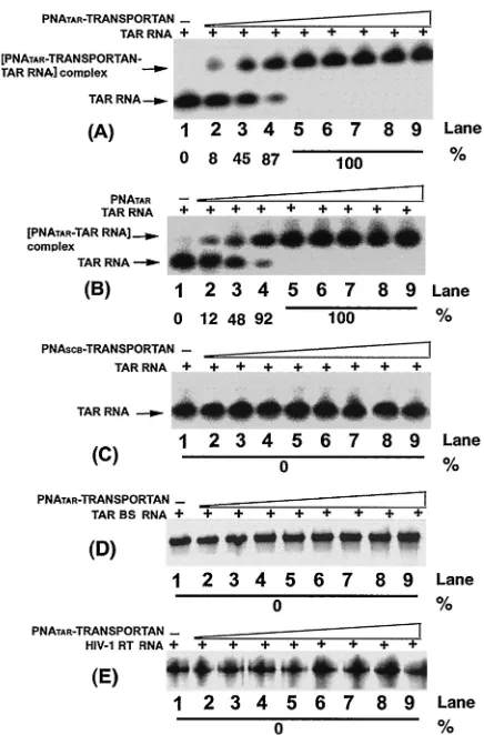
scrambled PNA-transportan conjugate, or unconjugated PNA TAR (Fig. 2A to C). Titration of PNATAR-transportan
conju-gate with TAR RNA at molar ratios of the conjuconju-gate to TAR RNA of less than 1 resulted in a stoichiometric band shift of the labeled TAR RNA (Fig. 2A, lanes 2 to 4).
[image:4.587.312.530.71.402.2]As shown in the figure, at molar ratios of 0.1, 0.5, and 0.8 of the conjugate to labeled TAR RNA, the extent of band shift corresponded to approximately 8, 45, and 87%, respectively. A complete shift in the mobility was achieved at an equimolar
FIG. 2. Binding specificity of PNATAR-transportan conjugate to its
target sequence. The affinity of the PNATAR-transportan conjugate
(A) for its target sequence on the TAR RNA was assessed by gel mobility shift analysis as described in Materials and Methods. Uncon-jugated PNATAR(B) was included as a positive control, and scrambled
PNA-transportan conjugate (PNASCB-transportan) (C) was included
as a negative control. The specificity of this interaction was determined by analyzing the relative gel mobility shift upon interaction of
PNA-TAR-transportan conjugate with a mutant TAR RNA (TAR BS; panel
D) as well as with an unrelated RNA such as the HIV-1 RT RNA (panel E). In panel A, lanes 1 through 9 represent molar ratios of PNATAR-transportan conjugate to TAR RNA of 0, 0.1, 0.5, 0.8, 1.0,
2.5, 5, 7.5, and 10, respectively. Panel B and panel C show gel mobility shifts performed at similar ratios as indicated in panel A except that unconjugated PNATAR and scrambled PNA-transportan conjugate,
respectively, were used. Similar ratios are also shown in panels D and E except that the mobility shift of the PNATAR-transportan conjugate
was carried out with the TAR BS and HIV-1 RT RNA probes, respec-tively. The extent of gel shift was determined by quantifying the probe RNA band on the PhosphorImager using Image-Quant software (Mo-lecular Dynamics). The percentage of labeled TAR RNA retarded due to PNA binding is indicated.
on November 8, 2019 by guest
http://jvi.asm.org/
ratio or a molar excess of the conjugate to TAR RNA (panel A, lanes 5 to 9). A similar titration was carried out in the presence of unconjugated PNATAR(panel B, lanes 2 to 9) and
scrambled PNA-transportan conjugate (panel C, lanes 2 to 9). As shown in the figure, the binding of PNATAR to the TAR
RNA was similar to that obtained with the PNATAR
-transpor-tan conjugate, suggesting that conjugation of PNATARwith the
transportan peptide vector had no effect on its binding affinity. Scrambled PNA-transportan conjugate at similar molar ratios did not result in any gel shift, suggesting the specificity of the interaction. Further evidence of this specificity was demon-strated by our observation that interaction of PNATAR
-trans-portan conjugate with TAR BS, a 63-base-long mutant TAR RNA carrying a deletion in the stem and bulge region (panel D), and with HIV-1 RT RNA, an unrelated RNA (panel E), also exhibited no shift in mobility.
Reverse transcription of TAR RNA region is blocked by PNATAR-transportan conjugate.Since the PNATAR
-transpor-tan conjugate displayed affinity for TAR RNA similar to un-conjugated PNATAR, it was interesting to examine if the
con-jugate could effectively block reverse transcription of TAR RNA. Ability to block reverse transcription, in turn, would have multiple impacts on viral replication besides influencing Tat-mediated transactivation. For this purpose, the TAR RNA transcript primed with the labeled 17-mer DNA primer was incubated in the absence or presence of 100 nM PNATAR
-transportan conjugate, PNATAR, or scrambled PNA at 37°C,
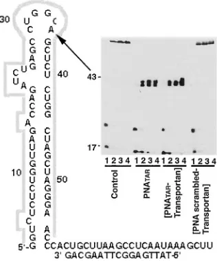
followed by initiation of reverse transcription by HIV-1 RT. The results are presented in Fig. 3. PNATARand PNATAR
-transportan conjugate showed a similar pattern of reverse tran-scription, with a prominent pause at nucleotide position 42, prior to the loop site targeted by these two PNAs. These results suggest that conjugation of PNATARwith transportan did not
alter the ability of PNATARto block reverse transcription of
TAR RNA.
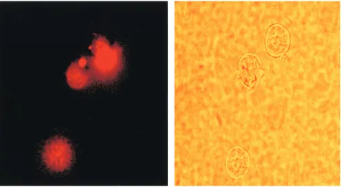
Cellular uptake of PNA-transportan and its intracellular localization.Although PNAs display strong binding affinity to their target sequence and have great potential as antisense agents, their biodelivery in the cell is very poor (47). The biodelivery of PNA into cells has been enhanced by cationic detergents (39) and by linking them with membrane-translo-cating peptides (51). To evaluate the uptake of the PNA-transportan conjugate in our system, we used a PNA- transportan-conjugated 10-mer PNA tagged with a TAMRA fluorophore probe. The entry of the dye-linked conjugate into the cells was then monitored by fluorescence microscopy and analyzed by fluorescence-activated cell sorting (FACS) analysis. The re-sults are shown in Fig. 4 and Table 1.
It was observed that the PNA-transportan conjugate is able to enter the cells in a time-dependent manner, confirming that transportan linked with PNA retained its ability to cross the cell membrane (Fig. 4). At 12 h, approximately 90% of the CEM cells were found to display fluorescence, as against 52% of the Jurkat cells (Table 1). This suggests that the PNA-transportan conjugate can efficiently cross the membrane of both cell types, although the kinetics of cell entry varied. Fur-thermore, the PNA-transportan conjugate had no detrimental effect on cell viability up to the 10M concentration tested in both CEM and Jurkat cells (results not shown). These results demonstrate that membrane-permeating peptide vectors such
as transportan may be effective carriers for intracellular deliv-ery of PNA.
Inhibition of Tat-mediated transactivation of HIV-1 LTR by PNATAR-transportan conjugate.Since the transporter peptide was found to be an efficient vehicle for biodelivery of PNA, we examined the ability of PNATAR-transportan conjugate to
block Tat-mediated transactivation of HIV-1 LTR in CEM and Jurkat cells. Using the reporter plasmid, we first established the optimum amounts of Tat required for the expression of the HIV-1 LTR in these cells. For this, we used the highly sensitive assay system involving expression ofRenillaluciferase and fire-fly luciferase. Expression of the firefire-fly luciferase (pHIV-1 LTR-Luc) and Renillaluciferase (pCMV-R.Luc) was under the control of the HIV-1 LTR and CMV promoter, respec-tively. The cells were transfected with these two plasmids in the presence of various concentrations of the Tat expression vector (pCMV-Tat).
[image:5.587.340.498.75.265.2]In the Jurkat cells, the transfected cells were activated with PMA and Ca2⫹ionophore for expression of the reporter plas-mids. When Tat was expressed intrans, the firefly luciferase was transactivated severalfold, while theRenillaluciferase ex-pression, used as an internal control to monitor transfection efficiency, remained constant. Expression of the firefly lucif-erase in CEM as well as Jurkat cells was found to be propor-tional to the concentration of pCMV-Tat used in the transfec-tion. The amount of pCMV-Tat for optimum stimulation of HIV-1 LTR in CEM cells (100-fold stimulation) and Jurkat
FIG. 3. Effect of PNATAR-transportan conjugate on reverse
tran-scription on TAR RNA primed with the 17-mer DNA primer. TAR RNA template primed with the 5⬘-32P-labeled 17-mer DNA primer
was incubated in the absence or presence of PNATAR-transportan
conjugate, unconjugated PNATAR, or scrambled PNA-transportan
conjugate at 37°C for 1 h and used in the gel extension reaction as described under Materials and Methods. The reaction products were analyzed on a denaturing 8% polyacrylamide–urea gel and subjected to PhosphorImager analysis. Lanes 1 through 4 in each set represent the extension reactions at 25°C for 5, 10, 15, and 20 min, respectively. The control set represents reactions carried out in the absence of PNATAR-transportan conjugate, unconjugated PNATAR, or scrambled
PNA-transportan conjugate. The arrow indicates the position on the gel corresponding to the sequence targeted by PNATAR-transportan
conjugate as well as unconjugated PNATARon TAR RNA.
on November 8, 2019 by guest
http://jvi.asm.org/
cells (150-fold stimulation) was 1g and 3 g, respectively (Fig. 5).
Using this standardized system, we determined the func-tional ability of the PNATAR-transportan conjugate to inhibit
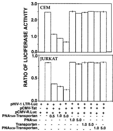
Tat-mediated transactivation of the HIV-1 LTR. The results are shown in Fig. 6. As shown in the figure, the PNATAR
-transportan conjugate substantially inhibited Tat-mediated transactivation of the HIV-1 LTR, as judged from the expres-sion levels of luciferase in both CEM and Jurkat cells. Approx-imately 55% inhibition was achieved at 500 nM PNA-peptide conjugate, which gradually leveled off to 70 to 80% inhibition at higher concentrations. Unconjugated PNATAR, scrambled
PNA-transportan, and transportan itself had no effect on the luciferase activity under identical conditions. These results demonstrate that PNATAR-transportan present in the culture
medium is able to enter the cells and prevent Tat-mediated transactivation by efficiently sequestering the TAR region of the HIV-1 LTR.
PNATAR-transportan inhibits at the level of transcription.A substantial reduction in the expression of the luciferase re-porter in both CEM and Jurkat cells suggested that the PNATAR-transportan inhibits the Tat-mediated transactivation
of the HIV-1 LTR. To determine if the inhibition was at the level of transcription, we performed RNase protection assays. CEM and Jurkat cells transfected with an HIV-1 LTR promot-er-driven luciferase reporter plasmid in the absence or pres-ence of a Tat expression plasmid were treated with various concentrations of PNATAR-transportan, PNATAR, scrambled
PNA-transportan, or transportan and harvested at the speci-fied times for total RNA isolation. In addition, the Renilla
luciferase reporter driven by a CMV promoter was included as a control for monitoring transfection efficiency. The RNA sam-ples were hybridized with the 32P-labeled pSPluc⫹ and
pSPrluc probes, digested with the RNase A and T1mix, and
examined by denaturing gel electrophoresis for protected frag-ments corresponding to the firefly andRenillatranscripts, re-spectively.
A representation of one such analysis is presented in Fig. 7. Our results indicated that the accumulation of firefly RNA was inversely proportional to the concentration of PNATAR
-transpor-tan in CEM and Jurkat cells (Fig. 7A). This provides direct evi-dence that the PNATAR-transportan interferes with the
transcrip-tion process, presumably by binding to the TAR region of the HIV-1 LTR, thereby preventing Tat-mediated transactivation. Neither the unconjugated PNATAR, transportan itself, nor
scram-bled PNA-transportan affected the firefly RNA expression levels, indicating the specificity of the interaction of PNATAR
[image:6.587.46.542.72.344.2]-transpor-FIG. 4. Cellular uptake of PNA-transportan conjugate. CEM cells were incubated with 70 nM TAMRA-tagged PNA-transportan conjugate in a six-well plate in RPMI 1640 medium in the absence of serum. At various times, aliquots of the cells were washed, resuspended in PBS, and examined by fluorescence microscopy for monitoring cellular uptake. The panel on the left is the fluorescence image, and that on the right is the phase-contrast image of the same field.
TABLE 1. Time course of TAMRA-tagged PNA-transportan conjugate uptake in cellsa
Cells Uptake of PNA-transportan (%) at time posttransfection:
1 h 2 h 4 h 6 h 12 h
CEM 5 12 35 55 90
Jurkat 0 3 16 27 52
aThe extent of uptake of TAMRA-tagged PNA-transportan conjugate was
monitored in CEM and Jurkat cells as described in Materials and Methods. The values represent averages of three independent experiments.
on November 8, 2019 by guest
http://jvi.asm.org/
[image:6.587.43.283.649.703.2]tan with its target sequence. Furthermore, the effect of PNATAR
-transportan on the HIV-1 LTR appeared to be promoter specific, as noted from our observation that PNATAR-transportan, PNA-TAR, transportan, and scrambled PNA-transportan did not affect
the expression levels of the RenillaRNA driven by the CMV promoter (Fig. 7B).
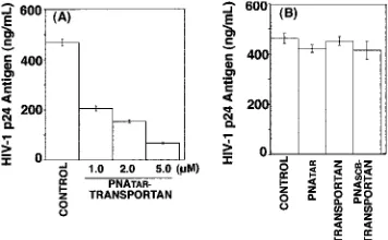
Effect of PNATAR-transportan on HIV-1 production in chronically HIV-1-infected H9 cells.Since PNATAR
-transpor-tan was able to enter the cells and interfere with Tat-mediated transactivation, we examined its antiviral efficacy in H9 cells chronically infected with HIV-1. These cells were grown in the absence or presence of PNATAR-transportan (1.0 to 5.0M),
unconjugated PNATAR (5.0 M), transportan (5.0 M), or
scrambled PNA-transportan (5.0 M) for 3 days, and their culture supernatants were analyzed for levels of p24 antigen by ELISA, since the levels of p24 antigen provided a measure of viral titers. The results are shown in Fig. 8.
The levels of p24 antigen decreased by 60% at 1 M PNATAR-transportan conjugate. Further increases in the
con-centration of the PNATAR-transportan conjugate resulted in a
substantial decrease (70 to 90%) in the p24 levels, suggesting its potential for blocking HIV-1 production (Fig. 8A). Our observation that unconjugated PNATARat similar
concentra-tions did not decrease viral production indicated the efficacy of PNATAR-transportan. Furthermore, the effect of PNATAR
-transportan appeared to be quite specific, since neither
scram-bled PNA-transportan nor transportan by itself decreased HIV-1 production under identical conditions (Fig. 8B).
PNATAR-transportan inhibits transcription of HIV-1 mRNA in chronically HIV-1-infected H9 cells.We also examined the mechanism by which PNATAR-transportan inhibited HIV-1
production in chronically HIV-1-infected H9 cells. Total RNA isolated from the cells treated with PNATAR-transportan,
un-conjugated PNATAR, transportan, or scrambled transportan
were hybridized to the radiolabeled probe synthesized from pGEM23 and subjected to an RNase protection assay. The results are shown in Fig. 9.
The mRNA initiating at⫹1 of the HIV-1 LTR protected an 83-nucleotide fragment of the riboprobe corresponding to the anti-TAR sequence. A decrease in levels of HIV-1 mRNA in cells treated with PNATAR-transportan suggested that the
in-hibition occurred at the transcription step. The specificity of the effect of PNATAR-transportan was seen from the
observa-tion that neither transportan itself nor scrambled PNA-trans-portan altered the HIV-1 mRNA levels. Furthermore, PNATAR alone also did not influence the levels of HIV-1
mRNA synthesis, indicating the efficiency of transportan as a vehicle for delivering PNATARto its target site.
PNATAR-transportan conjugate has no effect on cellular
pro-FIG. 5. Tat-mediated transactivation of HIV-1 LTR. The indicated amounts of pCMV-Tat were cotransfected along with a plasmid cock-tail comprising pHIV-1 LTR-Luc (4g) and pCMV-R.Luc (1g) in Jurkat and CEM cells (5.0⫻106cells) in order to establish the extent
of Tat-mediated transactivation of the HIV-1 LTR. The highly sensi-tive luciferase reporter was used for this analysis. The extent of stim-ulation of the HIV-1 LTR was monitored as a function of the expres-sion of the firefly luciferase gene, with theRenillaluciferase reporter serving as an internal control for normalizing the transfection effi-ciency. The results are expressed as the ratio of firefly to Renilla
[image:7.587.95.230.72.292.2]luciferase activity. Panels A and B represent the extent of transactiva-tion in CEM and Jurkat cells, respectively, as a functransactiva-tion of pCMV-Tat concentration. The results are presented as an average of three inde-pendent experiments. The bars represent the standard deviation.
FIG. 6. Effect of PNATAR-transportan conjugate on Tat-mediated
transactivation of HIV-1 LTR. CEM and Jurkat cells (5.0⫻106cells),
cotransfected with the plasmid cocktail comprising pHIV-1 LTR-Luc (4g) and pCMV-R.Luc (1g) in the presence or absence of the Tat expression clone pCMV-Tat, were incubated with the indicated con-centrations of the PNATAR-transportan, unconjugated PNATAR,
trans-portan, or scrambled PNA-transportan (PNASCB-transportan) under
the conditions described in Materials and Methods. At 20 h (CEM) or 28 h (Jurkat) posttransfection, cell lysates were assayed for firefly and
Renillaluciferase activities. The results are presented as the relative ratio of firefly toRenillaluciferase activity. The amounts of the various plasmids, transportan, and PNA used in the experiment are indicated. The results are presented as an average of three independent experi-ments. The bars represent the standard deviation.
on November 8, 2019 by guest
http://jvi.asm.org/
[image:7.587.311.527.349.599.2]liferation of chronically HIV-1-infected H9 cells.We examined the incorporation of [3H]thymidine into cellular DNA in order
to determine if the PNATAR-transportan conjugate impacted
cellular proliferation of chronically HIV-1-infected H9 cells. The cells were grown in the presence or absence of PNATAR
-transportan and supplemented with [3H]thymidine. At the
in-dicated time points, aliquots were withdrawn, and the DNA was precipitated with TCA to determine thymidine incorpora-tion. The results of this analysis are shown in Fig. 10. As shown in the figure, similar amounts of [3H]thymidine were
incorpo-rated into cellular DNA in both the treated and untreated cells at all time points analyzed, indicating that cellular proliferation was not altered in the presence of PNATAR-transportan.
DISCUSSION
In spite of massive efforts for the prevention of AIDS, the AIDS epidemic has reached alarming proportions worldwide. The current antiviral treatment for HIV-1 infection is re-stricted to drugs that mainly target two key viral enzymes in its life cycle, reverse transcriptase and protease (7, 22, 23, 28, 32, 42–44, 55, 67). Successful HIV-1 therapy, such as the highly active antiretroviral therapy, is hampered by the appearance of drug-resistant strains of HIV-1, which have been directly cor-related with mutations in the genes encoding the viral reverse transcriptase and protease (1, 5).
[image:8.587.120.464.73.305.2]In the present study we have used specific PNA-transportan conjugate to target the TAR RNA, an important regulatory element in the nontranslated 5⬘end of the retroviral genome. The rationale for targeting TAR was that it forms a stable nuclease-resistant stem-loop structure which is averse to mu-tational changes (25, 59). HIV-1 Tat, a nuclear transcriptional activator, interacts with TAR to enhance the processivity of
FIG. 7. RNase protection assay. (A) Jurkat cells were transfected with 4g of pHIV-1 LTR-Luc and 1g of pCMV-R.Luc in the absence or presence of pCMV-Tat (2.5g). The cells were grown in the presence of PNATAR-transportan (0.5 to 5.0M), PNATAR(1.0 and 5.0M),
transportan (1.0 and 5.0M), or scrambled PNA-transportan (1.0 and 5.0M). At 28 h posttransfection, total RNA was isolated from the cells and hybridized with RNA probes for the different reporter transcripts (firefly andRenillaluciferase). The pSPrluc probe coding for theRenilla
RNA served as a transfection control. The firefly RNA levels were normalized to theRenilla RNA signal. The lanes are as follows: mock-transfected cells (lane 1); luciferase expression in the absence of Tat (lane 2); and luciferase expression in the presence of Tat and the indicated inhibitors (lanes 3 to 12). (B) CEM and Jurkat cells were transfected with 1g of pCMV-R.Luc and incubated in the absence or presence of PNATAR-transportan, unconjugated PNATAR, transportan, or scrambled PNA-transportan (PNASCB-transportan) at 5M in order to investigate
the promoter specificity of PNATAR-transportan. Total RNA was isolated from cells and subjected to the RNase protection assay with the pSPrluc
probe. The protected fragments were analyzed on a denaturing polyacrylamide-urea gel and visualized on a PhosphorImager.RenillaRNA was normalized to total RNA in the cell. The lane marked control representsRenillaRNA signal in transfected cells in the absence of the inhibitor.
FIG. 8. Effect of PNATAR-transportan conjugate on HIV-1
produc-tion in chronically HIV-1-infected H9 cells. The indicated concentra-tions of PNATAR-transportan (A), unconjugated PNATAR,
transpor-tan, or scrambled PNA-transportan (PNASCB-transportan) (B) were
added to the culture medium of chronically HIV-1-infected H9 cells, and their ability to inhibit HIV-1 production was examined by moni-toring the levels of p24 antigen in the culture supernatant. The results are presented as relative amounts of HIV-1 p24 antigen present in the supernatants of H9 cells cultured in the absence (control) or presence of PNATAR-transportan (1.0 to 5.0M), unconjugated PNATAR(5.0
M), transportan (5.0M), or scrambled PNA-transportan (5.0M). The results are expressed as mean values along with standard deviation for three independent experiments.
on November 8, 2019 by guest
http://jvi.asm.org/
[image:8.587.74.252.497.607.2]RNA polymerase II complexes (25). The importance of the Tat-TAR interaction in regulation of gene expression has been well established (21, 25). We had earlier demonstrated that a novel inhibitor, PNA, targeted to the TAR sequences of the viral RNA genome blocks the Tat-TAR interaction, resulting in the inhibition of transactivation of the HIV-1 LTR (40). In another study, PNA has been employed to inhibit in vitro protein synthesis at both the transcriptional and translational levels (20, 31).
Effective antisense inhibition in cells requires efficient cell entry. Though PNAs have been shown to inhibit gene expres-sion and have several advantages over conventional DNA or RNA antisense oligomers, their use as an alternative therapy has been limited chiefly by their inability to cross the cell membrane efficiently (47). More recent findings demonstrating that PNAs conjugated to transporter peptides exhibit efficient biodelivery into cells have redefined its potential as a thera-peutic agent (52).
For the present studies, we used the transportan peptide vector for biodelivery of PNATAR. Transportan is a 27-amino-acid
chi-meric peptide consisting of 13 amino acids from the amino ter-minus of the neuropeptide galanin and 14 amino acids from the amino terminus of the wasp venom toxic peptide mastoparan (33). Transportan penetrates every cell type in a rapid and effi-cient way and is localized mostly in membranous structure; upon prolonged incubation, it concentrates in the nucleus (50). In mod-erate concentrations (10M), it does not affect the growth of cells in culture (50). These unique properties of the chimeric transpor-tan peptide make it potentially useful as a vector for biodelivery of different proteins or nonprotein drugs into cells. We used this approach to address the issues of (i) uptake of the PNA-trans-portan conjugate by cells and its effect on cell viability and (ii) functional efficacy of the PNATAR-transportan conjugate in
blocking the Tat-TAR interaction, thereby inhibiting viral tran-scription and replication.
To ascertain the ability of the PNA-transportan conjugate to enter the cells, we tagged it with TAMRA, a fluorescent probe, and monitored its uptake in the cell. A substantial accumula-tion of the fluorophore in the cell in 12 h (Table 1) indicated efficient uptake, though the overall kinetics of its internaliza-tion was slower than with transportan alone (50, 51). In marked contrast to an earlier report demonstrating that cell type did not influence the cell-penetrating ability of transpor-tan (51), the uptake of the PNA-transportranspor-tan conjugate by CEM and Jurkat cells varied significantly (Table 1). This may be attributed to a difference in the morphology and membrane composition of these two cells, or it is likely that the presence of the PNA moiety influences uptake. Nonetheless, the accu-mulation of the PNA-transportan conjugate in the cells is en-couraging and suggests that the transportan vector may be used effectively for biodelivery of this class of compounds.
PNATAR-transportan substantially inhibited Tat-mediated
transactivation of the HIV-1 LTR in both CEM and Jurkat cells (Fig. 5). The PNA-peptide conjugate may have dual func-tions; it may prevent the TAR-Tat interaction by sequestering the TAR sequence of the nascent RNA, or it may prevent transcription directly by interacting with the TAR sequence on the proviral DNA. One may visualize that the large transpor-tan moiety may interfere with the PNA, targeting its site inside the cell. However, this did not appear to be true in this study, since the PNATAR-transportan conjugate displayed similar
binding affinity for its target sequence on the TAR RNA (Fig. 2) and was also able to block reverse transcription to a similar extent as the unconjugated PNATAR (Fig. 3). Moreover, a
substantial inhibition of Tat-mediated transactivation was also achieved by PNATAR-transportan conjugate in cell culture
(Fig. 6). The mechanism of this inhibition appeared to be at the level of transcription (Fig. 7).
Our results demonstrating that PNATAR-transportan can
[image:9.587.92.235.74.227.2]enter chronically HIV-1-infected H9 cells and inhibit virus production are very promising (Fig. 8). This is the first report
FIG. 9. PNATAR-transportan inhibits transcription of the HIV-1
mRNA in chronically HIV-1-infected H9 cells. Total RNA was iso-lated from chronically HIV-1-infected H9 cells treated with the indi-cated concentrations of PNATAR-transportan or 5.0 M PNATAR,
transportan alone, or scrambled PNA-transportan (PNASCB
-transpor-tan). The RNA was hybridized with the pGEM23 probe and digested with the RNase A and T1 mix, and the protected fragments were
separated on a polyacrylamide-urea gel and detected by PhosphorIm-ager analysis. The undigested probe corresponds to a 195-base frag-ment, whereas HIV-1 mRNA initiating at ⫹1 of the HIV-1 LTR protected an 83-nucleotide fragment of the riboprobe corresponding to the anti-TAR sequence.
FIG. 10. [3H]thymidine incorporation into cellular DNA in
chron-ically HIV-1-infected H9 cells. Chronchron-ically HIV-1-infected H9 cells grown in the presence or absence of 5.0M PNATAR-transportan were
supplemented with 10Ci of [methyl-3H]thymidine/ml. At the
indi-cated time points, the cells were harvested and assayed for the amounts of [3H]thymidine (TdR) incorporated into DNA by TCA precipitation
of the acid-insoluble material onto glass fiber filters. The total amount of radioactivity was determined by scintillation counting, and the re-sults are represented as counts per minute incorporated per milligram of protein. The results are expressed as average values along with the standard deviation for three independent experiments.
on November 8, 2019 by guest
http://jvi.asm.org/
[image:9.587.347.492.512.626.2]on the use of a PNA-transporter peptide conjugate to inhibit the replication of HIV-1. Interestingly, recent studies by Sei et al. (58) have demonstrated that PNA targeted to the Gag-Pol transframe domain can arrest virus production. However, very high concentrations of PNA (100 M) were required to achieve this effect. In contrast, the PNA-transportan conju-gates used in this study were able to inhibit virus replication at significantly lower concentrations. It may also be noted that the effect of the PNATAR-transportan conjugate on transactivation
of the HIV-1 LTR appeared to be more prominent in the transfected cells than in H9 cells. This is not surprising, as the events associated with the replication of HIV-1 in H9 cells are far more complex than those associated with the expression of the luciferase gene in cells.
The choice of antisense targets appears quite challenging, since targeting the various regulatory genes of the HIV-1 ge-nome has not always been effective against diverse HIV-1 strains (2, 29, 30, 36–38, 66). The unique 5⬘untranslated region (U5) (nucleotides 1 to 333) of the HIV-1 genome, containing several critical domains essential for viral replication, may be an ideal target for drug intervention. These critical domains include (i) the primer-binding site (nucleotides 183 to 201), essential for tRNA3Lys-primed initiation of reverse
transcrip-tion (45, 53, 62); (ii) the A-loop region located upstream of the primer-binding site (nucleotides 168 to 173), essential for the selection and interaction of tRNA3Lysprimer (24, 35, 63); and
(iii) the LTR sequences at the 5⬘and 3⬘ends, essential for viral transcription and integration (61).
Studies in our laboratory are under way to analyze the effect of PNA-transportan conjugates targeted against these sites as effective antiviral agents. A PNA-transportan conjugate di-rected against galanin receptor mRNA has been shown to inhibit the expression levels of galanin receptor in Bowes mel-anoma cells in cell culture and also alleviate pain in the rat model (52). Recently, Good et al. have shown that PNA con-jugated with a 10-mer peptide vector targeted against rRNA and against mRNA of Acp protein of bacteria can effectively cure HeLa cells infected with Escherichia coli K-12 strains, raising the possibility of anti-infective drug development (17). In summary, our studies have demonstrated that cellular uptake of anti-TAR PNA is significantly enhanced upon con-jugation with a transportan peptide, resulting in effective blockage of the Tat-TAR interaction at relatively low inhibitor concentrations and inhibiting transcription. These studies with transportan peptides as the vector for biodelivery of PNA have created a therapeutic niche for the application of PNA against HIV-1. Thus, our approach using PNA-transportan to target the critical regions of the HIV-1 genome in the quest for novel inhibitors of HIV-1 appears quite promising. Optimal chemical modification of the existing transporter peptides by treatment with polyethylene glycol (64) in order to increase the circulat-ing half-life of PNA-transporter conjugates will further ad-vance the efforts to develop potent antiviral therapeutics tar-geting viral genes.
ACKNOWLEDGMENTS
The first two authors made an equal contribution.
We thank Michael Mathews and M. Peterlin for the kind gift of the plasmids and John Stephens for assistance with the HIV-1 p24 assay. This research was supported by a grant from the NIAID (AI42520).
REFERENCES
1.Aalen, O. O., V. T. Farewell, D. De Angelis, N. E. Day, and O. N. Gill.1999. New therapy explains the fall in AIDS incidence with a substantial rise in
number of persons on treatment expected. AIDS13:103–108.
2.Agrawal, S., T. Ikeuchi, D. Sun, P. S. Sarin, A. Konopka, J. Maizel, and P. C. Zamecnik.1989. Inhibition of human immunodeficiency virus in early in-fected and chronically inin-fected cells by antisense oligodeoxynucleotides and
their phosphorothioate analogues. Proc. Natl. Acad. Sci. USA86:7790–7794.
3.Berkhout, B., R. H. Silverman, and K. T. Jeang.1989. Tat transactivates the
human immunodeficiency virus through a nascent RNA target. Cell59:273–282.
4.Bohjanen, P. R., R. A. Colvin, M. Puttaraju, M. D. Been, and M. A. Garcia-Blanco.1996. A small circular TAR RNA decoy specifically inhibits
Tat-activated HIV-1 transcription. Nucleic Acids Res.24:3733–3738.
5.Centers for Disease Control and Prevention.1998. HIV/AIDS surveillance report, midyear ed., vol. 10. Centers for Disease Control and Prevention, Public Health Service, Department of Health and Human Services, Atlanta, Ga. 6.Cordingley, M. G., R. L. Lafemina, P. L. Callahan, J. H. Condra, V. V.
Sardana, D. J., Graham, T. M. Nguyen, K. Legrow, L. Gotlib, A. J. Schla-bach, and R. J. Colonno.1990. Sequence-specific interaction of Tat protein and Tat peptides with the transactivation-responsive sequence element of human immunodeficiency virus type 1 in vitro. Proc. Natl. Acad. Sci. USA
87:8985–8989.
7.De Clercq, E.1992. HIV inhibitors targeted at the reverse transcriptase.
AIDS Res. Hum. Retrovirol.8:119–134.
8.Demidov, V. V., V. N. Potaman, M. D. Frank-Kamenetskii, M. Egholm, O. Buchard, S. H. Sonnichsen, and P. E. Nielsen.1994. Stability of peptide nucleic acids in human serum and cellular extracts. Biochem. Pharmacol.
48:1310–1313.
9.Derossi, D., S. Calvet, A. Trembleau, A. Brunissen, G. Chassaing, and A. Prochiantz.1996. Cell internalization of the third helix of the Antennapedia
homeodomain is receptor-independent. J. Biol. Chem.271:18188–18193.
10.Dingwall, C., I. Ernberg, M. J. Gait, S. M. Green, S. Heaphy, J. Karn, A. D. Lowe, M. Singh, and M. A. Skinner.1990. HIV-1 Tat protein stimulates transcription by binding to a U-rich bulge in the stem of the TAR RNA
structure. EMBO J.12:4145–4153.
11.Faria, M., D. G. Spiller, C. Dubertret, J. S. Nelson, M. R. White, D. Scher-man, C. Helene, and C. Giovannangeli.2001. Phosphoramidate oligonucle-otides as potent antisense molecules in cells and in vivo. Nat. Biotechnol.
19:40–44.
12.Fawell, S., J. Serry, Y. Daikh, C. Moore, L. L. Chen, B. Pepinsky, and J. Barsoum.1994. Tat-mediated delivery of heterologous proteins into cells.
Proc. Natl. Acad. Sci. USA91:664–668.
13.Feng, S., and E. C. Holland.1988. HIV-1 Tat trans-activation requires the
loop sequence within tar. Nature334:165–167.
14.Frankel, A. D., S. Blancalana, and D. Hudson.1989. Activity of synthetic
peptides from the Tat protein of HIV-1. Proc. Natl. Acad. Sci. USA86:
7397–7401.
15.Fujinaga, K., R. Taube, J. Wimmer, T. P. Cujec, and B. M. Peterlin.1999. Interactions between human cyclin T, Tat, and the transactivation response element (TAR) are disrupted by a cysteine to tyrosine substitution found in
mouse cyclin T. Proc. Natl. Acad. Sci. USA96:1285–1290.
16.Futaki, H., T. Suzuki, W. Ohashi, T. Yagami, S. Tanaka, K. Ueda, and Y. Sugiura.2001. Arginine-rich peptides. An abundant source of membrane-permeable peptides having potential as carriers for intracellular protein
delivery. J. Biol. Chem.276:5836–5840.
17.Good, L., S. K. Awasthi, R. Dryselius, O. Larsson, and P. E. Nielsen.2001. Bactericidal antisense effects of peptide-PNA conjugates. Nat. Biotechnol.
19:360–364.
18.Gunnery, S., S. R., Green, and M. B. Matthews.1992. Tat-responsive region RNA of human immunodeficiency virus type 1 stimulates protein synthesis in vivo and in vitro: relationship between structure and function. Proc. Natl.
Acad. Sci. USA89:11557–11561.
19.Hamy, F., E. R. Felder, G. Heizmann, J. Lazdins, F. Aboul-ela, G. Varani, J. Karn, and T. Klimkait.1997. An inhibitor of the Tat/TAR RNA interaction that effectively suppresses HIV-1 replication. Proc. Natl. Acad. Sci. USA
94:3548–3553.
20.Hanvey, J. C., N. J. Peffer, J. E. Bisi, S. A. Thomas, R. Cadilla, J. A. Josey, et al.1992. Antisense and antigene properties of peptide nucleic acids.
Science258:1481–1485.
21.Jeang, K. T., H. Xiao, and E. A. Rich.1999. Multifaceted activities of the
HIV-1 transactivator of transcription, Tat. J. Biol. Chem.274:28837–28840.
22.Johnson, V. A., D. P. Merrill, T. C. Chou, and M. S. Hirsch.1992. Human immunodeficiency virus type 1 (HIV-1) inhibitory interactions between pro-tease inhibitor Ro 31-8959 and zidovudine, 29,39-dideoxycytidine, or recom-binant interferon-alpha A against zidovudine-sensitive or -resistant HIV-1 in
vitro. J. Infect. Dis.166:1143–1146.
23.Kageyama, S., J. N. Weinstein, T. Shirasaka, D. J. Kempf, D. W. Norbeck, J. J. Plattner, J. Erickson, and H. Mitsuya.1992. In vitro inhibition of
human immunodeficiency virus (HIV) type 1 replication byC2
symmetry-based HIV protease inhibitors as single agents or in combinations.
Antimi-crob. Agents Chemother.36:926–933.
on November 8, 2019 by guest
http://jvi.asm.org/
24.Kang, S. M., Z. Zhang, and C. D. Morrow.1997. Identification of a sequence within U5 required for human immunodeficiency virus type 1 to stably maintain
a primer binding site complementary to tRNAMet. J. Virol.71:207–217.
25.Karn, J.1999. Tackling Tat. J. Mol. Biol.293:235–254.
26.Kashanchi, F., M. R. Sadaie, and J. N. Brady.1997. Inhibition of HIV-1 transcription and virus replication using soluble Tat peptide analogs.
Virol-ogy227:431–438.
27.Kaushik, N., N. Rege, P. N. S. Yadav, S. G. Sarafianos, M. J. Modak, and V. N. Pandey.1996. Biochemical analysis of catalytically crucial aspartate mutants of human immunodeficiency virus type 1 reverse transcriptase.
Bio-chemistry35:11536–11546.
28.Kempf, D. J., K. C. Marsh, J. F. Denissen, E. McDonald, S. Vasavanonda, C. A. Flentge, B. E. Green, L. Fino, C. H. Park, X. P. Kong, et al.1995. ABT-538 is a potent inhibitor of human immunodeficiency virus protease and has high oral bioavailability in humans. Proc. Natl. Acad. Sci. USA
92:2484–2488.
29.Kim, S. G., T. Hatta, S. Tsukahara, H. Nakashima, N. Yamamoto, Y. Shoji, K. Takai, and H. Takaku.1995. Antiviral effect of phosphorothioate oli-godeoxyribonucleotides complementary to human immunodeficiency virus.
Bioorg. Med. Chem.3:49–54.
30.Kinchington, D., S. Galpin, J. W. Jaroszewski, K. Ghosh, C. Subasinghe, and J. S. Cohen.1992. A comparison of gag, pol and rev antisense
oligode-oxynucleotides as inhibitors of HIV-1. Antivir. Res.17:53–62.
31.Knudsen, H., and P. E. Nielsen.1996. Antisense properties of duplex- and
triplex-forming PNAs. Nucleic Acids Res.24:494–500.
32.Lam, P. Y., P. K. Jadhav, C. J. Eyermann, C. N. Hodge, Y. Ru, L. T. Bacheler, J. L. Meek, M. J. Otto, M. M. Rayner, Y. N. Wong, et al.1994. Rational design of potent, bioavailable, nonpeptide cyclic ureas as HIV protease
inhibitors. Science263:380–384.
33.Langel, Ü., M. Pooga, C., Kairane, M., Zilmer, and T. Bartfai.1996. A
galanin-mastoparan chimeric peptide activates the Na⫹, K⫹-ATPase and
reverses its inhibition by ouabain. Regul. Peptides62:47–52.
34.Laspia, M. F., A. P. Rice, and M. B. Mathews.1989. HIV-1 Tat protein
increases transcriptional initiation and stabilizes elongation. Cell59:283–292.
35.Li, Y., S. M. Kang, and C. D. Morrow.1997. Stability of HIV type 1 proviral genomes that contain two distinct primer-binding sites. AIDS Res. Hum.
Retrovir.13:253–262.
36.Lisziewicz, J., D. Sun, M. Klotman, S. Agrawal, P. Zamecnik, and R. Gallo.
1992. Specific inhibition of human immunodeficiency virus type 1 replication by antisense oligonucleotides: an in vitro model for treatment. Proc. Natl.
Acad. Sci. USA89:11209–11213.
37.Lisziewicz, J., D. Sun, J. Smythe, P. Lusso, F. Lori, A. Louie, P. Markham, J. Rossi, M. Reitz, and R. C. Gallo.1993. Inhibition of human immunode-ficiency virus type 1 replication by regulated expression of a polymeric Tat activation response RNA decoy as a strategy for gene therapy in AIDS. Proc.
Natl. Acad. Sci. USA90:8000–8004.
38.Lisziewicz, J., D. Sun, V. Metelev, P. Zamecnik, R. C. Gallo, and S. Agrawal.
1993. Long-term treatment of human immunodeficiency virus-infected cells with antisense oligonucleotide phosphorothioate. Proc. Natl. Acad. Sci. USA
90:3860–3864.
39.Ljungstrom, T., H. Knudsen, and P. E. Nielsen.1999. Cellular uptake of
adamantyl conjugated peptide nucleic acids. Bioconjug. Chem.10:965–972.
40.Mayhood, T., N. Kaushik, P. K. Pandey, F. Kashanchi, L Deng, and V. N. Pandey.2000. Inhibition of Tat-mediated transactivation of HIV-1 LTR transcription by polyamide nucleic acid targeted to TAR hairpin element.
Biochemistry39:11532–11539.
41.Mhashilkar, A. M., D. K. Biswas, J. LaVecchio, A. B. Pardee, and W. A. Marasco.1997. Inhibition of human immunodeficiency virus type 1 replica-tion in vitro by a novel combinareplica-tion of anti-Tat single-chain intrabodies and
NF-B antagonists. J. Virol.71:6486–6494.
42.Mitsuya, H., and S. Broder.1986. Inhibition of the in vitro infectivity and cytopathic effect of human T-lymphotrophic virus type III/lymphadenopa-thy-associated virus (HTLV-III/LAV) by 29,39-dideoxynucleosides. Proc.
Natl. Acad. Sci. USA83:1911–1915.
43.Mitsuya, H., R. F. Jarrett, M. Matsukura, F. Di Marzo Veronese, A. L. DeVico, M. G. Sarngadharan, D. G. Johns, M. S. Reitz, and S. Broder.1987. Long-term inhibition of human T-lymphotropic virus type III/lymph-adenop-athy-associated virus (human immunodeficiency virus) DNA synthesis and RNA expression in T cells protected by 29,39-dideoxynucleosides in vitro.
Proc. Natl. Acad. Sci. USA84:2033–2037.
44.Mitsuya, H., K. J. Weinhold, P. A. Furman, M. H. St. Clair, S. N. Lehrman, R. C. Gallo, D. Bolognesi, D. W. Barry, and S. Broder.1985. 39-Azido-39-deoxythymidine (BW A509U): an antiviral agent that inhibits the infectivity and cytopathic effect of human T-lymphotropic virus type III/lymphoid
nopa-thy-associated virus in vitro. Proc. Natl. Acad. Sci. USA82:7096–7100.
45.Muesing, M. A., D. H. Smith, C. D. Cabradilla, C. V. Benton, L. A. Lasky, and D. J. Capon.1985. Nucleic acid structure and expression of the human
AIDS/lymphadenopathy retrovirus. Nature313:450–458.
46.Muesing, M. A., D. H. Smith, and D. J. Capon.1987. Regulation of mRNA accumulation by a human immunodeficiency virus transactivator protein.
Cell48:691–701.
47.Nielsen, P. E.1999. Peptide nucleic acids as therapeutic agents. Curr. Opin.
Struct. Biol.9:353–357.
48.Nielsen, P. E., M. Egholm, R. H. Berg, and O. Buchardt.1991. Sequence-selective recognition of DNA by strand displacement with a
thymine-substi-tuted polyamide. Science254:1497–1500.
49.O’Brien, W. A., M. Sumner-Smith, S. H. Mao, S. Sadeghi, J. Q. Zhao, and I. S. Chen.1996. Anti-human immunodeficiency virus type 1 activity of an
oligocationic compound mediated via gp120 V3 interactions. J. Virol.70:
2825–2831.
50.Pooga, M., M. Hallbrink, M. Zorko, and U. Langel.1998. Cell penetration by
transportan. FASEB J.12:67–77.
51.Pooga, M., M. Lindgren, M. Hallbrink, E. Brakenhielm, and U. Langel.
1998. Galanin-based peptides, galparan and transportan, with
receptor-de-pendent and indereceptor-de-pendent activities. Ann. N. Y. Acad. Sci.863:450–453.
52.Pooga, M., U. Soomets, M. Hallbrink, A. Valkna, K. Saar, K. Rezaei, U. Kahl, J. X. Hao, X. J, Xu, Z Wiesenfeld-Hallin, T. Hokfelt Bartfai, and U. Langel.1998. Cell penetrating PNA constructs regulate galanin receptor
levels and modify pain transmission in vivo. Nat. Biotechnol.16:857–861.
53.Ratner, L., W. Haseltine, R. Patarca, K. J. Livak, B. Starcich, S. F. Josephs, E. R. Doran, J. A. Rafalski, E. A. Whitehorn, K. Baumeister, L. Ivanoff, J. S. R. Petteway, M. L. Pearson, J. A. Lautenberge, T. S. Papas, J. Ghrayeb, N. T. Chang, R. C. Gallo, and F. Wong-Staal.1985. Complete nucleotide
sequence of the AIDS virus, HTLV-III. Nature (London)313:277–284.
54.Re, M. C., G. Furlini, M. Vignoli, E. Ramazzotti, G. Roderigo, V. De Rosa, G. Zauli, S. Lolli, S. Capitani, and M. La Placa.1995. Effect of antibody to HIV-1 Tat protein on viral replication in vitro and progression of HIV-1
isease in vivo. J. Acquir. Immune Defic. Syndr. Hum. Retrovirol.10:408–416.
55.Roberts, N. A., J. A. Martin, D. Kinchington, A. V. Broadhurst, J. C. Craig, I. B. Duncan, S. A. Galpin, B. K. Handa, J. Kay, A. Krohn, et al.1990.
Rational design of peptide-based HIV proteinase inhibitors. Science248:
358–361.
56.Sanger, F., S. Nicklen, and A. R. Coulson.1977. DNA sequencing with
chain-terminating inhibitors. Proc. Natl. Acad. Sci. USA74:5463–5467.
57.Schwarze, S. R., A. Ho, A. Vocero-Akbani, and S. F. Dowdy.1999. In vivo protein transduction: delivery of a biologically active protein into the mouse.
Science285:1569–1572.
58.Sei, S., Q. Yang, D. O’Neill, K. Yoshimura, K. Nagashima, and H. Mitsuya.
2000. Identification of a key target sequence to block human
immunodefi-ciency virus type 1 replication within thegag-poltransframe domain. J. Virol.
74:4621–4633.
59.Selby, M. J., E. S. Bain, P. A. Luciw, and B. M. Peterlin.1989. Structure, sequence, and position of the stem-loop in tar determine transcriptional elongation by Tat through the HIV-1 long terminal repeat. Genes Dev.
3:547–558.
60.Smith, C. S., W. Lee, E. Wong, H. Gallardo, K. Page, O. Gaspar, J. Leb-kowski, and E. Gilboa.1996. Transient protection of human T-cells from human immunodeficiency virus type 1 infection by transduction with
adeno-associated viral vectors which express RNA decoys. Antivir. Res.32:99–115.
61.Vink, C., D. C. Van Gent, Y. Elgersma, and R. H. A. Plasterk.1991. Human immunodeficiency virus integrase protein requires a subterminal position of
its viral DNA recognition sequence for efficient cleavage. J. Virol.65:4636–4644.
62.Wakefield, J. K., A. G. Wolf, and C. D. Morrow.1995. Human immunode-ficiency virus type 1 can use different tRNAs as primers for reverse tran-scription but selectively maintains a primer binding site complementary to
tRNA3Lys. J. Virol.69:6021–6029.
63.Wakefield, J. K., S.-M. Kang, and C. D. Morrow.1996. Construction of a type 1 human immunodeficiency virus that maintains a primer binding site
complementary to tRNAHis. J. Virol.70:966–975.
64.Wang, J., S. Y. Huang, I. Choudhury, M. J. Leibowitz, and S. Stein.1995. Use of polyethylene glycol-peptide conjugate in a competition gel shift assay for screening potential antagonists of HIV-1 Tat protein binding to TAR
RNA. Anal. Biochem.232:238–242.
65.Wang, S., P. W. Huber, M. Cui, A. W. Czarnik, and H. Y. Mei.1998. Binding of neomycin to the TAR element of HIV-1 RNA induces dissociation of Tat
protein by an allosteric mechanism. Biochemistry37:5549–5557.
66.Weichold, F. F., J. Lisziewicz, R. A. Zeman, L. S. Nerurkar, S. Agrawal, M. S. Reitz, Jr., and R. C. Gallo. 1995. Antisense phosphorothioate oligode-oxynucleotides alter HIV type 1 replication in cultured human macrophages
and peripheral blood mononuclear cells. AIDS Res. Hum. Retrovirol.11:
863–867.
67.Yarchoan, R., R. W. Klecker, K. J. Weinhold, P. D. Markham, H. K. Lyerly, D. T. Durack, E. Gelmann, S. N. Lehrman, R. M. Blum, D. W. Barry, et al.
1986. Administration of 39-azido-39-deoxythymidine, an inhibitor of HTLV-III/LAV replication, to patients with AIDS or AIDS-related complex. Lancet
i:575–580.