STUDY OF FUNCTIONAL OUTCOME OF
MODIFIED TENSION BAND WIRING IN
TRANSVERSE FRACTURE PATELLA
Partial fulfilment of the regulations required for the award of
THE TAMIL NADU Dr M.G.R. MEDICAL
CHENNAI, TAMIL NADU
1
STUDY OF FUNCTIONAL OUTCOME OF
MODIFIED TENSION BAND WIRING IN
TRANSVERSE FRACTURE PATELLA
Dissertation submitted in
Partial fulfilment of the regulations required for the award of
M.S. Degree in Orthopaedics Surgery
THE TAMIL NADU Dr M.G.R. MEDICAL
UNIVERSITY
CHENNAI, TAMIL NADU
April 2015STUDY OF FUNCTIONAL OUTCOME OF
MODIFIED TENSION BAND WIRING IN
TRANSVERSE FRACTURE PATELLA
Partial fulfilment of the regulations required for the award of
Orthopaedics Surgery
2
CERTIFICATE
This is to certify that the dissertation entitled “STUDY OF FUNCTIONAL OUTCOME OF MODIFIED TENSION BAND
WIRING IN TRANSVERSE FRACTURE PATELLA ” is a bonafide
and genuine research work Carried out by Dr. Prakash. T.R in partial fulfilment of the requirement for the degree of Master of Surgery in Orthopaedics
Date : Prof. Dr.S.Dhandapani, M.S. Ortho, M.Ch.
Ortho
Unit Chief
Date : Prof. Dr.S.Dhandapani, M.S. Ortho, M.Ch.
Ortho
Professor & Head of the Department Department of Orthopaedics
Date : Dean
3
DECLARATION
I declare that this dissertation titled “STUDY OF FUNCTIONAL OUTCOME OF MODIFIED TENSION BAND WIRING IN
TRANSVERSE FRACTURE PATELLA” has been prepared by me, at Coimbatore Medical College Hospital under the guidance of
Prof. Dr. S. Dhandapani, Coimbatore Medical College Hospital, Coimbatore, in partial fulfilment of Dr. M.G.R. Tamilnadu Medical University, regulations for the award of M.S. Degree in Orthopaedics. I have not submitted this dissertation to any other university for the award of any degree or diploma previously.
Place: Date:
Dr. Prakash. T.R
Post graduate in Orthopaedics, Coimbatore Medical College Hospital,
4
ACKNOWLEDGEMENT
It gives me great satisfaction and pleasure in completing this dissertation.
Firstly I sincerely thank Dr. S. Revwathy M.D., D.G.O., DNB Dean- Coimbatore Medical College for permitting me to do this research work.
I express my deep sense of gratitude & heartfelt thanks to
Prof. Dr. S.Dhandapani, HOD of Orthopaedics, Coimbatore Medical
College Hospital, Coimbatore for his valuable guidance and constant
encouragement in bringing out this dissertation.
I also express my sincere thanks to Prof. Dr.S.Elangovan and
Prof. Dr. S. Vetrivel Chezian for their guidance and suggestions during
this dissertation. I sincerely thank all my assistant Professors namely
Dr. Major.K.Kamalanathan, Dr.S.Marimuthu, Dr.M.S.Mugundhan,
Dr. K. Gajendran, Dr.K.S.Maheswaran, , Dr.P.Balamurugan, for
helping me bringing out this dissertation.
I also thank my colleagues, tutors, staff nurses and other
members of the Department of Orthopaedics, Coimbatore Medical
5
Lastly, my sincere thanks to all my beloved patients and their
attenders, with their excellent co-operation became the backbone of this
dissertation.
Dr. Prakash. T.R.
ABSTRACT
INTRODUCTION
Fracture of the eccentrically loaded bone like patella is one of the most
common fracture encountered by an orthopaedic surgeon. They continue to pose
vexing problems as these being intraarticular are subjected to continuous
deforming forces from muscles. It is also difficult to restore the desired
anatomical continuity and congruity of their articular surfaces after reduction and
thereby causing complications like osteoarthritis, stiffness of joints, non-union
etc.In this era, fractures of patella are on an increasing note mainly resulting from
road traffic accidents and hectic working schedule of a common man. Hence,
with better operative techniques, internal fixation of these fractures with tension
band wiring has become an accepted mode of treatment with its matching
outcome results enabling the patient to smoothly resume his work without
hampering his day-to-day life. Internal fixation of fractures hastens healing and
rehabilitation. It also allows for early mobilization of the joint thereby preventing
stiffness of joints and other complications related to immobilization.
MATERIALS AND METHODS
The study will be conducted in Coimbatore medical college and hospital
during the period 2012-2015. 20 patients with transverse fractures will be
Inclusion criteria:
1. Age above 16 years
2. Includes both sex
3. Fresh and old fractures
Exclusion criteria:
1. Age less than 18 years
2. Infected fractures
CONCLUSION
It was concluded from the present study that:
1.
Tension band wiring by principle overcomes the deforming
force, achieves compression at the fracture site and maintains the
alignment by minimum hardware.
2.
The fixation enables early active movements of joints as early as four
weeks when the fracture is healing, which reduces joint stiffness.
3.
The
long-term
complications
of
prolonged
Joint
Stiffness,
Immobilization like Muscle Wasting, Pressure Sores, and osteoporosis
4.
By achieving compression at fracture site, the fracture heals faster and
the patient is back to work earlier.
5. Hence, it is concluded that tension band wiring is a simple, inexpensive
technique and effective means of fixing fracture based on biomechanical
6
CONTENTS
S.NO TITLE PAGE
1. INTRODUCTION 1
2. AIMS AND OBJECTIVES 2
3. REVIEW OF LITERATURE 3
4.
PATELLA
a. Anatomy
b. Biomechanics
c. Classification
d. Mechanism of Injury
e. Signs and Symptoms
f. Radiographic evaluation
g. Management h. Complications 16 26 36 34 35 38 44 51
5. MATERIALS AND METHODS 55
6. RESULTS 70
7. DISCUSSION 85
8. CONCLUSION 101
9. ANNEXURE
a. CASE ILLUSTRATIONS
b. BIBLIOGRAPHY
c. PROFORMA
d. MASTER SHEET
e. CONSENT FORM
7
Study of functional outcome of Modified Tension
Band Wiring in Transverse fracture patella
INTRODUCTION
8
AIM OF THE STUDY:
The aim of the study is to analyze the functional outcome of patients with transverse fracture patella treated by modified tension band wiring.
OBJECTIVES OF THE STUDY:
1. To analyze the clinical and functional outcome of patient with transverse fracture patella treated by modified tension band wiring
2. To evaluate the early benefits following surgery.
9
REVIEW OF LITERATURE:
Conservative methods of treatment were used before 1870. The affected limb was splinted in extended portion. This allowed the fracture fragments getting approximated and the quadriceps getting relaxed. This method of treatment did not serve good for displaced fracture. End results were fracture non-union and extensor mechanism loss.
Open reduction were attempted then. Earlier open reduction and internal fixation techniques were aimed to reduce and maintain the main fracture fragments in alignment. This was made possible with the help of pins which were used percutaneously, metal loops and allograft materials like tendoachilles, fascial strips.
Malgaigne towards the end of 1800 introduced the use of bone clamps in percutaneous manner and screws in maintaining reduction. Due to high incidence of pintrack infections and joint sepsis, this method was not followed.
10
In the year 1892, Berger employed the usage of wire loop in a circumferential manner. Trendelenburg and Lister employed the same in fixing patella fracture.
In the year 1936, Blodgett studied 35 patients with patella fractures being treated with open reduction and suturing of the fractured fragements with wires.
The results obtained were not satisfactory. He then opted for total patellectomy which yielded good results.
In the year 1937, Watson Jones concluded that patella interfered in the function of quadriceps and that the strength and function of knee can be improved by patellectomy. This became the choice of treatment for many years. Later lot of future studies questioned this choice of treatment. This ultimately ended in disproving Watson and Jones conclusions.
11
Later experimental studies in patellectomized rabbits by bruce et al. found that the anterior condyles were going for degenerative changes.
In the following year, Haxton et. al in their study in patients with patella and without patella, concluded the power of extension was greater in patients with patella they concluded that patella cannot be a vestigial organ.
In the year 1915, phemister employed silk sutures for direct repairing of patella fracture.
Kaufer et al, in their cadaveric study compared patellectomized with intact patella and found that the force of quardriceps required for extension in the patellectomized group was 15-30% greater. Peeples and Margo also reported extensor lag in patellectomized patients in a similar study.
In the early 1950's, Tension band technique was introduced by Pauwel.
12
In the year 1980, Weber MJ, Janecki, Mc Leod studied various methods of fixation of transverse fractures of patella. They studied the use of circumferential wiring, tension band wiring, modified tension band wiring and magnusson wiring. Failure of implant and fragment getting separated were taken into consideration. They found that this was less in the modified tension band wiring technique and Magnusson wiring technique.
In the year1980, Weber MJ, Janecki, Mc Leod.P., Nelson CL, Thompson JA in their study concluded that for early mobilization in the treatment of transverse fracture of patella, the method that needs to be used has to have the wire in direct contact with the bone. They also reported that retinacular repair in addition helps in early mobilization.
13
Levack et al in the year 1985, reported that good results can be obtained by achieving good anatomical reduction and stable fixation. This can best achieved with the help of K-wires and a tension band.Whenever this is not possible, one may opt for patellectomy which also gave good results in such situations.
Marya et al in the year 1987, compared two group of patients one with patellectomy done and another with osteosynthesis done. They found only half of the patellectomy patients had excellent results.Nearly 80% of patients of with osteosynthesis done had excellent results. This has led to their conclusion that whenever there is a possibility, patella fracture needs to be reduced accurately and treated with tension band wiring.
14
In the year 1990, Curtis M.J. et.al, studied both modified tension band wiring and the technique involving both simple tension band wiring with cerclage. They concluded that tension band wiring along with cerclage offered more stability.
In the year 1993, Hung LK et al studied the combination of partial patellectomy with figure of eight tension band extending from patella to tibial tuberosity. They reported a high incidence of osteoarthritis in this technique.
Burvant JG et al in the year 1994, studied various methods of fixation of patella fracture. They concluded that screw fixation together with tension band wiring offered more stability. The gap in the fracture site reduced significantly by employing this technique when compared to modified tension band wiring. They found that the use of screws brought about more compression when the knee is flexed and also resisted the tensile loading when the knee is extended. This mechanical advantage gave superior results compared to modified tension band wiring.
15
tissues and also because of their stability helped the patients to mobilize earlier. This technique also helped in three osteopenic patients where tension band technique failed.
In the year 1997, Carpenter et al studied three methods of fixation of transverse patella fracture. They concluded that screw fixation along with tension band offered more stability when compared to screw fixation alone and modified tension band wiring alone. They used cannulated screws and found them be simpler and more reliable.
In the year 1998, Chatakondu SC et al used a special non absorbable suture material made of polyester instead of SS wire in treatment of patella fracture. They gave good results with seven patients. All these patients had good union and knee function.
16
In the year 2001, Gosal HS et al studied 37 patients of patella fracture fixed using polyester non absorbable suture material and metal wires. They found that the group treated with metal wires had high infection rate. When compared with patients fixed with polyester non absorbable suture material. Thus groups fixed with metal wires had more patient morbidity.
In the year 2003, Yang KH et al studied comminuted fracture involving inferior pole of patella. They used vertical wires to fix them and observed healing around seven weeks.They reported that besides fixing the fragments, this technique helped in maintaining the patellar length and aided in earlier mobilization. Complications like wire breakage and infection were not observed.
In the year 2005, Gardener Michael J et al studied tension band wiring method in the treatment of patella fracture. They concluded that whenever possible this method has to be employed. Better results can be achieved in pure transverse fracture of patella.
17
manipulation of peripatellar soft tissues which aided in extensor mechanism. Though this procedure is not simple, they concluded that this may serve as superior fixation method when compared to other fixation techniques in cases of highly comminuted fracture with minimal displacement.
Shrestha et.al in 2009, studied 36 patients with displaced transverse fractures of patient treated by Modified tension band wiring concluded that proper following of rehabilitation protocol post operatively, better functions can be achieved earlier
Syed Mujahid Humail et.al in 2009 studied 45 patients with transverse fractures patella with displacement more than 3mm, concluded that Tension band wiring allows early motion and rehabilitation.
18
S.Abdulhussein Mehdinasab et.al in 2013,in their study of Assessment Results of patellar fractures treatment after Tension Band wiring with 24 patients concluded that fixation of patellar fractures with Tension Band wiring was associated with high rate of union.
K.A.Lefaivre et.al in 2010,in a study of 31 patients with patellar fractures treated with modified tension band wire technique has found the technique to be useful as a part of the treatment of comminuted and simple fractures of the patella.
Onder Baran et.al in 2008,in their study “Anatomical and biomechanical evaluation of the tension band technique in patellar fractures” with 30 patients concluded that tension band is the main stability factor in patellar fractures in flexion and more stability and durability can be achieved by transferring the tension force directly on to the bone .
19
Christopher Le Brun et. al in a study involving 40 patients surgically treated for patella fractures demonstrated that overall long term functional outcomes after patella fractures are not encouraging.
PRINCIPLES OF TENSION BAND WIRING
Principle
The distractive forces at fracture site are converted to compressive force. The implant absorbs the tension and the bone compression.
20
Dynamic compression with tension band
The dynamic compression is one when the fragments are not only compressed by pre-stress of the implant but also are subjected to additional compression, which results from harnessing forces generated at the level of the fracture when skeleton comes under normal physiological load.
21
Advantages of Tension Band Wiring
• It is inexpensive and effective method of internal fixation. • Less cumbersome
• Good patient compliance
• Early mobilization of the joint is possible.
22
ANATOMY PATELLA
24
25
The blood supply to the patella is formed by an extraosseus anastomotic ring within the loose connective tissue lying over the extensor mechanism connective tissue.
There are several vessels that contribute to this anastomotic ring, including a central superior geniculate vessel, medial and lateral superior and inferior geniculate vessels, and an inferior recurrent tibia vessel. The primary blood supply of the patella enters the bone by way of the anastomotic vessels through the middle of the anterior portion of the body of the patella and through the distal pole vessels. This relationship is important in understanding the mechanism of avascular necrosis as a sequela of patellar fractures.
26
pole and, to a lesser degree, the central portion of the patella at risk for avascular necrosis. The inferior pole tolerates this vascular insult better because of the dual blood supply originating from the midpatellar and inferior pole vessels that remain intact after transverse fractures. The incidence of avascular necrosis has been reported to range from 3.5% to 24%. Avascular necrosis may develop after conservative treatment but is more often seen after internal stabilization of fractures treated with circumferential repair that occludes peripatellar vessel blood supply.
Avascular necrosis is frequently diagnosed within 1 to 2 months after fracture with radiographic evidence of sclerosis of either the proximal or the distal pole fragment. The presence of avascular necrosis does not dramatically affect patellar function immediately.
27
28
RANGE OF MOTION
The knee joint is both a hinge and a pivot joint. The full range of motion of knee extends from 10 degree extension to 140 degree flexion.and Flexion and extension involve both rolling and sliding motions.
30
31
32
BIOMECHANICS OF PATELLA
33
The patellar cartilage articulates with the anterior surface of the femoral condyles. The area of contact between the patella and the femur varies according to the position of the knee. With the knee extended, the lower portion of the patella is in contact with the femur. Increasing knee flexion brings first the middle and then the upper portion of the patella into contact with the anterior femoral condyle. The posterior articular surface of the patella is divided by a vertical ridge into two major surfaces, the medial and lateral facets. This ridge articulates with the anterior distal femoral articular groove. Inferior to the patellar articular surface is a rough non-articular area, the distal pole. This portion of the patella provides the attachment of the patellar tendon.
34
The medial and lateral extensor retinacula, or "expansions," are composed of longitudinal fibers of the vastus medialis and vastus lateralis. The vastus lateralis combines with fibers of the fascialata that bypass the patella and insert directly into the upper tibia at Gerdy's tubercle. Preservation of these medial and lateral expansions along with intact anterior fascia and Sharpey's fibers allows active extension of the knee after patellar fracture. This is an important fact in both diagnosis and treatment.
36
CLASSIFICATION OF FRACTURES
PATELLA
Orthopaedic trauma association classification
1. Nn-displaced: Transverse Stellate Vertical
2. Displaced: Transverse Stellate
Polar - Proximal and Distal
37 AO Classification
A: Patella extra-articular
A1: Patella extra-articular avulsion A2: Patella extra-articular, isolated body
B:
Partial articular, extensor mechanism intact B1: Partial articular vertical, lateral
1. Simple
2. Multifragmentary B2:
Partial articular, vertical, medial 1. Simple
2. Multifragmentary B3:
Partial articular, multifragmentary
C:
Complete articular, disrupted extensor mechanism C1: Complete articular, transverse
38 C2:
Complete articular, transverse plus two fragments 1. Middle
2. Proximal 3. Distal C3:
Complete articular, complex 1. 3 fragments
2. > 3 fragments 3. Non-reconstructible
Displaced fractures: It is defined displaced, if fractures fragment separation is more than 3 mm or an articular incongruity of 2 mm or more.
Multifragmented fractures (Stellate fracture result from direct compression with varying degrees of communition and displacement.
39
40
PATELLAR FRACTURES
Mechanism of injury
Patella fractures account for 1% of all skeletal fractures and seen in age of 20 to 50 years.Incidence in men is almost twice that in women. Patella fractures result from direct or indirect forces. Majority occurs from direct injuries such as blow to the patella from a fall, motor vehicle crash. Indirect injuries occur from a near fall, fall from a height or combination. This type of injury occurs when the forces from extensor mechanism exceeds the intrinsic strength of patella.
Once bony failure occurs, the injury may continue through the medial and lateral expansions of quadriceps by the pull of the muscle. This injury usually results in a transverse fracture with some inferior pole comminution and fragment displacement is dependant on the amount of damage to the quadriceps retinaculum.
41
Signs and symptoms
Patients present with pain, swelling, defect and decreased strength of the knee. Palpation reveals tenderness, gap between the fracture fragment segments and hemarthrosis. Abrasion, contusion over the knee may be noticed.
42
ABRASION/CONTUSION
43
OPEN FRACTURE
Damage to the extensor mechanism is tested by active knee extension.
Injection of local anesthetic into the joint can eliminate the pain and facilitate the performance of test.
Full active extension against gravity only indicates an intact extensor mechanism and does not rule out presence of fracture.
44
Radiographic evaluation
Antero-posterior, lateral, axial (skyline) views are routinely used in the evaluation of patellar fractures.
AP radiographs may be difficult to evaluate due to secondary superimposition of distal femoral condyles.
Lateral views provide fracture displacement, patellar position and congruity of articular surface.
45
A low riding patella or patella Baja may indicate rupture of quadriceps tendon. A high riding patella or patella Alta may be a sign of patellar tendon rupture.
46
PATELLAR POSITION ASSESSMENT
47
The patella should lie in the middle of femoral sulcus.
In AP view the inferior pole or patella should lie within 2 cms of a plane formed by distal femoral condyles.
In lateral view, the proximal pole of the patella should lie below the anterior surface of the femoral shaft with knee flexed to 90 degrees. Axial view can demonstrate a vertical fracture and osteochondral defects.
48
49
SKYLINE VIEW SHOWING FRACTURE PATELLA
50
Management of patella fractures
It is based on the type of fracture. The goals of treatment are:
1. Preserve the patella function.
2 Restore continuity of extensor mechanism
3. Reduce complications associated with articular surface. Treatment options include:
1.Non-operative treatment: It is indicated for
• Non-displaced fractures with an intact extensor mechanism.
• Fragment displacement less than 3 mm or articular displacement more than 2 mm.
•A cylinder cast is applied with knee in extension from groin to just above ankle.
51
Operative treatment: It is indicated for:
Displaced fractures for more than 3 mm separation of fragments.
Articular displacement more than 2 mm
Comminuted fractures.
Osteochondral fracture with displacement of loose body in the joint.
Open fractures
Types of patellar fixation
1. Circumferential wire loop fixation
52 2. Magnusson wiring
53
MAGNUSSON WIRING
3. Tension band wiring
54
TENSION BAND WIRING
4. Modified tension band wiring
55
MODIFIED TENSION BAND WIRING
5. Lotke longitudinal anterior band wiring
56
LOTKE LONGITUDINAL ANTERIOR BAND WIRING
Partial patellectomy is indicated when there is severe comminution of one pole that is not amenable to internal fixation.
Total patellectomy has been recommended for highly displaced, comminuted patella fracture, where reconstruction of patellar surface is not possible and those fractures that are not amenable to internal fixation. After treatment: The limb is placed in extension in a posterior splint or knee brace. Isometric exercises are started on the first postoperative day. Active range of motion is performed when the wound had healed approximately after two to three weeks.
57
COMPLICATIONS OF PATELLAR FRACTURES
1. Fracture fragment separation and dehiscence of the fracture repair are uncommon.
They generally result from either inadequate internal fixation or, in some cases, an inadequate period of postoperative joint support. Nummi reported a 7.4%
incidence of late displacement after treatment of patellar fractures by closed methods. He noted an 11% incidence of loss of fragment position after osteosynthesis.
2. Refractures are rare in nearly all series; the incidence varies from less than 1% to 5%. The trauma responsible for refracture is usually minimal. Treatment must be individualized to the patient. Repeat osteosynthesis may be necessary for moderate displacement and extensor mechanism insufficiency.
3.Avascular necrosis is reported by most authors to be rare; however, Scapinelli reviewed 162 transverse fractures of which 41 showed partial evidence of necrosis; 38 of these involved the proximal fragment. Treatment consists of observation only. Patients usually regain full knee function within 6 to 8 months, and varying degrees of patellofemoral arthritis usually develop. Revascularization usually occurs spontaneously within 2 years.
58
4.Patellofemoral pain or osteoarthritic symptoms may develop as late sequelae of patellar fracture. Nummi reported a long-term incidence of patellofemoral arthritis of 56.4% in more than 700 fractures. Conservative management of patellofemoral joint pain using non-steroidal anti-inflammatory agents and physical therapy is the mainstay of treatment. Under certain circumstances, the anterior tibial tubercle advancement advocated by Maquet may provide resolution of symptoms and improved extensor mechanism function; this is usually indicated in young patients with intractable knee pain.
5.Postoperative wound infection is managed by debridement and evaluation of the stability of the fixation. Wound infection with stable fixation and viable bone fragments is treated by debridement, irrigation, and closure over drains with intravenous antibiotics. Persistent infection in the presence of devitalized bone fragments requires excision of nonviable bone and plastic repair of the extensor mechanism. Any exposure of the knee articular cartilage to chronic infection results in progressive deterioration of both knee function and joint space. After repair of the residual extensor mechanism, the knee is immobilized until healing occurs. Partial to total patellectomy may be required to gain control over the infection process. Loss of knee function and degenerative changes are common sequelae.
59
6.Non-union: There is a low incidence (2.4%) of patellar fracture nonunion. Nonunion may be well tolerated by patients with limited or decreased functional demands on the knee. Nummi reported 14 of 17 non-unions as having satisfactory results. Repeat osteosynthesis may be indicated to obtain union in more active, young individuals. Partial patellectomy may be considered in the painful nonunion associated with avascular necrosis.
7.Painful retained hardware is common and is usually related to tendon or capsular irritation from Kirschner-wire penetration or the twisted ends of cerclage wires.
60
61
MATERIALS AND METHODS:
The study will be conducted in Coimbatore medical college and hospital during the period 2012-2015. 20 patients with transverse fractures will be included in the study.
Inclusion criteria: 1. Age above 16 years 2. Includes both sex
3. Fresh and old fractures
Exclusion criteria:
62
63
SS WIRE PASSER AND PATELLAR CLAMP
64
Patellar clamp helps in maintaining reduction while introducing k-wire and ss wire
65
Patellar claw can also be used to maintain reduction intraoperatively
66
STUDY DESIGN:
This study was a prospective clinical study to be conducted at the Department of Orthopaedic Surgery, Coimbatore Medical College Hospital from 2012 to 2015
STUDY PROCEDURE:
Preoperatively the patient will be evaluated by taking detailed history, general, local and Radiographic evaluations. Patient will be prepared in a conventional way in operation theatre.
Through a midline longitudinal incision over the patella, Fracture fragments will be exposed. Fracture fragments will reduced by using patellar clamps and fixed with two parallel 2mm K-wires from inferior to superior borders. Then an 18 gauge ss wire will be introduced in a figure of 8 fashion and tightened at the upper end.
67
OPERATIVE TECHNIQUE
68
69
71
72 Skin closure
75
FOLLOW UP EVALUATION:
Follow up will be in, 4th, 8th , 12th and 16thweeks
The results were evaluated according to:
Reich and Rosenberg (1954) for patellar fractures.
Reich and Rosenberg (1954) - Patella
Results Pain Movement
Excellent No pain or occasional No limitation
Good Pain on prolonged activity Limitation of last 100-200 of flexion
Fair Pain while climbing or during work
Flexion > 750
76
RESULTS
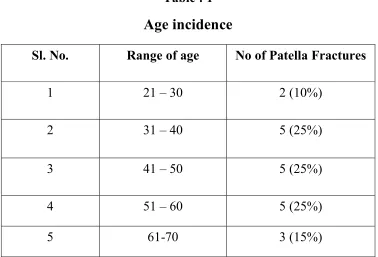
The present study consisted of 20 cases of fractures of Patella treated by modified tension band wiring Coimbatore medical college and hospital during the period 2012-2015. The following observations were made in the present study.
[image:80.612.135.513.266.523.2]Table : 1
Age incidence
Sl. No. Range of age No of Patella Fractures
1 21 – 30 2 (10%)
2 31 – 40 5 (25%)
3 41 – 50 5 (25%)
4 51 – 60 5 (25%)
0 0.5 1 1.5 2 2.5 3 3.5 4 4.5 5
21 – 30 31 –
77
40 41 – 50 51 – 60 61-70
Age Group
No of Patella Fractures
Total No of Patella Fractures 20 0 2 4 6 8 10 12 14 16 18 20
Total No of Patella Fractures
78
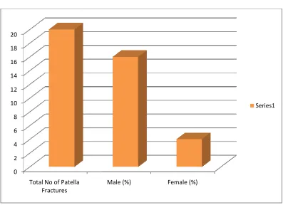
[image:82.612.128.525.306.594.2]Table : 2 Sex Incidence Total No of Patella
Fractures Male (%) Female (%)
16 (80%) 4 (20%)
Total No of Patella Male (%) Female (%)
Female (%)
4 (20%)
Total No of Patella Fractures 20 0 2 4 6 8 10 12 14 16 18 20
Total No of Patella Fractures
79
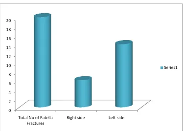
[image:83.612.137.511.290.558.2]Table : 3
Side Incidence Total No of Patella
Fractures
Right side (%) Left side (%)
1 6 (30%) 14 (70%)
Total No of Patella Right side Left side
Left side (%)
14 (70%)
Total No of Patella Fractures 20 0 2 4 6 8 10 12 14 16 18 20
Total No of Patella Fractures
80
[image:84.612.130.521.136.556.2]Table : 4
Mechanism of Injury Total No of Patella
Fractures
Direct trauma (%)
Indirect trauma (%)
8 (40%) 12 (60%)
Total No of Patella Direct trauma Indirect trauma
Indirect trauma (%)
12 (60%)
81
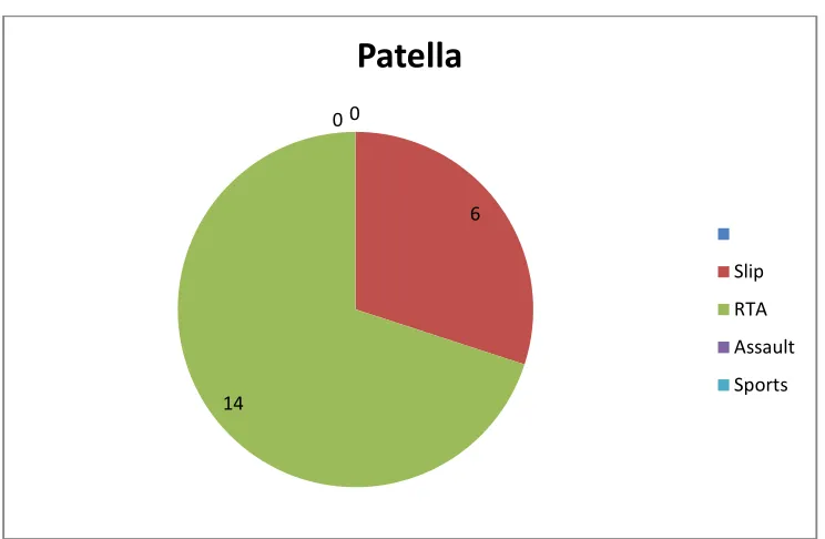
[image:85.612.138.510.365.608.2]Table : 5
Mode of injury
Sl. Mode of injury Patella
1 Slip 6
2 RTA 14
3 Assault -
4 Sports -
6
14
0 0
Patella
Slip
RTA
Assault
82
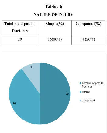
[image:86.612.147.499.82.516.2]Table : 6
NATURE OF INJURY Total no of patella
fractures
Simple(%) Compound(%)
20 16(80%) 4 (20%)
20
16
4
Total no of patella fractures
Simple
83
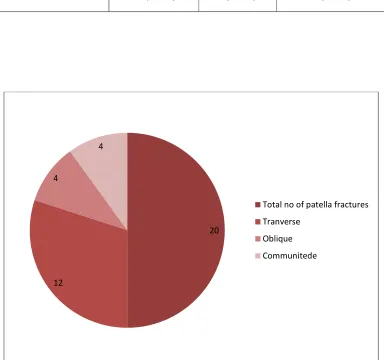
Table : 7
TYPE OF FRACTURE Total no of patella
fractures
Tranverse(%) Oblique(%) Communitede(%)
20 12(60%) 4(20%) 4(20%)
20
12 4
4
Total no of patella fractures
Tranverse
Oblique
TIME INTERVAL BETWEEN INJUERY AND SURGERY SLNO 1 2 3 0 2 4 6 8 10 12 14 16
within 48 hours
NO OF PATELLA OF FRACTURES
[image:88.612.130.519.75.586.2]84
Table : 8
TIME INTERVAL BETWEEN INJUERY AND SURGERY
DURATION NO OF PATELLA OF
FRACTURES
within 48 hours 2(10%)
2-10 days 16 (80%)
11-20 days 2 (10%)
2-10 days 11-20 days
NO OF PATELLA OF FRACTURES
NO OF PATELLA OF FRACTURES
TIME INTERVAL BETWEEN INJUERY AND SURGERY NO OF PATELLA OF
FRACTURES
2(10%) 16 (80%)
2 (10%)
0 2 4 6 8 10 12 14 16 18 20 No. of Patients followed Persistent Pain 4 Weeks Total No of Patella Fractures 4 Weeks No. of Patients followed Persistent
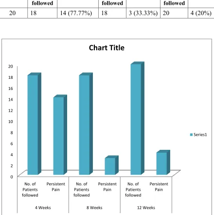
20 18 14 (77.77%)
85 No. of Patients followed Persistent Pain No. of Patients followed Persistent Pain
8 Weeks 12 Weeks
Chart Title
Table : 9
4 Weeks 8 Weeks
Persistent Pain No. of Patients followed Persistent Pain No. of Patients followed
14 (77.77%) 18 3 (33.33%) 20
Series1 12 Weeks No. of Patients followed Persisten t Pain
86
Pain and tenderness declined with progress of postoperative period but persisted in 26% of cases after 12 weeks. In most of patients pain persisted for 4 weeks, which decreased after 8 weeks.
87
Table : 10
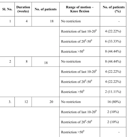
Movements - Patella
Sl. No. Duration
(weeks) No. of patients
Range of motion – Knee flexion
No. of patients (%)
1 4 18 No restriction -
Restriction of last 10-200 4 (22.22%) Restriction of 200-500 6 (33.33%) Restriction >500 8 (44.44%)
2 8 18 No restriction 8 (44.44%)
Restriction of last 10-200 4 (22.22%) Restriction of 200-500 4 (22.22%) Restriction >500 2 (11.11%)
3. 12 20 No restriction 16 (80%)
Restriction of last 10-200 2 (10%) Restriction of 200-500 2 (10%)
Restriction >500 -
Total No of
Patella Fractures
20
In our study 80% of radiological union of fracture was seen by 8 weeks
0 2 4 6 8 10 12 14 16 18 20
Total No of Patella Fractures
4 Weeks
[image:92.612.138.510.348.633.2]88
Table : 11
Radiological Union
4 Weeks 8 Weeks 12 Weeks
2 (10%) 16 (80%) 2 (10%)
In our study 80% of radiological union of fracture was seen by 8 weeks
4 Weeks 8 Weeks 12 Weeks 16 Weeks
16 Weeks
-
In our study 80% of radiological union of fracture was seen by 8 weeks
Sl. No.
1 Joint stiffness 2 Superficial infection 3 Deep infection 4. Osteoporosis 5. Migration of
One patient developed superficial infection of prepatellar bursa. Joint stiffness was seen in 1 patient of patellar fracture
[image:93.612.143.504.342.619.2]after physiotherapy. 0 0.2 0.4 0.6 0.81 1.2 1.4 1.6 1.82 89
Table : 12
Complications
Complications Patella
Joint stiffness 2 (10%)
Superficial infection 2 (10%)
infection Osteoporosis Migration of K-wire
One patient developed superficial infection of prepatellar bursa. Joint stiffness was seen in 1 patient of patellar fracture which recovered
Patella
Patella 2 (10%) 2 (10%) - - -One patient developed superficial infection of prepatellar bursa. which recovered
Total No of Patella Fractures
20
Results were done according to criteria outlined in the methodology. In our study, best results were obtained in fracture of patella 70%. 0 2 4 6 8 10 12 14 16 18 20
Total No of Patella Fractures
Excellent
[image:94.612.124.528.143.503.2]90
Table : 13
Evaluation of Results
Excellent Good Fair
14 (70%) 4 (20%) 2 (10%)
Results were done according to criteria outlined in the methodology. In our study, best results were obtained in fracture of
Excellent Good Fair Pour
Poor
-
Results were done according to criteria outlined in the methodology. In our study, best results were obtained in fracture of
91
DISCUSSION
The aim of treatment of fracture is not only achieving union but also to preserve the optimum function of adjacent joints. In intraarticular fractures of patella, it is important to maintain perfect anatomical reduction of fragments to obtain articular congruity by rigid fixation. The treatment of choice for these fractures by AO group is modified tension band wiring.
Modified tension band wiring was made use in our 20 cases. It has given favourable results in our experience. The findings, end results and other data will be analyzed and compared in the following discussion.
Age
[image:95.612.203.443.424.714.2]The average age in present series was found to be 40.1 years which is in accordance with series mentioned below:
Table 20 Age Incidence
Sl. No. Series Average age
1. Maini and Kochar 35.7 years
2. Bostman 42.0 years
3. Gary Wolfgang 40.5 years 4. Daniel F. Murphy 43.3 years
5. Levack 49.0 years
92
Sex
[image:96.612.154.495.190.384.2]In present study, fracture incidences are more in males (80%)and females (20%).Similar sex incidence was found in study made by Pandit and Shah, Maini and Kochar. This indicates males are more exposed to trauma.
Table 14
Sex Incidence
Series Fracture Male (%) Female (%)
Srinivasulu Patella 81.81 18.19
Maini and Kochar Patella 75 25
93
Side
In present study left side(70%) is involved more than right
[image:97.612.128.522.233.364.2]side(30%).Other series (Maini and Kochar, Gary Wolfgang) have reported higher incidence of fracture on left side. It may be due to the fact that the left non-dominant side makes vulnerable for trauma.
Table 15
Side Incidence
Series Fracture Right (%) Left (%)
Maini and Kochar Patella 55 45
94
Mechanism of injury
[image:98.612.223.475.230.418.2]Indirect trauma was more common in our study ( 6 0 % ) , the common mode of injury being road traffic accident..
Table 16
Mechanism of injury
Series Direct (%) Indirect (%)
Srinivasulu 50 50
Maini and Kochar 65 35
Pandit, Shah 20 80
Gary Wolfgang 7 93
Present study 40 60
Nature of injury
95
Table 17
Nature of injury
Series Simple (%) Compound (%)
Srinivasulu - -
Maini and Kochar 92 8
Pandit, Shah 80 20
Gary Wolfgang 68 32