ASSOCIATION BETWEEN SERUM TESTOSTERONE
CONCENTRATION AND CORONARY HEART DISEASE
Dissertation
Submitted in partial fulfillment of the regulations of
M.D. DEGREE EXAMINATION
BRANCH I GENERAL MEDICINE
Department of General Medicine
GOVT. STANLEY MEDICAL COLLEGE AND HOSPITAL
CHENNAI - 600001
THE TAMILNADU DR.M.G.R MEDICAL UNIVERSITY
CERTIFICATE
This is to certify that this dissertation titled
“
ASSOCIATION BETWEEN SERUM TESTOSTERONE
CONCENTRATION AND CORONARY HEART DISEASE”
is the bonafide work done by Dr. Sathesh Kumar. K.,Post Graduate student (2011 – 2014) in the Department of General Medicine, Government Stanley Medical College and Hospital, Chennai under my direct guidance and supervision, in partial fulfillment of the regulations of The Tamil Nadu Dr. M.G.R Medical University, Chennai for the award of M.D., Degree (General Medicine) Branch - I, Examination to be held in April 2014.
Prof.Dr.G.VASUMATHI, M.D., Prof.Dr.P.VIJAYARAGAVAN, M.D.,
Professor of Medicine, Professor and Head of Medicine, Dept. of General Medicine, Dept. of General Medicine,
Stanley Medical College, Stanley Medical College, Chennai-600001. Chennai-600001.
PROF. S. GEETHA LAKSHMI, M.D., PhD, The Dean,
DECLARATION
I, DR. K. SATHESH KUMAR solemnly declare that this
dissertation titled “
ASSOCIATION BETWEEN SERUM
TESTOSTERONE CONCENTRATION AND CORONARY
HEART DISEASE
” is a bonafide work done by me in the Department of General Surgery, Government Stanley Medical College and Hospital, Chennai under the guidance and supervision of my unit chief.Prof.Dr.G.VASUMATHI, M.D.,
Professor of Medicine
ACKNOWLEDGEMENT
I am grateful to Prof. Dr. S. Geethalakshmi, Dean, Govt. Stanley Medical College for permitting me to conduct the study and use the resources of the College.
My sincere thanks toProf. Dr.P.Vijayaragavan, M.D., Professor and HOD, Department of General Surgery, for his valuable guidance throughout the study.
I am highly indebted to my guide Prof.Dr.G.Vasumathi, M.D., Professor of Surgery for her constant help, inspiration and valuable advice in preparing this dissertation.
I express my deepest sense of thankfulness to my Assistant Professors Dr.Ramalingam,M.D., Dr.Sujatha,M.D.,,
Dr. Geetha,M.D., Dr. Raja Kumar,M.D., for their valuable inputs and
constant encouragement without which this dissertation could not have been completed.
I am particularly thankful to my fellow Super Specialty Post Graduate Dr. Azimudin Haja for his valuable support in the time of need throughout this study.
It is my earnest duty to thank my wife Mrs.S.Pavithra without whom accomplishing this task would have been impossible.
I am extremely thankful to my patients who consented and participated to make this study possible.
Overall it is the blessing of my father Mr.N.Kanagasabapathy
TABLE OF CONTENTS
INTRODUCTION 1
AIM & OBJECTIVES 3
REVIEW OF LITERATURE 4
SYNTHESIS OF TESTOSTERONE 6
TRANSPORT AND METABOLISM 12
TESTOSTERONE AND ATHEROSCLEROSIS 15
TESTOSTERONE AND CORONARY HEART DISEASE 18
CORONARY HEART DISEASE 23
MATERIALS AND METHODOLOGY 40
OBSERVATION AND RESEARCH 43
CONCLUSION 74
BIBLIOGRAPHY 75
ANNEXURES
ETHICAL COMMITTEE
PROFORMA
INFORMATION FORM
TURNTIN SCREEN SHOT
INTRODUCTION
Acute coronary syndrome includes unstable angina and non-ST elevated myocardial infarction and is defined as a spectrum of disease characterized by either:
1. New onset angina. 2. Angina at rest
3. Progression of angina of increasing frequency or severity. 4. Angina in response to lower levels of exertion.
5. STEMI
Acute coronary syndrome most often represents acute atherosclerotic plaque rupture with exposure of thrombogenic sub-endothelial matrix.
Coronary atheroma occurs as a result of inflammatory process. Cellular inflammation and local inflammation in the arterial wall occurs as a result of cytokines, which can further progress to cause vascular smooth muscle apoptosis, degradation of the fibrin cap and plaque rupture.
An anti-inflammatory effect of normal physiological levels of sex hormones may therefore, be important in atheroma protection. Cytokines and C-reactive proteins are found to be elevated in coronary heart disease patients.
Low serum testosterone levels is found in patients with coronary heart disease. Two most important activities in the pathogenesis of atheroma formation are cytokine activation and vascular smooth muscle cell apoptosis.
Main action of testosterone hormone is to deactivate cytokines as a result of which smooth muscle apoptosis is inhibited and leads to smooth muscle proliferation. So, as a consequence of this smooth muscle proliferation action by this hormone, it helps in maintaining the fibrous cap of the atherosclerotic plaque.
Testosterone hormone has calcium channel antagonism which results in coronary artery vasodilation. Various studies have proved the beneficial effect of testosterone hormone in coronary heart disease patients.
AIMS AND OBJECTIVES
To estimate the level of free testosterone level in male patient suffering acute coronary syndrome.
To correlate free testosterone level with mortality and morbidity in male patients presenting with acute coronary syndrome.
REVIEW OF LITERATURE
Atherosclerotic coronary artery disease (CAD) is a leading cause of mortality and morbidity all over the world. Men are more than twice as likely to develop CAD. Incidence of coronary heart disease in females when compared to male is low before menopause but it is of same incidence after menopause.
TESTOSTERONE
Leydig cells of testis produces testosterone which is synthesized from cholesterol and also from androstenidione secreted by the adrenal cortex. Leydig cells which is present in testis is controlled by LH for the secretion of hormone testosterone. Normal level of testosterone in adult – 4 to9ng/day secretion. Testosterone bound to protein is 98% and only 2% is free. Out of that 985 bound form65% is combined with beta globulin called gonadal steroid binding globulin and 33% bound to albumin.
Formula: C19H2802
SYNTHESIS
SYNTHESIS OF TESTOSTERONE
LH + 7-transmembrane, G protein–coupled receptor
LH receptor stimulated
Steroid acute regulatory protein produced ( rate limiting step)
Helps delivery of cholesterol to inner mitochondrial membrane
Formation of pregnenolone by CYP11A1
Testosterone produced
Testosterone has inverse relationship with visceral fat. Androgen receptors are more in number in visceral fat, as a result, it inhibits the action of lipoprotein lipase and fatty acid/triglyceride uptake. Thus limits the fat accumulation.
TESTOSTERONE AND CORONARY ARTERY DISEASE (CAD)
Serum testosterone has negative correlation with the extent of coronary heart disease which was proved by performing coronary angiogram. Patients who had angina and exercise induced ST segment depression have been benefited from testosterone therapy.
High androgen levels are presumed by many to explain the male predisposition to coronary artery disease. However, natural androgens inhibit male atherosclerosis.
As age advances, serum testosterone levels decrease with concurrent gonadotropin release with increased FSH and LH.
The ratio of estradiol to testosterone is raised in elderly male, which suggests that hyperestrogenemia may lead to M.I in men.
The ratio of male to female having CAD in old age group after menopause in female is 2:1. Premenopausal women are protected from endogenous estrogens while postmenopausal women are treated with exogenous estrogens. Replacement of natural analogues inhibit atheroma formation in men.
Patients who are having adverse lipid profile, raised blood pressure, obesity, insulin resistance and raised fibrinogen are found to have low testosterone levels.
Mortality in men is twice that of women who have protective effects of endogenous and exogenous estrogen. The abuse of anabolic steroid which resulted in sudden cardiac death leads the suggestion that it has delirious effects, but the persons who are actually having high testosterone levels have beneficial effect on cardiovascular system.
Studies have proved that intracoronary administration of testosterone have shown to increase the coronary blood flow.
males of same age group, who are having CAD have low testosterone levels when compared to men of same age group having normal coronary angiogram study.
Testosterone therapy delays the coronary event by their coronary vasodilator property.
Major function of testosterone hormone is to preserve sexual function, muscle tone, bone mineral density and post pubertal mood. Its level peaks during morning hours and also in spring, showing circadian and circannual rhythm. Testosterone is 68% bound to sex hormone binding globulin (SHBG),and 30% to albumin and remaining 2% is free form, which is biologically active.
Serum testosterone level was found to be low in established CAD patient compared to men of same age group having normal coronary angiogram.
Endogenous testosterone has direct relationship or correlation with HDL level but has indirect or negative correlation with total cholesterol, LDL and triglycerides.
Thus hypogonadal men have atherogenic dyslipidaemia whereas normal men with low testosterone have adverse lipid profile.
Various studies shown that testosterone treatment leads to symptomatic improvement in men with angina. The acute and chronic anti-ischemic properties of testosterone mediated by coronary artery vasodilatation, which appears to involve calcium channel antagonism in a gender- specific fashion.
The androgens possess immune-modulating properties and they suppress the activity of pro-inflammatory cytokines while enhancing that of anti-inflammatory factors. Thus hypogonadal men were found to have a greater degree of inflammatory activation compared with healthy controls, including higher serum cytokine levels. Androgen therapy in these patients led to a reduction in circulating cytokines.
CORONARY ARTERY DISEASE ; ACUTE CORONARY SYNDROME
Coronary artery disease (CAD) remains the number one cause of death in developed countries, however, the largest projected increases in disease rates are in developing countries. Angina pectoris is the principal manifestation of coronary atherosclerosis. Patients with stable angina are at increased risk of atherosclerotic plaque instability, the development of severe myocardial ischemia and the development of acute events such as myocardial infarction (MI). In line with the changing patterns of acute events, older descriptions such as sub endocardial and Q-wave MI have now been replaced with the term acute coronary syndrome (ACS). ACS include unstable angina (UA), non-ST segment elevation myocardial infarction (NSTEMI) and ST-segment elevation myocardial infarction (STEMI).
Epidemiology
ACS patients are divided into those without and those with ST-segment elevation. The ACS without ST elevation include both UA and NSTEMI. UA is further defined as ischemic pain at rest without elevation of biomarkers, whereas NSTEMI requires increased serum biomarkers. STEMI has both ST-segment and biomarker elevation. Using these criteria. MI (either NSTEMI or STEMI) is defined as myocardial cell death due to prolonged ischemia irrespective of electrocardiogram (ECG) changes. Myocardial necrosis can be confirmed by the appearance of different proteins (biomarkers) released into the circulation by damaged myocytes. These include myoglobin, cardiac troponins T (TnT) and I (TnI), creatine kinase (CK-MB) and total creatine kinase (CK).
Acute Coronary Syndrome
No ST Elevation ST Elevation
Non ST Elevation MI
Unstable Angina Myocardial Infarction
Pathophysiology
UA, NSTEMI and STEMI are closely related conditions with similar pathologies and clinical presentation, but with differing severity. They differ primarily in whether the ischemia is severe enough to cause sufficient myocardial damage to release detectable levels of indicators of myocardial injury, such as troponins.
The pathophysiology of atherosclerosis and ACS is well characterized. The earliest visible atherosclerotic lesion appears to be the fatty streak. Beginning in early life, these fatty streaks, or Type II lesions, are slightly raised above the intimal surface and predominantly contain foam cells derived from tissue macrophages and T lymphocytes.
Fatty steaks may progress to become mature plaques. Platelets adhere to the initial lesion and secrete growth factors including platelet-derived growth factor (PDGF) that initiate smooth muscle cell migration from the media to the intima. This is turn stimulates the production of collagen, elastin and glycoproteins, which provide the connective tissue matrix of the plaque and give it structural strength.
Plaque rupture
The rupture of an atherosclerotic plaque can initiate a range of complications from UA and acute MI through to sudden cardiac death.
Acute coronary occlusion usually occurs through one of two distinct processes. First, the endothelium covering the atherosclerotic plaque can become denuded (endothelial erosion) with exposure of the prothrombotic sub endothelium to the circulating blood. Second, plaque disruption may occur with the tearing open of the fibrous cap (plaque rupture) to expose the highly thrombogenic lipid core.
The type of thrombus (platelet plug or platelet-fibrin thrombus) and the extent and duration of obstruction to coronary blood flow determine the effect on the myocardium with the development of UA, NSTEMI, or STEMI.
Plaque rupture depends more on the type of plaque than its size. Major determinants of plaque vulnerability include both local and systemic factors. Local factors include size and consistency of the atheromatous core, thickness of the fibrous cap, ongoing inflammation and repair with the cap, eccentric, plaque shape, and shear forces.
with attendant increased adrenergic tone, shear forces, and generalized inflammation. The milieu of the plaque is also fundamental to its vulnerability.
Inflammation
Atherosclerosis is clearly an inflammatory disease and does not result simply from the accumulation of lipids. Low grade coronary inflammation is widespread throughout the coronary tree.
CRP, an acute phase reactant produced in the liver in response to interleukin-6 (I1-6), is both a marker and mediator of vascular inflammation as well as being a predictor of coronary events in patents with stable and unstable angina.
The most useful way of CRP testing in the emergency room setting will be among those with chest pain syndromes who have negative troponin levels.
Platelets and the coagulation system
Diagnosis of ACS
All chest pain patients should have a resting 12-lead ECG. Patients with chest pain can be categorized into those with
- Non-cardiac chest pain - Stable angina
- UA
- NSTEMI or STEMI
Diagnosis is initially based on: 1. Clinical signs and symptoms 2. Resting 12-lead ECG
3. Cardiac enzymes/biomarkers.
Clinical history is important for both the diagnosis and risk stratification of patients with ACS. Age, traditional CAD risk factors, use of aspirin, and frequency of rest chest pain episodes are independent risks for adverse outcome.
Electrocardiography
UA and NSTEMI the ST segments are normal, even during pain, with development of T wave inversion a few hours later. ECG findings further determine risk in ACS. In-hospital death has been reported as 8.7% with non-specific ECG changes and 11.5% with diagnostic results. Even a normal ECG carries a 5.7% 7-day mortality.
New or reversible ST-segment deviation of 0.5 mm from baseline or left bundle branch block on the admission ECG is associated with an increase in incidence of death or MI at 1 year, i.e. 15.8% versus 8.2% in patients without ECG changes. Reversible ST-segment depression is associated with a 3 to 6 fold increase in death, MI, ischemia at rest, or provocable ischemia on exercise. The ECG result and the CK level on admission can identify a difference in mortality (in the group with ST-segment elevation plus depression and elevated CK level.
Cardiac enzymes and biomarkers
Standard blood testing for the patient with suspected ACS includes TnI or TnT, total CK and CK-MB fraction. Elevated serum MB is an early marker of MI with detection as early as an hour after onset of myocardial injury.
Cardiac isoforms of TnT and TnI are more specific and sensitive indicators of myocardial damage and can detect the micro-infarctions typified by NSTEMI.
TIMI Risk Score
- Age > 65
- > 3 traditional risk factors for CAD (male sex, hyeprlipidemia, hypertension, smoking, diabetes mellitus, family history of premature CAD).
- Prior coronary stenosis > 50%.
- ST segment elevation or depression at presentation - > 2 anginal events in the prior 24 hours
- Aspirin use in the prior 7 days - Elevated serum cardiac biomarkers.
TIMI risk score can be used to define high risk (> 5 points), intermediate risk (3-4 points), and low risk (0-2 points). The TIMI risk score is a validated way to assess risk of cardiac events.
Inflammatory and other prognostic markers
Exercise testing
Testing should be performed in most cases within 72 hours of presentation in low or intermediate risk individuals who are free of active ischemia or heart failure symptoms for a minimum of 8-12 hours, and in intermediate risk patients after 2-3 days.
DANAMI-2 describes the prognostic importance of a pre-discharge
maximal exercise test following acute MI in the era of aggressive reperfusion treatment – thrombolysis and percutaneous coronary intervention (PCI).
Echocardiography
Resting and stress echo play role in the general assessment of CAD as ischemia results in immediate changes that can be detected by echo. These include:
Abnormalities of wall motion – hypokinetic, akinetic, dyskinetic :
Abnormalities of wall thickening – reduced or absent systolic thickening or systolic thinning.
Coronary angiography
Coronary angiography is regarded as the “gold standard” to define coronary anatomy. Coronary angiography should be performed in patients where the diagnosis cannot be reliably made using non-invasive testing. Patients with high-risk clinical markers, who tend to be older, have multiple high-risk factors and have previous MI, should undergo early coronary angiography. High-risk UA patients who are refractory to medical therapy should be referred urgently for angiography.
Management of UA/NSTEMI
General treatment guidelines
The use of combination evidence-based medical treatments including antiplatelet agents, beta-blockers, statins and ACE inhibitors has been shown to be independently and strongly associated with lower 6- month mortality in patients with ACS.
Anti-ischemic therapies
Nitrates: Their main role is to provide relief of pain. They act by arterial
Nicorandil: It is a compound with potassium-channel opener and a nitrate moiety. The IONA study showed that nicorandil 20 mg twice daily, in addition to standard anti anginal therapy, improved outcomes in terms of reducing events related to acute coronary disease and the associated requirement for admission to hospital.
Beta-blockers
Patients with ACS with ST-segment elevation, who do not have contraindications, should be treated with beta-blockers as soon as possible to prevent acute MI/re-infarction. Similarly, oral beta-blockers are recommended for long-term use, indefinitely, in all patients who recover from an acute MI.
Beta-blockers reduce the odds of death in long-term trials by 23% and in short term trials by 4% following acute MI. These agents were associated with a 40% improvement in survival at 2 years, and suggested that the specific beta-blocker chosen will have little influence on mortality.
Calcium antagonists
The anti anginal effects of calcium antagonists appear to be mediated through a reduction of myocardial oxygen demand secondary to decreased afterload and myocardial contractility.
Oral antiplatelet therapies
Aspirin
Acute treatment with aspirin is recommended in all patients with suspected ACS in the absence of contraindications. Aspirin should be administered as soon as possible after patient presentation of suspected ACS and continued indefinitely.
Analysis of aspirin dose in the CURE study showed that medium-dose (101-199 mg/day) and high-dose (> 200 mg/day) aspirin had no advantage over low-dose (< 100 mg/day) aspirin whether patients received combination therapy with clopidogrel or aspirin alone.
Clopidogrel
The CURE trial compared the benefit of aspirin plus clopidogrel (300 mg loading dose, then 75 mg/day) treatment versus aspirin alone in 12,652 patients with UA or NSTEMI treated for 3-12 months.
Clopidogrel was of benefit in the CURE trial irrespective of whether high-risk features were present.
Intravenous antiplatelet therapies
GP IIb/IIIa receptor inhibitors
Antithrombin therapy
Unfractionated heparin
UFH reduces thrombin generation and Xa activity. This underlies the rationale for its use in ACS.
Clinical experience
In UA, i/v UFH significantly reduced the risk of MI and recurrent refractory angina by 89% and 63% respectively.
Recommendations and dosing
A weight-based UFH regimen is preferable, with a bolus of 60 to 70 U/kg (maximum 5000 U) followed by an infusion of 12 to 15 U/kg/hour (maximum 1000 U) for 2 to 5 days.
Low molecular weight heparins
Enoxaparin is accepted as an anticoagulant in UA/NSTEMI.
Direct thrombin inhibitors
Hirudin can be used as an alternative to heparin among patients with HIT in UA/NSTEMI.
PCI ad CABG
Management of STEMI
Medical therapy
Nitrates
Oxygen
Analgesics
Morphine sulfate is the preferred analgesic agent to treat ongoing chest pain.
Antiplatelet agents
Patients should have immediate administration of non-enteric aspirin (162 mg or greater). Clopidogrel can be considered as an alternative antiplatelet agent for aspirin-sensitive patients. A 600 mg loading dose of clopidogrel may be superior. Clopidogrel enhances reperfusion with thrombolysis. Dual antiplatelet therapy reduced the composite endpoint of coronary artery occlusion, death or recurrent MI from 21.7% to 15.0%.
Infarct size limitation
- Beta-blockers
- Renin-angiotensin-aldosterone inhibition
Patients with a large anterior MI or left ventricular failure should be considered for early treatment (within first 24 hours) with an oral ACE inhibitor.
- Statin
Reperfusion options for treatment of STEMI Reperfusion: fibrinolytic agents
Fibrinolytic drug administration
Drug Dosing Features
Streptokinase 1.5 million units over 30-60 minutes
Lower reperfusionr ate than fibrin-specific agents, lowest cost
Alteplase 15 mg bolus
- then 0.75 mg/kg over 30 minutes (maximum 50 mg) - then 0.50 mg/kg over 60 minutes (maximum 35 mg)
Lower mortality than streptokinase
Short half-life requires infusion adjustment
Tenecteplase Weight-based single bolus over 10 seconds
< 60 kg = 30 mg 60-69 kg = 35 mg 70-79 kg = 40 mg 80-89 kg = 45 mg > 90kg = 50 mg
Efficacy similar to alteplase with lower bleeding
complications when weight adjusted.
Reteplase 10 units over 2 minutes - then repeat 10 units at 30 minutes
Adjunctive therapies
Antiplatelet therapy
Heparin therapy
GP IIb/IIIa inhibitors
Elective PCI after fibrinolysis
Primary PCI
Newer agents like Ranolazine
Prognosis
MATERIALS AND METHODS
Source:
For the study, male subjects presenting with acute coronary syndrome and those patients who have recovered from a recent acute coronary event attending Medicine OPD were taken as cases. Age & sex matched healthy control were taken from the medical ward. Study was done between October 2012 and November 2013.
Number of cases - 50 Number of controls – 25
Inclusion criteria:
All male patients presenting with acute coronary syndrome between the age group of 35-74 years.
Exclusion criteria:
Patient with liver disease
Patient with renal parenchymal disease Patient with diabetes mellitus
(a) History of angina:
Onset, situation, radiation, duration, aggravating and relieving factors were studied.
(b) ECG changes:
Resting ECG were obtained in all patients.
ECG studied for ST deviation, new onset left bundle branch block and significant Q waves.
(c) Method of serum free testosterone study:
50 male subjects with defined acute coronary syndrome were studied and serum free testosterone level were measured. Level of serum free testosterone was estimated by chemiluminescence Radio immuno-assay method.
Method of study
Informed consent were obtained from all patients.
Biochemical parameter included fasting and random blood sugar, blood urea, serum creatinine and lipid profile.
History of hypertension, diabetes mellitus, chronic kidney disease and dyslipidemia were noted.
Presence of hypertension defined as per the Joint National Committee (JNC) 7 criteria.
Presence of diabetes mellitus defined by American Diabetes Association Criteria.
Presence of chronic kidney disease defined by National Kidney Foundation Criteria.
Dyslipidemia as per ATP III guidelines.
BMI calculated as per latest WHO guidelines.
Statistical analysis:
In the present study, values are expressed as mean±2SD. Variables are compared by 't' test for 2 sample mean. Attributes are compared by odds ratio and standard error of difference between two proportions by Chi-square test.
OBSERVATION
[image:42.612.87.527.305.493.2]In this study, 50 patients who presented with acute coronary syndrome were studied, along with 25 age and sex matched controls, who did not have any evidence of coronary artery disease.
Table - 1
Distribution of patients according to age
Age Cases Controls
35-44 12 10
45-54 14 11
55-64 15 4
65-74 9 0
Total 50 25
Table - 2
Incidence of Different symptoms (n=50)
Symptoms No. of cases Percentage
Chest Pain 45 90%
Breathlessness 4 8%
Palpitation 28 56%
Syncope 3 6%
Table - 3
Distribution of risk factor in cases (n=50) & controls (n=25)
Risk Factor Cases Controls
Hypertension 17 7
Dyslipidemia 20 4
Smoking 23 5
Alcohol 2 2
Family History 3 2
(a) Dysplipidemia is defined by any or all of the below: T. cholesterol > 240 mg/dl
HDL-C < 40 or > 60 mg/dl LDL-C > 130 mg/dl
Triglycerides > 160 mg/dl
Dyslipidemia was seen in 20 cases and 4 control patients.
(b) In case group (n=50), 17 patients had history of hypertension (34%) whereas in control group 7 patients had hypertension (28%)
(c) In case group, 23 (46%) were chronic smokers, and among controls groups, 5(20%) were chronic smokers.
Table - 4
BMI in Cases & controls
BMI Cases Controls
18.5-24.9 37 25
25-29.9 13 0
>30 0 0
Table - 5
Mean BMI in cases and controls
Cases Controls p value
23.40±2.35 20.64±1.52 < 0.05
(p value = 0.000001)
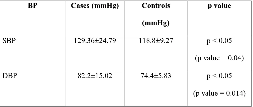
Table - 6
Mean BP in cases and controls
BP Cases (mmHg) Controls
(mmHg)
p value
SBP 129.36±24.79 118.8±9.27 p < 0.05
(p value = 0.04)
DBP 82.2±15.02 74.4±5.83 p < 0.05
(p value = 0.014)
Table - 7
Total cholesterol in case and controls
Total cholesterol (mg/dl) Cases Controls
< 200 19 20
201-239 8 1
> 240 23 4
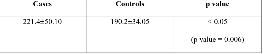
Table - 8
Mean total cholesterol (mg/dl) in cases and controls
Cases Controls p value
221.4±50.10 190.2±34.05 < 0.05
(p value = 0.006)
Table - 9
HDL-C level in cases and controls
HDL-C (mg/dl) Cases Controls
< 40 18 4
40-60 32 21
> 60 0 0
Table - 10
Mean HDL-C (mg/dl) in cases and controls
Cases Controls p value
40.34±9.91 45.16±5.96 < 0.05
(p value = 0.02)
Table - 11
LDL-cholesterol level in cases and controls
HDL-C (mg/dl) Cases Controls
< 100 7 19
100-129 26 6
130-159 12 0
160-189 5 0
> 190 0 0
Table - 12
Mean LDL-C (mg/dl) in cases sand control
Cases Controls p value
124.14±26.0 74.94±26.39 < 0.05
(p value = 0.0001)
Table - 13
TG level in cases and controls
HDL-C (mg/dl) Cases Controls
< 160 42 21
> 160 8 4
In the case group 42 (84%) had TG levels in normal range and 8 (16%) above normal.
Table - 14
Mean TG (mg/dl) level in cases and controls
Cases Controls p value
136.9±34.8 101.2±33.07 < 0.05
(p value = 0.00006)
Table - 15
Serum free testosterone level and coronary artery disease (ACS) cross tabulation
S. free testosterone Cases (ACS) Controls (without ACS)
< 9 ng/dl 40 1
> 9 ng/dl 10 24
Yates corrected Chi-square test : 35.84 Risk ratio (95% CI) : 3.32
Odds ratio (75% CI) : 96.00 p value = 0.000001 significant
In the present study group, the normal level of serum free testosterone was 9-30 ng/dl.
Table - 16
Mean serum free testosterone level (ng/dl) in cases and controls
Cases Controls p value
8.36±1.80 12.94±3.06 < 0.05
(p value = 0.000001)
Table - 17
Mean serum free testosterone level (ng/dl) cross tabulation according to
number of cardiovascular risk factors in cases (n=50)
Cases with 0-2
risk factors
Cases with > 3 risk
factors
p value
Mean serum free testosterone
8.02±1.23 8.58±2.08 0.33
Table - 18
Mean serum free testosterone (ng/dl) in hypertensive cases and hypertensive
controls
Cases Controls p value
8.38±1.56 11.27±1.85 < 0.05
(p value = 0. 0001)
Table - 19
Mean serum free testosterone level (ng/dl) in smokers (cases and controls)
Cases Controls p value
8.72±1.96 11.1±2.21 < 0.05
(p value = 0. 00001)
Table No. 20
Correlation of mean serum free testosterone level with pattern of ACS
ACS Cases Mean serum free
testosterone (ng/dl)
Percentage
AWMI 24 8.67±2.16 48%
IWMI 16 8.24±1.37 32%
UA 7 8.03±1.30 14%
AWMI+IWMI 3 7.31±1.83 6%
In the present study, most patients presented with AWMI (48%) followed by IWMI (32%).
The mean serum free testosterone level in AWMI and IWMI were 8.67±2.16 ng/dl and 8.24±1.37 ng/dl respectively.
Table No. 21
Correlation of mean serum free testosterone with mortality in acute coronary
syndrome
No. of cases Mean Serum free
testosterone (ng/dl)
Percentage
Mortality 2 10.1±4.6 4%
Survivors 48 8.28±1.66 96%
DISCUSSION
Coronary artery disease (CAD) is a major cause of mortality and morbidity in developed world, and in developing countries, the incidence is rising. Atherosclerosis is the major cause of CAD. CAD manifests as acute coronary syndrome (ACS) and stable angina.
Men are more than twice as likely as women to die from coronary heart disease, and this ratio is consistent in all population and is not related to differences in risk factors. Low testosterone level are associated with CAD and low serum testosterone is associated with increased aortic atheroma.
Furthermore, low testosterone levels are associated with several risk factors for the development of CAD including systolic and diastolic hypertension, adverse lipid profile, and high levels of fibrinogen and procoagulable factors.
The present study is undertaken in order to correlate the role of serum free testosterone in patients with ACS.
Age
Clinical presentation
In the present study, most common presenting symptoms was chest pain (90%) followed by palpitation (56%).
Hypertension
In the present study, 34% patients had hypertension while only 28% of controls had hypertension.
The mean systolic blood pressure (SBP) in cases was 129.36±24.79 mmHg and mean diastolic blood pressure (DBP) was 82.2±15.02 mmHg.
Various other studies have shown the following parameters:
In 1994, the mean SBP was 134±22 mmHg and mean DBP was 74±14 mmHg.
In 2000 study, hypertension was found in 40.5% of cases.
In 2003 study, hypertension was found in 21.5% of cases.
In 2007 study, hypertension was found in 43.4% of cases.
The mean SBP was comparable to that seen in 2000 study was 131±7 mmHg.
Smoking status
In the present study, 46% of cases and 20% of control were smokers. In 2003 study, the incidence of smoking was 19%.
In 2007 study, the incidence of smoking was 37.2%.
Smoking incidence was comparatively higher in our study group, and is in accordance with community trends.
Body Mass Index (BMI)
The mean BMI in the present study was 23.40±2.35 in cases and 20.64±1.52 in controls.
The mean BMI in 2007 study was 26.2±3.1 which is higher compared to our study. The mean BMI in 2003 study was 25.5±3 and comparable to our study. The mean BMI in 2000 study was 27.7±0.7.
In 2003 study, there was a negative correlation between free testosterone and body mass index.
Lipid profile
In the present study, the mean total cholesterol in cases was 221.4±50.10 mg/dl and in controls 190.2±34.05 mg/dl. The total cholesterol was higher in cases than controls and the difference was statistically significant.
In 2007 study, total cholesterol was 247±25 mg/dl.
In 2003 study, total cholesterol was 226.6±1.11 mg/dl and is comparable to our study.
The result of the present study and previous studies in accordance with cardiovascular risk attributed to total cholesterol.
HDL CHOLESTEROL:
The mean HDL cholesterol level in cases was 40.34±9.91 mg/dl and control was 45.16±5.96 mg/dl.
The mean HDL-C level in 2003 study was 51.81±0.33 mg/dl. The mean HDL-C level in 2007 study was 47.95±0.32 mg/dl.
The lower level of HDL-C in case and control in our study is in accordance to the prevalent lipid status of South Asian population.
LDL CHOLESTEROL:
In 2007 study, the mean LDL-C level in case was 156±23 mg/dl. In 2003 study, the mean LDL-C level was 150.42±0.98 mg/dl.
TRIGLYCERIDES:
The mean triglyceride level in the present case was 136.9±34.8 mg/dl. The mean TG level in 2000 study was 76.95±1.09 mg/dl.
In 2003 study, the mean TG level in cases was 52.97±0.75 mg/dl.
The mean TG level in the present study was comparatively higher.
Serum free testosterone (FT)
In the present study, serum free testosterone in patients with ACS was studied and compared with controls who had cardiovascular risk factors but no evidence of CAD.
The mean serum free testosterone in cases was 8.36±1.80 ng/dl and control was 12.94±3.06 ng/dl. The difference was statistically significant (p value < 0.001).
In 2003 study, men with coronary artery disease had significantly lower levels of free testosterone than did controls.
In 1994 study, it was found out that free testosterone correlated negatively with the degree of coronary artery disease.
In 2007 study, it was found out that the patients had significantly lower levels of testosterone than controls (9.8±6.5 mmol/l, p<0.01).
In 2000 study, it was concluded that men with coronary artery disease had significantly lower level of free testosterone 47.95±13.77 pmol (p value = 0.027).
In 2009 study, it was found that total and free testosterone levels of the patients with coronary artery disease were significantly lower than those of controls.
In the present study, we found that the mean serum free testosterone in patients with 0-2 cardiovascular risk factors was 8.02±1.23 ng/dl.
The difference between these 2 subgroups of ACS patients was not statistically significant which means that the level of serum free testosterone level decreases irrespective of the number of cardiovascular risk factors.
The inverse relationship between testosterone levels and coronary atherosclerosis found in the present study suggests a possible protective role of the hormone on the progression of atherosclerosis.
Serum free testosterone and hypertension
The mean serum free testosterone level in hypertensive cases was 8.38±1.56 ng/dl and hypertensive control was 11.5±1.85. The difference was statistically significant.
In 2005 study, it was found that testosterone values were significantly lower in hypertensive cases.
In 1994 study, it was demonstrated that testosterone is lower in populations of men with hypertension than in normal men.
Serum free testosterone and smoking
The mean serum free testosterone in smokers with ACS was 8.72±1.96 ng/dl and serum free testosterone of smokers in control group was 11.1±2.21 ng/dl. The difference was statistically significant.
The 1990 study, 1993 study and 2000 study reported that smoking is a strong risk factors for atherosclerosis.
Serum free testosterone and patterns of acute coronary syndrome
In the present study, most patients had AWMI (48%) and IWMI (32%). Mean serum free testosterone was highest in AWMI (8.67±2.16 ng/dl) and lowest in AWMI+IWMI (7.31±1.83 ng/dl).
Serum free testosterone and mortality in ACS
In the present study, most of the patients (96%) survived the illness while 2 patients died due to ACS.
SUMMARY
The present study included 50 patients who presented with acute coronary syndrome and 25 patients of age matched controls without evidence of CAD.
In the study group, the mean age of presentation was 53.3 yrs.
The commonest presenting symptom was chest pain followed by palpitation.
Hypertension present in 34% of cases and 28% of controls. Smoking incidence was 46% in cases and 20% in controls.
Higher levels of total cholesterol, triglyceride and LDL cholesterol and lower level of HDL cholesterol were seen in cases. They significantly differ in comparison to controls.
Of the cases studied, about 86% had STEMI and 14% had NSTEMI.
The mean serum free testosterone was significantly lower in patients with ACS.
Serum free testosterone level was also significantly lower in patients with hypertension and smokers.
Mean serum free testosterone level was lowest in patient who had IWMI+AWMI as compared with other patterns of ACS.
CONCLUSION
The serum free testosterone was found to be lower in patients with acute coronary syndrome as compared to controls. The difference between cases and controls was statistically significant.
The serum free testosterone level was lowest in patients who presented with AWMI+IWMI as compared with other patterns of ACS.
There was an inverse correlation found between serum free testosterone level and mortality and morbidity in patients with acute coronary syndrome.
BIBLIOGRAPHY
1. C.J. Malkin et al : Testosterone for secondary prevention in men with ischaemic heart disease? Q J Med 2003;96:521-529.
2. Shalender Basin et al : Disorders of the testes and male reproductive system. Harrison 17th Edition.
3. Majon Muller et al : Endogenous sex hormones and Cardiavascular disease in men.J Clin Endocrinol Metab. 88:5076-5086, 2003.
4. Chou TM et al : Testosterone induces dilatation of canine coronary conductance and resistance arteriesin vivo. Circulation 94:2614-2619. 5. Nathan L, Chaudhuri G et al 2001 : Testosterone inhibits early
atherogenesis by conversion to estradiol: critical role of aromatase. 6. Ramsay LE et al : Lancet 1996; 348:1251.
7. Oppenheim DS et al : Elevated serum lipids in hypogonadal men with and without hyperprolactinemia. Ann Intern Med 1989.
8. Pugh PJ et al : Acute haemodynamic effects of testosterone administration in men with heart failure. Euro Heart J 2002.
9. Maresca G et al : Measuring plasma fibrinogen to predict stroke and myocardial infarction- an update. Arterioscler Thromb Vasc Biol 1999. 10. Glueck CJ et al: Endogenous testosterone, fibrinolysis, and coronary heart
11. Phillips GB, Pinkernell BH, Jing TY. The association of hypotestosteronemia with coronary artery disease in men. Arterioscler
Thromb. 1994;14:701–706.
12. Hak AE et al . Low levels of androgens increase the risk of atherosclerosis in elderly men : the Rotterddam study. J Clin Endocrinol Metab 2002. 13. Hanke H, Lenz C, Hess B et al . Effect of Testosterone on plaque
development and and androgen receptor expression in the arterial wall. Circulation 2001.
14. Moreno PR, Falk E et al. Macrophage infiltration in acute coronary syndrome:implications for plaque rupture. Circulation 1994.
15. George SJ. Tissue inhibitors of metalloproteinases in atherosclerosis. Curr Opin Lipidol 1998.
16. Geng YJ et al. Apoptosis of vascular smooth muscle cells induced by in vitro stimulation with interferon gamma, tumour necrosis factor alpha and interleukin-1 Beta. Arterioscler Thromb Vasc Biol 1996.
17. Gornstein RA et al. Androgens modulate interleukin-6 production by gingival fibroblasts in vitro. J Periodontal 1999
18. Khosla S, Atkinson EJ et al. Effect of estrogen versus testosterone on circulating osteoprotegerin and other cytokine levels in normal elderly men. J Clin Endocrinol Metab 2002.
20. Rebuffe-Scrive M et al. Metoblism of adipose tissue in intra-abdominal depots of non-obese men and women. Metabolism 1989.
21. Marin P et al . Assimilation and mobilization of triglycerides in subcutaneous abdominal and femoral adipose tissue in vivo in men: effects of androgens. J Clin Endocrinol Metab 1995.
22. Schneider G, Kirshner A et al. Increased estrogen production in obese men. J Clin Endocrinol Metab 1979.
23. Snyder PJ et al. Effect of testosterone treatment on body composition and muscle strength in men over 65 years of age. J Clin Endocrinol Metab 1999.
24. Wang C et al. Transdermal testosterone gel improves sexual function, mood, muscle strength, and body composition parameters in hypogonadal men. J Clin Endocrinol Metab 2000.
25. Webb CM, Adamson DL, de Ziegler D, Collins P. Effect of acute testosterone on myocardial ischemia in men with coronary artery disease.
Am J Cardiol. 1999;83:437–439.
26. Jaffe MD. Effect of testosterone cypionate on postexercise ST segment depression.Br Heart J. 1977;39:1217–1222.
27. Gray A et al . Age, disease, and changing sex hormone levels in middle-aged men. J Clin Endocrinol Metab 1991.
29. Harman SM, Tsitouras PD, Costa PT. Reproductive hormones in aging men, II: basal pituitary gonadotropins and gonadotropin responses to luteinizing hormone-releasing hormone. J Clin Endocrinol Metab.
1982;54:547–551.
30. Alexandersen P, Haarbo J, Byrjalsen I, Lawaetz H, Christiansen C. Natural androgens inhibit male atherosclerosis: A study in castrated, cholesterol fed rabbits. Circ Res 1999; 84: 813–19.
31. British Heart Foundation Statistics Database. BHF, 1996: 21–2.
32. Kannel WB, Hjortland MC, McNamara PM, Gordon T. Menopause and the risk of cardiovascular disease. The Framingham Study. Ann Intern Med 1976; 85: 447–52.
33. Stampfer MJ, Colditz GA, Willett WC et al. Post menopausal estrogen therapy and cardiovascular disease. N Engl J Med 1991; 325: 756–62. 34. Heller R, Wheeler MJ, Micallef J. Relationship of HDL cholesterol with
total and free testosterone and SHBG. Acta Endocrinologica 1983; 194: 253–6.
35. Phillips GB, Jing TY, Resnick LM, Barbagallo M, Laragh JH, Sealey JE. Sex hormones and haemostatic risk factors for coronary heart disease in men with hypertension. J Hypertens 1993; 11: 699–702.
37. Haffner SM, Karhapaa P, Mykkanen L, Laakso M. Insulin Resistance, body fat distribution and sex hormones in men. Diabetes 1994; 43: 212–19. 38. Glueck CJ, Glueck HI, Stroop D, Spiers J, Hamer T, Tracy T. Endogenous
testosterone, fibrinolysis and coronary heart disease risk in hyperlipidaemic men. J Lab Clin Med 1993; 122: 412–20.
39. Stigler LH, Tulgan J. Treatment of angina pectoris by testosterone propionate. NY State J Med 1943; 43: 1424–8.
40. Rosano GMC, Leonardo F, Pagnotta P et al. Acute antiischaemic e.ect of testosterone in men with coronary artery disease. Circulation 1999; 99: 1666–70.
41. Yue P, Chatterjee K, Beale C, Poole-Wilson, Collins P. Testosterone Relaxes Rabbit Coronary Arteries and aorta. Circulation 1995; 91: 1154– 60.
42. Webb CM, Adamson DL, de Zeigler D, Collins P. E.ects of testosterone on coronary vasomotor regulation in men with coronary heart disease. Circulation 1999; 100: 1690–6.
43. Mendelsohn ME, Karas RH. The protective effects of estrogen on the cardiovascular system. N Engl J Med. 1999;340:1801–1811.
44. Sullivan ML, Martinez CM, Gennis P, et al. The cardiac toxicity of anabolic steroids.Prog Cardiovasc Dis. 1998;41:1–15.
46. Alexandersen P, Haarbo J, Byrjalsen I, et al. Natural androgens inhibit male atherosclerosis: a study in castrated, cholesterol fed rabbits. Circ Res.
1999;84:813–819.
47. Lesser MA. Testosterone propionate therapy in one hundred cases of angina pectoris.J Clin Endocrinol. 1946;6:549–557.
48. Jaffe MD. Effect of testosterone cypionate on postexercise ST segment depression.Br Heart J. 1977;39:1217–1222.
49. Wu SZ, Weng XZ. Therapeutic effects of an androgenic preparation on myocardial ischemia and cardiac function in 62 elderly male coronary heart disease patients.Chin Med J. 1993;106:415–418.
50. Rosano GMC, Leonardo F, Pagnotta P, et al. Acute anti-ischemic effect of testosterone in men with coronary artery disease. Circulation.
1999;99:1666–1670.
51. Webb CM, Adamson DL, de Zeigler D, et al. Effect of acute testosterone on myocardial ischemia in men with coronary artery disease.Am J Cardiol.
1999;83:437–439.
52. Webb CM, McNeill JG, Hayward CS, et al. Effects of testosterone on coronary vasomotor regulation in men with coronary heart disease.
Circulation. 1999;100:1690–1696.
55. English KM, Mandour O, Steeds RP, Diver MJ, Jones TH, Channer KS. Men with coronary artery disease have lower levels of androgens than men with normal coronary angiograms. Eur Heart J 2000; 21:890–4.
56. English KM, Steeds RP, Jones TH, Diver MJ, Channer KS. Low-dose transdermal testosterone therapy improves angina threshold in men with chronic stable angina: A randomized double-blind placebo controlled study. Circulation 2000;102:1906–11.
57. Webb CM, McNeill JG, Hayward CS, de Zeigler D, Collins P. Effects of Testosterone on Coronary Vasomotor Regulation in men with Coronary Heart Disease. Circulation 1999; 100:1690–6.
58. Levine Sa, Likoff WB. The therapeutic value of testosterone propionate in angina pectoris. N Engl J Med 1943;229:770–2.
59. Tremblay RR, Dube JY. Plasma concentrations of free and non Te-BG bound testosterone in women on oral contraceptives. Contraception 1974;
10:599–605.
60. Dai WS, Gutai JP, Kuller LH, Falvo-Gerard L. Relation between plasma high-density lipoprotein cholesterol and sex hormone concentrations in men. Am J Cardiol 1984;53:1259–63.
61. Lichtenstsin MJ, Yarnell JWG, Elwood PC, Beswick AD, Sweetnam PM, Marks V, Teale D, Riad-Fahmy D. Sex hormones, insulin, lipids and prevalent ischemic heart disease. Am J of Epidemiol 1987;126:647–57. 62. Hamalainen E, Adlercreutz H, Ehnholm C, Puska P. Relationship of serum
sex hormone binding globulin in healthy Finnish men. Metabolism 1986;
35:535–41.
63. Hromadova M, Hacik T, Malatinsky E, Riecansky I. Alterations of lipid metabolism in men with hypotestosteronemia. Horm Metab Res 1991;
32:392–4.
64. Heller RF, Wheeler MJ, Micallef J, Miller NE, Lewis B. Relationship of high density lipoprotein with total and free testosterone and sex hormone binding globulin. Acta Endocrinol (Copenh) 1983;104:253–6.
65. Simon D, Charles M-A, Nahoul K, Orssaud G, Kremski J, Joubert E, Papoz L, Eschwege E. Association between plasma total testosterone and cardiovascular risk factors on healthy adult men: The Telecom Study. J Clin Endocrinol Metab 1997;82:682–5.
66. Haffner SM, Mykkanen l, Valdez RA, Katz MS. Relationship of sex hormones to lipids and lipoproteins in nondiabetic men. J Clin Endocrinol Metab 1993;77:1610–15.
67. Barrett-Connor E, Khaw KT. Endogenous sex hormones and cardiovascular disease in men. a prospective population-based study. Circulation 1988;
78:539–45.
68. Barrett-Connor E. Lower endogenous androgen levels and dyslipidaemia in men with non-insulin-dependent diabetes mellitus. Ann Intern Med 1992;
69. Oppenheim DS, Greenspan SL, Zervas NT et al. Elevated serum lipids in hypogonadal men with and without hyperprolactinaemia. Ann Intern Med 1989;111:288–92.
70. Zmunda JM, Thompson PD, Dickenson, Bausserman LL. Testosterone decreases lipoprotein (A) in men. Am J Cardiol 1996;77:1244–7.
71. Sewdarsen M, Vythilingum S, Jialal I, Desai RK. Abnormalities in sex hormones are a risk factor for premature manifestation of coronary artery disease in South African Indian men. Atherosclerosis 1990; 83: 111–7. 72. Jeppesen LL, Jorgensen HS, Nakayama H, Raaschou HO, Olsen TS,
Winther K. Decreased serum testosterone in men with acute ischemic stroke. Arterioscler Thromb Vasc Biol 1996; 16: 749–54.
73. Phillips GB, Castelli WP, Abbott RD, McNamara PM. Association of hyperestrogenemia and coronary heart disease in men in the Framingham cohort. Am J Med 1983; 74: 863–9.
74. Zhao SP, Li XP. The association of low plasma testosterone level with coronary artery disease in Chinese men. Int J Cardiol 1998; 63: 161–4. 75. English KM, Mandour O, Steeds RP, Diver MJ, Jones TH, Channer KS.
Men with coronary artery disease have lower levels of androgens than men with normal coronary angiograms. Eur Heart J 2000; 21; 890–4.
77. Rosano GM, Leonardo F, Pagnotta P et al. Acute antiischemic effect of testosterone in men with coronary artery disease. Circulation 1999; 99: 1666–70.
78. Journals of American college of Cardiology 2003.
79. Sethi et al ‘Ischaemic heart disease in” : Siddarth N Shah, Paulananad N. Acharya et al (eds.). API text book of medicine 7th Edition 2003; p:432. 80. Fox KAA et al; Findings from GRACE. Eur. Heart J. 2002. 323.
81. Michael J. Davies; Pathophysiology of acute coronary syndrome. Indian Heart J. 2000; Vol. 52; P: 473-479.
82. JACC 1993, Clin Card 1997 .
83. Ridker et al; CRP and LDL-C in cardiovascular events ;N. Eng. J. Med. 2002 ; 347.
84. Welch RD et al ;JAMA 2001 : 286.
85. Yeghiazarians et al; N. Eng. J. Med. 2000; 342. 86. Stone PH et al; Eur. H. J. 1999 ; 20.
87. Savonitto et al; Prognostic value of admission ECG in ACS ;JAMA 1999 : 281.
88. Bertrand et al; Eur. H.J. 2002 : 23 Task force on management of ACS. 89. Antman et al; The Timi Risk Score; JAMA 2000; 284.
94. Mukherjee et al ;Circulation 2004.; Evidence based medical therapy in ACS.
95. Impact of Nicorandil in Angina; Lancet 2002. 96. Lopez Sendon et al; Eur. Heart J. 2004 : 25. 97. Freemantle N. et al; BMJ 1999.
98. Journals of American college of cardiology 2002. 99. New England Journal of Medicine 2001.
100. Braunwald E et al ;ACC./AHA guidelines.
FBS RBS 1 12621 Kandhasamy 35 Driver 12.10.12 19.10.12 Chest Pain
Palpitation x 1 day
Smoker 104 80/60 18 175 67 21.83 NR Afebrile ST in I,II, III aVL. aVF, V1-V6 5.2 84 145 +ve AWMI + IWMI
TC 246, TG 147 HDL 30 LDL 126 VLDL -29
Urea 32 Creat.0.90 2 12732 Gurumoorthy 50 Shopkeeper 18.10.12 24.10.12 Chest Pain
Palpitation x 1 day
Smoker 76 110/70 18 170 75 25.95 NR Afebrile ST in II, III aVF, 8.4 90 103 +ve IWMI TC 246, TG 130 HDL 51 LDL 113 VLDL27
Urea -28 Creat. 0.81 3 12789 Sudhakaran 65 Driver 07.02.13 16.02.13 Chest pain
Palpitation x 4 hrs.
Tobacco Chewing 88 150/90 16 184 70 20.63 NR Afebrile ST in precordial leads 7.1 92 130 +ve AWMI TC 178, TG 194,HDL 46, LDL 120 VLDL 26
Urea - 30 Creat. 0.52 4 12801 Gopalakrishn
an
70 Driver 13.02.13 16.02.13 Chest Pain Nausea, Vomiting, Abdominal pain , Dizziness, Palpitation x 4 days
Smoker 58 160/110 18 175 70 22.86 NR Afebrile ST in V1 V2 V3 7 96 136 +ve AWMI TC 252, TG 148, HDL 39, LDL 130, VLDL 38
Urea - 14 Creat. 0.3
5 12815 Muthusamy 40 Shopkeeper 10.02.13 18.02.13 Chest pain x 4 hrs. Not Significant 74 160/110 24 180 74 22.79 NR Afebrile ST in II, III aVF 6.8 78 102 +ve IWMI TC 260, TG 133, HDL 40, LDL 111, VLDL 33
Urea - 20 Creat-6 12847 Mahendran 45 Worker 28.02.13 07.03.13 Chest pain,
uneasiness x 6 hrs.
Smoker 102 120/70 18 172 66 22.26 NR Afebrile ST in aVL, V1 to V6 9 84 96 "+ve AWMI TC 210, TG 142, HDL 48, LDL 112, VLDL 42
Urea -30 7 12901 Arockiasamy 50 Shopkeeper 05.03.13 12.03.13 Chest pain,
Breathlessness, Palpitation x 2 days
Chronic smoker 120 200/120 18 171 70 23.89 NR Afebrile ST in V1 to V3 14.3 76 110 +ve ASMI TC 265, TG 140, HDL 20, LDL 110, VLDL 24
Urea -25 Creat-0.6 8 12935 Krishnaraj 70 Businessman 28.03.13 02.04.13 Chestpain,
Palpitationx 1 day
Tobacco Chewing 88 140/90 16 165 68 24.92 NR Afebrile ST in II, III aVF 7.9 74 92 +ve IWMI TC 270, TG 145, HDL 40, LDL 120, VLDL 42
Urea -26 Creat-0.4 9 12967 Ramgopal 50 Shopkeeper 28.03.13 02.04.13 Chestpain,
Palpitationx 1 day
Smoker 90 140/80 16 170 65 22.4 NR Afebrile ST in II, III aVF 9.6 84 90 +ve IWMI TC 211, TG 130, HDL 54, LDL 126, VLDL 16
Urea -32 Creat-0.9 10 13112 Gopalsamy 50 Businessman 12.04.13 18.04.13 Chestpain x 5 hrs. Not Significant 120 130/80 16 172 60 20.24 NR Afebrile ST in I, aVL precordial leads 8.5 80 79 '+ve Ext. AWMITC 173., TG 214, HDL
40, LDL 90, VLDL 42 Urea -48 Creat-0.9 11 13215 Hariharan 59 Fisherman 12.04.13 20.04.13 Chestpain x 1 day Not Significant 84 118/80 16 176 80 25.77 NR Afebrile ST in I aVL V1 to V6 9.3 70 78 +ve NST EMI TC 290, TG 90, HDL 40,
LDL 111, VLDL 38
Urea -30 Creat-0.9 12 13298 Prakash 64 Coolie 13.04.13 21.04.13 Chestpain since 5
hrs
Not Significant 60 150/80 16 180 70 21.56 NR Afebrile ST in II, III aVF 11.2 86 91 +ve IWMI TC 255, TG 145, HDL 30, LDL 125, VLDL 18
Urea -32 Creat-0.5 13 13356 Balamurugan 47 Businessman 12.04.13 18.04.13 Chestpain x 1 day Chronic smoker 88 140/100 16 177 70 22.29 NR Afebrile ST in II, III aVF 8.45 95 107 +ve IWMI TC 167, TG 130, HDL
38, LDL 103, VLDL 26 Urea -33 Creat-0.7 14 13398 Ahamed 45 Driver 18.04.13 24.04.13 Chestpain x 8 days Chronic smoker 104 80/60 16 170 68 23.48 Raised Afebrile ST in I, II, III aVF, aVL 8.45 78 108 +ve IWMI+
AWMI
TC 172, TG 130, HDL 47, LDL 136, VLDL 29
Urea -32 Creat-0.6 15 13421 Manoharan 45 Plumber 18.04.13 24.04.13 Chestpain x 4 hrs. Smoker 88 120/80 15 177 70 22.29 NR Afebrile ST in II, III aVF & RsR' in v1 8.15 87 102 +ve IWMI+
RBB
TC 280, TG 139, HDL 43, LDL 112, VLDL 33
Urea -21 Creat-1 16 13498 Muthuraman 65 Electrician 06.04.13 11.04.13 Chestpain x 1 day Smoker 78 140/90 16 174 66 22.8 NR Afebrile ST in II, III aVF 9.8 84 97 +ve IWMI TC 237, TG 137, HDL
52, LDL 114, VLDL 27 Urea -33 Creat-0.8 17 13526 Ashokan 43 Policeman 23.04.13 26.04.13 Chestpain x 1 day Not Significant 100 90/60 15 178 65 20.5 NR Afebrile Q wave in I & aVL, ST inV5 V6 8.6 79 130 +ve ALMI TC 169, TG 80, HDL 48,
LDL 105, VLDL 16
Urea -46 Creat-1.59 18 13701 Moorthy 64 Plumber 17.04.13 23.04.13 Chestpain x 1 day Not Significant 86 120/80 18 173 80 26.67 NR Afebrile ST in precordial leads 8.67 84 99 +ve AWMI TC 261, TG 147, HDL
35, LDL 126, VLDL 35 Urea -27 Creat-0.7 19 13820 Ramanujam 64 Coolie 15.04.13 Exp. Chestpain,
heaviness of chest, dizziness, Palpitation, vomiting x 1day
Not Significant 74 110/80 16 168 70 24.8 NR Afebrile ST in I,aVL V4 to V6 13.4 80 86 +ve ALMI TC 300, TG 160, HDL 30, LDL 140, VLDL 46
Urea -30 Creat-0.1 Occupation Presenting
complaints S.No. I.P.No Name Age DOA DOD/
Death ECG on admission Pulse b.p.m. B.P. mmHg R/R Hight c.m. General examination
BMI JVP Temp.
MASTER CHART - CASE
FBS RBS Occupation Presenting
complaints S.No. I.P.No Name Age DOA DOD/
Death ECG on admission Pulse b.p.m. B.P. mmHg R/R Hight c.m. General examination
BMI JVP Temp.
RFT Diagnosis Serum free testosteron e level (ng/dL) Blood Sugar (mg%) T roponin T Personal History Lipid Profile Investigations Weight kg
20 13917 Krishnan 75 Driver 23.04.13 Exp. Breathlessness, chest discomfort x 1 day
Not Significant 92 110/70 18 170 60 20.8 NR Afebrile ST in precordial leads 6.8 72 90 +ve AWMI TC 200, TG 130, HDL 40, LDL 156, VLDL 44
Urea -12 Creat-0.4 21 14002 Muthukumar 64 Electrician 24.04.13 28.04.13 Chestpain,
palpitation x 1 day
Tobacco Chewing 100 130/80 16 174 74 25 NR Afebrile ST in II,III aVF 8.6 86 120 '+ve IWMI TC 262, TG 151, HDL 51, LDL 146, VLDL 45
Urea -37 Creat-0.56 22 14119 Raman 45 Fisherman 02.05.13 10.05.13 Chestpain x 1 day Chronic smoker 90 150/90 16 180 70 21.6 NR Afebrile ST in I aVL V1 to V6 7.2 74 103 +ve AWMI TC 210, TG 156, HDL
40, LDL 110, VLDL 42 Urea -45 Creat-0.81 23 14185 Govindhan 50 Plumber 02.05.13 09.05.13 Chestpain,
palpitation, vomiting since 1 day
Not Significant 100 90/60 16 174 70 23.1 NR Afebrile ST in II, III aVF 5.2 74 108 +ve IWMI TC 272, TG 171, HDL 50, LDL 148, VLDL 42
Urea -40 Creat-0.31 24 14325 Sundaram 40 Electrician 09.05.13 16.05.13 Chestpain,
Palpitation x 2 hrs.
Smoker 80 120/70 16 178 70 22.1 NR Afebrile ST in V1 to V6 8.2 81 90 +ve AWMI TC 300, TG 152, HDL 46, LDL 120, VLDL 42
Urea -36 Creat-0.4 25 14506 Suresh 58 Mill Worker 09.05.13 16.05.13 Chestpain x 1 day Tobacco Chewing 78 170/110 16 179 62 19.4 NR Afebrile ST in V1 to V3 7.7 72 84 +ve ASMI TC 287, TG 151, HDL
26, LDL 150, VLDL 40 Urea -38 Creat-0.6 26 14611 Ramkumar 65 Coolie 13.05.13 21.05.13 Chestpain,
palpitation x 1 day
Not Significant 85 120/70 16 174 74 24.4 NR Afebrile ST in I aVL II, III aVF, V1 to V6 8.3 76 98 +ve IWMI+ AWMI
TC 171, TG 151, HDL 38, LDL 102, VLDL 30
Urea -16 Creat-0.03 27 14678 Suresh 55 Mill Worker 16.05.13 24.05.13 Not Significant 136 160/80 18 171 70 23.9 NR Afebrile ST in II, III aVF 8.2 74 90 +ve IWMI TC 270, TG 200, HDL
38, LDL 140, VLDL 42 Urea -32 Creat-0.4 28 14721 Manoharan 43 Plumber 21.05.13 29.05.13 Chestpain,
palpitation x 1day
Not Significant 100 110/70 16 178 80 25.2 NR Afebrile ST in I aVL V1 to V6 7.8 79 104 +ve AWMI TC 147, TG 95, HDL 32, LDL 96, VLDL 19
Urea -32 Creat-1.01 29 14890 Sreeram 60 Driver 21.05.13 29.05.13 Chestpain,
palpitation x 1 day
Smoker and alcoholics
70 140/90 20 168 60 21.3 NR Afebrile ST in precordial leads 8 86 99 +ve AWMI TC 200, TG 130, HDL 43, LDL 127, VLDL 18
Urea -37 Creat-0.5 30 15200 Ramasamy 65 Pensioner 22.05.13 29.05.13 Chestpain,
Palpitation, Dizziness x 1 day
Tobacco Chewing 90 130/90 16 172 72 24.3 NR Afebrile Normal ECG 8 85 106 +ve NST EMI TC 163, TG 75, HDL 47, LDL 130, VLDL 13
Urea -30 Creat-0.7 31 15401 Dinesh 41 Driver 22.05.13 30.05.13 Chestpain,
palpitation x 1 day
Not Significant 86 130/80 16 170 62 21.5 NR Afebrile ST in V1, V2,V3 6.7 80 96 +ve ASMI TC 239, TG 154, HDL 45, LDL 163, VLDL 30
Urea -21 Creat-0.7 32 15721 Gurusamy 40 Mill Worker 30.05.13 06.06.13 Chestpain,
palpitation x 1 day
Smoker 62 130/80 16 158 70 28 NR Afebrile ST in precordial leads 8.5 81 102 +ve AWMI TC 268, TG 182, HDL 25, LDL 142, VLDL 52
Urea -24 Creat-0.6 33 15832 Faisal 63 Driver 07.06.13 14.06.13 Chestpain,
palpitation x 1 day
Not Significant 88 170/110 18 175 70 22.9 NR Afebrile ST in precordial leads 8 82 130 '+ve AWMI TC 208, TG 92, HDL 42, LDL 107, VLDL 18
Urea -19 Creat-0.1 34 16010 Azhagesan 40 Policeman 09.06.13 20.06.13 Chestpain,
palpitation, sweating x 1 day
Not Significant 100 100/60 18 178 64 20.2 NR Afebrile ST in I aVL V1 to V6 8.4 70 98 +ve AWMI TC 128, TG 62, HDL 30, LDL 75, VLDL 12
Urea -32 Creat-0.7 35 16125 Muthuraman 62 Teacher 09.06.13 16.06.13 Chestpain,
palpitation, sweating, vomiting since 1 day
Not Significant 90 130/90 14 170 67 23.2 NR Afebrile ST in II, III aVF 8.8 90 102 +ve IWMI TC 126, TG 56, HDL 27, LDL 87, VLDL 11
Urea -20 Creat-0.5
36 16303 Mohan 50 Teacher 13.6.13 19.06.13 Chestpain, Palpitation x ½ hrs.
Not Significant 100 120/80 16 168 76 26.9 NR Afebrile Normal ECG 5.9 99 102 +ve NST EMI TC 175, TG 129, HDL 30, LDL 119, VLDL 25
Urea -18 Creat-0.77 37 16534 Ashokan 42 Coolie 13.06.13 18.06.13 Chestpain x 1 day Smoking 80 130/80 14 169 78 27.3 NR Afebrile T in I aVL V3 to V6 7.2 90 108 +ve NST EMI TC 178, TG 133, HDL
FBS RBS Occupation Presenting
complaints S.No. I.P.No Name Age DOA DOD/
Death ECG on admission Pulse b.p.m. B.P. mmHg R/R Hight c.m. General examination
BMI JVP Temp.
RFT Diagnosis Serum free testosteron e level (ng/dL) Blood Sugar (mg%) T roponin T Personal History Lipid Profile Investigations Weight kg
40 17475 Rajkumar 40 Govt. Servant 21.06.13 28.06.13 Chestpain, palpitation x 1day
Not Significant 90 170/110 18 176 70 22.6 NR Afebrile ST in precordial leads 8.9 74 108 +ve AWMI TC 153, TG 142, HDL 27, LDL 97, VLDL 28
Urea -20 Creat-0.5 41 17633 Muthusamy 40 Shopkeeper 21.06.13 28.06.13 Chestpain, x 1 day Smoking 98 100/80 16 174 65 21.5 NR Afebrile ST in I aVL V1 to V6 13.4 94 111 +ve AWMI TC 246, TG 118, HDL
41, LDL 181, VLDL 23 Urea -31 Creat-1 42 18011 Singaram 56 Coolie 21.06.13 30.06.13 Chestpain x 10 days Not Significant 94 100/70 18 176 70 22.6 NR Afebrile ST in II, III aVF 8.2 76 94 +ve IWMI TC 225, TG 163, HDL
62, LDL 160, VLDL 32 Urea -22 Creat-0.7 43 18305 Ramesh 45 Govt. Servant 23.06.13 30.06.13 Chestpain x 1 day Smoking 76 140/80 16 170 56 19.4 NR Afebrile ST in II, III aVF 8.6 82 113 +ve IWMI TC 252, TG 110, HDL
42, LDL 152, VLDL 13 Urea -22 Creat-0.6 44 18567 Kalimuthu 55 Farmer 26.06.13 04.07.13 Chestpain,
palpitation x 1 day
Smoking 90 110/70 18 160 70 27.3 NR Afebrile ST in II, III aVF 10.2 80 95 +ve AWMI TC 181, TG 120, HDL 45, LDL 123, VLDL 20
Urea -40 Creat-0.8 45 18987 Krishan 55 Govt. Servant 26.06.13 02.07.13 Chest, Palpitation, x
1 day
Smoking 106 110/70 14 174 80 26.4 NR Afebrile ST in precordial leads 8.6 72 79 +ve NST EMI TC 268, TG 133, HDL 54, LDL 188, VLDL 26
Urea -16 Creat-0.3 46 19017 Muralidharan 65 Mill Worker 27.06.13 02.07.13 Breathlessness,
palpitation x 1 day
Not Significant 100 110/70 16 176 71 22.9 NR Afebrile ST in precordial leads 5.6 66 96 +ve AWMI TC 270, TG 130, HDL 42, LDL 100, VLDL 27
Urea -16 Creat-0.72 47 19219 Shanmugam 40 Govt. Servant 07.07.13 16.07.13 Breathlessness,
palpitation x 1 day
Smoking 76 130/80 16 180 68 21 NR Afebrile ST in V2 V3 8.8 70 84 +ve ASMI TC 118, TG 63, HDL 19, LDL 85, VLDL 12
Urea -28 Creat-0.79 48 19325 Ramesh 56 Shopkeeper 09.07.13 16.07.13 Chestpain x 15 days Smoking 70 130/80 16 169 73 25.6 NR Afebrile ST in I aVL, V2, -V6 7.3 76 85 +ve NST EMI TC 191, TG 215, HDL
46, LDL 101, VLDL 43 Urea -18 Creat-0.74 49 19567 Sulthan 61 Plumber 01.12.13 08.12.13 Chestpain x 1 day Not Significant 80 140/80 16 166 60 21.8 NR Afebrile ST in II, III aVF 6.4 84 98 +ve IWMI TC 270, TG 164, HDL
20, LDL 157, VLDL 36 Urea -21 Creat-0.24 50 19702 Rajan 50 Mill Worker 01.12.13 07.12.13 Chestpain x 10 days Not Significant 84 120/70 16 174 72 23.8 NR Afebrile ST in I, avL, V1-V6 7.4 84 95 +ve AWMI TC 210, TG 155, HDL