AN IN VITRO STUDY
Dissertation submitted to
THE TAMILNADU Dr. M.G.R. MEDICAL UNIVERSITY
In partial fulfillment for the Degree of MASTER OF DENTAL SURGERY
BRANCH IV
teacher and my guideDr. M. Rajasekaran, M.D.S., Professor, Department of Conservative Dentistry andEndodontics, Ragas Dental College and Hospital, for his perseverance in motivating and supporting me throughout my study period.
My sincere thanks toDr. R. Indira, M.D.S., Professor andHOD, Department of Conservative Dentistry andEndodontics, Ragas Dental College and Hospital, who have helped me with her guidance, support and constant encouragement throughout my study period wherever and whenever needed.
My sincere thanks toDr. S.Ramachandran, M.D.S.,Professor& Principal, Department of Conservative Dentistry andEndodontics, Ragas Dental College and Hospital, who have helped me with his advice and immense support throughout my post graduate curriculum.
I would also like to thank Dr. S.M. Venkatesan, M.D.S., Dr. Shankar Narayan, M.D.S.,andDrJanani, M.D.S., Senior lecturers for their friendly guidance and support
I am grateful to DrUshaAnandRao,PhDfor helping and guiding me in the microbiological part of my study. .
I also wish to thank the management of Ragas Dental College and Hospital, Chennai for their help and support.
I am extremely indebted to Dr.VidhyaHari, Diploma in Laser Dentistry,who helped me withDIODE LASER, without which my study would not have been possible.
Iam profoundly thankful to DrMalini, Central Research Center, Sri RamachandraUniversity for assisting me in my Confocal Laser scanning microscope study.
Mysincere thanks toDr.Ravanan, Ph.D for his guidance in biostatistics.
love, understanding, support and encouragement throughout these years without which, I would not have never reached so far.
Mysincere thanks to Mr.K.ThavamaniandMiss.R.Sudhafor their guidance and support in DTP and Binding works.
S. NO. TITLE PAGE NO.
1. INTRODUCTION 1
2. REVIEW OF LITERATURE 6
3. MATERIALS AND METHODS 19
4. RESULTS 30
5. DISCUSSION 36
6. SUMMARY 57
7. CONCLUSION 58
NO. TITLE
1. ONE WAY ANOVA FOR COMPARISON OF COLONY FORMING UNIT’S AMONG ALL THE GROUPS.
2. POST HOC TUKEY ANALYSIS FOR INTRAGROUP COMPARISON OF COLONY FORMING UNIT’S
3. ONE- WAY ANOVA FOR COMPARISON OF
SURVIVAL FRACTION AMONG ALL THE GROUPS 4. POST HOC TUKEY ANALYSIS FOR INTERGROUP
Graph
No. TITLE
1 COMPARISON OF COLONY FORMING UNITS AMONG ALL THE GROUPS.
PAD Photoactivated Disinfection
PDT Photodynamic Therapy
PS Photosensitizer
MB Methylene blue
PLGA Polylactic- co- glycolic Acid
NP Nano particle
GP Gutta Percha
1 AUTOCLAVE 2 INCUBATOR 3 FREEZE DRYER 4 MAGNETIC STIRRER 5 DIODE LASER
6 CONFOCAL LASER SCANNING MICROSCOPE 7 BACLIGHT VIABILITY KIT
8 SYTO9 & PROPIDIUM IODIDE 9 SODIUM OLEATE
10 POLYLACTIC CO GLYCOIC ACID 11 PLURONIC
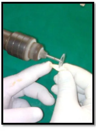
12 METHYLENE BLUE 13 TEETH SPECIMENS 14 DECORONATION
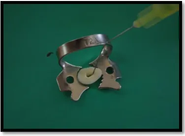
15 AUTOCLAVED SPECIMENS 16 INFECTION OF ROOT CANALS 17 ARMAMENTARIUM
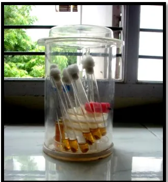
18 CHEMO-MECHANICAL PREPARATION 19 BIOFILM GROWTH IN SPECIMENS 20 INCUBATION IN PS
21 LASER TIP
22 WL FOR PHOTO IRRADIATION 23 PHOTOSENSITIZATION
24 FLUSHING OF ROOT CANALS
Aim & Objective: The objective of the study was to evaluate the in-vitro effects Polylactic-co- glycolic acid (PLGA) nanoparticles loaded with the Photosensitiser Methylene blue (MB) and light against E. faecalis biofilm.
Materials and methods: 50 extracted mandibular premolar teeth were decoronated and chemomechanically prepared. Biofilm was allowed to be formed in the root canals of specimens by incubating it for 2 weeks in BHI broth infected with E faecalis. Based on the mode of treatment, the specimens were divided into 5 groups (n=10): Group I- MB encapsulated with PLGA nanoparticles (MB+NP), Group II – MB with passive ultrasonic activation (MB+US), Group III- MB with GP activation (MB+GP), Group IV – MB without any activation and Group V- specimens without any treatment (control). The specimens were incubated with the Photosensitiser for 10 mins and subjected to Photoactivated disinfection using Diode laser (665nm), with a power density of 100mW/cm2 and total energy fluence of 30 J/cm2. The groups were subdivided, 8 specimens were subjected to microbiological analysis and 2 specimens were analysed under CLSM to evaluate the alive/dead bacteria.
Results: The MB+NP group showed least number of CFU’s after PAD, and was followed by MB+US group. The control group exhibited maximum number of CFU’s. All the groups showed significant difference when compared to the control group. The CLSM images mirrored and justified the results obtained by microbiological analysis. Conclusion: The ultilisation of PLGA nanoparticles encapsulated with photosensitive dye proved to be effective against E faecalis biofilm and can be a promising adjunct in antimicrobial endodontic therapy.
1
INTRODUCTION
The success of endodontic therapy depends on the effective
control of bacterial infection within the root canal system.33 Accepted
treatment procedures to eliminate the infection include
chemo-mechanical preparation which comprises of removal of infected hard
tissue, disinfection by one or more irrigants and intracanal
medicaments, followed by obturation of the canal with an inert
material to provide fluid impervious seal.12 However, the main cause
of treatment failures are the presence of persistent microorganism and
recontamination of canals due to inadequate coronal seal.29
An ideal irrigant or combination of irrigants, kills bacteria,
dissolves necrotic tissue, lubricates canal, removes smear layer and
does not irritate healthy tissue. Most commonly used irrigants are
2.5% to 5.25% Sodium hypochlorite, 0.2% Chlorhexidine, 15% to
17% EDTA, Hydrogen peroxide. In addition, inter-appointment
placement of intracanal medicaments such as Calcium hydroxide, has
been suggested for disinfection of the root canal system.29 In spite of
2
disinfection has not been possible and poses a challenge to the
endodontist.
The microorganisms present in the root canal system can be
either in the form of planktonic species i.e. free floating or in the form
of biofilm. Eradication of biofilm with conventional treatment has
been challenging due to biological complexity of the biofilm structure
and the variety of inanimate surface that microbial cells colonize.8
Incidence of treatment failures associated with non-surgical
endodontic treatment are associated with high proportions of Gram
positive anaerobic organisms, especially Enterococcus faecalis, and
have been reported to show resistance to common intracanal
medication.37E. faecalis has the ability to survive in the root canal as
a single organism without the support of other bacteria, in harsh
ecology of environment34. In addition, E-faecalis can form intra and
extra radicular biofilms, which make it even harder to control them.35
Despite the improvements in instrumentation techniques and
use of intracanal medicaments, failure of endodontic treatment has
3
the use of antibiotics serves an alternative approach but its long term
use can be rendered ineffective by the resistance developed in the
target organisms.10 Considering this, disinfection of root canal,
including the most distant areas of the tubular system is a major
challenge in endodontic treatment.
Contemporary approaches to disinfect root canal have been
proposed that include the use of ultrasonics, negative pressure syringe
and photodynamic theory.12 Low power lasers within visible range
along with dyes or Photosensitiser (PS) have been used for root canal
disinfection, and is termed as photoactivated disinfection (PAD). It is
based on the concept that non-toxic photosensitisers can be
preferentially localised in certain tissues and subsequently activated
by a light source of appropriate wavelength, to generate singlet
oxygen and free radicals that are cytotoxic to the target tissue.25
Methylene blue (MB) a well established photosensitiser has
been used in PAD for targeting various gram positive and negative
oral bacteria, as well as in endodontic disinfection. However, studies
have demonstrated incomplete destruction of oral biofilms using
4
attributed to reduced penetration of dye.20 In addition, it has been
shown that dyes such as MB are substrates of multidrug resistance
pumps in bacteria thus decreasing the effectiveness of the
photosensitiser.16 Therefore, to overcome these deficiencies a drug
delivery system has to be developed that can improve the
pharmacological characteristics of MB.
Recent studies on PAD have focused on the use of
polymer-based nanoparticles for photosensitizer delivery and release
systems.20,30,1 Nanoparticles containing photosensitizers have
exhibited several advantages over photosensitizing molecules not
encapsulated in nanoparticles.20
FDA-approved biodegradable polymeric nanoparticle Poly
lactic co-glycolic acid (PLGA), has been used as a drug delivery
system for photosensitizers. Once encapsulated within PLGA, the
excited state of the photosensitizer is quenched, which results in loss
of phototoxicity. When the nanoparticles were incubated with the
targeted cells, they showed a time-dependent release of the
photosensitizer, which then regained its phototoxicity and resulted in
5
AIM:
The aim of the present study was to determine the efficiency of
photoactivated disinfection against E. faecalis biofilm.
The hypothesis of the study was that, encapsulation of
Methylene blue with PLGA nanoparticle may offer a novel design of
nanoplatform for enhanced drug delivery in the root canal system and
photodestruction of E. faecalis biofilm.
OBJECTIVE:
The objective of the study was to explore the:
i. Efficacy of photoactivated disinfection against E. faecalis
biofilm using photosensitiser MB loaded with PLGA
nanoparticle and blank MB, by Colony Forming Unit
technique.
ii. Demonstrate the viable and dead bacteria in the dentinal
6
REVIEW OF LITERATURE
Siqueira et al (1999)34 reviewed the function of calcium
hydroxide as a routine intracanal medicament. He elaborated the
physic-chemical properties of the substance that may limit its effectiveness in
disinfecting the entire root canal system. Calcium hydroxide was not
effective against all bacterial species found in root canal infections. Its
association with other medicaments enhanced the efficacy of the
intracanal medication in eliminating residual bacteria in the root canal
system.
Lee et al (2004)19 investigated the usefulness of Class IV lasers
(such as Nd:YAG, diode, KTP and Er:YAG) for photo-thermal
disinfection of the root canal using photosensitisers such as tolonium
chloride. They concluded that while PAD can be undertaken as part of
the routine disinfection of the root canal system, it also has potential use
for eradicating persistent endodontic infections for which conventional
methods have been unsuccessful.
Ugo Bilati et al (2005)3 formulated and modified the process to
improve the versatility of the nanoprecipitation technique for
7
and the non-solvent nature, the solvent/non-solvent volume ratio and the
polymer concentration. It was shown that the mean particle size was
closely dependent on the type of non-solvent selected. High polymer
concentration in the solvent prevented nanoparticle formation. Thus they
concluded, both poly lactic acid (PLA) and polylactic-co-glycolic acid
(PLGA) could be used as a efficient carrier by accurately choosing the
polymer solvent.
Wei Tang et al (2005)42 developed and characterised the
nanoparticles loaded with methylene blue (MB), which are designed to
be administered to tumor cells externally and deliver singlet oxygen for
photodynamic therapy. Induced by light irradiation, the entrapped MB
generated singlet oxygen which was measured quantitatively.
Polyacrylamide nanoparticles showed the most efficient delivery of
oxygen, but its loading of MB was low. In contrast, the sol-gel
nanoparticles had the best MB loading but the least efficient oxygen
delivery. The encapsulation of MB in nanoparticles diminished the
interaction of this PS with the biological milieu, thus facilitating its
8
Garcez et al (2006)9 in their invitro study proved that photosensitizer alone or laser alone did not have any bactericidal effect.
Chemical solution (0.5% NaOCl) reduced viable bacteria in 93.25%.
Laser photosensitization resulted in a reduction of 99.2%, a significantly
higher bacterial reduction than NaOCl. Laser photosensitization was
effective for reducing E. faecalis in root canals and could be an adjunct
to endodontic treatment.
Willam et al (2006)44 measured the antibacterial action of
photoactivated disinfection (PAD) on endodontic bacteria in planktonic
suspension and root canals using Fusobacterium nucleatum,
Peptostreptococcus micros, Prevotella intermedia and Streptococcus
intermedius. Bacteria in canals were sampled before and after light
irradiation. In suspension, reductions in bacteria were highly significant
for light/Tolonium chloride combinations compared to light or Tolonium
chloride alone. Antibacterial action was increased by energy dose
increase, but not by Tolonium chloride concentration. PAD killed
endodontic bacteria at statistically significant levels and the kills varied
9
Soukos et al (2006)37 investigated the effects of photodynamic therapy on endodontic pathogens in planktonic phase as well as on
Enterococcus faecalis biofilms in experimentally infected root canals of
extracted teeth. Methylene blue fully eliminated all bacterial species with
the exception of E. faecalis (53% killing). The same concentration of
methylene blue in combination with red light was able to eliminate 97%,
using an optical fiber with multiple cylindrical diffusers that uniformly
distributed light at 360 degrees.
Tada et al (2007)42 prepared and characterised methylene
blue-containing silica-coated magnetic particles by transmission electron
microscopy, light scattering, and X-ray diffraction. The immobilized
drug can generate singlet oxygen, which was detected by its
characteristic phosphorescence decay curve in the near-infrared and by a
chemical method using 1, 3-diphenylisobenzofuran to trap singlet
oxygen. The lifetime of singlet oxygen was determined to be 52 µsec (in
acetonitrile) and 3µsec (in water). The release of singlet oxygen was
affected by the encapsulation of MB in the silica matrix, which caused a
10
Saji et al (2007)13 evaluated the cytotoxicity and selectivity of an
advanced noninvasive light-activated disinfection against fibroblast
cells. Simultaneous evaluation of cytotoxicity and antibacterial effect
was also conducted to study the specificity of light activated therapy
toward prokaryotic cells (Enterococcus faecalis). Data revealed that
cytotoxicity was significantly less in LAT compared with NaOCl. E
faecalis cells were killed at a faster rate than fibroblasts. An irradiation
dose producing 97.7% bacterial killing showed only 30% fibroblast
dysfunction.
Jason M. Duggan et al (2007)6 tested the hypothesis that the
ability of Enterococcus faecalis to form biofilms is related to the source
of the strains. They evaluated the E. faecalis strains recovered from root
canals, oral cavity, and non-oral/non-endodontic sources and concluded
that there were no significant associations between biofilm formation
and the presence of the virulence determinants asa, cylA, esp, and gelE.
Sulis et al (2007)36 reviewed the use of Passive ultrasonic
irrigation in endodontics. Irrigation with sodium hypochlorite is more
effective than with water and ultrasonic irrigation is more effective than
11
role of cavitation during PUI remains inconclusive. The influence of
irrigation frequency and intensity on the streaming pattern as well as the
complicated interaction of acoustic streaming with the adherent biofilm
has been clarified to reveal the underlying physical mechanisms of PUI.
Garcez et al (2008)12 analyzed the antimicrobial effect of
photodynamic therapy (PDT) in association with endodontic treatment
and suggested that the use of PDT added to endodontic treatment leads
to an enhanced decrease of bacterial load and may be an appropriate
approach for the treatment of oral infections.
Bechet et al (2008)1 described and compared the different
individual types of nanoparticles that are currently in use for PDT
applications. They highlighted on the recent advances in the use of
nanoparticles, including inorganic oxide-, metallic-, ceramic-, and
biodegradable polymer-based nanomaterials as carriers of
photosensitizing agents. The nanoparticles were described in terms of
stability, photocytotoxic efficiency, biodistribution and therapeutic
efficiency. Finally, they summarized with new results concerning the
improvement of the photophysical properties of nanoparticles by means
12
Pinheiro et al (2008)23 evaluated photodynamic therapy in
deciduous teeth with necrotic pulp by means of fully quantifying viable
bacteria, before and after instrumentation and after the use of
photodynamic therapy. The instrumentation resulted in a reduction of
82.59% of viable bacteria, and, after PDT, the microbial reduction
observed was 98.37%.
Fimble et al (2008)8 investigated the photodynamic effects of
methylene blue on multispecies root canal biofilms comprising
Actinomyces israeli, Fusobacterium nucleatum subspecies nucleatum,
Porphyromonas gingivalis, and Prevotella intermedia in experimentally
infected root canals of extracted human teeth in vitro. Root canal
systems were incubated with methylene blue (25 µg/mL) for 10 minutes
followed by exposure to red light at 665 nm with an energy fluence of 30
J/cm2 by diode laser via a 250m diameter polymethyl methacrylate
optical fiber. Photodynamic therapy (PDT) achieved up to 80%
reduction of colony-forming unit counts.
Saji et al (2008)14 tested the hypothesis that the inclusion of an
oxidizer and oxygen carrier in the photosensitization formulation would
13
light activated disinfection. MB in emulsion was overall the most
effective photosensitizer formulation for photooxidation, generation of
singlet oxygen, and in disinfecting biofilm bacteria. Advanced
noninvasive PAD using a photosensitizer formulation containing
oxidizer and oxygen carrier disrupted the biofilm matrix and facilitated
comprehensive inactivation of biofilm bacteria.
Souza et al (2009)39 aimed to investigate the antibacterial effects
of photodynamic therapy with methylene blue (MB) or toluidine blue
(TB) as a supplement to instrumentation/irrigation of root canals
experimentally contaminated with Enterococcus faecalis. PDT with
either MB or TB did not significantly enhance disinfection after
chemomechanical preparation using NaOCl as irrigant. No significant
differences were observed between the two photosensitizers. Thus the
results suggest that PDT with either MB or TB may not exert a
significant supplemental effect to instrumentation/ irrigation procedures
with regard to intracanal disinfection.
Reddy et al (2009)27 reviewed Photodynamic therapy (PDT) as a powerful laser-initiated photochemical reaction and demonstrated the
14
bacterial and fungal infections and photodynamic diagnosis of malignant
transformation of oral lesions. PACT (photodynamic antimicrobial
chemotherapy) has been efficacious in the management of
peri-implantitis, endodontic infections and oral biofilms such as plaque. The
absence of genotoxic and mutagenic effects, no risk of developing
resistance to its antimicrobial action and increased healing process
favors its long-term safety and use.
Xu et al (2009)45 assessed the in vitro synergistic effect of
methylene blue (MB) and red light on human gingival fibroblasts and
osteoblasts. Assessment of PDT-induced apoptosis was investigated.
Light at 20 and 40 mW/cm2 with MB had modest effects at 24 hours on
osteoblasts in both assays, whereas sodium hypochlorite (NaOCl)
completely eliminated cells. Western blot analysis revealed no signs of
apoptosis in either cell type. The data suggest that there is a safe
therapeutic window whereby PDT can inactivate endodontic pathogens
without affecting host cell viability.
Avinesh Kumari et al (2010)18 reviewed the biodegradable
nanoparticles that have been used frequently as drug delivery vehicles.
15
related drugs on biodegradable nanoparticles such as PLGA, PLA,
chitosan, gelatin, polycaprolactone and poly-alkyl-cyanoacrylates.
Schlafer et at (2010)29 evaluated the antimicrobial effect of
photoactivated disinfection (PAD) using toluidine blue and an LED lamp
on endodontic pathogens in planktonic suspension and after inoculation
into extracted teeth. Photoactivated disinfection yielded significant
reductions in the viable counts of all organisms in planktonic suspension
as well as in root canals.
Anil Kishen et al (2010)16 evaluated the efficacy of antimicrobial
photodynamic inactivation of Enterococcus faecalis biofilms using a
cationic, methylene blue (MB) and an anion, rose bengal (RB). The role
of a specific microbial efflux pump inhibitor (EPI), verapamil
hydrochloride in the MB-mediated PDT on E. faecalis biofilms was also
investigated. PDT with cationic MB produced superior inactivation of E.
faecalis strains in a biofilm along with significant destruction of biofilm
structure when compared to anionic RB. The ability to inactivate biofilm
16
Garcez et al (2010)10 reported the antimicrobial effect of
photodynamic therapy (PDT) combined with endodontic treatment.
Endodontic therapy alone produced a significant reduction in numbers of
microbial species but only 3 teeth were free of bacteria, whereas the
combination of endodontic therapy with PDT eliminated all
drug-resistant species and all teeth were bacteria-free.
Shinde et al (2011)32 formulated nanoparticles for simvastatin
drug used as a lipid lowering agent. Nanoparticles were prepared by
precipitation-solvent deposition method and were found to be effective
in sustained drug release for a prolonged period.
S Rajesh et al (2011)27 have reviewed the use of therapy for
management of periodontal disease. They have enumerated the need to
develop an evidence based approach to the use of this therapy for
management of periodontitis, periimpantitis and endodontic therapy.
Rios et al (2011)28 evaluated the antimicrobial effect of PDT
using toluidine blue O (TBO) and a low-energy light emitting diode
(LED) lamp after the conventional disinfection protocol of 6% NaOCl.
The bacterial survival rate of the NaOCl/TBO/light group was
17
TBO and a LED lamp has the potential to be used as an adjunctive
antimicrobial procedure in conventional endodontic therapy.
Steier et al (2011)40 evaluated the bovine pulp tissue dissolution
ability of photodynamic therapy and concluded that only NaOCl was
able to promote complete dissolution of pulp tissue whereas
photodynamic therapy does not show any ability to dissolve pulp tissue.
Raymond et al (2011)26 evaluated the anti-microbial effects of
photodynamic therapy (PDT) on infected human teeth ex vivo. They
divided the study into two groups- canals treated with chemomechanical
debridement (CMD) and canals treated with CMD followed by treatment
with Photoactivated disinfection (PAD) using Methylene blue. The
results suggested that post-treatment detection levels for all species were
markedly lower for canals treated by CMD+PDT than were for those
treated by CMD alone.
Jarza et al (2011)15 reviewed the application of nanotechnology
for enhancement of PAD. In the article influence of silver-doped
nanomaterials addition on the fluorescence intensity of photosensitizers
18
spectroscopy. Influence of sonication on the fluorescence enhancement
was also investigated. It was demonstrated that the fluorescence
enhancement of photosensitizers depends on the concentration of both:
photosensitizer and silver-doped nanoparticles.
Shimab Shahin et al (2012)30 reviewed the potential of
nanotechnology to offer solutions to obstacles in cancer therapies.
Nanoscale devices have impacted cancer biology at three levels: early
detection, tumour imaging using radio contrast nanoparticles or quantum
dots; and drug delivery using nanovectors and hybrid nanoparticles.
Nanocrystals labeling with immune cells can act as a platform
technology for nanoimmunotherapy.
Silva et al (2012)33 evaluated the in vivo response of apical and
periapical tissues of dogs teeth with apical periodontitis after one-session
endodontic treatment with and without antimicrobial photodynamic
therapy. In the PDT-treated groups, the periapical region was
moderately/severely enlarged with no inflammatory cells, moderate
neoangiogenesis and fibrogenesis, and the small periapical lesions was
detected suggesting PDT to be a promising adjunct therapy to cleaning
19
MATERIALS
1. Extracted mandibular premolars
2. Enterococcus faecalis (ATCC 29212)
3. Blood agar
4. Sodium hypochloride – 6%, 0.5%
5. Methylene blue (Bio Lab systems)
6. Poly lactic-co-glycolic acid(PLGA) {Sigma Aldrich, Germany}
7. Pluronic (Sigma Aldrich, Germany)
8. Sodium oleate (Sigma Aldrich, Germany)
9. Diamond saw
10.Glyde(Dentsply, Tulsa)
11.Gutta Percha (Dentsply,Tulsa)
12.17% EDTA (Tulsa,USA)
13.Micro centrifuge tube
14.Micro pipette (Eppendorf, Germany)
15.Phosphate buffered saline (PBS)
16.Nail polish
20 18.Rubber dam
19.SYTO-9 and Propidium iodide stains
ARMAMENTARIUM:
20.Hand piece (PAN AIR, NSK,Japan)
21.Incubator (Biotechniques, India)
22.Access opening bur (Mani Inc, Japan)
23.K- File- size 15, 25 (Mani inc, Japan)
24.ProTaper- S1,S2,F1,F2,F3,F4(Dentsply Maillefer,Tulsa)
25.X- smart (Dentsply Maillefer,Tulsa)
26.Ultrasonic instrument ( Satlec, USA)
SPECIAL EQUIPMENTS:
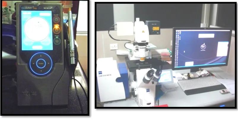
27.Laser unit (Ezlase Diode laser, san Clemente, USA)
28.Magnetic stirrer (Remi Elektrotechnite Ltd)
29.Cryo-centrifuge (Rotor F-45)
30.Freeze dryer (Delvac)
21
METHODOLOGY
Preparation of the Poly lactic co-glycolic acid (PLGA) nanocarriers:
76mg of PLGA was mixed with 14mg of Pluronic in 5ml of
acetone. The solution was heated and stirred until it became clear. 10mg
of Methylene blue and 90mg of Sodium oleate was introduced into the
acetone solution and kept for overnight vigorous magnetic stirring. The
nanoparticle solution was centrifuged at 10000rpm for 20 minutes. The
sediment part was collected, washed twice with water and freeze dried.
Preparation of the tooth specimens:
Fifty freshly extracted single-rooted mandibular premolar teeth
with straight canals were stored in 0.5% sodium hypochlorite for two
weeks. Specimens were decoronated to a standard 12 mm root segment
length with a rotating diamond saw at 20,000 rpm under water-coolant.
Patency of apical foramina was established by inserting a size 15K-file.
A file measurement was taken at the point where the size 15 K-file
became visible at the apical foramen and 0.5 mm was subtracted to set
22
S1, S2, F1, F2, F3 and F4 following a crown-down technique. X-smart
hand piece was used, following a pecking motion. The final apical
patency was established with a size 25 K-file in order to allow for an
adequate apical aperture for flushing of microbial aggregates. Glyde was
used as a lubricant and canals were irrigated with 6% sodium
hypochlorite throughout the instrumentation sequence. The final
irrigation consisted of 1 ml of 17% ethylene demine tetra acetic acid
(EDTA) solution for 3 min for smear layer removal, deactivated with 1
ml of 6% NaOCl for 3 minutes. Each tooth specimen was then placed in
a micro centrifuge tube containing 500 μl of phosphate buffered saline
(PBS). Teeth were subsequently autoclaved at 121°C for 20 minutes.
Following autoclave sterilization, PBS was aspirated from the
microcentrifuge tubes under sterile conditions. The root surface was
coated with nail polish to avoid external microbial contamination.
E.faecalis biofilm growth:
Fifty root specimens were transferred into sterile microcentrifuge
tubes. One millilitre of BHI broth containing 109 microorganisms of
23
Canal-Clean 30 gauge irrigation needle. After injection, each specimen
was entirely submerged in BHI broth, and the tubes were incubated
anaerobically for 10 days.
Grouping & Photodisinfection of root canals:
The specimens were randomly divided into 5 groups, each
containing 10 specimens (n=10).
Group I - MB with Nanoparticle carrier + Laser (photoactivation)
Group II – MB + Ultrasonic activation + Laser
Group III – MB + GP activation + Laser
Group IV – MB +Laser
Group V – No PAD (control)
Parameters for Laser irradiation:
In the present study, the source was a diode laser (Ezlase), with an
output power of 1 Watt and a central wavelength of 665 nm. The power
density was 100mW/cm2 and the total energy fluence dose was 30 J/cm2.
24
optical fiber that was mechanically notched over a one-centimeter length
at approximately one-millimeter intervals.
Group I
The root specimens were placed in microcentrifuge tubes under
sterile conditions. The canals were loaded till the access cavity with
Methylene blue (MB) encapsulated with PLGA nanoparticle at
concentration of 50 μg/ml equivalent to MB, using Canal clean 30 gauge
needle. The entire specimen was immersed in the nanoparticle solution
for 10 minutes. Excess drug solution was aspirated and the root
specimens were removed from the tubes. Canals were irradiated with
laser for 5 minutes with a break at 2.5 minutes, for 2.5 minutes, placing
the tip at 1mm short of the working length following a spiral outward
motion.
Group II
The root specimens were placed in microcentrifuge tubes under
sterile conditions. The canals were loaded till the access cavity with
25
containing MB were ultrasonically activated for 20secs. The entire
specimen was immersed in MB for 10 minutes. Excess MB was
aspirated and the root specimens were removed from the tubes. Canals
were irradiated with laser for 5 minutes with a break at 2.5 minutes, for
2.5 minutes, placing the tip at 1mm short of the working length
following a spiral outward motion.
Group III
The root specimens were placed in microcentrifuge tubes under
sterile conditions. The canals were loaded till the access cavity with
Methylene blue (MB), using Canal clean 30 gauge needle. The canals
containing MB was activated using size #35 Gutta-percha following 20
up and down movements till the working length at a frequency of 3 per
sec. The entire specimen was immersed in MB for 10 minutes. Excess
MB was aspirated and the root specimens were removed from the tubes.
Canals were irradiated with laser for 5 minutes with a break at 2.5
minutes for 2.5 minutes, placing the tip at 1mm short of the working
26
Group IV
The root specimens were placed in microcentrifuge tubes under
sterile conditions. The canals were loaded till the access cavity with
Methylene blue (MB), using Canal clean 30 gauge needle. The entire
specimen was immersed in MB for 5 minutes. Excess MB was aspirated
and the root specimens were removed from the tubes. Canals were
irradiated with laser for 10 minutes with a break at 2.5 minutes for 2.5
mins, placing the tip at 1mm short of the working length following a
spiral outward motion.
Group V
The specimens of this group were not exposed to Photoactivated
disinfection and served as the control for the study.
Following all treatments, the 8 specimens from each group were
subjected to microbiological analysis and 2 specimens were subjected to
27
Microbiological analysis:
Specimens was aseptically mounted on a rubber dam, by utilizing
a plastic u-shaped rubber dam frame attached to a rack and oriented
parallel to the lab bench top. The coronal 4 mm of each specimen was
above the surface of the dam. The contents of root canals were sampled
by flushing the root canals with a coronal application of 1-ml of BHI
broth with a Canal clean 30 gauge irrigation needle. The bacterial
suspension was collected in a 1.5 ml microcentrifuge tube positioned
below the apical foramen. After vortexing for 20 seconds, serial dilutions
were prepared and 100 μl aliquots were inoculated onto blood agar and
incubated anaerobically for 7 days. Observations were obtained from
each treatment group and colony forming units (CFU’s) were calculated.
Confocal Laser Scanning Microscopic analysis:
Tooth specimens were split longitudinally into two equal halves
and rinsed with 10mL of PBS for three times. Sections were then stained
immediately with the SYTO9 and Propidium iodide (PI) reagents which
28
Carlsbad, CA, USA) and examined under the Confocal Laser Scanning
Microscope.
Sections were scanned using Confocal Laser Scanning
Microscope with illumination by a Krypton/Argon laser (488 nm). The
border of the root canal was first located with the microscope, and five
randomly selected places were scanned with the CLSM for each section.
The mounted specimens were observed at Magnification of 63×. The
dimension of each scanned field was 0.70 ×0.70 mm. A 477/543-nm
double dichroic mirror was used as an excitation beam splitter and a
545-nm short-pass filter divided green (SYTO9) and red fluorescence
(Propidium Iodide) between the photomultipliers. A 505- to 550-nm
band-pass filter was used to visualize SYTO9 and a 650-nm long-pass
filter for PI.
The Confocal laser scanning microscopic (CLSM) images were
recorded in the fluorescent mode. Fluorescence images were analysed
with Amira 5.0 (Visage Imaging Inc., Andover, MA, USA), and image
stacks were viewed with LSM Image Browser (Carl Zeiss Ltd). The
29
individual component colour channels and saved as grey-scale images.
For each greyscale image, fluorescence was adjusted (‘thresholded’)
such that signals of intensity less than 20% were regarded as
background. The split greyscale images were then combined and
calibrated to form a single fluorescent image which was qualitatively
analyzed by three independent blinded observers to determine the
proportion of green and red fluorescence denoting the presence of live
Fig 1Autoclave Fig 2 Incubator
Fig 13 Teeth specimens
Fig 14 Decoronation
Fig 15 Autoclaved specimens Fig 16 Infection of root canals
[image:43.612.351.510.102.318.2] [image:43.612.107.302.147.304.2]
Fig 23 Photosensitization Fig 24 Flushing of the root canals
Fig 25 Collection in Fig 26 Aliquots plated on
-glycoic acid
Fig 5 Diode laser Fig.6 Confocal Laser Scanning
Microscope (CLSM)
Fig. 7 BacLight Viability Kit Fig.8 SYTO9 &
Teeth were decoronated, chemo mechanically prepared & divided into 5 groups (n=10)
Group I :
NP +MB+ Laser
Group II : MB+ Ultrasonic+ Laser
Group III: MB+GP activation+Laser
Group V: Control - No treatment Group IV: MB+ Laser Root canals were incubated with NP loaded MB for 10 minutes Root canals containing MB were US activated and incubated for 10 minutes Root canals containing MB were activated with GP and incubated for
10 minutes
Root canals were incubated with MB for 10
minutes
Specimens were irradiated with diode laser (665nm) for 5 minutes, followed by pause at 2.5 minutes for 2.5 minutes .
The contents of the root canal was flushed with 1ml BHI broth and bacterial suspension
was collected in micro centrifuge tube.
Serial dilutions were prepared, inoculated on blood agar and incubated anaerobically for 7
days.
CFU’s were calculated and statistically analyzed.
The specimens were cut longitudinally with diamond saw
disc & washed in 10mL PBS.
Samples were stained with BacLight Bacterial viability kit for
2 minutes.
30 RESULTS
The colony forming units (CFU’s) and survival fraction was
calculated and statistically analyzed.
The data values of CFU’s were log 10 transformed to reduce
variance heterogeneity.
The survival fraction was calculated from CFU’s counted on
the plates:
Survival fraction = No. of CFU in the treated group/No. of
CFU in the untreated group x 100
Cell death = 100 - Survival fraction
The untreated group (Group V) was used as a reference to
calculate the survival fraction.
Cell death denotes exactly the efficiency of treatment or the
reduction of bacteria, in terms of percentage, which gives better
31
The results of the present study were subjected to statistical
analysis to interpret the significant differences among various
treatment groups. One-Way ANOVA and post hoc Tukey HSD tests
were used for statistical analysis in the present study.
One-way Analysis Of Variance (ANOVA) is used to study the
overall variance within groups. It is the extension of the between
groups t-test to the situation in which more than two groups are
compared simultaneously. However, it is not possible to identify the
difference between the various subgroups with the help of the P
values obtained from ANOVA. Hence, the Tukey HSD is done in
order to determine which groups differ from each other. The Tukey
Test Honestly Significant Difference or HSD test is a post hoc test
designed to perform a pair wise comparison of the means to identify
the specific sub groups in which significant differential expression
occurs.
In this study one way ANOVA followed by Tukey HSD test
showed statistically significant difference among various subgroups
32
I. Comparison of growth of CFU’s in all the Groups:
Groups Mean ± Standard deviation P-value
I 2.45 ± 0.24
< 0.001**
II 3.05 ± 0.11
III 4.07 ± 0.12
IV 4.75 ± 0.16
V 6.77 ± 0.08
Note: ** denotes significance at 1% level
II. Comparison of growth of Colony forming units between each
group:
Groups Mean difference Significance
Gr I x Gr II -.6001* .000
Gr I x Gr III -2.2482* .000
Gr I x Gr IV -2.2962* .000
Gr I x Gr V -4.3203* .000
Gr II x Gr III -1.6480* .000
Gr II x Gr IV -1.6961* .000
Gr II x Gr V -3.7201* .000
Gr III x Gr IV -.0480 .964
Gr III x Gr IV -2.0721* .000
Gr IV x Gr V -2.0241* .000
33
III. Comparison of the Survivability Fraction of all the groups:
Groups Mean ± Standard deviation P-value
I 0.06± .028
< 0.001**
II 5.46± .48
III 9.13± 3.05
IV 10.30± 3.41
Note: ** denotes significance at 1% level.
IV. Comparison of Survivability Fraction in between the groups:
Groups Mean difference Significance
Gr I x Gr II -5.4075* .000
Gr I x Gr III -9.0738* .000
Gr I x Gr IV -10.2512* .000
Gr II x Gr III -3.6663* .018
Gr II x Gr IV -4.8437* .001
Gr III x Gr IV -1.1775 .738
34
Table I: denotes the Colony forming units in all the groups.
Photoactivated disinfection with MB loaded with nanoparticles
showed least value. It was followed by the group treated with MB
activated with ultrasonic, MB+GP and MB. Group V (control)
showed maximum CFU’s. The P-value was <0.001, which implies
statistically significant difference between the groups.
Table II demonstrates the significance of P values in all the
groups. When the group treated with MB activated with GP was
compared to the group treated with blank MB, no statistical
significance was observed. The comparison of all the other groups
were statistically significant (P < 0.5%).
Table III denotes the Survivability Fraction in all the groups.
Photoactivated disinfection with MB loaded with nanoparticles
showed least value. It was followed by the group treated with MB
activated with ultrasonic and MB+GP. Group treated with blank MB
showed maximum Survival Fraction. The P-value was <0.001, which
implies statistically significant difference between the groups.
Table IV demonstrates the significance of P values in all the
35
compared to the group treated with blank MB, no statistical
significance was observed. The comparison of all the other groups
were statistically significant (P < 0.5%).
Colony Forming Unit’s CLSM image (63x)
Colony Forming Unit’s CLSM image (63x)
Graph II: Comparison of mean Survivable fraction in all the groups:
0 1 2 3 4 5 6 7 8
MB+NP MB+US MB+GP MB Control
Log 10 CFU's
0 2 4 6 8 10 12
MB+NP MB+US MB+GP MB
36
DISCUSSION
The main goal of endodontic treatment is effective control of
bacterial infection within the root canal system by elimination of
pathogenic microflora, toxins and tissue debris.33 Literature reports that
endodontic therapy will have a 94% success rate when a negative
microbiological culture is obtained from the root canal at the time of
obturation. Whereas, when obturation is performed and the cultures are
positive, the success rate is reduced to 68%.11, 12 Studies have shown the
shoddier healing of periapical lesions in cases where obturation has been
performed with positive cultures.12 Undoubtedly, the major factors
associated with endodontic failure are the persistence of microbial
infection in the root canal system and in the periradicular area.33, 35
Chemo-mechanical preparation can be considered to be an
essential step in the root canal disinfection. It includes debriding of the
infected dentinal walls of the root canal system using files in a sequence
and use of irrigants. Most commonly used irrigants are hydrogen
peroxide, sodium hypochlorite (2.5-5.2%), EDTA (15-17%),
Chlorhexidine 0.2%.28 The depth of penetration of irrigants such as
37
whereas tubular infection may occur closer to cementum-dentin junction
at 1000 µm.28 Moreover, studies have demonstrated that part of root
canal space often remains untouched during chemomechanical
preparation, regardless of the technique or instruments employed.35
Thus complete elimination of the bacteria was difficult to accomplish.
To overcome this, intrancanal medicaments such as Calcium Hydroxide
were introduced.
Action of Calcium hydroxide is mainly by increase in pH in the
root canals by release of hydroxyl ions. But certain bacteria such as
enterococci tolerate very high pH value, ranging from 9-11.Several
studies have attested the inefficiency of calcium hydroxide in
eliminating the bacterial cells inside dentinal tubules.22,34Haapasalo and
Orstavik (1987) reported that Calcium hydroxide paste failed to
eliminate even superficial E faecalis in the tubules.35
E.faecalis is a gram positive bacterium, member of the
commensal human flora and an opportunistic pathogen implicated as one
of the leading causes of nosocomial infection. It is mostly associated
with persistent endodontic infection and is highly associated with
38
when present in the form of biofilms.16 A biofilm can be defined as
microbial population attached to an organic or inorganic substrate,
surrounded by microbial extracellular products, which form an inter
microbial matrix.35
According to George et al, 2008 the phenotypic and genotypic
variation of biofilm bacteria when compared to their free flowing
counterpart, complemented by the structure and composition of biofilm
matrix contribute to their high microbial resistance.16 The surface protein
(Esp), the transcriptional regulator (BopD), the quorum sensing locus fsr
and gelatinase (GelE) have been reported to be involved in promoting
biofilm formation in E faecalis.14
Inability to completely eradicate biofilm structures in proximity to
host immune cells will result in persistent infection and subsequent
re-establishment of infection, with probably more treatment-resistant
resident bacteria. Epidemiological studies have reported that 30%- 50%
of root canal treatments fail from residual infection.33 This has led to the
quest of novel disinfection procedures that can be an adjunct to standard
39
orthograde endodontic treatment and retreatment procedures. (Fimble
2008)8.
Contemporary approaches to disinfect root canal include the use
of ultrasonics and lasers. Ultrasonics in endodontics was introduced by
Richman in the year 1955.This technique appeared promising and files
had the potential to prepare as well as debride root canals mechanically.
Unfortunately, it proved to be difficult to control the cutting of dentine
during ultrasonic preparation, which resulted in irregularly shaped root
canals and also apical perforations (Sluis 2007)36.Passive ultrasonic
irrigation has recently led to a renaissance in the use of ultrasonics
during root canal treatment. The technique utilizes an ultrasonically
activated file or smooth wire within the root canal space following the
completion of canal preparation.36
Laser is a device which transforms light of various frequencies
into a chromatic radiation in the visible, infrared, ultraviolet regions with
all the waves in phase capable of mobilizing immense heat and power
when focused at close range. Stern and Sognnaes (1964) and Goldman et
al (1964) were first to investigate the potential use of ruby lasers in
40
using other high power lasers such as Argon, Carbon dioxide, Nd: YAG
and Er: YAG. The first lasers used in endodontics were reported by
Weichman and Johnson, 1971.
Most of the lasers used for root canal disinfection were high
power laser which were dose dependent and generated heat. Thus in
addition to killing microorganisms they caused collateral damage such as
charring of dentin, ankylosis of root, melting of cementum, root
resorption and periradicular necrosis.28 To overcome these problems a
new antibacterial strategy that involves the combination of a non toxic
photo sensitizer and a laser light source within visible region (400
-700nm), has been adopted and was termed as Photoactivated disinfection
(PAD) 27. Dickers et al, 20095 demonstrated that after 150s of PAD
irradiation, the average temperature rise was 0.16±0.08ºC, the recorded
values were lower than 7ºC which was within the safety level for
periodontal injury.
The origin of light as therapy in medicine and surgery can be
traced from antiquity to modern day. It began in ancient Egypt, Greece
and India but disappeared for many centuries only to be rediscovered by
41
physician Friedrich Mayer-Betz performed the first study, with what was
first called photoradiation therapy (PRT) with porphyrins in 1913 on
humans. But it was John Toth, who acknowledged the photodynamic
chemical effect of the therapy with early clinical argon dye lasers and
renamed it as photodynamic therapy (PDT). It received even greater
interest as Thomas Dougherty formed the International Photodynamic
Association. Its use first started in dermatology (1992), then oncology
(1995), and recently in microbiology (1996).27
Photoactivated disinfection can be defined as an oxygen
dependent photochemical reaction that occurs upon light mediated
activation of the photosensitizing compound, leading to generation of
cytotoxic reactive oxygen species, predominantly reactive oxygen. The
treatment procedure involves three basic components: a photosensitizer,
a light source and tissue oxygen. A photosensitizer (PS) is a chemical
compound (usually a dye) that can be excited by light of specific
wavelength (visible or infra-red light). The photosensitizer is
administered (injected or applied externally) to the patient and gets
accumulated in the targeted tissues. The tissue is then exposed to the
42
state which then undergoes an intersystem crossing forming a longer
lived excited triplet state. In the presence of endogenous oxygen, energy
transfer then takes place from this activated agent to the oxygen
molecule forming excited singlet state oxygen or other reactive oxygen
species (ROS), causing a rapid and selective destruction of the target tissues.25,27
During PAD, cytotoxic reactive oxygen species (ROS) are formed
by two mechanisms. Type I reaction involves electron transfer directly
from the photosensitizer producing ions, or electron/hydrogen removal
from a substrate molecule to form free radicals. These radicals react
rapidly with oxygen, resulting in the production of highly reactive
oxygen species (superoxide, hydroxyl radicals, and hydrogen peroxide).
Type II reactions produces the electronically excited and highly reactive
state of oxygen known as singlet oxygen. Usually the procedure involves
a contribution from both the mechanisms.27
PAD requires a source of light that activates the photosensitizer
by exposure to low-power visible light at a specific wavelength. Various
light sources for PAD are Diode laser systems, Non-coherent light
43
Diode laser is the most widely used laser. It emits a bandwidth of
laser (Central wavelength: 665nm) which matches with the required
wavelength for photosensitization.8,11,39 Thus, in the present study diode
laser has been used.
An optimal Photosensitizer (PS) should possess photophysical,
chemical, and biological characteristics. The properties of an ideal
photosensitizer are, stable composition, minimal self aggregation
tendency, nontoxic in the absence of light, target specificity and can be
easily cleared from the body.1 Most of the sensitizers used for medical
purposes belong to the following basic structures: a) Tricyclic dye i.e.
Methylene Blue. b) Tetrapyrroles i.e. Porphyrins c) Furocoumarins i.e.
Xanthotoxin.25
Methylene blue (MB) has been used as a photosensitizing agent
for almost nine decades and is used routinely as a marker dye in surgery.
The clinical use of MB for photodynamic therapy of bladder and
oesophageal cancer along with its use in case of methaemoglobinamea
suggests the local use of MB is safe.26
Methylene blue (MB) is a well-established photosensitizer and has
44
of MB, along with its low molecular weight and positive charge, allows
passage across the porin-protein channels in the outer membrane of
gram-negative bacteria. MB predominantly interacts with the anionic
macromolecule lipopolysaccharide, resulting in the generation of MB
dimers, which participate in the photosensitization process.8 MB has
been successfully used in PAD for targeting various gram-positive and
gram-negative oral bacteria and has the ability to infiltrate dentinal
tubules.46
However, studies by Soukos et al 200038, have demonstrated
incomplete destruction of oral biofilms using MB-mediated PAD. The
reduced susceptibility of biofilms to PAD was attributed to reduced
penetration of dye. In addition, Kishen et al, 201016 have shown that
dyes such as MB are substrates of multidrug resistance pumps in bacteria
thus decreasing the effectiveness of the photosensitiser. These resistant
microorganisms have microbial efflux pumps that have the ability to
extrude or expel antibiotics. Thus the penetrability of drugs into the
microorganism biofilm is impaired, and as a result disinfectant action is
45
has to be developed which can improve the pharmacological action of
MB.
Nanoparticles represent an emerging photosensitiser carrier that
show great promise for PAD. Bechet et el 20081, have enumerated the
overcoming of drawbacks of classic photosensitisers by use of
nanoparticles and explained the improved pharmacokinetic properties of
the drug. It can be achieved by either surface bounding of
photosensitizer particles to a nanoparticle or confinement of
photosensitizer in nanocapsules. The advantages of a nanoparticle carrier
for PS has been described by Pagonis 201020, i.e. they include a larger
critical mass (concentrated package of photosensitizer) for the
production of reactive oxygen species (ROS) that destroy cells, it limits
the target cell’s ability to pump the drug molecule back out thus reducing
the possibility of multiple-drug-resistance, selectivity of treatment by
localized delivery agents, and having a non-immunogenic nanoparticle
matrix.
In this study PLGA nanoparticles was used, which are synthetic
and biodegradable carriers. Synthetic polymers have the advantage of
46
of days to several weeks.30 Bechet et al 20081 have enumerated that
biodegradable carriers, in addition of being non immunogenic and non
inflammatory, are aqueous in composition and can disintegrate readily to
release the photosensitisers when light is irradiated.
PLGA polymers have a number of advantages over other
polymers used in drug and gene delivery, such as their biodegradability,
biocompatibility, and approval by the FDA for human use. PLGA
polymers degrade in the body through hydrolytic cleavage of the ester
linkage to lactic and glycolic acid. These monomers are easily
metabolized in the body via Krebs’ cycle and eliminated as carbon
dioxide and water. Biodegradation products of PLGA are formed at a
very slow rate, and they therefore do not affect normal cell function.
Furthermore, these polymers have been tested for toxicity and safety in
extensive animal studies and are currently used in humans for resorbable
sutures, bone implants and screws, contraceptive implants, and also as
graft materials for artificial organs and supporting scaffolds in tissue
47
Confocal Laser Scanning Microscopy (CLSM) has become an
invaluable tool for a wide range of investigations in the biological and
medical sciences for imaging thin optical sections in living and fixed
specimens ranging in thickness up to 100 micrometer. CLSM works on
the principle of fluorescence. The advantage of fluorescence for
microscopy is that fluorescent dye molecules can be attached to specific
parts of any sample, so that only those parts are the ones seen in the
microscope. Therefore, it is possible to distinguish two different parts of
a particular sample. The CLSM can determine the viable and dead
microorganism immobilized in the dentinal tubules.14,16
The objective of the present study was to investigate the efficacy
of photoactivated disinfection, by MB-loaded PLGA nanoparticles
against E. Faecalis biofilm, using CFU technique, and to assess the dead
and viable bacteria by Confocal laser scanning microscopy.
In the present study fifty freshly extracted single-rooted human
mandibular premolar teeth with straight canals were used. Teeth were
decoronated to a standard 12 mm root segment length and chemo
mechanically prepared. Teeth were subsequently autoclaved at 121°C for
48
polish to avoid external microbial contamination.22 Biofilm was allowed
to be formed on the specimens by the procedure described by Rios et al,
2011.28
The nanocarriers were prepared following the solvent
displacement procedure as described by Shenoy et al 200531.
Nanoprecipitation technique or solvent displacement method for
nanoparticle was first developed and patented by Fessi et al in 1989.Its
advantages are that it is straight forward, rapid and easy. It enables
production of small nanoparticles i.e. 100-300nm. Nanoprecipatition
occurs by rapid dissolvation of the polymer when the polymer solvent is
added to the nonsolvent. As soon as the polymer containing solvent has
diffused into the dispersing medium, the polymer precipitates involving
immediate drug entrapment.
The specimens were randomly divided into 5 groups (n=10) from
which 8 specimens were subjected to microbial analysis and 2 specimens
were used for confocal laser scanning microscopy. Group I [MB+NP]
comprises of specimens being photo activated with Methylene blue
(MB) loaded PLGA nanoparticle (NP).Group II [MB+US] comprises of
49
followed by photoactivation. Group III [MB+GP] comprises of
Methylene blue(MB) being activated using size #35 Gutta-percha
following 20 up and down movements till the working length at a
frequency of 3 per sec., followed by photoactivation. Group IV [MB]
comprises of specimens being photoactivated using only Methylene
blue. Group V [C] serves as the control, and is not treated with
photoactivation.
The incubation time in photosensitiser used in the present study
was in accordance to Xu et al, 200945, were he described short incubation
times up to 10 minutes with low concentrations of photosensitizer led to
bacterial killing while human cells (fibroblasts and keratinocytes) were
spared.
The parameters for diode laser specifications in this study were a
power density of 100 mW/cm2 and the total energy fluence dose of 30
J/cm2. These values were in accordance to the specifications described
by Xu et al,200945 and provides a safe therapeutic window without
affecting the normal periapical cells. The output power used is 1 Watt
50
Canals were irradiated with laser for 5 minutes with a pause at 2.5
minutes, for 2.5 minutes. Fractionating the exposure to light may
enhance the efficacy of the PAD treatment (Fimble et al,2008)8. The
PAD effects are abolished under anoxic conditions, and the dark interval
of 2.5 minutes might allow time for oxygen to diffuse back into the
anoxic root canal system.
The laser tip was placed at 1mm short of the working length
following a spiral outward motion. The spiral movements, from apical to
cervical, were manually performed to ensure even diffusion of the light
inside the canal lumen (Garcez,2010).10
In the present study notched optical fibre was used as it has been
enumerated by Fimble et al, 20088 that notches provided a mechanical
interruption to light propagation through the fiber and was able to
uniformly distribute light over 360 degrees in the entire root canal
system.
Following all treatments, the 8 specimens from each group were
subjected to microbiological analysis and 2 specimens were subjected to