The epidemiology of gill-associated virus in Penaeus monodon and the development of alternative detection methods
139
0
0
Full text
(2) ELECTRONIC COPY. I, the undersigned, the author of this work, declare that the electronic copy of this thesis provided to the James Cook University Library, is an accurate copy of the print thesis submitted, within the limits of the technology available.. _______________________________ Signature. _______________ Date.
(3) STATEMENT OF ACCESS I, the undersigned, the author of this work, understand that James Cook University will make it available for use within the University Library and, via the Australian Digital Thesis network, for use elsewhere. I understand that, as an unpublished work, a thesis has significant protection under the Copyright Act and I do not wish to place any further restriction on access to this work.. James Munro. STATEMENT OF SOURCES DECLARATION. I declare that this thesis is my own work and has not been submitted in any form for another degree or diploma at any university or other institution of tertiary education. Information derived from the published or unpublished work of others has been acknowledged in the text and a list of references is given.. James Munro. i.
(4) DECLARATION ON ETHICS The research presented and reported in this thesis was conducted within the guidelines for research ethics outlined in the National Statement on Ethics Conduct in Research Involving Humans (1999), the Joint NHMRC/AVCC Statement and Guidelines on Research Practice (1997), the James Cook University Policy on Experimentation Ethics; Standard Practices and Guidelines (2001), and the James Cook University Statement and Guidelines on Research Practice (2001). The proposed research methodology received clearance from the James Cook University Experimentation Ethics Review Committee (approval numbers A746, A832, A947).. James Munro 06/03/2006. ii.
(5) ACKNOWLEDGMENTS Firstly, I would like to thank my supervisor Leigh Owens, not only for his support and enthusiasm for this project but also for giving me a lot of freedom in my PhD to investigate different aspects of the project. I would like to thank Dick Callinan for helping to direct the project and obtaining funding from ACIAR. Without him, the project would never have been possible. I always enjoyed our meetings and found his overwhelming support for the project, inspiring. I would like to thank Graham Burgess and Jan Smith for their help with questions I often had about antibodies and proteins. The graduate students definitely need a big acknowledgement. Especially my fellow suffering room mate Kezza (a.k.a Crofty). Thanks to you, Court and Gin, I am sure my name will always be a legend in the Equatorial Hotel, Penang. I would also like to give a big thank you to my sidekick and partner in crime, Ginny. Your support throughout my PhD, brainstorming and proof reading was amazing. Outside of uni, your companionship was as equally enjoyed. I would like to thank my parents for their support, not only through my PhD but throughout my entire university education. Without their support I would never have been able to complete it. Last but definitely not least, I would like to thank Dominion coffee shop. After a soured friendship with Hungry Jacks (according to the experts it was due to a gluten allergy, however I think we just grew apart), you were always there to fill a void. Without you I would have found it hard to gather the enthusiasm for this PhD.. iii.
(6) ABSTRACT The hypothesis for this project was that gill-associated virus (GAV) affects production of Penaeus monodon on Australian farms. The main objectives of the research were, firstly; to determine if there is a relationship between the degree of GAV infection and the production of P. monodon from the examined farms. Secondly, to develop low cost detection methods for GAV and by inference, yellow head virus (YHV) which are applicable to the prawn farming industry in both Australia and developing countries. The first objective was achieved by sampling a total of forty five prawn ponds from three prawn farms at both one month poststocking and at one week preharvest for the prevalence and semi-quantitative load of GAV using reverse transcription nested polymerase chain reaction (RT-nPCR) as the detection method. The three prawn farms were situated at different geographical positions along the east coast of Australia to reduce the possibility of location bias. The three locations were: 1) Palmers Island (northern New South Wales) - 29° 44’ South, 153° 26’ East; 2) Woongoolba (southern Queensland) - 27° 73’ South, 153° 32´East and 3) Cardwell (northern Queensland) - 18° 27’ South, 146° 02’ East. Prior to screening the three prawn farms for the affect of GAV, two criteria needed to be met. Firstly, to be able to determine if plasmid contamination had occurred within the PCR procedure, due to the high prevalence of GAV in P. monodon within Australia a high number of positives were expected and it would be difficult to detect false positives from plasmid contamination and secondly; to determine if the published RT-nPCR detection method for GAV could be used semi-quantitatively to compare levels of GAV infection between prawns. To fulfil these two criteria, firstly, a synthetic positive control for GAV RT- nPCR was developed. This PCR control produced larger amplified products in the outer and inner nest than the diagnostic test with the same primers. This technique is advantageous over traditional cloning of the diagnostic PCR product itself by making it visually easy to detect plasmid contamination and thus, prevent false positives.. iv.
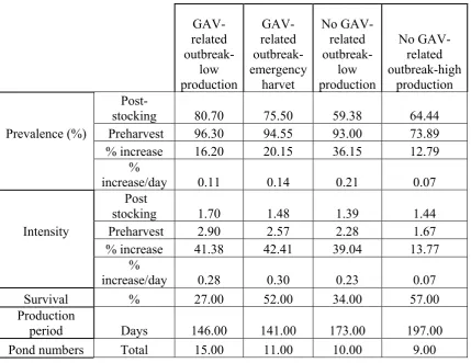
(7) To determine if RT-nPCR could be used to determine the semi-quantitative load, GAV from homogenised P. monodon gill tissue was purified on a continuous sucrose gradient. The RNA was then isolated from GAV and ten-fold serially diluted into crayfish (Cherax quadricarinatus) RNA that was free of GAV. These serial dilutions were then reverse transcribed into cDNA and amplified with PCR. It could be seen that as the original sample of GAV became increasingly diluted, the corresponding amplified product became progressively lighter when visualised on an agarose gel containing 0.5 μg/ml ethidium bromide under ultraviolet light. It was concluded that the RT-nPCR could be used as a semi-quantitative tool for the subsequent cohort study. The ponds examined were categorised into four groups; i) GAV related outbreak – low production, ii) GAV related outbreak – emergency harvest, iii) no GAV related outbreak – low production, and iv) no GAV related outbreak – high production. The study found that GAV had a strong association with reduced production from the farms. Ponds with GAV related outbreaks had a statistically higher initial prevalence (75.5 % – 80.7 %) and a higher increase in prevalence (16.3 % - 20.1 %) over the production period. While the ponds with no GAV outbreak – low production had the lowest initial prevalence (59.4 %) but the largest increase in prevalence (36.2 %) over the production period and the ponds with no GAV outbreak – high production had the lowest increase in prevalence (12.8 %) over the production period. The association of GAV with reduced production was also seen with respect to the semi-quantitative loading of the infected prawns, as the ponds with GAV related outbreaks had the highest loading (2.6 – 2.9) (maximum being 4) and the highest percentage increase (41.8 % - 42.3 %) at harvest. While the ponds with no GAV outbreak – low production had a moderate increase (39 %) in GAV load and the ponds with no GAV outbreak – high production had the lowest increase (14 %) in load. It was concluded that the prevalence and loading of GAV are strongly associated with the severity of disease; with a increase in GAV correlating with a decreased production. Ponds with a higher initial prevalence and higher increase in load of v.
(8) GAV suffered GAV-related outbreaks. Ponds with low initial prevalence of GAV but with a high increase in prevalence and viral load over the production period suffered low level mortality resulting in no outbreak being identified, yet low production. The ponds that had moderate to low initial prevalence of GAV with a low increase in prevalence and load of GAV over the production period incurred no GAV related outbreak – high production. The second objective was achieved through the development of two alternative detection methods for GAV. Firstly, polyclonal antibodies (PAbs) from chickens and monoclonal antibodies (MAbs) derived from mice were produced against GAV and a capture ELISA was developed. Secondly, haemagglutination (HA) using chicken erythrocytes was used to detect GAV. These diagnostic tests for GAV were then tested for sensitivity, specificity, positive predictive value (PPV), negative predictive value (NPV), and overall accuracy when compared to the RT-nPCR as the gold standard for agreement with the ELISA or compared to the ELISA for comparison with HA. PAbs from chickens were produced against the 116 kDa and 64 kDa protein of GAV seen in Western blots. The development of the PAbs was based on the report that YHV consisted of four structural proteins of approximately 170, 135, 67 and 20 kDa. However it was later reported that the 170 kDa protein may have originated from prawn cells. The PAbs reacted to this 170 kDa. However, due to the specificity of the MAbs, this did not interfere with the developed capture ELISA. Of the 11 MAbs developed against the 20 kDa protein of GAV, all were IgM isotype. Monoclonal antibody 3K5-11 was used in immunohistochemical studies, Western blot analysis and affinity purification to demonstrate specificity to GAV. Haemagglutination using chicken erythrocytes tested the haemolymph, gill, lymphoid organ, heart, subcutaneous tissue, eye stalk, pleopods, uropods and the central nerve cord for agglutination activity in 100 P. monodon, with the haemolymph and gill tissue giving the highest end-point titres of 1:1370 and 1:361, respectively. The sensitivity of HA was demonstrated by testing two different populations of P. monodon, which had a highly significant difference (F = 56.4, DF = 4, 88, P<0.001) in HA activity, indicating a difference in viral load. By testing vi.
(9) three other penaeid species (n = 20 each), Penaeus esculentus, Penaeus merguiensis and Penaeus longistylis, and the crayfish, C. quadricarinatus, it was demonstrated that natural agglutinins were not causing the high agglutination in the population of P. monodon. There was no effect of freezing and thawing of samples on HA activity. The speed and low cost of HA makes it a very useful tool, particularly in the developing world, for on-farm testing of penaeid prawns to indicate YHV and GAV loads which can contribute to management practices with respect to the harvesting of ponds. The two developed tests were compared for agreement using 120 P. monodon for the presence of GAV. Initially, the ELISA was compared to the RT-nPCR and then the HA was compared to the ELISA. For the ELISA, the sensitivity was 97 %, the specificity was 65 %, the PPV was 93.3 %, the NPV was 81.3 % and the overall accuracy was 91.7% when using an optical density of greater than 0.75 as a positive result. The HA had a sensitivity of 88 %, specificity of 75 %, PPV of 79%, NPV of 61 % and an overall accuracy of 73 % compared to the ELISA or an estimated accuracy of 66.9 % when compared to the RT-nPCR when using an HA titre of greater than 16 as a positive result.. vii.
(10) TABLE OF CONTENTS STATEMENT OF ACCESS ...................................................................................... i DECLARATION......................................................................................................... i DECLARATION ON ETHICS................................................................................. ii ACKNOWLEDGMENTS ........................................................................................ iii ABSTRACT ............................................................................................................... iv LIST OF TABLES .................................................................................................. xiv LIST OF FIGURES ................................................................................................ xvi LIST OF ABBREVIATIONS ............................................................................... xxii CHAPTER 1 ............................................................................................................... 1 General Introduction ................................................................................................. 1 CHAPTER 2 ............................................................................................................... 4 REVIEW OF LITERATURE ................................................................................... 4 2.1. Properties of the Okavirus.......................................................................... 4. 2.2. Viruses of the yellow head complex .......................................................... 5. 2.3. Infection and morphology of yellow head-like viruses ............................. 7. 2.4. Disease and distribution of yellow head-like viruses............................... 10. 2.5. Mode of infection of yellow head-like viruses ........................................ 14. 2.6. Interactions with other viruses ................................................................. 16. 2.7. Yellow head-like infections in other crustaceans..................................... 20. 2.8. Signs of infection and detection methods of YHLV................................ 22. 2.9. Conclusion ............................................................................................... 26. viii.
(11) CHAPTER 3 ............................................................................................................. 29 DEVELOPMENT OF A SYNTHETIC POSITIVE CONTROL WHICH DETECTS PLASMID CONTAMINATION IN DIAGNOSTIC POLYMERASE CHAIN REACTION FOR GILL-ASSOCIATED VIRUS................................... 29 3.1. Introduction .................................................................................................. 29. 3.2. Material and Methods ................................................................................. 30. 3.2.1. Diagnostic primers ................................................................................... 30. 3.2.2. PCR plasmid positive control .................................................................. 30. 3.3. Results ........................................................................................................... 33. 3.3.1 3.4. Plasmid positive control........................................................................... 33 Discussion...................................................................................................... 35. CHAPTER 4 ............................................................................................................. 36 IS GILL-ASSOCIATED VIRUS RELATED TO DECREASED PRODUCTION OF Penaeus monodon IN AUSTRALIAN PRAWN FARMS .............................. 36 4.1. Introduction .................................................................................................. 36. 4.2. Materials and Methods ................................................................................ 38. 4.2.1. Purification of gill-associated virus ......................................................... 38. 4.2.2. RNA isolation .......................................................................................... 38. 4.2.3. Serial dilution of GAV RNA ................................................................... 38. 4.2.4. Reverse transcription - nested PCR.......................................................... 39. 4.2.5. PCR primers ............................................................................................. 39. 4.2.6. Prawn samples from this farm study........................................................ 39. 4.2.7. Positive control ........................................................................................ 39. 4.2.8. Negative control ....................................................................................... 40. 4.2.9. Case definitions of GAV-related outbreak............................................... 40. 4.2.10. Grading system ........................................................................................ 40. ix.
(12) 4.2.11 4.3. Statistical analysis .................................................................................... 41. Results ........................................................................................................... 42. 4.3.1. Determining if PCR grading system gives a semi-quantitative result ..... 42. 4.3.2. Component two – Determining if GAV has an effect on production ...... 43. 4.4. Discussion...................................................................................................... 50. CHAPTER 5 ............................................................................................................. 54 PRODUCTION OF POLYCLONAL AND MONOCLONAL ANTIBODIES AGAINST GILL-ASSOCIATED VIRUS AND THE DEVELOPMENT OF AN ELISA........................................................................................................................ 54 5.1. Introduction .................................................................................................. 54. 5.2. Materials and Methods ................................................................................ 55. 5.2.1. Purification of gill-associated virus ......................................................... 55. 5.2.2. Polyacrylamide gel electrophoresis.......................................................... 55. 5.2.3. Production of polyclonal antibodies in chickens ..................................... 56. 5.2.4. Western blots............................................................................................ 56. 5.2.4.1 Protein transfer ......................................................................................... 56 5.2.4.2 Immunostaining Western blots ................................................................ 57 5.2.5. Visualisation of affinity purified protein from MAbs.............................. 57. 5.2.6. Fixation of tissue ...................................................................................... 57. 5.2.7. Preparation of paraffin sections for immunostaining............................... 58. 5.2.8. Immunostaining of tissue sections ........................................................... 58. 5.2.9. Antigen preparation for development of indirect and capture ELISA..... 59. 5.2.10. Optimisation of indirect ELISA ............................................................... 59. 5.2.11. Cell count ................................................................................................. 60. 5.2.12. Production of monoclonal antibodies in Balb/c mice .............................. 60. 5.2.12.1 Immunisation schedule ............................................................................ 60. x.
(13) 5.2.12.2 Culture of myeloma cells ......................................................................... 60 5.2.12.3 Optimisation of capture ELISA ............................................................... 61 5.2.12.4 Screening for GAV using capture ELISA................................................ 62 5.2.12.5 Preparation of spleen cells ....................................................................... 62 5.2.12.6 Fusion protocol ........................................................................................ 63 5.2.12.7 Screening for antibody production........................................................... 63 5.2.12.8 Cloning of monoclones ............................................................................ 64 5.2.13. Cryopreservation of cell lines .................................................................. 64. 5.2.13.1 Freezing cultured cells ............................................................................. 64 5.2.13.2 Thawing cells from liquid nitrogen storage ............................................. 64 5.2.14. Antibody isotyping................................................................................... 65. 5.2.15. RNA isolation .......................................................................................... 65. 5.2.16. Reverse transcription (RT) – PCR ........................................................... 65. 5.2.17. PCR primers ............................................................................................. 65. 5.2.18. Animal Ethics........................................................................................... 65. 5.3. Results ........................................................................................................... 66. 5.3.1. Development of an ELISA for GAV ....................................................... 66. 5.3.2. Confirmation that the developed MAbs and PAbs antibodies are specific for GAV ...................................................................................... 67. 5.4. Discussion...................................................................................................... 70. CHAPTER 6 ............................................................................................................. 73 HAEMAGGLUTINATION AS A LOW COST DETECTION METHOD FOR GILL-ASSOCIATED VIRUS AND BY INFERENCE, YELLOW HEAD VIRUS IN Penaeus monodon .................................................................................. 73 6.1. Introduction .................................................................................................. 73. 6.2. Materials and Methods ................................................................................ 75. xi.
(14) 6.2.1. Viral acquisition ....................................................................................... 75. 6.2.2. Determination of optimal tissue/organ to obtain virus............................. 75. 6.2.3. Obtaining and storing chicken erythrocytes............................................. 75. 6.2.4. Haemagglutination ................................................................................... 75. 6.2.5. Controls .................................................................................................... 76. 6.2.6. Determination of the effect of freezing samples ...................................... 76. 6.2.7. Determination of natural agglutinins in Penaeus esculentus, Penaeus. merguiensis and Penaeus longistylis ...................................................................... 76 6.2.8. RNA isolation to determine prevalence of GAV ..................................... 76. 6.2.9. Reverse transcription for GAV ................................................................ 77. 6.2.10. PCR primers towards GAV...................................................................... 77. 6.2.11. Statistical analysis .................................................................................... 77. 6.2.11. Animal Ethics........................................................................................... 77. 6.3. Results ........................................................................................................... 78. 6.3.1. Prevalence of GAV in studied penaeids................................................... 78. 6.3.2. Determining optimal tissue/organ from haemagglutination .................... 78. 6.3.3. Determining haemagglutination titre from other penaeid prawns ........... 79. 6.3.3. Determining the effect of freezing samples on haemagglutination titre .. 81. 6.4. Discussion...................................................................................................... 81. CHAPTER 7 ............................................................................................................. 85 SENSITIVITY AND SPECIFICITY OF CURRENT DIAGNOSTIC TESTS FOR GILL-ASSOCIATED VIRUS ....................................................................... 85 7.1. Introduction .................................................................................................. 85. 7.2. Materials and Methods ................................................................................ 86. 7.2.1. Sample preparation of gill tissue for comparison between tests .............. 86. 7.2.2. RNA isolation .......................................................................................... 86. xii.
(15) 7.2.3. Reverse transcription (RT) – nested PCR ................................................ 86. 7.2.4. PCR primers ............................................................................................. 86. 7.2.5. Semi-quantitative grading with RT-nPCR ............................................... 86. 7.2.6. Screening for GAV using a capture ELISA............................................. 87. 7.2.7. Haemagglutination .................................................................................. 88. 7.2.8. Obtaining and storing chicken erythrocytes............................................. 88. 7.2.9. Controls .................................................................................................... 88. 7.2.10. Comparison clarification.......................................................................... 89. 7.2.11. Optical density cut off.............................................................................. 89. 7.3. Results ........................................................................................................... 90. 7.3.1. Comparison of the ELISA with RT-nPCR .............................................. 90. 7.3.2. Comparison of haemagglutination with RT-nPCR .................................. 92. 7.4. Discussion...................................................................................................... 95. CHAPTER 8 ............................................................................................................. 97 General Discussion ................................................................................................... 97 LITERATURE CITED.......................................................................................... 104 APPENDIX 1 – Papers and Presentations........................................................... 113. xiii.
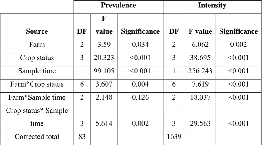
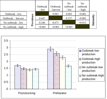
(16) LIST OF TABLES Page Table 2.1. Natural and experimental host range for YHLV. Table 4.1. The effect of husbandry factors on the prevalence and. 22. intensity of GAV over the entire production period. Note: Crop status = GAV-related outbreak-low production, GAV-related outbreak-emergency harvest, No GAV-related outbreak- low production, No GAV-related outbreak-high production, Sample time = poststocking and preharvest Table 4.2. 44. Average prevalence and intensity of GAV in the three farms at one month poststocking, one week preharvest and the percentage increase between the sampling periods and percentage of daily increase (n = 1800). Table 4.3. 45. Significant differences in prevalence (lower triangle) and intensity (upper triangle) of GAV between ponds poststocking. Note: Crop status = GAV-related outbreak-low production, GAV-related outbreakemergency harvest, No GAV-related outbreaklow production, No GAV-related outbreak-high production, Sample time = poststocking and preharvest. Table 4.4. 48. Significant differences in prevalence (lower triangle) and intensity (upper triangle) of GAV between ponds preharvest. Note: Crop status = GAV-related outbreak-low production, GAV-related outbreakemergency harvest, No GAV-related outbreak- low production, No GAV-related outbreak-high production, Sample time = poststocking and preharvest. 49. xiv.
(17) Table 4.5. Significant differences in survival in ponds that had GAV-related outbreaks-low production, GAV-related outbreaks-emergency harvest, no GAV-related outbreaks-low production and no GAV-related outbreaks-high production. Note: Crop status = GAV-related outbreak-low production, GAV-related outbreak-emergency harvest, No GAV-related outbreak- low production, No GAV-related outbreak-high production, Sample time = poststocking and preharvest. Table 6.1. 50. Significant differences (P value) of haemagglutination titre from individual organs/tissue compared to other individual organs/tissue from Penaeus monodon. Note: (L.O.) lymphoid organ, (Subcut) subcutaneous tissue, (CNC) central nerve cord. Table 6.2. 79. Significant differences (P value) of haemagglutination titre using gill tissue between different species of penaeid prawns. Note: (*) frozen for unknown period. Table 8.1. 80. Cost analysis of reverse transcription nested polymerase chain reaction, enzyme linked immunosorbent assay and haemagglutination when used as detection tests for GAV based on 40 ponds, with 20 sample prawns/pond and two sample periods. Note *- Technician cost can dramatically vary depending on the labour costs of individual countries. 102. xv.
(18) LIST OF FIGURES Page Figure 2.1. Family tree of the genera and families within the order Nidovirales. Figure 2.2. 5. A YHLV-infected Penaeus monodon lymphoid organ cell showing paracrystalline arrays of enveloped virions (A). Figure 2.3. 8. Organisation of the 26235 nt (+) ssRNA YHLV genome indicating translation features and deduced open reading frame (ORF) functions. Figure 2.4. 9. Diagrammatic representation of the structural components and schematic morphology of coronavirus virions. Note: N, nucleocapsid protein; sM, small-membrane protein; HE, haemagglutinin-esterase protein. Figure 2.5. 10. Model for the infection and disease cycle of yellow head complex viruses. Figure 2.6. 14. TEM of vesicle showing particles (PAR) of 50 - 96 nm in diameter and rod-shaped structures (ROD) 155 – 207 nm long. Figure 2.7. 19. TEM of vesicles within nerve cells of the fasciculated zone of the eye of moribund Penaeus monodon. The vesicles are 3 μm in diameter and contain unidentified particles (PAR) of 20 nm in diameter. Some particles also appear to be free in the cytoplasm. Figure 2.8. 20. The world prawn aquaculture production and value up to 2004 (FAO).. 26 xvi.
(19) Figure 3.1. A diagrammatic representation of the oligonucleotide developed for the PCR positive control using primers developed for the detection of gill-associated virus. Figure 3.2. 31. RT-PCR (A) and RT-nPCR (B) of clinical samples of Penaeus monodon. A: RT – PCR products (618 bp), using primers GAV5 and GAV6 (lanes 1 – 20) B: RT – nPCR products (317 bp) (lanes 1 – 13), using primers GAV1 and GAV2. (M) molecular weight marker, (-C) negative control, (+C) plasmid positive control. Figure 4.1. 34. RT-PCR (A) and RT-nPCR (B) of RNA of GAV from gills of Penaeus monodon 10-fold serially diluted in RNA of Cherax quadricarinatus A: RT – PCR products (618 bp), using primers GAV5 and GAV6 (lanes A - E) B: RT – nPCR products (317 bp) (lanes D - J), using primers GAV1 and GAV2. Note: (M) molecular weight marker, (-) negative control, (+) plasmid positive control. Figure 4.2. 43. Daily percentage increase in prevalence and intensity of the farms over the production period. Note: Outbreak – low = GAV related outbreak – low production, Outbreak – emergency harvest = GAV related outbreak – emergency harvest, No outbreak – low = no GAV related outbreak – low production, No outbreak – high = no GAV related outbreak – high production. 46. xvii.
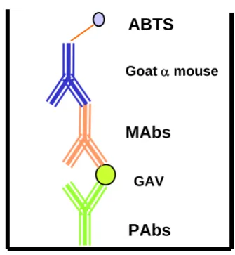
(20) Figure 4.3. Percentage increase of GAV intensity over the production period. Note: Outbreak – low = GAV related outbreak – low production, Outbreak – emergency harvest = GAV related outbreak – emergency harvest, No outbreak – low = no GAV related outbreak – low production, No outbreak – high = no GAV related outbreak – high production. Figure 4.4. 47. Prevalence of GAV from the ponds at poststocking, preharvest and the percentage increase over the production period. Note: Outbreak – low = GAV related outbreak – low production, Outbreak – emergency harvest = GAV related outbreak – emergency harvest, No outbreak – low = no GAV related outbreak – low production, No outbreak – high = no GAV related outbreak – high production. Figure 4.5. 47. GAV intensity in ponds at poststocking and at preharvest. Note: Outbreak – low = GAV related outbreak – low production, Outbreak – emergency harvest = GAV related outbreak – emergency harvest, No outbreak – low = no GAV related outbreak – low production, No outbreak – high = no GAV related outbreak – high production. Figure 5.1. 49. A diagrammatic representation of the capture ELISA used in this study to detect GAV. Figure 5.2. 61. Titration of antigen for the optimisation of a capture ELISA. Figure 5.3. 66. Titration of polyclonal antibodies for the optimisation of the capture ELISA. Figure 5.4. 66. Measured optical density readings of the developed monoclonal antibodies to GAV in a capture ELISA. 67 xviii.
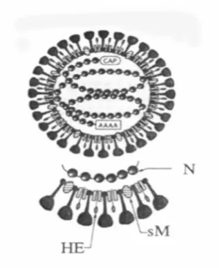
(21) Figure 5.5. Western blot using 3K5-11 MAb against purified GAV detecting a single protein band of 20 kDa. Figure 5.6. 67. Western blot of PAbs against purified GAV showing three protein bands of 170, 110 and 63 kDa respectively. Figure 5.7. 68. Affinity purified GAV protein eluted from the capture 3K5-11 MAb, visualised by PAGE and stained with silver stain. Figure 5.8. 68. Immunostaining gills of P. monodon using 3K5-11 MAb raised against GAV to demonstrate potential specificity towards GAV. Figure 5.9. 69. A lack of immunostaining with gills of P. merguiensis using 3K5-11 MAb raised against GAV demonstrating the antibodies are not binding to prawn gills. Figure 5.10. 69. Capture ELISA using a single prawn gill filament to obtain the antigen. The filament was homogenised and diluted to 1:128. Note: prawns 1 – 20 are Penaeus monodon. (C) – Control = P. merguiensis. Figure 6.1. 70. A reaction between the agglutinating virus and a receptor of the erythrocyte. Note: The virus can attach to more than one erythrocyte causing viral bridging indicating a positive result when testing for haemagglutination. RBC = red blood cell. Figure 6.2. 74. Haemagglutination titre of various organs/tissue from Penaeus monodon. Note: (CNC) central nerve cord, (L.O) lymphoid organ. 78. xix.
(22) Figure 6.3. Haemagglutination titre of gill tissue from penaeid prawns. Note: (*) frozen Penaeus monodon. Figure 6.4. 80. The effects of serial freezing of P. monodon gill tissue (n=20) on haemagglutination titre. Figure 7.1. 81. Reverse transcription nested polymerase chain reaction (gold standard) results from 120 P. monodon showing the prevalence and semi-quantitative viral load of GAV for comparison with the ELISA results (Figure 7.3). Figure 7.2. 90. ELISA readings from 120 P. monodon showing the percentage of prawns recorded as positive at different optical density readings. Figure 7.3. 91. Prevalence and comparative level of infection of GAV when using the ELISA for GAV (n = 120) for comparison with the reverse transcription nested polymerase chain reaction results (Figure 7.1). Figure 7.4. 91. The sensitivity, specificity, positive predictive value, negative predictive value and the accuracy of the ELISA for GAV using different cut-off values as a negative result when compared to the reverse transcription nested polymerase chain reaction. Figure 7.5. 92. Prevalence and comparative level of infection of GAV when using the ELISA for GAV (n = 120) for comparison with the haemagglutination results (Figure 7.7). Figure 7.6. 93. Showing the affect of increasing the haemagglutination titre cut off on the percentage of prawns that were recorded positive for gill-associated virus. 93 xx.
(23) Figure 7.7. Prevalence and recorded level of infection of gill associated virus when using the haemagglutination for the detection of GAV (n = 120) in comparison to the ELISA (Figure 7.5). Figure 7.8. 94. The sensitivity, specificity, positive predictive value, negative predictive value and the accuracy of haemagglutination for gill-associated virus using different cut off titres compared to the ELISA. Note: Optical density used for ELISA was <0.4 = negative. 94. xxi.
(24) LIST OF ABBREVIATIONS. A260. absorbance at 260 nm. A280. absorbance at 280 nm. ABTS. 2,2'-azino-di-(3-ethylbenzthiazoline-6-sulphonic acid). APS. ammonium persulphate. AU. Australian. BDS. bovine donor serum. bp. base pairs. BSA. bovine serum albumin. cDNA. complimentary deoxyribonucleic acid. CO2. Carbon dioxide. d. days. DAB. 3, 3'-diaminobenzidine tetrahydrochloride. D.P.X. di-n-butylphethalate polysyrene xylene. DMSO. dimethyl sulfoxide. DNA. deoxyribonucleic acid. EDTA. ethylenediamine tetraacetic acid. ELISA. enzyme linked immunosorbent assay. FAO. food and agriculture organisation of the United Nations. FBS. foetal bovine serum. g. gram. g. gravity. GAV. gill-associated virus. h. hours. HA. haemagglutination. HAT. hypoxanthine-aminopterin-thymidine. H&E. haematoxylin and eosin. HRP. horseradish peroxidase. HT. hypoxanthine-thymidine. ICTV. International Committee on Taxonomy of Viruses. IHHNV. infectious hypodermal and haematopoietic necrosis virus. kb. kilobase. xxii.
(25) kDa. kiloDalton. LOV. lymphoid organ virus. LO. lymphoid organ. LPV. lymphoid parvo-like virus. LSD. least significant difference. MAbs. monoclonal antibodies. MAb. monoclonal antibody. MBV. monodon baculovirus. MCMS. mid-crop mortality syndrome. MoV. Mourilyan virus. MW. molecular weight. NDV. newcastle disease virus. NPV. negative predictive value. O.D. optical density. OPI. oxaloacetate-pyruvate-insulin media supplement. ORF. open reading frame. PAbs. polyclonal antbodies. PBS. phosphate buffered saline. PCR. polymerase chain reaction. PEG. polyethylene glycol. PL. postlarvae. PPV. positive predictive value. PVDF. polyvinylidene fluoride. RBC. red blood cell. RNA. ribonucleic acid. RT-nPCR. reverse transcription nested polymerase chain reaction. RT-PCR. reverse transcription polymerase chain reaction. SDS. sodium-dodecyl-sulphate. SDS-PAGE. sodium-dodecyl-sulphate poly-acrylamide gel electrophoresis. SEM. scanning electron microscopy. SEMBV. systemic ectodermal and mesodermal baculovirus. SMV. spawner-isolated mortality virus. SPSS. statistical package of social sciences. TAE. tris/acetate/EDTA xxiii.
(26) TCID50. 50% tissue culture infectious dose assay. TE. tetracycline. TEM. transmission electron microscope. TEMED. tetramethylethylenediamine. TNE. tris/NaCl/EDTA. U. units. US. United States. WSSV. white spot syndrome virus. YHLV. yellow head-like virus. YHV. yellow head virus. v. volts. xxiv.
(27) CHAPTER 1 General Introduction Penaeid aquaculture was originally performed as “catch and hold” culture systems. For centuries, Southeast Asian farms have been producing incidental crops of wild prawns in tidal ponds. With the advent of ever advancing technology and the increasing requirement for low cost protein as a food source, the penaeid prawn culture has advanced from its experimental beginnings into major industries providing hundreds of thousands of jobs, billions of dollars in revenue, and an expansion of the world’s food supply with a high value crop (Lightner and Redman 1998). The importance of disease within the industry has increased proportionally with the growth of the prawn industry. Until the early 1990’s, prawn aquaculture exhibited astonishing growth of 16.8 % per annum between 1984 and 1995 (Subasinghe, Bartley, McGladdery, and Barg 1998). Recently, however, diseases have had devastating impacts on prawn farming. An economic impact assessment from Lundin (1997) reported that in 1994, just over two billion United States (US) dollars were lost due to disease. Disease outbreaks have continued to cause major losses. Diseases with viral aetiologies have been the most important cause of economic loss in the majority of countries (Fegan and Clifford 2001). The emergence of ‘new’ viruses is rapidly increasing. For example, in 1990, Lightner, Bell, and Redman reported six viruses affecting penaeid prawns. By 1992, the list of known viruses affecting penaeid prawns had increased to 12 (Lightner 1996) and presently, approximately 20 viruses have been reported in penaeid prawns. Three of these viruses have been responsible for the most severe losses. These are, in order of their discovery, Taura syndrome virus (TSV), yellow head virus (YHV) and white spot syndrome virus (WSSV). Australia’s zoogeographical isolation from Southeast Asia enabled it to be fairly late with respect to the major viral disease outbreaks. However by 1994, major disease outbreaks were occurring within the Australian prawn farming industry, with. 1.
(28) mortalities reaching as high as 80% (Owens, Haqshenas, McElnea, and Coelen 1998). This disease outbreak was dubbed mid-crop mortality syndrome (MCMS). There were four viruses associated with this syndrome. Initially it was thought to consist of only two viruses, spawner-isolated mortality virus (SMV) and gillassociated virus (GAV). However, recently it was reported that infectious hypodermal and haematopoietic necrosis virus (IHHNV) (Krabsetsve, Cullen, and Owens 2004) which is similar to SMV with respect to being a non-enveloped DNA parvo-like virus, and Mourilyan virus (MoV) (Cowley, McCulloch, Spann, Cadogan, and Walker 2005) which is similar to GAV with respect to being a enveloped RNA virus, were also present during the outbreaks. These viruses could have equally influenced the mortality of the infected prawns as they have all been associated with disease. There were two main objectives of this study. Firstly, to determine if GAV and by inference, YHV have an association with disease in Penaeus monodon within the prawn farming industry. GAV and YHV are reported to be geographic topotypes, sharing 85.1% sequence identity (Cowley et al., 1999) and have been reported to be highly pathogenic to P. monodon. The pathogenicity of GAV was determined by inoculation of filtered homogenates of lymphoid organ, gills and whole cephalothoraces from P. monodon that were positive for GAV, resulting in mortality to other P. monodon which were already infected with GAV (Spann, Cowley, Walker, and Lester 1997a; Vega, Degnan, Hall, Cowley, and Wilson 2004). The method of determining the pathogenicity of a virus via crude inoculations should be obsolete due to our current knowledge of multiple infections of viruses within penaeid prawns. Determining pathogenesis with this method is only accurate when either pathogen-free stock or crustacean cell lines are available with viable pure virus. At present, there are approximately twenty research papers describing different aspects of GAV and only one of those papers by Callinan, Jiang, Smith, and Soowannayan (2003a) attempts to determine if GAV is associated with causing an economic impact on Australian prawn farms.. 2.
(29) In the present study, the first objective investigates the association of GAV with survival on three prawn farms located over three different geographic locations along the eastern coast of Australia. This aims to determine whether GAV is associated with disease and mortality within the Australian P. monodon aquaculture industry. The second objective was to develop detection methods for GAV, and by inference YHV, which are cost effective for small scale prawn farmers in Australia and developing countries. There are currently many molecular techniques for the detection of GAV/YHV. Despite these PCR techniques being highly sensitive for the detection of the viruses, there are practical limitations to their widespread application. These limitations include the requirement for specialised equipment, expensive molecular reagents and well-trained personnel. Consequently, these high costs result in the assay being non-viable for the majority of prawn farmers or small support laboratories that screen for viruses. The following study aimed at developing non-molecular detection methods which were both sensitive and specific whilst being relatively low cost.. 3.
(30) CHAPTER 2 REVIEW OF LITERATURE 2.1. Properties of the Okavirus. The purpose of this literature review is to focus on the yellow head-like viruses. There has been considerable confusion with respect to the taxonomic classification of these viruses. Initially, the yellow head-like viruses were proposed to be a baculovirus due to their size and enveloped rod-shaped appearance (Chantanachookin, Boonyaratpalin, Kasornchandra, Direkbusarakom, Ekpanithanpong, Supamataya, Sriurairatana, and Flegel 1993). However, upon the discovery that the genome consisted of ssRNA, it was suggested that the virus was either a rhabdovirus or a coronavirus (Wongteerasupaya, Sriurairatana, Vickers, Akrajamorn, Boonsaeng, Panyim, Tassanakajon, Withyachumnarnkul, and Flegel 1995a). Loh, Tapay, Lu, and Nadia (1997) reported the genome to possibly be negative in polarity resulting in the virus being classified as Rhabdoviridae. Tang and Lightner (1999) subsequently reported that yellow head virus (YHV) was a plusstrand RNA virus via in situ hybridisation and sequence analysis. These latest results placed YHV into the corona-like viruses. Cowley, Dimmock, Spann, and Walker (2000a) reported that the yellow head-like virus genomes contain a ORF1a polyprotein containing a 3C-like Cys protease, an ORF1b coding sequence with replicase functions including an SDD polyprotein, and a helicase domain, an efficient – 1 ribosomal frameshift site at the ORF1a/1b overlap that will facilitate translation of a 759 kDa ORF1ab polyprotein. From this they concluded that the yellow headlike viruses were a unique member of the Nidovirales. The yellow head-like viruses have subsequently been placed as members of a new genus Okavirus of a new family Roniviridae, within the order Nidovirales (Mayo 2002). There are two other families within the order Nidovirales. These are Coronaviridae and Arteriviridae (Figure 2.1).. 4.
(31) Equine arteritis virus Arterividae. Lactate dehydrogenaseelevating virus. Arterivirus. Porcine respiratory and reproductive syndrome virus Simian hemorrhagic fever virus Group 1 species Coronavirus Nidovirales. Group 2 species. Coronaviridae Group 3 species unclassified coronavirues Bovine torovirus Equine torovirus Torovirus. Human torovirus Porcine torovirus. Roniviridae. Okavirus. Yellow head-like virus. Figure 2.1. Family tree of the genera and families within the order Nidovirales. 2.2. Viruses of the yellow head complex. Currently, the literature isolates the yellow head-like viruses into two distinct viruses, being gill-associated virus (GAV) and YHV. GAV is the junior synonym of lymphoid organ virus (LOV) which was reported in 1995 by Spann, Vickers, and Lester. LOV was reported to be found only in the lymphoid organ and was similar to YHV with respect to ultrastructural and cytopathological features. However, LOV had no association with disease and mortality (Spann et al., 1995; Spann, Cowley, Walker, and Lester 1997a). GAV was subsequently reported as a pathogenic relative of LOV, found both in the lymphoid organ and the gills of infected Penaeus monodon (Spann et al. 1997a). With the development of a reverse transcription nested polymerase chain reaction (RT-nPCR) towards GAV it was reported that LOV had a 98.9% nucleotide identity to the GAV sequence, indicating that they are the same virus (Cowley, Dimmock, Spann, and Walker 2000b). However, the. 5.
(32) method to determine the level of nucleotide similarity used only two clones to determine the 1.1 % (3/274) nucleotide variation. In contrast, GAV had an 85.1 % nucleotide identity to YHV from a 577 bp region and a 83 % nucleotide identity to YHV from a 135 bp sequence of a cDNA clone. From this, YHV was reported to be a closely related geographic topotype of GAV. However, this study only performed sequences analysis on three YHV clones (Cowley, Dimmock, Wongteerasupaya, Boonsaeng, Panyim, and Walker 1999). Even though GAV and YHV are currently classified as distinct viruses, it appears that the research applied to form these conclusions was very limited. Due to the extremely small number of clones that were sequenced and the small sequence region, the research did not take into account the possibility of the thousands of mutants within the clones which can constitute so-called quasi-species within a population (van Regenmortel 2000) not to mention the possible natural genomic variation within the so-called two distinct viruses. Therefore, due to the small sample size, the sequence variation was not representative of the actual population and the actual nucleotide variation could be a lot larger or smaller than the reported variation resulting in either the viruses being the same virus or distinctly different. Since 1991, the International Committee on Taxonomy of Viruses (ICTV) has accepted the definition that “a virus species is a polythetic class of viruses that can constitute a replicating lineage and occupy a particular ecological niche”. van Regenmortel (2000) lists the following characteristics for discriminating between virus species: •. Relatedness of genome sequence. •. Natural host range. •. Cell and tissue tropism. •. Pathogenicity and cytopathology. •. Mode of transmission. •. Physicochemical properties of virions. •. Antigenic properties of viral proteins. Due to this, and the fact that the viruses are morphologically indistinguishable and cause the same gross disease, in this literature review, GAV and YHV will be referred to as the same virus, being ‘yellow head-like virus’ (YHLV).. 6.
(33) 2.3. Infection and morphology of yellow head-like viruses. A study by Lu, Tapay, Loh, Brock, and Gose (1995) reported that YHLV particles were detected in the gill, lymphoid organ, head soft tissue, heart, midgut, hepatopancreas, abdominal muscle, eyestalk and nerve cord of an experimentally infected Penaeus vannamei. Lu et al. (1995) reported that the lymphoid organ, gill and head muscle had a 50 % tissue culture infectious dose assay (TCID50) titer (ml-1) of 106, while the midgut, abdominal muscle and heart had a TCID50 titer (ml-1) of 105 and the nerve cord, hepatopancreas and the eyestalk had a TCID50 titer (ml-1) of 104. These findings suggest that the lymphoid organ, gill and head muscle contained the highest number of infectious virions compared to the other tested tissue/organs. These results also indicated that the viral infection was systemic. Virions of similar morphological appearance to YHLV have also been reported in the optic nerve fibres and in the nerve cord of P. monodon (Smith 2000; Callinan et al., 2003a). To confirm the entire viral distribution in prawn tissues, a more comprehensive examination needs to be conducted in other organs and tissues such as haematopoietic tissue, Y-organ, stomach, antennal gland, periopods, pleopods and uropods. YHLV replication occurs in the cell cytoplasm, primarily in the prawn lymphoid organ, gills, haemocytes and connective tissues (Cowley, Dimmock, Spann, and Walker 2001). The YHLV virions are rod-shaped, enveloped particles containing helical nucleocapsids that mature by the process of budding through intracytoplasmic membranes (Chantanachookin et al. 1993, Spann et al. 1997a). The nucleocapsids exhibit striations with a periodicity of approximately 7 nm and are often observed in association with the distended endoplasmic reticulum (Spann et al. 1997a). The virions vary between 160-186 nm by 38-50 nm and 183-200 nm by 34-42 nm in size and are often packed densely into vesicles, resembling paracrystalline arrays (Figure 2.2). Free virions are also observed in intercellular spaces probably via release from disintegrating cells (Chantanachookin et al 1993; Spann and Lester 1997c). Within all stages of YHLV infected cells, virogenic stroma and filamentous YHLV nucleocapsids, 116-435 nm by 16-18 nm in size are often observed scattered. 7.
(34) randomly within the cytoplasm (Spann and Lester 1997c). The nucleocapsid of YHLV becomes enveloped by passage through the endoplasmic reticulum or the virions have occasionally been observed invading the interstitial spaces of the lymphoid organ and gain their envelope by passage through the plasma membrane (Spann and Lester 1997c).. THIS IMAGE HAS BEEN REMOVED DUE TO COPYRIGHT RESTRICTIONS. Figure 2.2. A YHLV infected Penaeus monodon lymphoid organ cell showing paracrystalline arrays of enveloped virions (A). Scales bar = 200 nm (Spann et al. 1995) YHLV consists of 26,235 nucleotides that are organised into four open reading frames (Figure 2.3) (Cowley and Walker 2002). Initially YHLV was thought to consist of four structural proteins with the following estimated molecular weights: 170, 135, 67, and 22 kDa (Nadala, Tapay, and Loh 1997). The proteins were thought to represent, respectively the L (RNA transcriptase), G (spike), N (nucleocapsid), and M (matrix). However, Wang and Chang (2000) reported only three major YHLV proteins, being: 110, 63, and 20 kDa in size and suggested that the larger protein (170 kDa) reported by Nadala et al. (1997) was cellular in origin. However, it is possible that due to the techniques used to purify the virus by Wang and Chang (2000), the 170 kDa polyprotein may have been cleaved to produce the reported 110 and 63 kDa proteins.. 8.
(35) THIS IMAGE HAS BEEN REMOVED DUE TO COPYRIGHT RESTRICTIONS. Figure 2.3. Organisation of the 26235 nt (+) ssRNA YHLV genome indicating translation features and deduced open reading frame (ORF) functions (Cowley and Walker 2002). Haemagglutination (HA) activity from YHLV has been reported by Nadala et al. (1997). They reported that purified YHLV agglutinated chicken erythrocytes yielding a HA end-point titre of 1:256 and the virus was not eluted after 24 hours, suggesting the reaction was stable and that the virus lacked receptor-destroying enzymes. HA activity from YHLV infected prawns was confirmed by Munro and Owens (2005) while YHLV-free prawns demonstrated negligible HA activity. The protein responsible for the HA is thought to be similar to the HA protein on the outside of some Coronaviruses (Figure 2.4). 9.
(36) Figure 2.4. Diagrammatic representation of the structural components and schematic morphology of coronavirus virions. Note: N, nucleocapsid protein; sM, smallmembrane protein; HE, haemagglutinin-esterase protein.. 2.4. Disease and distribution of yellow head-like viruses. The YHLV throughout most of Southeast Asia is reported to be a highly pathogenic agent for cultured P. monodon, causing significant mortalities and adversely affecting the mariculture prawns in Thailand (Chantanachookin et al. 1993). The YHLV was first described in Thailand in 1990 by Limsuwan (Chantanachookin et al. 1993). Limsuwan named the new syndrome based on the light yellow colouration of the dorsal cephalothorax area and the general pale appearance of the infected prawn. The yellow appearance was a result of the underlying enlarged yellow hepatopancreas. Since this time, the disease has been associated with epizootic mortalities in Thailand. YHLV is reported to be one of the most highly virulent viruses of the causative agents in Thailand, since it is associated with massive mortality of P. monodon, the principle penaeid species cultured in Thailand (Sithigorngul, Chauychuwong, Sithigorngul, Longyant, Chaivisuthangkura, and Menasveta 2000). Since the identification of the causative agent as YHLV in Thailand in 1990 and the resulting epizootic mortalities it was associated with, the virus has been associated with mortalities in penaeid prawns in Taiwan, Indonesia, Malaysia, China, Philippines, India and the Americas (Lightner 1996; Mohan 1996).. 10.
(37) There has been only one reported occurrence of YHLV in the Americas at a Penaeus setiferus farm in Texas. The farm was in close proximity to a prawn processing plant and it was suggested that the virus was imported from Asia (Lightner, Redman, Poulos, Nunan, Mari, and Hasson 1997). The YHLV has also been detected in frozen prawns that had been imported into the US from Asia. At present, there is no evidence to show that YHLV is now present in wild or farmed prawns in the Americas. Initially, YHLV principally infected pond-reared juvenile to sub-adult prawns of 5 to 15 g in size, especially at 50 - 70 days of grow-out (Lightner 1996). Since the initial epizootic, it has been suggested that the disease is now less severe than when YHLV was first isolated in Thailand. YHLV infection is now common in healthy prawns. This disease resistance is proposed to be from “active accommodation” by a tolerance mechanism involving binding of viral antigens to cellular receptors during the early life stages of the prawn (Flegel and Pasharawipas 1998). This theory of “active accommodation” is supported in the literature. For example, transmission electron microscopy (TEM) of broodstock collected in Thailand prior to the initial reports of the virus, indicated that YHLV was present in one out of seven healthy broodstock sampled. During the peak of the YHLV epidemic in Thailand, YHLV was detected by TEM in gill samples from at least one prawn from 15 ponds with gross signs and a pond history that indicated YHLV was present. In the three ponds with no signs of YHLV disease, six out of six prawns sampled negative for YHLV by TEM. However, later in the YHLV epidemic, YHLV could be detected by TEM in gill samples from 33 out of 44 healthy prawns sampled from 11 ponds without signs of YHV (Walker, Cowley, Spann, Hodgson, Hall and Withyachumnarnkul 2001). A study of 19 P. monodon broodstock collected from hatcheries in Thailand was conducted using RT-PCR on total nucleic acid extracted from gill tissue (Wongteerasupaya, Tongchuea, Boonsaeng, Panyim, Tassanakajon, Withyachumnarnkul, and Flegel 1997). All 19 prawns tested negative for YHLV. However, using a 2-step RT-PCR on the same prawns resulted in 15 of the 19 prawns (78.9%) testing positive for YHLV (Wongteerasupaya et al 1997). A survey 11.
(38) conducted in the Philippines on 219 healthy prawns with Western blot analysis also indicated a relatively high prevalence (24.2%) of YHLV infection (Natividad, Magbanua, Migo, Alfafara, Albaladejo, Nadala, Loh, and Tapay. 1999). The prevalence varied between 0 – 66.7% depending upon which districts were sampled. There was also evidence of a higher prevalence of infection in postlarvae (54.5%) than in broodstock (16.9%). With respect to the limited sensitivity of Western blot methods compared to 2-step PCR, the true level of chronic YHLV infection in the Philippines may be much higher. Yang, Shariff, Lee, Hassan (2000) also reported that YHLV occurs in high prevalence in Malaysia but it does not appear to have been associated with significant mortalities. These studies suggest that a high proportion of apparently healthy P. monodon broodstock from some areas of Asia carry chronic infections of YHLV. It is highly likely that the previous TEM studies of broodstock and farmed prawns in Thailand would not have detected this low level of infection that is often only evident in 2-step PCR. Therefore, chronic YHV infection may have been highly prevalent in healthy prawns prior to the appearance of the disease. The increase in prevalence of infection detected by TEM could have been due to a general increase in viral load in the farmed prawns. The relationship between viral load and susceptibility to disease is the subject of ongoing research. Within Australia, YHLV has been associated with significant mortalities which have adversely affected the prawn farm industry in Australia since at least 1996 (Spann, Donaldson, Cowley, and Walker 2000). YHLV infections can be chronic or acute. A chronically infected P. monodon with YHLV displays no gross signs of disease or tissue necrosis, while acute infections result in necrosis, disease and mortalities. Under experimental conditions YHLV is reportedly highly pathogenic, causing mortalities from 4 to 5 days after infection (Walker et al. 2001). In the farming environment, mortalities from YHLV are generally observed in prawns between 8 and 15 g, although prawns up to 40 g have exhibited signs of disease (Spann and Lester 1997). The reported prevalence of YHLV within Australia in P. monodon broodstock from a sample size of 148 prawns captured in north eastern Queensland was 97.3%. 12.
(39) Prevalence of YHLV in postlarvae from a sample size of 50 was 100% and prevalence of YHLV in juveniles from a sample size of 56 was 98.2% (Walker et al. 2001). However, it was not reported how these samples were obtained, so it is unknown if they are from the same hatchery and/or the same broodstock. Even with this high prevalence of YHLV, not all YHLV infected P. monodon express disease. Currently, the factors involved for YHLV-related disease to be expressed are poorly understood. Some hypotheses of these factors are; the viral load of parental broodstock, the initial viral load of postlarvae or an unknown environmental factor acting as a stressing agent. YHLV has been reported to be highly pathogenic to P. monodon within Australia. The pathogenicity of YHLV was determined by inoculation of filtered homogenates of lymphoid organ, gills and whole cephalothoraces from P. monodon that were positive to YHLV, resulting in mortality from 7 to 8 days (d) post-inoculation (Spann et al., 1997a). However, at the time of the pathogenicity trial there were at least five other viruses infecting Australian P. monodon. These viruses consisted of monodon baculovirus (MBV) (Doubrovsky, Paynter, Sambhi, Atherton, and Lester 1988), lymphoid parvo-like virus (LPV) (Owens, De Beer, and Smith 1991), IHHNV (Owens, Anderson, Kenway, Trott, and Benzie 1992), spawner-isolated mortality virus (SMV) (Fraser and Owens 1996) and Mourilyan virus (MoV) (Cowley, et al., 2005) which all could have possibly influenced the mortality of the P. monodon in this trial. This infection trial was repeated in 2004 by Vega et al. In that study, the prawns were again inoculated with filtered prawn homogenate containing levels of YHLV (not purified virus). They reported that 100% (15/15) of the YHLV injected prawns died compared to 40 % (2/5) of the controls while there was a significant increase (P = 0.010) in YHLV for both the infected prawns and the controls during the trial. However, the YHLV increase was significantly higher (P = 0.047) in the YHLV injected prawns than the control prawns. Again, no other viruses were tested for in that study or referred to as potential pathogens. There are many papers that report YHLV to be pathogenic. However there is no substantial evidence to support this. Currently there is only an association with disease. Many papers report that severe necrosis of the lymphoid organ (LO) is a 13.
(40) typical lesion caused by YHLV (Boonyaratpalin et al., 1993; Chantanachookin et al., 1993; Lu, Tapay, Brock, and Loh 1994; Wang, Tang, Kou, and Chen 1996). However, penaeid prawns with severe white spot syndrome virus (WSSV) exhibit the same marked LO necrosis (Pantoja and Lightner 2003). The current method of injecting YHLV infected crude homogenate of prawn tissue to determine pathogenicity should be obsolete with our current knowledge of possible multiple viral infections within the same prawn or homogenate sample. To demonstrate a direct pathogenic effect of YHLV, either pathogen-free stock would be required or a viable penaeid cell line would be needed with purified viable YHLV. Until these requirements are available, YHLV can only be said to be associated with disease.. 2.5. Mode of infection of yellow head-like viruses. The modes of infection of the yellow head-like viruses have been placed into two general groups, horizontal transmission and vertical transmission (Figure 2.5).. THIS IMAGE HAS BEEN REMOVED DUE TO COPYRIGHT RESTRICTIONS. Figure 2.5. Model for the infection and disease cycle of yellow head complex viruses (Walker et al. 2001) Horizontal transmission can occur when YHLV-free P. monodon either feed on infected carcasses, experience bath exposure to membrane-filtered tissue extracts, by cohabitation with infected prawns, or by direct experimental injection of the viral. 14.
(41) inoculum (Walker et al. 2001). An experiment to determine the susceptibility of postlarval (PL) P. monodon of YHLV by ingestion showed that PL20 died 7-10 days post-infection but PL15 survived the exposure (Spann, Donaldson, McCulloch, Cowley, and Walker submitted). The available data suggested that disease was associated with viral loading. Walker et al. (2001) reported that YHLV from a diseased P. monodon caused mortalities after ingestion or immersion exposure but extracts from YHLV-infected healthy P. monodon caused infection without mortality. However, after the viral concentration of the healthy prawns had an equivalent titre to the diseased prawn, YHLV extracts from chronically infected, healthy P. monodon also induced disease, indicating disease is associated with viral loading. The potential for vertical transmission was first reported by Chantanachookin et al. (1993). They reported that YHLV infection in larval offspring could occur from latent, asymptomatic infected, broodstock prawns. This theory was originally dismissed for YHLV because TEM screening of P. monodon in Thailand suggested that the prevalence of YHLV was low, and therefore, that vertical transmission was unlikely to contribute significantly to the occurrence of infection and disease on farms (Flegel, Boonyaratpalin, and Withyachumnarnkul. 1997). With more recent screening of broodstock with RT-nPCR, Walker et al. (2001) suggested that the prevalence of YHLV infection in Thailand may be significantly higher than originally indicated using TEM. To determine if vertical transmission of YHLV contributes to the high prevalence of chronic infections in wild and farmed P. monodon in eastern Australia, Cowley, Hall, Cadogan, Spann, and Walker (2002) tested gonads and lymphoid organs for signs of YHLV from healthy male and female P. monodon broodstock and in fertilized eggs and from nauplii spawned from wild-fertilized females using RT- nPCR. They reported that the level of YHLV in wild P. monodon was generally low. However, high levels of YHLV were detected in moribund male broodstock reared in captivity for more than 12 months. The RT-nPCR product from spermatophore in these prawns were also significantly greater than those amplified from the lymphoid organ, which had previously been identified as the primary site of YHLV replication in chronically infected P. monodon (Spann et al. 1995). It was also reported that in 1 of 15.
(42) 3 spermatophores examined by TEM, mature YHLV virions were detected in the seminal fluid but not in the sperm cells. The RT- nPCR for YHLV in eggs were positive, however, nauplii and protozoea were generally negative. This suggested that YHLV is associated with the egg surface and the majority of the virus is lost when the nauplii hatch and that the infection levels in the protozoea remain low. Cowley et al. (2002) suggested that this could be due to the lack of development of the lymphoid organ in larval and early postlarval life stages and is likely to limit potential infection levels. Walker et al. (2001) reported that RT- nPCR has detected YHLV in PL5 to PL15 both from hatcheries and experimental spawnings of P. monodon. Walker et al. (2001) suggested that viral replication in postlarvae is occurring at sufficient levels to be detected. Cowley et al. (2002) reported that the identification of lymphoid organ spheroid bodies and YHLV particles in ∼1.2 g juvenile P. monodon grown from hatchery stocks (PL6 and PL20) suggests that at least some of the postlarvae were infected with YHLV. However, since individual postlarvae were not grown in isolation, it cannot be discounted that some juvenile infections may have occurred during the course of the grow-out through cannibalism or water-borne transmission. This horizontal transmission could promote translocation of YHLV and the potential infection of wild P. monodon in the vicinity of farms via water or through the escape of infected farmed prawns. The authors concluded that the high prevalence of chronic YHLV infection in P. monodon broodstock from northeastern Queensland and farmed prawns produced from these broodstock strongly suggests that this is perpetuated primarily by vertical transmission both in the wild and in hatcheries. However, in that paper, the authors did not comment on the likely survival of the YHLV infected progeny. They only tested eggs and nauplii. It is only an assumption that these postlarvae survive through to adulthood.. 2.6. Interactions with other viruses. At present there are approximately 18 viruses that have been reported in penaeids. Not all of these viruses have been shown to cause disease or to interact with each other once they have infected the prawn. Within the YHLV, there are two main groups of viruses that have been indicated to cause disease when a dual infection. 16.
(43) occurs. The two groups of viruses that interact with each other are the YHLV with WSSV and YHLV with SMV. WSSV was previously classed as a baculo-like virus (Nadala, Tapay, and Loh 1998). However, it has recently been placed into a new family and genus, being family Nimaviridae, genus Whispovirus (Mayo 2002). It was first seen as a dual infection with YHLV in Thailand in P. monodon in 1993 and was originally named Systemic Ectodermal and Mesodermal Baculovirus (SEMBV) (Wongteerasupaya, Vickers, Sriurairatana, Nash, Akrajamorn, Boonsaeng, Panyim, Tassanakajon, Withyachumnarnkul, and Flegel 1995b). The virus was first characterised as WSSV from an outbreak in a Penaeus japonicus in Japan in 1993 (Flegel 1997). The naming of the virus derived from the gross examination displaying white spots. However, Chou, Huang, Wang, Chiang, and Lo (1995) reported that the first epizootic of WSSV occurred in Taiwan in 1992. The dual infection of WSSV and YHLV has since been reported in India and Taiwan (Mohan, Shankar, Kulkarni, and Sudha. 1998; Wang and Chang 2000) Both WSSV and YHLV can cause significant mortalities in penaeid prawns (Chantanachookin et al. 1993; Liu, Wang, Tian, Yin, and Kwang 2002) and are currently the most serious diseases threatening the prawn farming industry in Thailand (Flegel et al. 1997). At present there is no further information on the interaction between YHLV and WSSV in cultured prawns. Wang and Chang (2000) suggested that the reason for mass loss in the prawn culture industry in Taiwan between 1996 and 1999 was not only WSSV, but a dual infection of WSSV and YHLV. Prawns with dual infection, generally exhibit only typical signs of white spot syndrome with the YHLV symptoms being less obvious. This would result in the mixed disease being diagnosed as only WSSV infection. In 1994, prawn farms in northern Australia experienced increased mortality rates in 12 to 15 g prawns, with mortality reaching as high as 80% in some ponds (Owens et al. 1998). This disease outbreak was dubbed mid-crop mortality syndrome (MCMS). An investigation into the syndrome initially revealed two distinct viral types using TEM (Owens et al. 1998). The two viruses that were implicated as being involved in MCMS were YHLV and SMV (Anderson and Owens 2001). SMV is a 17.
(44) parvo-like virus which had been first identified by Fraser and Owens (1996). This virus was isolated from prawns affected with MCMS by Owens et al. (1998). Using a bioassay, Owens et al. (1998) reported that this parvo-like virus was capable of causing mortality. The gross symptoms of SMV were lethargy, reduced feeding, and redness of the carapace and pleopod (Fraser and Owens 1996). Owens et al. (1998) reported that SMV virulence in MCMS affected prawns was enhanced by the presence of other coinfecting viruses such as an enveloped, filiform virus. The coinfecting virus was YHLV, which had first been identified by Spann et al. (1995). The enhanced virulence from YHLV was demonstrated when Owens et al. (1998) treated prawn extract with ether prior to injecting it into P. monodon. As SMV is unenveloped, the ether should not have harmed the parvovirus. The extract killed at a slower rate than untreated extract, suggesting that the SMV was capable of causing mortality but that its virulence was enhanced by the presence of other coinfecting viruses such as an enveloped, filiform virus like YHLV. Spann et al. (1997a) reported that YHLV was also isolated from prawns affected by MCMS. They reported that diseased prawns were observed swimming at the surface and edge of ponds and displayed varying degrees of red body colouration. Symptoms from both of these viruses were apparent in MCMS affected prawns. In recent years, two other viruses have been reported being present during the MCMS. These viruses are IHHNV (Krabsetsve et al., 2004) which is similar to SMV with respect to being a non-enveloped DNA virus and MoV (Cowley et al., 2005) which is similar to YHLV with respect to being an enveloped RNA virus. These two viruses could both have influenced the mortality of the infected prawns. In 2000, Smith reported that lesions were found in a P. monodon displaying nonspecific signs of disease. He reported that the causative agents of lesions appeared to be Vibrio spp. and a rod shaped virus similar to YHLV. With the use of TEM it was observed that the nerve cells in the fasciculated zone contained cytoplasmic vesicles with particles and rod shaped nucleocapsids. These rods were similar to YHLV and were 130 to 260 nm long by 10 to 16 nm in diameter and had a helical symmetry with a screw like thread. Also an unidentified enveloped virus, ranging from 50 – 96 nm in diameter, was observed in cytoplasmic vesicles in the fasciculated zone (Figure 2.6). Smith (2000) reported that the unidentified enveloped virus was a. 18.
(45) possible aetiological agent in one of the disease outbreaks. In the paper, Smith (2000) did not suggest what the virus was. It is likely that this virus was MoV which is an enveloped virus, averaging 85 – 100 nm in diameter. From the TEM photograph (Figure 2.7), Smith (2000) also reports particles of 20 nm in diameter. However, Smith (2000) does not suggest to what these could be. It is highly likely that these particles were the parvovirus SMV which is reported to be non-enveloped, averaging 20 nm in diameter. This would suggest triple viral interaction between YHLV, SMV and MoV to cause disease.. THIS IMAGE HAS BEEN REMOVED DUE TO COPYRIGHT RESTRICTIONS. Figure 2.6. TEM of vesicle showing particles (PAR) of 50 - 96 nm in diameter and rod-shaped structures (ROD) 155 – 207 nm long (Smith 2000).. 19.
(46) THIS IMAGE HAS BEEN REMOVED DUE TO COPYRIGHT RESTRICTIONS. Figure 2.7. TEM of vesicles within nerve cells of the fasciculated zone of the eye of moribund Penaeus monodon. The vesicles are 3 μm in diameter and contain unidentified particles (PAR) of 20 nm in diameter. Some particles also appear to be free in the cytoplasm (Smith 2000). 2.7. Yellow head-like infections in other crustaceans. A range of crustaceans can be infected with YHLV (Table 2.1). P. monodon is the only crustacean that is commonly affected by YHLV (Walker et al. 2001). However, several other penaeid prawns and other crustaceans have been reported to be susceptible by either natural or experimental infection. Flegel (1997) reported that, unlike WSSV, YHLV has not been seen to infect crabs or freshwater prawns and that the wide spectrum of hosts within the penaeid species suggests there is a common mechanism of infection that results in preventative methods being hard to develop. Natural infection from YHLV has only been detected in Penaeus esculentus that were co-cultivated with P. monodon (Table 2.1). However, only 14 prawns from one location were tested with 8 being positive. For a more substantial conclusion to be drawn, larger sample numbers are needed. The YHLV infection in P. esculentus was reported to be chronic and there were no signs of gross disease. In Thailand, it has. 20.
Figure


+7


Related documents