Copyright © 2004, American Society for Microbiology. All Rights Reserved.
Identification of an Envelope Protein from the FRD Family of Human
Endogenous Retroviruses (HERV-FRD) Conferring Infectivity and
Functional Conservation among Simians
Sandra Blaise,
1Alessia Ruggieri,
2Marie Dewannieux,
1Franc¸ois-Loic Cosset,
2and Thierry Heidmann
1*
Unite´ des Re´trovirus Endoge`nes et Ele´ments Re´troïdes des Eucaryotes Supe´rieurs, UMR 8122 CNRS, Institut Gustave Roussy,
94805 Villejuif,1and Laboratoire de Vectorologie Re´trovirale et The´rapie Ge´nique, INSERM U412, Ecole Normale
Supe´rieure de Lyon, 69364 Lyon,2France
Received 15 July 2003/Accepted 30 September 2003
A member of the HERV-W family of human endogenous retroviruses (HERV) had previously been demon-strated to encode a functional envelope which can form pseudotypes with human immunodeficiency virus type 1 virions and confer infectivity on the resulting retrovirus particles. Here we show that a second envelope protein sorted out by a systematic search for fusogenic proteins that we made among all the HERV coding envelope genes and belonging to the HERV-FRD family can also make pseudotypes and confer infectivity. We further show that the orthologous envelope genes that were isolated from simians—from New World monkeys to humans—are also functional in the infectivity assay, with one singular exception for the gibbon HERV-FRD gene, which is found to be fusogenic in a cell-cell fusion assay, as observed for the other simian envelopes, but which is not infectious. Sequence comparison of the FRD envelopes revealed a limited number of mutations among simians, and one point mutation—located in the TM subunit—was shown to be responsible for the loss of infectivity of the gibbon envelope. The functional characterization of the identified envelopes is strongly indicative of an ancestral retrovirus infection and endogenization, with some of the envelope functions subsequently retained in evolution.
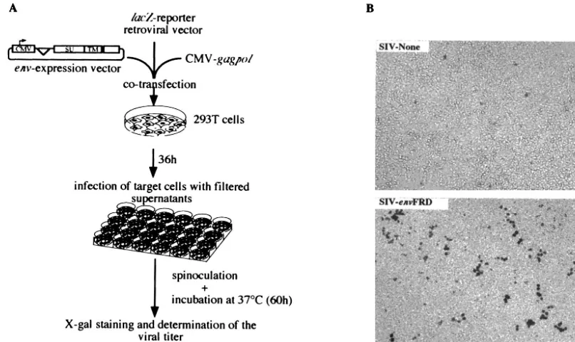
Human endogenous retroviruses (HERV) comprise approx-imately 8% of the human genome (9). Most of the identified elements are defective due to mutations and/or deletions within their genes, but some elements have conserved fully open reading frames (3, 12, 17, 18). A systematic search for coding envelope genes has led to the identification of 16 such genes (8). It had previously been demonstrated that one of these genes, the HERV-W envelope gene, is indeed functional, since it can induce cell-cell fusion when expressed in cells possessing the RDR receptors (5, 10) and since pseudotypes generated with human immunodeficiency virus type 1 (HIV-1) virions are infectious (2, 10). In this study, we show that an-other envelope protein, associated with an endogenous retro-virus of the so-called HERV-FRD family (see references 16 and 17) and sorted out by a systematic screen of the human genome for fusogenic envelopes (4), can also confer infectivity on pseudotypes generated with lentiviral virions. To do so, we cloned the HERV-FRD envelope gene into a human cytomeg-alovirus (hCMV) promoter-driven expression vector and used a previously described assay (10), as schematized in Fig. 1A. In this assay, human 293T cells are cotransfected with an expres-sion vector for the retroviral proteins—except the envelope— from type C viruses (murine leukemia virus [MLV]) or lenti-viruses (HIV-1 and simian immunodeficiency virus [SIV]), a corresponding lacZ gene-marked defective retroviral vector, and an expression vector for theenvgene to be tested (or an
empty vector as a negative control and a vector for the am-photropic MLV envelope as a positive control). Then, the pseudotype virions are assayed for infectivity after recovery of the transfected-cell supernatant 36 h posttransfection, transfer of the supernatant onto target test cells, and centrifugation of the plates (i.e., spinoculation [see reference 10]) and, after an additional 60-h period, counting thelacZ-positive test cell col-onies by in situ histochemical staining for-galactosidase ac-tivity. The results of such an assay are given in Fig. 1B and Table 1, for the various viral cores (MLV, SIV, and HIV-1) and target cells tested, which included murine, feline, and human cells. It can be clearly observed that the HERV-FRD envelope, when expressed on lentiviral particles, confers infec-tivity on the virions, with evidence for viral titers on feline (G355-5) and human (TE671 and 293T) cells in the 100- to 500-CFU/ml range. Interestingly, the murine 3T3 cells and the human HeLa cells are refractory to infection (although they are positive for the control amphotropic MLV envelope), a result consistent with the absence of any detectable syncytium formation in a fusion assay carried out with these cells (see below). It can also be noted that the MLV core is much less efficient—if at all—than the lentiviral cores, as similarly ob-served for the HERV-W envelope (2, 5, 10). Finally, since some envelopes are sensitive to shearing, viral titers were also measured in the absence of the spinoculation step: in all cases, its omission (data not shown) resulted in a 5- to 20-fold reduc-tion of the titers in Table 1 (and similarly for the simian FRD envelopes assayed as shown in Fig. 2).
Previous experiments had shown that HERV-FRD is a very ancient family, which entered the primate branch after the prosimian and simian divergence but before the split between
* Corresponding author. Mailing address: Unite´ des Re´trovirus En-doge`nes et Ele´ments Re´troïdes des Eucaryotes Supe´rieurs, UMR 8122 CNRS, Institut Gustave Roussy, 94805 Villejuif, France. Phone: 33/1-42-11-49-70. Fax: 33/1-42-11-53-42. E-mail: heidmann@igr.fr.
1050
on November 8, 2019 by guest
http://jvi.asm.org/
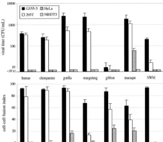
New World and Old World monkeys, 40 million years ago (4). Moreover, we had identified in the simian genomes—by PCR using primers in the HERV-FRD provirus flanking DNA—the gene orthologous to the humanenvFRD gene and shown that
[image:2.603.92.495.73.313.2]it is present in a fully coding state in all simians, from New World monkeys to humans (4). We therefore assayed whether the simian orthologous env genes also confer infectivity on SIV-pseudotype virions. The results of an experiment similar to that carried out for Fig. 1 for the human gene are given in Fig. 2, which discloses the following features: (i) all simian ERV-FRD envelopes (with one exception [see below]) confer infectivity on the pseudotype virions, with closely related viral titers (up to 2,000 CFU/ml) and target cell specificity, thus demonstrating conservation over ⬎40 million years of the ERV-FRD envelope function, but (ii) there is one exception to this rule for the gibbon envelope, which is negative for all the target cells tested. Interestingly, this negative result is not due to a severe defect in the gibbon envelope, since the protein is still capable of promoting cell-cell fusion, as illustrated in the lower panel of the figure by an experiment carried out in parallel using the same expression vectors and cells: the results show that for all simian envelopes (except the gibbon enve-lope) there is a good correlation, as expected, between infec-tivity and cell-cell fusion and that the gibbon envelope is clearly positive for cell-cell fusion, with the same cell specificity as that of the other simian envelopes. These features, which de facto uncouple the processes of cell-cell fusion and viral entry, could
FIG. 1. Infection assays using retroviral particles pseudotyped with the HERV-FRD and control envelopes. (A) Rationale of the assay. To determine whether the HERV-FRD envelope could confer infectivity on MLV, HIV, or SIV Env-defective pseudoparticles, corresponding pseudotype viruses were produced by cotransfecting 7.5 ⫻ 105293T cells with 0.55 g of the phCMV-HERV-FRD vector expressing the
HERV-FRD envelope (or pcDNA3 for the negative control and phCMVampho for the amphotropic MLV envelope positive control); 1.75g of a vector encoding the retroviral proteins (except the envelope) of MLV (phCMVintron) (13), HIV (pCMV⌬R8.91) (20), or SIV (pSIV3⫹) (13); and 1.75g of the corresponding defective retroviral vectors marked with a-galactosidase reporter gene (pMFGsnlslacZ[7], pHR⬘CMVlacZ [20], and R9SA [14]), by calcium phosphate precipitation (Invitrogen). At 36 h posttransfection, viral supernatants were collected and filtered through 0.45-m-pore-size membranes. Target cells (murine 3T3; feline G355-5; or human TE671, HeLa, and 293T cells) were seeded in 24-well plates at a density of 104cells per well and incubated overnight at 37°C. Five to 500l of virus samples containing 4g of Polybrene per ml was
[image:2.603.43.283.547.691.2]added to the cells and centrifuged for spinoculation at 1,200⫻gfor 2 h 30 min at 25°C (10). After removal of the supernatants, the cells were incubated in regular medium for 60 h at 37°C. Viral titers were then measured by X-Gal (5-bromo-4-chloro-3-indolylphosphate) staining of the cells and expressed as lacZ CFU per milliliter of viral supernatant. (B) Infectivity of SIV-based retroviral particles pseudotyped with the HERV-FRD envelope (or with no protein in the “SIV-None” control), as assayed using human 293T cells as target cells and X-Gal staining, following the protocol described for panel A.
TABLE 1. Infection assay of the HERV-FRD envelopea
Core Env
Viral titer (CFU/ml) NIH
3T3 G355-5 TE671 HeLa 293T
MLV FRD 0 0 1.9⫻101 0 2.0⫻101
None 0 0 0 0 6
Ampho 4⫻105 2⫻106 4⫻106 1⫻106 1.1⫻106
HIV FRD 1 1⫻102 2.1⫻101 0 8.8⫻101
None 0 0 0 0 6
Ampho 2⫻105 1.4⫻106 1⫻105 1.2⫻104 6⫻104
SIV FRD 0 3.6⫻102 2.3⫻102 4 3.4⫻102
None 0 5 0 0 6
Ampho 5⫻105 1.2⫻106 6.8⫻105 4.3⫻105 8⫻105 aExperimental conditions were the same as described for Fig. 1. The retroviral
particles (core) were pseudotyped with the HERV-FRD envelope (FRD), no protein (None), or the amphotropic MLV envelope (Ampho), and the target test cells are indicated. Viral titers are the means from two independent experiments.
on November 8, 2019 by guest
http://jvi.asm.org/
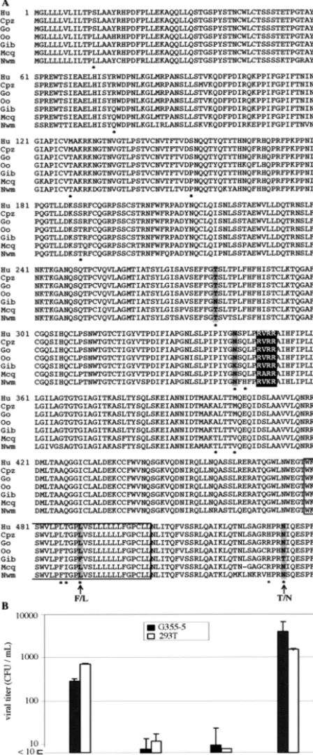
be accounted for by either intrinsic differences in the molecular requirements for the two processes or differences, possibly, in the envelope viral load and/or stability (see discussion in ref-erences 1, 10, and 11). At the sequence level, it is noteworthy that the gibbon envelope discloses strong conservation with the other simian FRD envelopes, as it is 97% identical to the human sequence, i.e., it is closer than the infection-positive marmoset New World monkey envelope, which is only 88% identical to the human sequence (Fig. 3A). Actually, compar-ison of the gibbon and human sequences reveals only four mutations which are gibbon specific, two within the SU and two within the TM subunits (shaded in Fig. 3A). Preliminary experiments using a chimeric construct with the human SU domain fused to the gibbon TM moiety strongly suggested that mutations in the TM subunit were responsible for the loss of infectivity of the gibbon envelope, as infectivity of the chimeric envelope remained low (data not shown). Two constructs with reversion of the identified TM point mutations were therefore tested for infectivity, one with the F-to-L reversion at position 490 and one with the T-to-N reversion at position 532. As illustrated in Fig. 3B, the T-to-N reversion fully restored in-fectivity to the gibbon envelope, with no effect of the F-to-L mutation located in the hydrophobic transmembrane domain
of the TM subunit. Due to its specific location in the cytoplas-mic tail of the envelope protein, it is likely that the N-to-T mutation alters some of the functions and interactions associ-ated, in infectious retroviruses, with this specific domain and involved in the loading, stability, and conformational transition of the Env proteins (e.g., see references 6, 15, and 19; see also references in reference 1). In this respect, the gibbon envelope together with the other currently characterized simian enve-lopes can be considered natural mutants and be of use in studying the refined molecular processes associated with enve-lope function and virus entry.
[image:3.603.130.454.70.353.2]Whatever the underlying molecular events, the present func-tional characterization of the ERV-FRD envelopes is strongly suggestive of an ancestral infection event by a functional ret-rovirus and its subsequent endogenization, with some of the envelope functions retained in evolution. Close examination of the primate FRD envelope sequences indeed reveals a preva-lence of synonymous rather than nonsynonymous substitutions (ratios for all pairwise comparisons,⬎3, by the SNAP.pl pro-gram at the www.hiv.lanl.gov site), thus suggesting positive selection for a physiological function which remains to be un-raveled.
FIG. 2. Infection and cell-cell fusion activities and host range of the HERV-FRD and simian orthologous envelopes. Upper panel: infection of feline (G355-5), human (293T and HeLa), and murine (3T3) cells by SIV-based particles pseudotyped with the indicated ERV-FRD envelopes. Experimental conditions were the same as in Fig. 1, with negative and positive control values as in Table 1. NWM, New World monkey (Callithrix jacchusmarmoset). Lower panel: cell-cell fusion assay for the ERV-FRD envelopes and cells as in the upper panel. The G355-5 and HeLa cells were transfected using Lipofectamine (Invitrogen; 2g of DNA for 5⫻105cells), and the 293T and TE671 cells were transfected using calcium
phosphate precipitation (Invitrogen; 5 g of DNA for 5⫻ 105cells). Fusion activities were measured 12 to 36 h posttransfection of the
corresponding expression vectors. To visualize syncytia, cells were fixed in methanol and stained by adding May-Gru¨nwald and Giemsa solutions (Sigma) according to the manufacturer’s instructions. The fusion index, which represents the percentage of fusion events in a cell population, is defined as [(N⫺S)/T]⫻100, whereNis the number of nuclei in the syncytia,Sis the number of syncytia, andTis the total number of nuclei counted.
on November 8, 2019 by guest
http://jvi.asm.org/
This work was supported by the CNRS and by grants from the Ligue Nationale contre Le Cancer (Equipe Labelise´e).
REFERENCES
1. Aguilar, H. C., W. F. Anderson, and P. M. Cannon.2003. Cytoplasmic tail of Moloney murine leukemia virus envelope protein influences the conforma-tion of the extracellular domain: implicaconforma-tions for mechanism of acconforma-tion of the R peptide. J. Virol.77:1281–1291.
2. An, D. S., Y.-M. Xie, and I. S. Y. Chen.2001. Envelope gene of the human endogenous retrovirus HERV-W encodes a functional retrovirus envelope. J. Virol.75:3488–3489.
3. Benit, L., P. Dessen, and T. Heidmann.2001. Identification, phylogeny, and evolution of retroviral elements based on their envelope genes. J. Virol.
75:11709–11719.
4. Blaise, S., N. de Parseval, L. Be´nit, and T. Heidmann.2003. Genomewide screening for fusogenic human endogenous retrovirus envelopes identifies syncytin 2, a gene conserved on primate evolution. Proc. Natl. Acad. Sci. USA100:13013–13018.
5. Blond, J.-L., D. Lavillette, V. Cheynet, O. Bouton, G. Oriol, S. Chapel-Fernandes, B. Mandrand, F. Mallet, and F.-L. Cosset.2000. An envelope glycoprotein of the human endogenous retrovirus HERV-W is expressed in the human placenta and fuses cells expressing the type D mammalian ret-rovirus receptor. J. Virol.74:3321–3329.
6. Brody, B. A., S. S. Rhee, and E. Hunter.1994. Postassembly cleavage of a retroviral glycoprotein cytoplasmic domain removes a necessary incorpora-tion signal and activates fusion activity. J. Virol.68:4620–4627.
7. Cosset, F.-L., Y. Takeuchi, J.-L. Battini, R. A. Weiss, and M. K. Collins.
1995. High-titer packaging cells producing recombinant retroviruses resis-tant to human serum. J. Virol.69:7430–7436.
8. de Parseval, N., V. Lazar, J. F. Casella, L. Benit, and T. Heidmann.2003. Survey of human genes of retroviral origin: identification and transcriptome of these genes with coding capacity for complete envelope proteins. J. Virol.
77:10414–10422.
9. Lander, E. S., L. M. Linton, B. Birren, C. Nusbaum, M. C. Zody, J. Baldwin, K. Devon, K. Dewar, M. Doyle, W. FitzHugh, et al.2001. Initial sequencing and analysis of the human genome. Nature409:860–921.
10. Lavillette, D., M. Marin, A. Ruggieri, F. Mallet, F. L. Cosset, and D. Kabat.
2002. The envelope glycoprotein of human endogenous retrovirus type W uses a divergent family of amino acid transporters/cell surface receptors. J. Virol.76:6442–6452.
11. Lavillette, D., M. Maurice, C. Roche, S. J. Russell, M. Sitbon, and F.-L. Cosset.1998. A proline-rich motif downstream of the receptor binding do-main modulates conformation and fusogenicity of murine retroviral enve-lopes. J. Virol.72:9955–9965.
12. Lo¨wer, R., J. Lo¨wer, and R. Kurth.1996. The viruses in all of us: character-istics and biological significance of human endogenous retrovirus sequences. Proc. Natl. Acad. Sci. USA93:5177–5184.
13. Negre, D., P. E. Mangeot, G. Duisit, S. Blanchard, P. O. Vidalain, P. Leiss-ner, A. J. Winter, C. Rabourdin-Combe, M. Mehtali, P. Moullier, J. L. Darlix, and F.-L. Cosset.2000. Characterization of novel safe lentiviral vectors derived from simian immunodeficiency virus (SIVmac251) that effi-ciently transduce mature human dendritic cells. Gene Ther.7:1613–1623. 14. Negre, D., P. E. Mangeot, G. Duisit, P. Moullier, J. L. Darlix, and F.-L
Cosset.2002. Lentiviral vectors derived from simian immunodeficiency virus (SIV). Curr. Top. Microbiol. Immunol.261:53–74.
15. Rein, A., J. Mirro, J. G. Haynes, S. M. Ernst, and K. Nagashima.1994. Function of the cytoplasmic domain of a retroviral transmembrane protein: p15E-p2E cleavage activates the membrane fusion capability of the murine leukemia virus Env protein. J. Virol.68:1773–1781.
16. Seifarth, W., H. Skladny, F. Krieg-Schneider, A. Reichert, R. Hehlmann, and C. Leib-Mosch.1995. Retrovirus-like particles released from the human breast cancer cell line T47-D display type B- and C-related endogenous retroviral sequences. J. Virol.69:6408–6416.
17. Tristem, M.2000. Identification and characterization of novel human
en-FIG. 3. Comparison of the simian ERV-FRD envelopes and iden-tification of a unique point mutation responsible for the gibbon enve-lope loss of function. (A) Aligned primary sequences of the HERV-FRD and simian orthologous envelopes. Abbreviations: Hu, Homo sapiens; Cpz, chimpanzee; Go, gorilla; Oo, orangutan; Gib, gibbon; Mcq, macaque; Nwm, New World monkey. Accession numbers are AL136139 for the human gene and AJ577595 to AJ577600 for the simian genes. The RVRR canonical motif for SU-TM cleavage is highlighted, and the predicted transmembrane hydrophobic domain is boxed. Amino acids differing between the human and gibbon proteins are indicated with an asterisk, among which those unique to the gibbon
sequence are shaded. The two amino acids in the TM moiety of the gibbon protein that were mutated and assayed for reversion to the infectious phenotype are indicated with an arrow. (B) Infection of feline (G355-5) and human (293T) cells by SIV-based particles pseudotyped with the wild-type (wt) and mutant (F4903L or T5323N) gibbon FRD envelopes, with the human FRD envelope as a control. The indicated amino acid changes in the gibbon envelope were performed by a three-fragment ligation, using PCR fragments gener-ated with oligonucleotides containing the gibbon-to-human single-base mutations (TTT3CTT and AAT3ACT for amino acid positions 490 and 532, respectively), and subsequent sequencing of the resulting constructs. Infection assays were performed as described for Fig. 1. The viral titers are averages from two independent experiments.
on November 8, 2019 by guest
http://jvi.asm.org/
[image:4.603.53.277.79.619.2]dogenous retrovirus families by phylogenetic screening of the human ge-nome mapping project database. J. Virol.74:3715–3730.
18. Turner, G., M. Barbulescu, M. Su, M. I. Jensen-Seaman, K. K. Kidd, and J. Lenz.2001. Insertional polymorphisms of full-length endogenous retrovi-ruses in humans. Curr. Biol.11:1531–1535.
19. Yu, X., X. Yuan, M. F. McLane, T. H. Lee, and M. Essex.1993. Mutations in
the cytoplasmic domain of human immunodeficiency virus type 1 transmem-brane protein impair the incorporation of Env proteins into mature virions. J. Virol.67:213–221.
20. Zufferey, R., D. Nagy, R. J. Mandel, L. Naldini, and D. Trono.1997. Multiply attenuated lentiviral vector achieves efficient gene delivery in vivo. Nat. Biotechnol.15:871–875.