Inorganic nitrogen and phosphorus nutrition in tridacnid clams and their algal symbionts
219
0
0
Full text
(2) INORGANIC NITROGEN AND PHOSPHORUS NUTRITION IN TRIDACNID CLAMS AND THEIR ALGAL SYMBIONTS. Thesis submitted by CARMELITA A. BELDA, B.Sc., M.Sc. (Phil.) in May 1994. for the degree of Doctor of Philosophy in the Department of Zoology at James Cook University of North Queensland.
(3) ii. ABSTRACT This study examined inorganic nutrition in tridacnids to address the question of nutrient limitation of their zooxanthellae, and the influence of elevated nutrient concentrations on calcification. Experiments consisted of three-monthlong exposures of cultured Tridacna gigas in outdoor tanks and indigenous T.. maxima in situ to increased levels of ammonia (N) and phosphate (P), alone or in combination (N+P). Following the incubation experiments, biomass and nutrientdepletion of the symbiotic partners were measured. In addition, a more detailed study was carried out on the effect of elevated P on the zooxanthellae, both in situ and in culture.. Major findings from the outdoor experiments were confirmed in the field study, while laboratory experiments elucidated host involvement in nutrient limitation of the zooxanthellae. In particular, zooxanthellae are N-limited in vivo, as demonstrated by the increased zooxanthellae density in N-supplemented clams. The decrease in ammonium-depletion by zooxanthellae isolated from these clams demonstrates that zooxanthellae have access to increased N in sea water. Nlimitation of zooxanthellae, therefore, is a function of sea water concentrations of inorganic N, particularly ammonia, as this nutrient can diffuse across cell membranes depending on its concentration gradient.. Also, zooxanthellae are P-limited in vivo. Regardless of ambient phosphate concentrations in sea water, zooxanthellae inside the animal host were not affected, and they exhibited N:P ratios (>30:1) and P-depletion rates similar to.
(4) iii those of P-starved cultured zooxanthellae. Host involvement in P availability to the zooxanthellae is strongly implicated by: (1) the unaffected acid phosphatase activity (P-liberating enzymes) and undetectable polyphosphates (P reserves) in zooxanthellae, regardless of the clams' P environment; and (2) rapid growth, decreased N:P ratio, and decreased P-depletion by zooxanthellae grown under Psufficient culture conditions. Host influence on the algae's P environment in vivo is further substantiated by the consistently low levels of inorganic phosphate (< 0.1 pM)) in the haemolymph surrounding the zooxanthellal tubes, despite the clams' uptake and assimilation of P from sea water (i.e., decreased N:P ratio). Plimitation of zooxanthellae, therefore, is independent of ambient sea water concentrations of phosphate, which cannot passively cross cell membranes. The host's role in P availability to its zooxanthellae is either: (1) a natural consequence of the morphological and spatial relationships between the symbiotic partners, with the host retaining phosphate for its own use before the nutrient can reach the zooxanthellae; or (2) through active P restriction by the host to control its algal population.. Investigation of calcification demonstrated that increased ambient levels of N and P modified shell formation in T. gigas as follows: (1) enhanced shellextension rates, but reduced shell weights at equivalent size; (2) changes in crystal lattice parameters based on X-ray diffractometry on the shells; and (3) structural alterations in the outer shell layer, with misshapen aragonite crystals, irregular crossed-lamellar orientation, and increased porosity. Such skeletal weakening has adverse implications for calcifying organisms in eutrophic reef waters..
(5) iv. STATEMENT ON SOURCES. I declare that this thesis is my own work and has not been submitted in any form for another degree or diploma at any university or other institution of tertiary education. Information deriVed from the published or unpublished work of others has been acknowledged in the text and a list of references is given.. C. A. BELDA May 1994.
(6) V. STATEMENT ON ACCESS TO THESIS. I, the undersigned, the author of this thesis, understand that James Cook University of North Queensland will make it available for use within the University Library and, by microfilm or other photographic means, allow access to users in other approved libraries. All users consulting this thesis will have to sign the following statement:. "In consulting this thesis I agree not to copy or closely paraphrase it in whole or in part without the written consent of the author; and to-make proper written acknowledgement for any assistance which I have obtained from it.". Beyond this, I do not wish to place any restriction on access to this thesis.. C. A. Belda May 1994.
(7) vi. ACKNOWLEDGEMENTS. This study was supported by my Fellowship Grant from the Australian International Development Assistance Bureau - Australian Centre for International Agricultural Research (AIDAB/ACIAR) and an Augmentative Research Grant from the Great Barrier Reef Marine Park Authority (GBRMPA). Additional support from Prof. John Lucas' URG and A/Prof. Dave Yellowlees' ENCORE, MRG, and URG grants are likewise gratefully acknowledged.. The ACIAR Giant Clam Project of John Lucas provided the cultured. Tridacna gigas I used in a substantial part of this study; while, GBRMPA gave permission to use part of a natural population of T. maxima in an experiment at One Tree Island Reef. Dr. John Norton's and ACIAR's permission to reproduce in this thesis a diagram of the zooxanthellal tube system from their atlas publication is also appreciated.. I sincerely thank my supervisors, John Lucas and Dave Yellowlees, for their guidance and assistance in all aspects of this research. John Lucas facilitated the early and trying years of my study, provided stimulating discussions on various aspects of the project, and imparted excellent lessons in scientific writing. Dave Yellowlees ably steered my work in the right direction, gave a helping hand on countless occasions, and greatly improved the logical presentation of this thesis. Both proved committed to their students and were encouraging all throughout despite their tight schedules.. Dr. Bill Fitt of the University of Georgia, in the course of a collaborative research with Dave Yellowlees and John Lucas, helped get myself started in an excellent project. His generous and patient guidance during the early years of my research is greatly valued. Dr. Chris Cuff of the Geology Department was instrumental in making the chapter on calcification possible. I sincerely appreciate his encouragement and selfless enthusiasm in the study..
(8) vii. The following friends also helped in the course of this study in many special ways: volunteers at Orpheus Island, namely, Jamie Whitford, Steve Lindsay, Rick Braley, Jeremy Barker, Geoff Charles, Russell, 'Snowy', Virginia, Chris Farr, Mary Flanagan, and Kevin Pery; volunteers at One Tree Island, namely, Dave Hawkings, Mabs Johnston, Alwyn Rees, and Tina Tentorie. I thank Dr. Ove Hoegh-Guldberg, in particular, for collecting T. maxima clams and placing them in the micro-atolls at One Tree Island. I also acknowledge the help of Sharon Ness and Allan Chappell of the Geology Department, Jim Darley of the Electron Microscopy Unit, and Alan Diamond of the Sir George Fisher Centre. The fun company of my peers in the laboratory, namely, Brett Baillie, Spencer Whitney, Jack Puanglarp, Karl Skewes, Danielle Johnston, Bill Leggat, and Amanda Fowler, will especially be remembered.. I cannot thank enough my unfailing source of encouragement and motivation all these years, Prof. Edgardo Gomez of the University of the Philippines Marine Science Institute. I thank him for sending me over here for a chance to enhance my professional skills and meet special people.. Finally, a most special thanks go to my best mate, Brett Baillie. Brett has helped me in a myriad of ways, generously assisting in many field trips, patiently sorting out for me many confusing calculations and computer operations, and providing enlightening conversations on many biochemical aspects of my research. His loving and reassuring company every step of the way is most surely cherished..
(9) viii. TABLE OF CONTENTS. ii. ABSTRACT. STATEMENT ON SOURCES. iv. STATEMENT ON ACCESS TO THESIS. v. ACKNOWLEDGEMENTS. vi. LIST OF FIGURES. xiii. LIST OF TABLES. xvii. Chapter 1 GENERAL INTRODUCTION 1.1 PARTNERS IN SYMBIOSIS 1.1.1 The Invertebrate Host. 1 2 2. 1.1.2 The Zooxanthella. 4. 1.1.3 Establishment of Symbiosis. 10. 1.2 NUTRITION OF THE INVERTEBRATE HOST. 11. 1.2.1 Autotrophic Sources of Nutrition. 11. 1.2.2 Heterotrophic Sources of Nutrition. 14. 1.2.3 Autotrophy Versus Heterotrophy. 18. 1.3 NUTRITION OF THE ZOOXANTHELLAE. 20. 1.3.1 Nutrient Requirements. 20. 1.3.2 Nutrient Acquisition Through The Host. 23. 1.3.3 Nutrient Limitation. 29. 1.3.4 Nutrient Recycling. 33. 1.4 EFFECTS OF P ON CALCIFICATION. 36. 1.5 ASSESSMENT OF STATUS OF RESEARCH. 38. 1.6 OBJECTIVES OF THIS STUDY. 40.
(10) ix. Chapter 2 GENERAL MATERIALS AND METHODS 2.1 EXPERIMENTAL ORGANISMS. 41 42. 2.1.1 Tridacna gigas. 42. 2.1.2 Tridacna maxima. 42. 2.1.3 Symbiodinium sp.. 43. 2.2 MEASUREMENT OF BIOMASS PARAMETERS 2.2.1 Clam Host. 44 44. 2.2.1.1 Shell length and soft-tissue weight. 45. 2.2.1.2 C:N:P ratio of soft tissues. 45. 2.2.1.3 Nutrient levels in haemolymph. 45. 2.2.2 Zooxanthellae. 46. 2.2.2.1 Density and size. 47. 2.2.2.2 C:N:P ratio. 47. 2.3 MEASUREMENT OF NUTRIENT UPTAKE BY FIZ. 47. 2.4 DETERMINATION OF DISSOLVED NUTRIENTS. 48. 2.4.1 Ammonia and Phosphate in Sea Water. 48. 2.4.2 Ammonia, Phosphate, and Total Phosphorus in the Haemolymph. 49. Chapter 3 N- AND P-SUPPLEMENTED Tridacna gigas IN TANKS: NUTRIENT-LIMITED GROWTH OF SYMBIOTIC PARTNERS . 49 3.1 INTRODUCTION. 49. 3.2 MATERIALS AND METHODS. 52. 3.2.1 Experimental Design and Maintenance. 52. 3.2.2 Weight and C:N:P Ratio of Soft Tissues. 56. 3.2.3 Chlorophyll a Content, Density, and C:N:P Ratio of Zooxanthellae. 56. 3.2.4 Mitotic Index, Specific Growth Rate, and Doubling Time of Zooxanthellae 3.2.5 Polyphosphate Content in Zooxanthellae. 57 58.
(11) 3.2.6 Acid-phosphatase Activity in Zooxanthellae. 60. 3.2.7 Statistical Analyses of Data. 60. 3.3 RESULTS. 62. 3.3.1 Whole-clam Parameters. 62. 3.3.2 Zooxanthellae Parameters. 64. 3.4 DISCUSSION. 71. 3.4.1 Clam Growth. 71. 3.4.2 C:N:P Ratio in Host Tissue. 72. 3.4.3 Zooxanthellae Population. 74. 3.4.4 Chl a Levels. 76. 3.4.5 Host Control Over Zooxanthellae Growth. 78. Chapter 4 N- AND P-SUPPLEMENTED Tridacna gigas IN TANKS: PERTURBATION OF CALCIFICATION. 82. 4.1 INTRODUCTION. 83. 4.2 MATERIALS AND METHODS. 84. .. 4.2.1 Experimental Design and Maintenance. 84. 4.2.2 Measurement of Shell Growth. 85. 4.2.3 Scanning-electron Microscopy (SEM). 85. 4.2.4 X-ray Diffractometry (XRD). 85. 4.2.5 Statistical Analyses of Data. 86. 4.3 RESULTS. 87. 4.3.1 Shell-extension Rate and Shell Weight. 88. 4.3.2 Shell Microstructure Based on SEM. 91. 4.3.3 Crystallographic Structure Based on XRD. 99. 4.4 DISCUSSION. 101. Chapter 5 N- AND P-SUPPLEMENTED Tridacna maxima IN SITU: NUTRIENT-LIMITED GROWTH OF SYMBIOTIC PARTNERS . . 107 5.1 INTRODUCTION. 107. 5.2 MATERIALS AND METHODS. 110.
(12) xi. 5.2.1 Experimental Design and Maintenance. 110. 5.2.2 Measurement of Biomass Parameters. 115. 5.2.2.1 Clam host. 115. 5.2.2.2 Freshly-isolated zooxanthellae (FIZ). 115. 5.2.2.3 Nutrient levels in haemolymph. 115. 5.2.3 Measurement of N-uptake by FIZ. 116. 5.2.4 Statistical Analyses of Data. 116. 5.3 RESULTS. 117. 5.3.1 N:P Ratios in Clam Viscera and Zooxanthellae. 117. 5.3.2 N-uptake By FIZ. 120. 5.3.3 Zooxanthellae Density and Size. 121. 5.3.4 Ammonia, Phosphate, and Total Phosphorus Levels in the Haemolymph 5.4 DISCUSSION. 123 124. 5.4.1 N:P Ratios; Zooxanthellae Density and Size. 124. 5.4.2 N-uptake by FIZ. 125. 5.4.3 Nutrient Concentrations in the Haemolymph. 126. 5.4.4 Conclusions. 127. Chapter 6 ZOOXANTHELLAE IN VITRO AND IN VIVO: ROLE OF THE ANIMAL HOST IN THE SUPPLY OF P. 129. 6.1 INTRODUCTION. 129. 6.2 MATERIALS AND METHODS. 131. 6.2.1 The Clam Host. 131. 6.2.1.1 Collection and maintenance of clams. 131. 6.2.1.2 P-depletion by clams. 132. 6.2.1.3 Clam biomass. 133. 6.2.2 The Zooxanthellae 6.2.2.1 Collection and maintenance of zooxanthellae. 133 133. 6.2.2.2 P-depletion by zooxanthellae. 135. 6.2.2.3 Zooxanthellae biomass. 136. 6.2.3 Statistical Analyses of Data. 136.
(13) xi i. 6.3 RESULTS. 137. 6.3.1 The Animal Host. 137. 6.3.2 The Zooxanthellae. 141. 6.4 DISCUSSION. 146. Chapter 7 GENERAL CONCLUSIONS. 150. REFERENCES. 160. APPENDICES (Publications). 181.
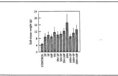
(14) LIST OF FIGURES Figure 1.1.. Dorsal diagrammatic view of a giant clam showing the zooxanthellal tube system. 9. Figure 3.1.. Experimental set-up at Orpheus Island Research Station. Figure 3.2.. Mean C:N and C:P atomic ratios in soft tissues of Tridacna gigas. 54. with or without ammonia and phosphate supplement in outdoor tanks on Orpheus Island. Figure 3.3.. 62. Mean soft-tissue weights of Tridacna gigas with or without ammonia and phosphate supplement in outdoor tanks on Orpheus Island. Figure 3.4.. 62. Total zooxanthellae numbers in Tridacna gigas with or without ammonia and phosphate supplement in outdoor tanks on Orpheus Island. Figure 3.5.. 64. Mean Chl a content per zooxanthella in Tridacna gigas with or without ammonia and phosphate supplement in outdoor tanks on Orpheus Island 64. Figure 3.6.. Chl a per clam and Chl a per zooxanthella as a function of the total zooxanthellae per clam. Figure 3.7.. 65. Preliminary data on mean mitotic indices of zooxanthellae isolated from juvenile Tridacna gigas incubated in ammonia and phosphate on Orpheus Island. 67.
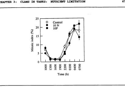
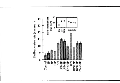
(15) xiv. Figure 3.8.. Mean mitotic indices of zooxanthellae isolated from Tridacna gigas with or without ammonia and phosphate supplement in outdoor tanks on Orpheus Island. Figure 3.9.. 67. NMR spectra of zooxanthellae extracts from Tridacna gigas with or without phosphate supplement in outdoor tanks on Orpheus Island. Figure 4.1.. 68. Mean shell-extension rates of Tridacna gigas with or without ammonia and phosphate supplement in outdoor tanks on Orpheus Island. Figure 4.2.. 88. Shell length versus shell weight in Tridacna gigas with or without ammonia and phosphate supplement in outdoor tanks on Orpheus Island. Figure 4.3.. 89. Shell weight adjusted to shell size of Tridacna gigas with or without ammonia and phosphate supplement in outdoor tanks on Orpheus Island. Figure 4.4.. 90. Representative SEMs of fractured sections of outer shell layer, showing shape and packing of aragonite crystals in Tridacna gigas with or without ammonia and phosphate supplement in outdoor tanks on Orpheus Island. Figure 4.5.. 93. Representative SEMs of fractured sections of outer shell layer, showing packing and growth orientation of aragonite crystals in. Tridacna gigas with or without ammonia and phosphate supplement in outdoor tanks on Orpheus Island. 95.
(16) Figure 4.6.. Representative SEMs of fractured sections of outer shell layer, showing detailed features of crystal morphology and fabric in. Tridacna gigas with or without ammonia and phosphate supplement in outdoor tanks on Orpheus Island. Figure 4.7.. 97. Mean angle 28 values at reflection planes with Miller indices of (012) and (200) in the aragonite crystal of Tridacna gigas with or without ammonia and phosphate supplement in outdoor tanks on Orpheus Island. Figure 5.1.. An aerial view of one of the twelve experimental micro-atolls with its nutrient-dispersal unit at One Tree Island Reef. Figure 5.2.. 100. 113. Mean N:P atomic ratios of the viscera of Tridacna maxima with or without ammonia and phosphate supplement in micro-atolls on One Tree Island Reef 117. Figure 5.3.. Mean ammonium-depletion rates of zooxanthellae isolated from. Tridacna maxima with or without ammonia and phosphate supplement in micro-atolls on One Tree Island Reef. Figure 5.4.. 119. Mean densities of zooxanthellae isolated from Tridacna maxima with or without ammonia and phosphate supplement in microatolls on One Tree Island Reef. Figure 5.5.. 121. Mean diameters of zooxanthellae isolated from Tridacna maxima with or without ammonia and phosphate supplement in microatolls on One Tree Island Reef. Figure 6.1.. 121. Growth curves of cultured zooxanthellae originally isolated from. Tridacna gigas. 134.
(17) xvi. Figure 6.2.. Mean phosphate-depletion rates of Tridacna gigas previously with or without phosphate supplement in raceways on Orpheus Island. Figure 6.3.. 137. Mean densities of zooxanthellae from Tridacna gigas with or without phosphate supplement in raceways on Orpheus Island. Figure 6.4.. Mean densities of zooxanthellae cultured with or without phosphate. Figure 6.5.. 141. 141. Mean N:P atomic ratios of zooxanthellae from Tridacna gigas with or without phosphate supplement in raceways on Orpheus Island. Figure 6.6.. Mean N:P atomic ratios of zooxanthellae cultured with or without phosphate. Figure 6.7.. 142. 142. Mean P-depletion rates of zooxanthellae isolated from Tridacna. gigas with or without phosphate supplement in raceways on Orpheus Island. Figure 6.8.. 144. Mean P-depletion rates in the light and in the dark of zooxanthellae previously cultured with or without phosphate. 144.
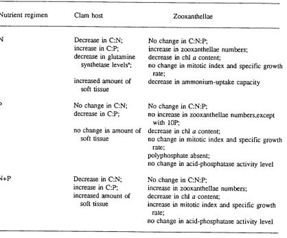
(18) xvii. LIST OF TABLES Table 3.1.. Mean C:N:P atomic ratios in zooxanthellae isolated from Tridacna. gigas with or without ammonia and phosphate supplement in outdoor tanks on Orpheus Island. Table 3.2.. 66. Mean acid-phosphatase activity in zooxanthellae isolated from. Tridacna gigas with or without ammonia and phosphate supplement in outdoor tanks on Orpheus Island. Table 3.3.. 69. Summary of responses of the symbiotic partners to nutrient supplementation. Table 5.1.. 82. Mean N:P atomic ratios of zooxanthellae isolated from Tridacna. maxima with or without ammonia and phosphate supplement in micro-atolls on One Tree Island Reef. Table 5.2.. 118. Mean concentrations of inorganic phosphate, total phosphorus, and ammonia in the haemolymph of Tridacna maxima with or without ammonia and phosphate supplement in micro-atolls on One Tree Island Reef. Table 6.1.. 122. Mean concentrations of inorganic phosphate and total phosphorus in the haemolymph of Tridacna gigas with or without phosphate supplement in raceways on Orpheus Island. Table 6.2.. 138. Mean N:P atomic ratios of viscera (minus kidneys) and kidneys from Tridacna gigas with or without phosphate supplement in raceways on Orpheus Island. Table 6.3.. 139. Mean N and P composition of viscera (minus kidneys) and kidneys from Tridacna gigas with or without phosphate supplement in raceways on Orpheus Island. 139.
(19) 1. Chapter 1 GENERAL INTRODUCTION. Giant clams of the family Tridacnidae are a unique group of bivalves. Apart from their enormous size, they differ from ordinary bivalves in having an exposed fleshy mantle densely populated by unicellular dinoflagellates, commonly known as zooxanthellae. These zooxanthellae photosynthesize carbon-rich compounds (Griffiths and Streamer, 1988), much of which is translocated to the animal host for use in its respiration, growth, and reproduction (Gladfelter, 1985). In return, the clam host shelters its algal symbionts and provides them with dissolved nutrients, such as inorganic nitrogen (see Wilkerson and Trench, 1986), phosphorus (see Yonge, 1936), and inorganic carbon (see Yellowlees et al., 1993) from sea water and host metabolic processes. Thus, this symbiotic relationship confers to giant clams a nutritional advantage over other bivalves in that they are not only heterotrophic bivalves that filter-feed on particulates (Klumpp et al., 1992) and take up dissolved organic molecules from sea water (Fankboner, 1971; Goreau et al., 1973; Southgate, 1988); but, they are also autotrophic bivalves capable of taking up dissolved inorganic molecules from sea water (Yonge, 1936; Wilkerson and Trench, 1986; Yellowlees et al., 1993). Such dual mode of nutrition in giant clams is the key factor in their survival in the nutrient-poor, tropical reef waters of the Indo-Pacific region (Yonge, 1975).. Recently, heavy exploitation and habitat degradation have threatened the status of giant clams throughout most of their geographic range, leading to international efforts on their conservation, management, and mariculture (Copland.
(20) CHAPTER 1: GENERAL INTRODUCTION. 2. and Lucas, 1988). This concern, coupled with the continuing scientific interest in symbiosis as a biological phenomenon, has intensified investigations of the clamzooxanthellae symbiosis.. 1.1 PARTNERS IN SYMBIOSIS. 1.1.1 The Invertebrate Host. There are eight extant species of giant clams, six of which belong to the genus Tridacna (Tridacna gigas, T. derasa, T. tevoroa, T. squamosa, T. maxima, and T. crocea), and two to the genus Hippopus (Hippopus hippopus and H.. porcellanus) (Rosewater, 1965; Rosewater, 1982; Lucas et al., 1991). A ninth species, T. rosewateri, is only known from shell type specimens (Sirenko and Scarlato, 1991). T. gigas is the largest (Yonge, 1975) and fastest-growing of all bivalves, reaching a size of up to 137 cm (Rosewater, 1965) with a growth rate (exponential growth stage) of up to 1 cm mo' (Belda, 1989; Gomez and Mingoa, 1993). T. crocea, on the other hand, is the smallest, reaching a maximum size of 14 cm (Lucas, in press). Attainment of such large sizes of these bivalves has been attributed to their dual mode of nutrition (Yonge, 1975).. Scientific interest in the nutrition of giant clams originated more than a century ago as a result of their unusual bivalve morphology (Brock, 1888)..
(21) CHAPTER 1: GENERAL INTRODUCTION. 3. Association with zooxanthellae has profoundly modified tridacnid structure in that the siphonal mantle regions, where the zooxanthellae predominate, have been extended over the upper surface for maximum exposure to light (Yonge, 1936). Yonge postulated that such change occurred through a 180° rotation of the visceropedal complex in relation to the mantle-shell complex, with a corresponding displacement of the hinge and ligament to a mid-ventral position. Stasek (1962, 1963) disputed Yonge's view, however, as it would suggest that the shell and mantle were independent of the rest of the clam's body components. Instead, he proposed that a morphologically-posterior direction of growth, instead of a ventral direction typical of bivalves, gave rise to the unique tridacnid morphology. Stasek's hypothesis is supported by the juvenile tridacnids' pattern of growth, which proceeds through a series of functional stages (LaBarbera, 1975).. The only other bivalves which have developed symbiosis with zooxanthellae are some cockles, which also belong to the superfamily Cardiacea. There are at least three known species, namely, Corculum cardissa and Fragum. fragum, which have translucent shell regions for exposure of their symbionts to light (see Goreau et al., 1973), and F. unedo, where the zooxanthellae are packed within the posterior mantle spreading over the substrate beyond the shell margins (Kawaguti, 1983). Among other molluscs, symbiosis is found in Gastropoda (in nudibranchs) (reviewed in Kempf, 1984)..
(22) CHAPTER 1: GENERAL INTRODUCTION. Apart from Mollusca, symbiotic associations with dinoflagellates are confined to four other phyla, namely, Cnidaria, Porifera, Platyhelminthes, and Protozoa (reviewed in Kokke and Spero, 1987; Hinde, 1988). Most tropical cnidarians are symbiotic with zooxanthellae, symbiosis being universal among hermatypic corals (Goreau et al., 1973; reviewed in Szmant-Froelich and Pilson, 1980). In temperate regions, the association is confined to some sea anemones (reviewed in Fitt et al., 1982), hydroids, and a few species of scleractinian corals (e.g., see Szmant-Froelich and Pilson, 1980). Porifera and Platyhelminthes have only a few known symbiotic associations with zooxanthellae (see Nicol, 1960; Hinde, 1988). In Protozoa, the symbionts are found in foraminiferans (McEnery and Lee, 1981; Jorgensen et al., 1985), radiolarians (reviewed in Rogerson et al., 1989), ciliates, and flagellates (Nicol, 1960).. 1.1.2 The Zooxanthella. Among the various algal groups symbiotic with marine invertebrates, the gymnodinioid dinoflagellates of the class Dinophyceae predominate in the marine environment (Taylor, 1973; Hofmann and Kremer, 1981; Trench and Blank, 1987). Association of these dinoflagellates with reef communities has made them predominantly circumtropical in distribution (Muscatine, 1980a).. These unicellular algae are coccoid in shape and yellow-brown in colour (Taylor, 1973). Inside the animal host, they are in a vegetative state, with no. 4.
(23) CHAPTER 1: GENERAL INTRODUCTION. flagella nor a distinct girdle; outside the host, they are typical motile (flagellated) gymnodinioid swarmers (Zahl and McLaughlin, 1957; McLaughlin and Zahl, 1959). Their photosynthetic pigments include chlorophylls a and c, 13-carotene, peridinin, neo-peridinin, dinoxanthin, neo-dinoxanthin, diadinoxanthin, and xanthophyll (Jeffrey and Haxo, 1968).. Until recently, Symbiodinium (=Gymnodinium) microadriaticum was regarded as a single, pandemic species of symbiotic dinoflagellates. The use of independent biochemical, physiological, morphological, and behavioural studies, however, resulted in a re-evaluation of the taxonomy of these zooxanthellae. Trench and Blank (1987) described four new species, although the authors acknowledged that evidence for sexual recombination remains to be presented before the existence of distinct biological species can be experimentally assessed. Recently, Rowan and Powers (1991a, b) characterised zooxanthellae isolated from taxonomically diverse hosts (corals, anemones, gorgonians, zoanthids, and jellyfishes), based on genetic sequences of the small ribosomal subunit of ribonucleic acid (ssRNA). They found the same alga in individuals of the same host species from single localities, but similar zooxanthellae in different host species, and estimated several distinct taxa distributed among the different hosts, regardless of host taxonomic status.. Overall, there is a consensus of a clear variability within the genus Symbiodinium, and recent advances in taxonomic studies show promise in. 5.
(24) CHAPTER 1: GENERAL INTRODUCTION. 6. demonstrating the existence of distinct biological species of symbiotic zooxanthellae. Until this task is completed, it is appropriate to refer to symbiotic dinoflagellates as Symbiodinium sp, with the exception of those species described by Trench and Blank (1987).. Until recently, the exact location of zooxanthellae in giant clams was controversial. Yonge (1936) initially observed that the zooxanthellae were contained within the haemolymph cells in the haemal spaces of the mantle. Later on, his own electron-microscopy studies showed that the zooxanthellae were not within haemolymph cells (Yonge, 1980), but he nevertheless continued to argue for the presence of zooxanthellae in the haemolymph. This was despite Mansour's (1946) observation, based on serial sections, that the zooxanthellae were contained within tubular structures, which he hypothesised to be connecting the mantle regions with the stomach. Mansour's observation of a complex duct system originating from the stomach was dismissed as extremely atypical among molluscs (Yonge, 1953) and even absurd (Morton, 1978). More recently, however, Norton et al. (1992) and Norton and Jones (1992) confirmed Mansour's findings when they demonstrated from their extensive histological studies on giant clams that the zooxanthellae are mainly in the mantle contained within the blind terminal branches of a diverticulum of the gut. This duct system arises as a single primary tube, extending from a diverticular duct of the stomach. It splits into right and left tubes above the digestive organs, both tubes passing ventrally through the kidney, crossing posteriorly through the connective tissue sheath of.
(25) CHAPTER 1: GENERAL INTRODUCTION. 7. the adductor muscle, and proceeding dorsally into the root of the siphonal mantle. Then each tube splits anteriorly and posteriorly on each side of the mantle into two secondary branches, which further divide into fine tertiary branches with blind ends within the exposed mantle surface (Fig. 1.1).. This confirmation brings the clam-zooxanthellae association in common with many other symbioses where the zooxanthellae are associated with the host's digestive system (Norton et al, 1992). In invertebrate hosts other than the clams and cockles, zooxanthellae are intracellular, residing within vacuoles surrounded by host-produced membranes within endodermal or gastrodermal cells (Trench et al., 1981; Trench and Blank, 1987)..
(26) 8. Fig. 1.1. Dorsal diagrammatic view of a giant clam showing the zooxanthellal tube system. AM - adductor muscle; CTN - ctenidia (gill); K - kidney; P - pericardium (heart); PZT - primary zooxanthellal tube; S - stomach; SZT - secondary zooxanthellal tube; TZT - tertiary zooxanthellal tubes. (reproduced with permission from Norton and Jones, 1992).
(27)
(28) CHAPTER 1: GENERAL INTRODUCTION. 10. 1.1.3 Establishment of Symbiosis. Acquisition of dinoflagellate symbionts by most animal hosts is by reinfection of offspring at each successive generation (Trench, 1987). This occurs in two ways. One is through access of the free-swimming algae to the host, possibly through some chemotrophic behaviour (Fitt, 1984). This has been shown in tridacnids (Fitt and Trench, 1981; Fitt et al., 1984), gorgonians, jellyfish (see Trench, 1987), nudibranchs (Hoegh-Guldberg and Hinde, 1986; Kempf, 1984), and cockles (see Fitt and Trench, 1981). Another is through ingestion of intermediate hosts, such as protozoans, plankton, and planulae harbouring algal symbionts, as has been demonstrated for Aiptasia sp. (Taylor, 1973). A less common mode of symbiont acquisition may be through maternal or cytoplasmic inheritance, where the algae are either partitioned between daughter cells or polyps during fission or budding (Trench, 1987), or transmitted directly via the egg or indirectly prior to the release of the offspring (Taylor, 1973). This has been demonstrated for many corals (reviewed in Taylor, 1973), alcyonaceans (Benayahu et al., 1988), and hydroids (reviewed in Trench, 1987).. In larval tridacnids, ingested zooxanthellae remain in the stomach for over a week (Fitt et al., 1986), and after metamorphosis, eventually migrate to microscopic channels within the developing mantle, via a duct (Fitt and Trench, 1981), which is the beginning of the zooxanthellal tube system described by Norton et al. (1992) and Norton and Jones (1992). Similarly, in coelenterates,.
(29) CHAPTER 1: GENERAL INTRODUCTION. 11. Symbiodinium sp. is phagocytosed by endodermal cells (Fitt and Trench, 1983), which eventually migrate into the mesoglea to become the "amoebocytes" where algal proliferation occurs (Colley and Trench, 1985). In both cases, the algae resist digestion by their hosts (Fitt and Trench, 1981; Fitt and Trench, 1983).. 1.2 NUTRITION OF THE INVERTEBRATE HOST. 1.2.1 Autotrophic Sources of Nutrition. Evidence for translocation of photosynthate from the zooxanthellae to the host comes from a wide range of studies employing radioisotopes. Use of this technique is based on the assumption that the zooxanthellae exclusively fix inorganic carbon into organic carbon and translocate some of it to the animal host. In in vitro studies, isolated zooxanthellae are incubated with radioactively-labelled bicarbonate and the incubation medium analysed for labelled products released by the algae. In in vivo studies, the whole organism is incubated with labelled bicarbonate and subsequently separated into its plant and animal tissue constituents. Examination of the extent of translocation is based on HC levels detected in the animal-tissue fraction (Muscatine, 1980b; Trench, 1987).. Short-term experiments demonstrated that 20-95% of the total fixed ' 4C is released in vitro and in vivo by zooxanthellae from hosts ranging from protozoa to.
(30) CHAPTER 1: GENERAL INTRODUCTION. 12. molluscs (Muscatine, 1967; Muscatine and Cernichiari, 1969; Schmitz and Kremer, 1977; Trench, 1971a; Steen and Muscatine, 1984; Hoegh-Guldberg and Hinde, 1986; Drits et al., 1987; Griffiths and Streamer, 1988). Evidence has been presented that the most commonly translocated photosynthetic products are glycerol, lipids, glucose, and alanine, with some leucine, glutamine, glutamate, and organic acids. Presence of host tissues or some "host factor" is believed to augment the release of photosynthate in vitro, but the nature of such augmentation is unknown (Muscatine and Cernichiari, 1969; Trench, 1971a, b, c; Patton and Burris, 1983; Gladfelter, 1985).. More recent studies have provided evidence for qualitative and quantitative translocation differences among various hosts. For the giant clam Tridacna gigas, Griffiths and Streamer (1988) showed that glucose is the major photosynthate translocated to the host in vivo, and not glycerol, as previously shown by Muscatine (1967) in vitro. Griffiths and Streamer found that glycerol only occurs in significant quantities in vitro in the presence of host tissue extracts. That glucose is the major photosynthate released by the algae to their clam host is supported by the much higher glucose concentrations (up to 620 pM) in the haemolymph of T. gigas, varying diurnally with day/night light regimes the way photosynthesis does, whereas glycerol concentrations were consistently low (< 4. M) (Rees et al, 1993a)..
(31) CHAPTER 1: GENERAL INTRODUCTION. 13. Photosynthesis: respiration ratio (P:R), while useful in investigating community metabolism, has limited applications in symbiotic associations because it yields limited information about the nutritional contribution of the zooxanthellae to the host (Gladfelter, 1985). For this reason, Muscatine et al. (1981) developed the concept of CZAR (contribution of zooxanthellae to animal respiration), which quantitatively assesses the portion of the host's daily metabolic energy demand that could be satisfied by algal production. Reported CZAR values range from over 92-100% for tridacnids (Fisher et al., 1985; Fitt et al., 1986; Mingoa, 1988; Klumpp et al., 1992), 9-158% for corals under various conditions of illumination or depth (Muscatine et al., 1981; McCloskey and Muscatine, 1984 in Gladfelter, 1985; Muscatine et al., 1984), 13-79% for sea anemones depending on the organism's nutritional history or intertidal position in the field (Fitt et al., 1982; Shick and Dykens, 1984; Zamer and Shick, 1987), and 13-48% for zoanthids (Steen and Muscatine, 1984).. Apart from respiration, translocated carbon may be used for other purposes. It may serve as a lipid reserve and reproductive energy source, and it may also be used for synthesis of new tissues, organic skeletal matrix, and mucus (Gladfelter, 1985; Rinkevich, 1989)..
(32) CHAPTER 1: GENERAL INTRODUCTION. 14. 1.2.2 Heterotrophic Sources of Nutrition. The exogenous sources of nutrition for symbiotic hosts are dissolved organic compounds and particulate food. In the aquatic environment, dissolved organic compounds include amino acids, carbohydrates, and vitamins; while particulate food consists of phytoplankton, zooplankton, bacteria, and animal detrital material (Taylor, 1973).. Tridacnid mantle, mantle cavity, and gills constitute a relatively large and potentially permeable surface area for uptake and loss of dissolved organic matter (Lucas, in press). For instance, evidence has been presented for pinocytosis of amino acids, possibly through the micropinocytic channels of the microvillous border of the clams' siphonal mantle epidermis (Fankboner, 1971). Similarly, Goreau et al. (1973) showed that [ 3H]-leucine in ambient sea water was rapidly taken up by T. maxima into its mantle and ctenidial (gill) epithelia. Such uptake of dissolved organic matter has been suggested to be of most nutritional benefit to tridacnids during their larval and early juvenile stages, when they have the highest ratio of surface area to body mass (Lucas, in press). In fact, other bivalve larvae are known to take up dissolved organic matter from sea water (Manahan, 1983).. Other invertebrate hosts, such as corals, have been shown under laboratory conditions to accumulate amino acids and [ 14C] glucose from modest concentrations in seawater (Stephen, 1962 in Fankboner, 1976). Such.
(33) CHAPTER 1: GENERAL INTRODUCTION. 15. heterotrophic transfer of dissolved substances may also occur in situ, as indicated by the transfer of kelp-exudated dissolved carbon to a coral (Fankboner, 1976). Similarly, zoanthids can absorb amino acids and sugars from very low concentrations in seawater, and it has been suggested that these substances may be incorporated into the hosts' reproductive tissue (Trench, 1974).. For a majority of symbiotic hosts, ingestion of particulate food such as phytoplankton, zooplankton, bacteria, and animal detrital material may constitute a significant source of essential metabolites (Goreau et al., 1973; Taylor, 1973). It has been suggested that phytoplankton is a major source of particulate food for tridacnids (Goreau et al., 1973), although this is questionable considering the low phytoplankton levels in reef waters. They are also capable of taking zooplankton into their stomach, where "rapid disintegration and probably digestion" were observed by Mansour (1945), in contrast to Ricard and Salvat (1977) who noted that T. maxima filter, but do not assimilate, phytoplankton and zooplankton. Fitt et al. (1986), however, demonstrated that T. gigas ingested and digested ['C]labelled Isochrysis galbana in sea water. Furthermore, unicellular algae have been used successfully in culture of clam larvae (Braley, 1990).. More recently, T. gigas has been shown to efficiently utilise particulate organic matter, mostly in the form of detritus, available in reef waters (Klumpp et al., 1992). The clams filtered three quarters of 2-50 um particles passing through their mantle cavity, and absorbed at least half of the particulates' carbon content..
(34) CHAPTER 1: GENERAL INTRODUCTION. 16. Such particulate food provides from a quarter to over half of the carbon used for growth and respiration in moderate- (ca. 190 mm shell length) and small-sized clams (ca. 35 mm), respectively, with the nutritional benefit obtained declining with clam size (Klumpp et al., 1992).. In the case of other symbioses, corals and zoanthids are capable of ingesting and assimilating animal detrital material and bacteria, but they feed primarily on zooplankton (reviewed in Muscatine, 1973; Johannes, 1974). For foraminiferans, zooplankton appears to be the main food source (Be et al., 1981 in Jorgensen et al., 1985). Similarly, it has been suggested that symbiosis in poriferans is probably sustained to a large extent by host filter-feeding (see Taylor, 1973); while in anemones, host phagotrophy is evidently indispensable for the total growth of the association (see Muscatine, 1973).. As regards the possible digestion of the zooxanthellae by the host, Yonge (1936) claimed that zooxanthellae are "farmed" by giant clams in their mantle, conveyed by amoebocytes via blood vessels, and digested en route to or in the digestive gland's interdiverticular spaces. This has been invalidated by the recent confirmation of Mansour's (1945) zooxanthellal tube system (Norton et al., 1992; Norton and Jones, 1992). Similarly, Fankboner (1971) interpreted the "culling" of senescent and atypical zooxanthellae by amoebocytes of the digestive gland as indicative of lysosome-derived hydrolytic activity. This histochemical demonstration of acid phosphatase activity, near or in the algae, is also rendered.
(35) CHAPTER 1: GENERAL INTRODUCTION. 17. ambiguous by the presence of intrinsic acid phosphatases in the algae themselves (see Trench, 1987; Jackson et al., 1989) and possible algal autolysis (Trench, 1974). Muscatine and Greene (1973) and Muscatine (1973) pointed out that there was no direct evidence for such hydrolysis of algal substrates, nor had there been any demonstration of host assimilation of the digestive products (Muscatine, 1973). More importantly, however, long before Norton and co-workers' significant histological findings, Mansour (1945) argued that the algae are not contained within amoebocytes and are defaecated undigested. Indeed, Fitt et al. (1986) showed that T. gigas juveniles released up to 80% of [ 14C]-labelled freshlyisolated zooxanthellae within 72 h following feeding. The persistent release of dark-brown faeces, packed with viable zooxanthellae, was documented by Ricard and Salvat (1977), Trench et al. (1981), and Lucas (in press). Lucas (in press) suggested that the zooxanthellae travel down the zooxanthellal tube system into the alimentary canal and are expelled undigested. Also, the epithelial cells lining the primary and secondary sections of the tube system are equipped with long cilia (Norton and Jones, 1992). This suggests that the clam host drives the zooxanthellae out of the tubular system, as one way of controlling their population size.. A 'medium' sized T. maxima was estimated to annually release 17-26 g of protein as zooxanthellae in its faeces (Ricard and Salvat, 1977). Lucas (in press) pointed out that such non-digestion of zooxanthellae is a surprising 'waste' of potential food. Algal densities are approximately 10 6 cells cm-2 colony surface in.
(36) CHAPTER 1: GENERAL INTRODUCTION. 18. corals, 109 cells g"' tissue in sea anemones, and 2 x 10 8 cells g"' mantle tissue in giant clams, comprising 3 to 14% of these associations' protein biomass (see Muscatine, 1980a). While digestion of the zooxanthellae constitutes a potential source of food, available evidence suggests that it is a minor source of nutrition to the clam host, if at all.. 1.2.3 Autotrophy Versus Heterotrophy. The nutritional benefits derived by the host from autotrophy, which are much easier to quantify even in field situations, have received more attention than the host's gains from exogenous sources. Most investigators have assessed the zooxanthellae's nutritional contribution and considered the rest of the host's nutritional needs as satisfied by heterotrophy.. The contribution of the zooxanthellae to the nutrition of their symbiotic hosts cannot be over-emphasised. However, while there is substantial evidence that energy-rich compounds derived from zooxanthellae contribute significantly to the metabolic energy demands of symbiotic hosts, the adequacy of symbiont production to fully meet the metabolic energy requirements in many associations, particularly under suboptimal conditions, has not been demonstrated (see CZAR values above). More importantly, there is no evidence for the ability of the symbionts to supply all the nutrients necessary to support host tissue synthesis. While carbon production by the zooxanthellae may meet the clam host's.
(37) CHAPTER 1: GENERAL INTRODUCTION. 19. requirements for respiration and structural carbon, heterotrophic sources of nutrition would certainly play a major supplementing role.. Yonge (1975) and Fankboner and Reid (1990) drew attention to the intact and functional filter-feeding and digestive apparatuses of giant clams, being comparable to those of heterotrophic bivalves. Fankboner and Reid (1990) pointed out the potential nutritional benefit that can be derived by clams from filtering large volumes of reef waters which contain dissolved organic carbon, particulates (zooplankton, little phytoplankton, and detritus), and mucus flocs. Indeed Klumpp et al. (1992) demonstrated the considerable nutritional benefit derived by T. gigas from its highly-efficient filtering activity, although these workers dealt mainly with carbon.. While inorganic nutrients, such as nitrogen and phosphorus, are typically present at very low concentrations in reef waters (Crossland, 1983; Kinsey, 1991), symbiotic associations may take advantage of occasional large inputs from natural sources such as oceanic upwellings and intrusions. The suggestion that the tridacnids may be capable of storing phosphorus in phosphorite concretions in the kidney (Trench et al., 1981), for instance, deserves exploration. Such potential capacity to store nutrients, in addition to nutrient recycling and conservation, would contribute to the tridacnids' success in a nutrient-poor environment..
(38) CHAPTER 1: GENERAL INTRODUCTION. 20. 1.3 NUTRITION OF THE ZOOXANTHELLAE. 1.3.1 Nutrient Requirements. Growth studies on cultured zooxanthellae have shown that photosynthesis can provide their total energy requirements (reviewed in Taylor, 1973). In addition, the zooxanthellae obtain inorganic and organic substances from their hosts and the seawater.. Host-derived substances useful to the zooxanthellae include end-products of metabolism and directed fluxes of specific metabolites from the host. In tridacnids and coelenterates, studies suggest that the zooxanthellae may receive nitrogen and phosphorus from their hosts' metabolic waste products (Yonge, 1936; D'Elia, 1977; Cates and McLaughlin, 1979; Wilkerson and Trench, 1986). Ammonium ions may be derived by zooxanthellae from host metabolic sources such as deamination, pyrimidine breakdown, and adenosine monophosphate (AMP) deaminase activity (McGilvery, 1983). As regards phosphate, Taylor (1973) suggested that glycero-phosphoric, cytidylic, adenylic, and guanylic acids may serve as phosphorus sources, just as urea, uric acid, guanine, adenine, and several amino acids can serve as nitrogen sources. In vitro studies on nutrient uptake by cultured zooxanthellae suggest that zooxanthellae are capable of uptake and utilisation of organic compounds potentially present in the host (reviewed in Steen, 1986). There is some evidence for an active transport mechanism for.
(39) CHAPTER 1: GENERAL INTRODUCTION. 21. uptake of cysteine, methionine, and taurine in cultured zooxanthellae from the giant clam T. maxima, and of alanine in zooxanthellae isolated from the jellyfish. Cassiopeia xamachana (reviewed in Steen, 1986). Coelenterate zooxanthellae, which are incapable of synthesising their own glycine from CO 2 or serine (von Holt, 1968), have been reported to show preferential uptake and assimilation of host glycine. Also, the "back transport" technique, where labelled food substrates are introduced to the hosts, followed by assay of the algae, has shown that zooxanthellae in anemones can assimilate 35S-amino acids from their hosts (Carroll and Blanquet, 1984; Steen, 1986).. The preceding studies, however, mostly dealt with determining the capacity of zooxanthellae for uptake of substances potentially present in the host by: (1) monitoring host excretion of inorganic nutrients, (2) measurement of uptake by zooxanthellae in culture, or (3) measurement of uptake by zooxanthellae isolated from the host. Detailed analysis of actual metabolite movement from host to algae, such as the "back transport" study of Carroll and Blanquet (1984), is wanting. Available evidence for host-derived nutrition of the zooxanthellae pales in comparison with that for zooxanthellae-derived nutrition of the host. It is important to clarify and quantify this less-studied aspect of symbiosis nutrition to further understand nutrient recycling and conservation, as well as the relative contribution of exogenous sources of nutrition to the symbiotic association..
(40) CHAPTER 1: GENERAL INTRODUCTION. 22. Nutrients from seawater, such as dissolved inorganic and organic compounds, may be taken up by the zooxanthellae via the host (see Cates and McLaughlin, 1979; Hinde, 1988) through diffusion or active transport (see Taylor, 1973; Wilkerson and Trench, 1986). Of the mineral nutrients available, nitrogen and phosphorus have received the most attention because of their possible limiting effects on zooxanthellae growth. While these nutrients may be acquired through the host, the ultimate source is the sea water.. Crossland (1983) presented an extensive tabulation of nutrient concentrations in coral-reef waters prior to 1980, showing wide ranges of values around different island types and locations (e.g., oceanic reefs, high islands). Dissolved organic nutrient concentrations ranged from 0.1 to 7.5 pM for nitrogen, and 0.02 to 5 pM for phosphorus. Nitrate concentrations were from 0.01 to 5 pM; nitrite from undetectable values to 5 pM; ammonia from 0.07 to 11 pM; and phosphate from 0.01 to 5.5 AM. Nevertheless, typical values for these dissolved inorganic nutrients were < 1 pM. In areas of known chronic reef pollution (e.g., Kaneohe Bay), typical ranges for inorganic nitrogen were 2 to 4 pM, and 0.3 to 1.2 pM for inorganic phosphorus (see Kinsey, 1985)..
(41) CHAPTER 1: GENERAL INTRODUCTION. 23. 1.3.2 Nutrient Acquisition Through The Host. The zooxanthellae are separated from the clam's haemolymph by a thin layer of epithelial cells, which comprise the tertiary tube branches in the mantle (Norton et al., 1992; Norton and Jones, 1992). These epithelial cells are likely to be similar to those lining the digestive tract of most organisms, in that they are permeable to nutrients (Yellowlees, pers. comm.). Exchange of nutrients or metabolites can, therefore, occur between the zooxanthellae and the haemolymph across the zooxanthellal tube (Rees et al., 1993b). As discussed previously, nutrients may be taken up by the intact association from sea water through the gills and other body surfaces (e.g., mantle) via diffusion or active transport. These nutrients are likely made available to the zooxanthellae through the haemolymph.. Active transport is generally held responsible for nutrient uptake by zooxanthellae (reviewed in Muscatine, 1980b). Giant clams, other corals, and some anemones, however, do not show clear-cut saturation kinetics for nitrogen uptake (see Wilkerson and Trench, 1986). One explanation for this is that net uptake has both diffusive and active components, with the diffusive component of uptake through the animal tissue masking any active uptake at the site of the algae (see Wilkerson and Trench, 1986).. D'Elia et al., (1983) proposed the "depletion-diffusion" model as a mechanism of nutrient uptake in symbiosis. That is, zooxanthellae deplete the.
(42) CHAPTER 1: GENERAL INTRODUCTION. 24. animal tissue of inorganic nutrients, creating a concentration gradient through which nutrients from seawater passively diffuse into the animal tissue. D'Elia and Cook (1988) further extended this hypothesis to include regulation of the host's cytoplasmic nutrient concentration by the nutritional status of the symbiotic partners. That is, when algal uptake exceeds host nutrient regeneration, host cytoplasmic concentration must decrease and vice versa.. Evidence presented in favour of this hypothesis includes localisation in zooxanthellae of assimilatory enzymes for inorganic nitrogen, and similarity of nutrient uptake kinetics between freshly isolated zooxanthellae and intact symbioses (D'Elia. et al., 1983). Other observations which implied the role of the zooxanthellae in the uptake of inorganic nitrogen are: ammonium-uptake by symbiotic hosts and ammonia-excretion by aposymbiotic and non-symbiotic hosts (e.g., Muscatine and D'Elia, 1978; Muscatine and Marian, 1982); reduction of ammonia release by the host in the presence of zooxanthellae (reviewed in Muscatine, 1980b); and ammonium-uptake by freshly-isolated zooxanthellae (e.g., Wilkerson and Muscatine, 1984).. This depletion-diffusion hypothesis, however, was developed to account for nutrient uptake by intracellular zooxanthellae. In this model, D'Elia and Cook (1988) emphasised that uptake kinetics of symbioses may be complicated by the spatial and morphological relationships of the symbiotic partners. As discussed previously, tridacnid zooxanthellae are intercellular, being separated from the.
(43) CHAPTER 1: GENERAL INTRODUCTION. 25. haemolymph by a tube system. Thus, any hypothesis explaining the acquisition of nutrients by zooxanthellae in the clam has to account for the morphological relationship between the symbiotic partners. That is, nutrients taken up from sea water will have to go across the gill or mantle membranes, into the haemolymph, across the zooxanthellal tube cells, and through the zooxanthellae membranes via diffusion or active transport, whichever is applicable.. Recently, Miller and Yellowlees (1989) suggested that the depletiondiffusion model is adequate to describe uptake of ammonia (i.e.,_uncharged species NH3), which can readily diffuse across membranes, but not phosphate and nitrate, which are always present as charged species. Additionally, intracellular phosphate concentrations are always high relative to those in sea water (McGilvery, 1983). Thus, they proposed that uptake of these charged nutrients from sea water and across the perialgal membrane (in the case of intracellular zooxanthellae) must be carrier-mediated (i.e:, active transport or facilitated diffusion via a non-specific carrier). The predominant species of ammonia (NH 3), ammonium (NH 4+), nitrate, and phosphate within the physiological pH range have been tabulated by Miller and Yellowlees (1989). At pH 7.4, NH 3 constitutes only about 1% of the total NH3-NH4+ species; at pH 8.2, its concentration increases to 8%. In contrast to ammonia, nitrate and phosphate, are always charged regardless of pH, the predominant species being NO3- , HP042- , and H2PO4- (Miller and Yellowlees,.
(44) CHAPTER 1: GENERAL INTRODUCTION. 26. 1989). Thus, uptake of these nutrients against a concentration gradient would only be via active transport across host membranes (Miller and Yellowlees, 1989).. In T. gigas, haemolymph pH varied on a diel cycle from 7.4 to 8.1, with highest values at noon and lowest at night, reflecting ambient light levels and zooxanthellae photosynthetic activity (Fitt et al., in prep.). Similarly, the pH of the culture medium surrounding zooxanthellae isolated from T. crocea increased during photosynthesis (D'Elia et al., 1983). The increase in pH is understood to be brought about by the zooxanthellae's removal of CO2 from the haemolymph (or culture medium) during photosynthesis (i.e., lower [HT. Natural ammonium levels in the haemolymph vary inversely with haemolymph pH, with greatest ammonium concentration found at night when haemolymph pH was lowest (Fitt et al., in prep.). Such increase in ammonium concentration at lower pH is understood to be brought about the the availability of more 1-1+ for protonation of ammonia (into ammonium) present in the haemolymph. It has also been observed that in clams exposed to increased ammonia in sea water, ammonium accumulated faster in the haemolymph at a lower than a higher haemolymph pH, suggesting that ammonia diffuses from sea water (pH 8.1) down a concentration gradient and accumulates in the haemolymph at night (Fitt et. al., in prep.). On the other hand, ammonium concentration in the haemolymph was lower at noon when pH in the haemolymph was similar to that of sea water..
(45) CHAPTER 1: GENERAL INTRODUCTION. 27. These observations support the depletion-diffusion hypothesis in clams. Ammonia, being an uncharged molecule, diffuses across the animal membrane (gill, mantle, or zooxanthellal tube system) depending on the pH gradient, which in turn determines this nutrient's concentration gradient across the animal membrane. On the other hand, ammonium, being a charged molecule, cannot diffuse back from the haemolymph into the sea water (or from the tube system back into the haemolymph), and instead accumulates inside the host for active uptake by the zooxanthellae and/or the host (see following discussion of host ammonium-assimilation enzymes).. There are two basic requirements that would enable the zooxanthellae to induce a net uptake of ammonium by the intact symbiosis, as proposed by the depletion-diffusion of D'Elia and co-workers. These are: (1) a system for active uptake, and (2) an ability to assimilate ammonium (Miller and Yellowlees, 1989). Indeed, cultured and freshly-isolated zooxanthellae from corals take up ammonium from sea water (e.g., Gunnersen et al., 1988). Also, assimilation enzymes (glutamine synthetase, GS; glutamine dehydrogenase, GDH) are present in the zooxanthellae of corals (Summons and Osmond, 1981; Gunnersen et al., 1988; Dudler and Miller, 1988) and tridacnids (Rees et al., in press).. However, recent studies also showed very high levels of ammonium assimilation enzymes in tridacnid hosts (GS) (Rees et al, in press) and some corals hosts (NADPH-GDH: Catmull et al., 1987; GS: Yellowlees et al., in press)..
(46) CHAPTER 1: GENERAL INTRODUCTION. 28. Miller and Yellowlees (1989) and Rees et al. (in press) pointed out that available enzyme-activity data imply the involvement of symbiotic host tissue in ammonium assimilation, as has previously been suggested for the Hydra-Chlorella symbioses (Rees, 1987). It has also been suggested that ammonium uptake and retention by zooxanthellae may only be indirectly dependent on the zooxanthellae. Instead, the host may assimilate ammonium, using translocated carbon from its zooxanthellae as amino-group acceptors (Miller and Yellowlees, 1989). Miller and Yellowlees (1989) pointed out that this host-mediated assimilation is compatible with available evidence for dependence of nutrient uptake by intact associations on light and presence of zooxanthellae. It is evident that both partners in the symbiosis have the capacity to both acquire and assimilate ammonium.. Light stimulation of uptake and retention of ammonium and nitrate has been demonstrated for tridacnids (Wilkerson and Trench, 1986), reef corals (Muscatine and D'Elia, 1978), anemones (Wilkerson and Muscatine, 1984), and cultured zooxanthellae (Domotor and D'Elia, 1984). This light stimulation of nutrient uptake is believed to occur through photoreduction, supply of energy for uptake via photophosphorylation, or photosynthetic supply of carbon skeletons as nitrogen acceptors (see Muscatine, 1980b). Ammonium-assimilation rate by T.. gigas decreased with time in continuous darkness, with net ammonia release occurring after several days (Wilkerson and Trench, 1986). Such release of ammonia under prolonged incubation in darkness has also been shown for symbiotic corals and anemones, suggesting that ammonium assimilation by the.
(47) CHAPTER 1: GENERAL INTRODUCTION. 29. zooxanthellae is prevented due to depletion of zooxanthellae carbon reserves (Muscatine and D'Elia, 1978; Wilkerson and Muscatine, 1984). Similarly, inorganic phosphorus is taken up in the light only by symbiotic corals, while uptake may diminish in the dark (D'Elia, 1977).. 1.3.3 Nutrient Limitation. It has generally been assumed that the host cell, where most zooxanthellae live, is a nutrient-rich environment (see Taylor, 1973). Recently, however, some investigators suggested that zooxanthellae, including those located intercellularly, may be nutrient-limited.. Evidence consistent with such a view includes: a) high zooxanthellae densities inside the host, suggesting a great nutrient demand that may deplete intracellular concentrations for further algal growth; b) intact associations taking up dissolved inorganic nutrients from very low concentrations in seawater, and uptake kinetics implying low nutrient concentrations within host tissues; and c) nitrate uptake, a characteristic of nitrogen-starved algae, (Cook and D'Elia, 1987), since nitrate must first be reduced to ammonium before it can be assimilated, making ammonium the preferred species for assimilation.. Of the major nutrients, nitrogen is considered by many investigators as the most limiting. The possibility that the clam zooxanthellae may be isolated from.
(48) CHAPTER 1: GENERAL INTRODUCTION. 30. host ammonium sources is suggested by T. gigas being capable of taking up nitrate (Wilkerson and Trench, 1986; Fitt et al., in prep.), and that ammonium was undetectable in the haemolymph (Deane and O'Brien, 1980). Furthermore, zooxanthellae freshly-isolated from T. gigas took up nitrate from haemolymph only when ammonium concentration was low enough not to inhibit nitrate uptake (Fitt et al, in prep.). Addition of ammonium promoted photosynthesis by zooxanthellae in clam tissue slices (Summons et al., 1986), and exposure of clams to increased ammonia or nitrate in sea water increased their shell-extension rates and tissue weights (Braley, 1992; Hastie et al, 1992; Fitt et al., 1993). Such increased growth of the clam host has generally been assumed to be a direct consequence of increased translocation of photosynthate from the zooxanthellae. Furthermore, surge uptake for ammonium and nitrate have been demonstrated for tridacnids, with nitrate uptake being saturated at 8-10 pM (Wilkerson and Trench, 1986).. For cnidarians such as corals and anemones, ammonium concentrations in tissue homogenates have been reported to be about 5-50 pM and 40 pM (Crossland and Barnes, 1977; Wilkerson and Muscatine, 1984). However, Wilkerson and Muscatine, (1984) pointed out that disruption of animal tissue generates ammonium from deamination reactions when cellular structure is destroyed. Furthermore, homogenisation evenly distributes tissue metabolites, thus precluding detection of possible small-scale local depletion of ammonium existing near endosymbionts (see D'Elia and Cook, 1988)..
(49) CHAPTER 1: GENERAL INTRODUCTION. 31. Also, nitrogen limitation occurs at high zooxanthellae densities, as indicated by further increase in population density, chlorophyll a content, and maximum photosynthetic rate in coral colonies incubated in ammonium-enriched seawater (Hoegh-Guldberg and Smith, 1989). It has also been suggested that intracellular ammonium concentration is very low and dependent on the feeding history of the host, as corroborated by the ammonium uptake rate by zooxanthellae being greatest in starved anemones, and internal levels of ammonium in well-fed hosts being elevated enough to competitively inhibit uptake of the ammonium analogue, methylammonium (D'Elia and Cook,_1988).. While these studies suggest nitrogen limitation in zooxanthellae, other studies presented contradictory evidence. For instance, the ammonium-uptake activity of zooxanthellae isolated from corals was lower than that of nitrogenstarved cultured zooxanthellae (Gunnersen et al., 1989). The same isolated zooxanthellae had very low levels of ammonium-assimilation enzymes. Furthermore, cnidarian zooxanthellae have a general carbon to nitrogen ratio of 6, which is typical of algae grown in nitrogen-sufficient conditions (D'Elia et al., 1983).. In contrast to nitrogen, phosphorus nutrition in symbiosis has received little attention. While it has been demonstrated that the giant clam T. crocea took up more phosphate than the non-symbiotic clam, Spondylus (Yonge, 1936); and that cultured zooxanthellae from tridacnids took up phosphate from the medium (see.
(50) CHAPTER 1: GENERAL INTRODUCTION. 32. Jackson et al., 1989), the possibility of phosphorus limitation in clam zooxanthellae has not been investigated.. Recently, Miller and Yellowlees (1989) pointed out that available evidence favours phosphate limitation over nitrogen limitation as a means of host control over algal growth. Unlike ammonium, phosphate is always present as a charged species (H 2P0,,- and HP042-) in the normal cytoplasmic pH range, making it simpler for the host to regulate phosphate availability to the algae through a carrier system. Furthermore, by regulating the activity of this carrier system, the animal host can sustain its own normally high (millimolar) cytoplasmic concentrations of phosphate (Miller and Yellowlees, 1989).. Relevant data are provided by Jackson et al., (1989), showing very high phosphate uptake activities of freshly isolated zooxanthellae from the coral. Acropora formosa and suggesting that the bulk of phosphatase activity associated with zooxanthellae reflects algal response to phosphate limitation (cf. Fankboner, 1971; Fitt and Trench, 1983). They observed that phosphatase activity, which makes inorganic phosphate available to the zooxanthellae, was always high in freshly isolated zooxanthellae, but repressed when incubated in a medium containing 2 mM phosphate (similar to host cytoplasmic phosphate concentration). This implies that the perialgal phosphate concentration experienced by the zooxanthellae in vivo may be below that of the host cytoplasm..
(51) CHAPTER 1: GENERAL INTRODUCTION. 33. To date, there is a general disagreement as to which nutrient is actually limiting the growth of zooxanthellae. Externally-supplied inorganic nitrogen enhances densities of coral zooxanthellae in vivo (Muscatine et al., 1989; HoeghGuldberg and Smith, 1989), while inorganic phosphorus does not (Muscatine et al., 1989). On the other hand, carbon:nitrogen ratios of zooxanthellae are comparable to those of algae grown in nutrient-sufficient conditions (D'Elia et al., 1983), while phosphate-uptake and phosphatase activities of zooxanthellae freshly isolated from corals suggest phosphorus limitation (Jackson et al., 1989). As has been pointed out by Miller and Yellowlees (1989) and others for other algae and plankton, however, it is possible that limitation by a single nutrient may be an over-simplification. Instead, they suggested a possible oscillation between nitrogen and phosphorus limitation as a consequence of host nutritional status and light availability, or concurrent limitation by more than one nutrient. Recently, Rees et al. (in press) suggested that both the clam host and its zooxanthellae are nitrogen-deficient, and that the glutamine-synthetase activity by the host has a major role in assimilating available ammonia from sea water. Clearly, further research is needed before this major issue in marine symbiosis can be resolved.. 1.3.4 Nutrient Recycling. The question of whether symbiotic associations are nutrient-limited seems to be confounded by the apparent disparity between the high rates of production in coral reef communities and the low concentrations of nutrients in the surrounding.
(52) CHAPTER 1: GENERAL INTRODUCTION. 34. waters. Such seeming incongruity lends much appeal to the widely accepted notion of tight nutrient recycling within symbiotic associations.. That nitrogen is recycled in tridacnids is consistent with available evidence. The zooxanthellae release some amino acids to their animal host, as has been shown in vivo for T. gigas (e.g., Griffiths and Streamer, 1988). Oxidative and biosynthetic metabolism by the animal host, in turn, produces ammonia, which may be taken up by the zooxanthellae for incorporation into amino acids. Indeed, tridacnids and some cnidarians do not release, or have reduced excretion of, ammonia in the presence of their zooxanthellae (see Muscatine, 1980b; Wilkerson and Trench, 1986), except under prolonged incubation in the dark (Muscatine and D'Elia, 1978; Wilkerson and Muscatine, 1984); while aposymbiotic and non-symbiotic cnidarians release ammonia in significant amounts (Muscatine and D'Elia, 1978; Muscatine and Marian, 1982). These results have been interpreted to suggest that the zooxanthellae retain the latter's metabolic waste product (i.e., ammonia) that might otherwise be excreted.. Phosphorus recycling within the intact association is not clear. Of interest, however, is the presence of numerous phosphorite concretions in the tridacnid kidney. Trench et al. (1981) pointed out that such phosphorus pools seem incongruous with the clams' existence in a phosphate-depleted environment, unless these concretions serve as a reservoir of phosphorus (Trench et al., 1981)..
(53) CHAPTER 1: GENERAL INTRODUCTION. 35. Although nutrient recycling is plausible, more detailed studies are needed to demonstrate the actual fate of these nutrients within the intact association. In particular, considering that the photosynthate translocated by the zooxanthellae to their animal host is largely non-nitrogenous (e.g., Griffiths and Streamer, 1988), it would appear that only a small amount of nitrogen is recycled between the symbiotic partners. Also, the observation of significant amounts of nitrogenassimilation enzymes (glutamine synthetase) in tridacnids (Rees et al, 1989; Rees et al., in press) and some cnidarians (Catmull et al., 1987; Yellowlees et al., in press) would indicate that retention of ammonia, as host waste product, may not only be attributable to the zooxanthellae, but also to the host (see Miller and Yellowlees, 1989).. While significant nutrient recycling may occur, recycled nutrients are inadequate for the total growth of the association. As mentioned earlier, amino acids translocated by the algae to the host seem to be but a minor fraction of the largely non-nitrogenous photosynthate.. For tridacnids and some corals, algal supply of reduced carbon, which may be more than enough to meet the hosts' metabolic energy demands (see CZAR values in earlier sections), is of possible additional benefit in terms of conserving essential amino acids from respiration. Nevertheless, heterotrophic contribution to the nutrition of symbiotic organisms must play an important role..
(54) CHAPTER 1: GENERAL INTRODUCTION. 36. Marine symbioses, unlike their non-symbiotic counterparts, are capable of occupying different trophic levels within their ecosystems, e.g. as primary producers, primary consumers, or secondary consumers, enabling them to more conveniently avail themselves of autotrophic and various heterotrophic nutritional sources at all times. It is, thus, likely that such trophic adaptability, coupled with nutrient recycling and conservation, is behind their success in tolerating relatively low nutrient concentrations in their environment.. 1.4 EFFECTS OF P ON CALCIFICATION. While zooxanthellae in symbiotic associations benefit from increased availability of dissolved inorganic nutrients from sea water, several studies have suggested that elevated levels of these nutrients may be harmful to the calcification process of the animal host. In particular, increased phosphorus may markedly suppress deposition of calcium carbonate in corals.. Kinsey and Domm (1974) carried out an eight-month long, discontinuous fertilisation (i.e., for 3 h during low tide each day) of a patch reef at One Tree Island, Great Barrier Reef, Australia, with 2.0 pM phosphate and 20 pM urea plus ammonium. They found a 50% increase in the rate of net community photosynthesis, and attributed such effect to increased production by benthic algae, possibly including the zooxanthellae. However, the calcification data from the.
(55) CHAPTER 1: GENERAL INTRODUCTION. 37. same experiment, analysed based on an alkalinity anomaly approach (Kinsey and Davies, 1979), showed > 50% suppression of net calcification of corals during the fertilisation period. Kinsey and Davies (1979) attributed this effect primarily to phosphate, which is in accordance with laboratory studies suggesting that phosphates may act as crystal poisons of calcification (Simkiss, 1964). There was no available experimental evidence for suppression (or otherwise) of calcification by increased nitrogen. Similarly, recent work by Rasmussen (1989) showed rapid skeletal extension of branches of Acropora formosa coral colonies exposed to up to 4 pM phosphate (superphosphate) in outdoor tanks, but this extension was accompanied by considerable thinning of skeletal diameter. Also, scanningelectron micrography indicated significant alteration of the internal morphological structure of the corals exposed to increased phosphate concentrations.. To date, no similar investigations on tridacnids have been carried out by other workers. Giant clams, like hermatypic corals, are among the coral reefs' calcifying constituents, which live in symbiosis with zooxanthellae. Potential effects of elevated nutrients on these different symbiotic associations may be comparable, and together, may have significant implications for calcification in reef waters receiving enhanced inputs of nutrients from anthropogenic sources..
(56) CHAPTER 1: GENERAL INTRODUCTION. 38. 1.5 ASSESSMENT OF STATUS OF RESEARCH. Some of the major issues in tridacnid nutritional physiology have now been satisfactorily resolved. These questions, which had prevailed over the decades, include: (1) the location of zooxanthellae in the clams and (2) whether clams derived a major portion of their nutrition from translocated photosynthate or digestion of the zooxanthellae themselves. The confirmation of the existence of the zooxanthellar tube system has much to do with helping to clarify the nutritional relationship between the clam host and its algal symbionts.. Nevertheless, a great scope for investigation remains, with the current trend of research gearing toward symbiotic interactions, in the context of nutrient fluxes and metabolism. In particular, there is a need to quantify many aspects of the nutritional physiology of tridacnids. Whereas the contribution of zooxanthellae photosynthesis to host nutrition has been widely addressed in many types of hosts, little work has been carried out on the contribution of host-derived substances, as well as the contribution of particulates and exogenous sources of dissolved organic and inorganic molecules on zooxanthellae nutrition. This information is necessary in order to determine the relative significance of the various sources of autotrophic and heterotrophic nutrition of the clam-zooxanthellae symbiosis. Other interesting aspects for further investigation include pathways of nutrient assimilation, detailed mechanisms of nutrient uptake, nutrient recycling and conservation, and the possible detrimental effects of nutrients (e.g. pollution). More importantly, the.
(57) CHAPTER 1: GENERAL INTRODUCTION. question of nutrient limitation of the zooxanthellae still remains. Which nutrient is actually limiting? Or is it possible that more than one nutrient is limiting?. These still unresolved issues are not unique to tridacnids. In fact, similar investigations are presently being carried out on other invertebrate-zooxanthellae symbioses, notably, the hermatypic corals. Tridacnids do not only offer a wide open field for investigations along these lines; but, they also present an excellent opportunity for comparing the nutritional features of their intercellular zooxanthellae with those of the coelenterate's intracellular algal symbionts.. 39.
(58) CHAPTER 1: GENERAL INTRODUCTION. 40. 1.6 OBJECTIVES OF THIS STUDY. This study was undertaken to further understand the nature of inorganic nutrition in giant clams, with respect to exogenous nitrogen (N) and phosphorus (P), as these are the two major nutrients shown to limit the growth of marine phytoplankton. In particular, the study aimed to:. determine whether growth of clams and their zooxanthellae is N- and/or P-limited under normal environmental conditions based on a number of growth parameters; ascertain the extent to which the host regulates the availability of nutrients to its algal symbionts; and demonstrate any effects of elevated N and P concentrations on clam calcification.. This study is significant in having both scientific and practical relevance. Apart from its scientific value in helping elucidate the nutritional physiology of tridacnids and their zooxanthellae, it has applications to clam mariculture in terms of growth enhancement of clam stocks, as well as potential implications for calcifying organisms in reef waters with elevated nutrient concentrations..
Figure


![Table 3.1. Mean C:N:P atomic ratios [± SE] in zooxanthellae from clams exposed to different](https://thumb-us.123doks.com/thumbv2/123dok_us/297782.62264/86.533.59.470.147.379/table-mean-atomic-ratios-zooxanthellae-clams-exposed-different.webp)
+7

![Table 3.2. Mean ft SE] acid-phosphatase activity (pmol PNPP hydrolysed min' me protein) in](https://thumb-us.123doks.com/thumbv2/123dok_us/297782.62264/89.534.66.445.357.583/table-mean-acid-phosphatase-activity-pnpp-hydrolysed-protein.webp)

Related documents