“A STUDY ON EVALUATION OF POSSUM SCORING SYSTEM IN PATIENTS UNDERGOING LAPAROTOMY”
A Dissertation submitted to
THE TAMIL NADU Dr.M.G.R.MEDICAL UNIVERISTY CHENNAI
with partial fulfilment of the regulations for the Award of the degree
M.S. (General Surgery) Branch – I
DEPARTMENT OF GENERAL SURGERY
,
STANLEY MEDICAL COLLEGE AND HOSPITAL,
CHENNAI.
CERTIFICATE
This is to certify that the dissertation entitled “A STUDY ON EVALUATION OF
POSSUM SCORING SYSTEM IN PATIENTS UNDERGOING
LAPAROTOMY” is a bonafide original work of
Dr. G. VIJAYALAKSHMI., in partial fulfilment of the requirements for
M.S.Branch– I (General Surgery) Examination of the Tamil Nadu Dr. M.G.R.
Medical University to be held in APRIL 2016 under my guidance and supervision
in 2014-15.
Prof.Dr.A.K.RAJENDRAN M.S., Professor of General Surgery, Guide and supervisor Department of General Surgery,
Stanley Medical College, Chennai – 600001.
Prof. Dr. ISSAC CHRISTIAN MOSES, M.D, Dean
Stanley Medical College and Government General Hospital, Chennai-600001
Prof.Dr.S.VISWANATHAN M.S Professor and Head of Surgery Department of General Surgery,
DECLARATION
I hereby solemnly declare that the dissertation titled “A STUDY ON
EVALUATION OF POSSUM SCORING SYSTEM IN PATIENTS
UNDERGOING LAPAROTOMY” is done by me at Stanley Medical College &
Govt. General Hospital, Chennai during 2014-15 under the guidance and
s u p e r v i s i o n o f P r o f . D r. A . K . R A J E N D R A N , P r o f . D r. P. D A RW I N ,
Prof.Dr.V.RUKMANGATHAN and my head of the department Prof
Dr.S.VISWANATHAN. The dissertation is submitted to The Tamilnadu Dr.M.G.R.
Medical University, Chennai towards the partial fulfillment of requirements for the
award of M.S. Degree (Branch-I) in General Surgery.
Place: DR.G. VIJAYALAKSHMI,
Date: Post Graduate Student ,
M.S. General Surgery
Department of General Surgery, Stanley Medical College,
ACKNOWLEDGEMENT
I am grateful to the Dean PROF.Dr.ISSAC CHRISTIAN MOSES for permitting me to conduct the study and utilise the resources of the college.
I consider it a privilege to have done this study under the supervision of my beloved professor and head of the department Prof.DR.S.VISWANATHAN, who has been a source of constant inspiration and encouragement to accomplish this work. I am highly indebted to my Chiefs Prof.Dr.A.K.RAJENDRAN, Prof.Dr.V.RUKMANGATHAN, Prof.Dr.P.DARWIN, Professors of General Surgery for their constant help, inspiration and valuable advice in preparing this dissertation.
I express my deepest sense of thankfulness to my assistant professors Dr.C.Arunbabu , Dr.D.S.Kumaresan, Dr.C.Manimegalai for the valuable inputs and constant encouragement without which this dissertation could not have been completed.
CONTENTS
S. No Title Page
Certificate II
Declaration III
Acknowledgement IV
Contents V
List of Tables VII
List of Figures VIII
Abbreviations IX
1 INTRODUCTION
1.1 Background 2
1.2 Objectives 4
2 REVIEW OF LITERATURE
2.1 Scores in General Surgery 6
2.2 Preoperative Evaluation and Optimisation 17
2.3 Principles of Laparotomy 30
2.5 Surgical Audit 67
3 MATERIALS AND METHODS 69
4 RESULTS 73
5 DISCUSSION
5.1 Discussion 89
5.2 Conclusion 95
5.3 Limitations 97
5.4 Recommendations 98
6 REFERENCES 99
7 APPENDICES
7.1 Appendix - I : IEC Approval 106
7.2 Appendix - II : Proforma 107
7.3 Appendix - III : Statistical Formula 116
7.4 Appendix - IV : Plagiarism 119
LIST OF TABLES
Table No. Name of Table Page No.
Table-I Prevalence of Elective and Emergency Surgeries 74
Table-II Age and Sex distribution 75
Table-III Prevalence of Risk Factors 76
Table-IV Analysis of Vital Parameters 76
Table-V Analysis of Blood Investigations 77
Table-VI Analysis of Procedural Details 78
Table-VII Prevalence of Morbidity 81
Table-VIII Prevalence of Mortality 81
Table-IX Comparison of POSSUM predicted morbidity
with observed morbidity 82
Table-X Comparison of POSSUM predicted mortality
with observed mortalilty 83
Table - XI Analysis for Significance of POSSUM score for
morbidity 85
Table - XII
Analysis for Significance of POSSUM score for
LIST OF FIGURES Figure
No. Name of Figure
Page No. Fig. 1 Distal Gastrectomy with Gastrojejunostomy
33 Fig. 2 Small bowel resection - -Mesentric Ligation
34 Fig. 3 Small bowel resection - -Anastomosis
35 Fig. 4 Appendicectomy
36 Fig. 5 Right Hemicolectomy
38 Fig. 6 Sigmoid Colectomy
40 Fig. 7 Total Mesorectal Excision
42 Fig. 8 Low Anterior resection
43 Fig. 9 Abdominoperineal Resection
45 Fig. 10 Stoma fashioning
47 Fig. 11 Cholecystectomy
48 Fig. 12 Classical Pancreaticodudenectomy
51 Fig. 13 Splenectomy
LIST OF ABBREVIATIONS
POSSUM - - Physiological and Operative Severity Score for enUmeration of Mortality and Morbidity
ASA - - American Society of Anesthiologists JVP - - Jugular Venous Pulse
CVA - - Cerebrovascular Accident GCS - - Glasgow Coma Scale ECG - - Electrocardiogram
DVT - - Deep Venous Thrombosis GFR - - Glomerular Filtration Rate
ARDS - - Acute Respiratory Distress Syndrome MODS - - Multi Organ Dysfunction Syndrome
SIRS - - Systemic Inflammatory Response Syndrome ABG - - Arterial Blood Gas Analysis
CHAPTER 1
1.INTRODUCTION
1.1 BACKGROUND
Risk management is an important health care issue. Prediction of complications is an essential part of risk management in surgery. Knowing which patient is at risk of developing complications contributes to the quality of surgical care and cost reduction in surgery. It is therefore essential to identify and make appropriate decision on those patients who are at high risk of developing serious complications.
It was developed by Copeland et al in 1991 and has since been applied to a number of surgical groups including orthopaedic patients, vascular surgery (AAA, carotid endarterectomy etc), head and neck surgery and GI/Colorectal surgery. POSSUM is becoming more widely used in the UK as surgical culture moves more towards outcome measures and providing the patient with as much information as possible to make fully informed consent. Furthermore a system that uses risk adjusted prediction is going to become an essential tool for clinical governance reviews to 'prove' a units’ performance and also for an individual consultant surgeons appraisal process for much the same reason.
There have been reports of overprediction in different surgical specialities. This has led some to produce specialty-specific POSSUM such as V-POSSUM for use in elective vascular surgery (Prytherch 2001)
1.2 OBJECTIVES
The Physiological and Operative Severity Score for the enUmeration of Mortality and morbidity (POSSUM) is a patient risk prediction model based on 12 patient characteristics and 6 characteristics of the surgery performed.
CHAPTER 2
REVIEW OF LITERATURE
2.1 SCORES IN GENERAL SURGERY
The modern preparation of a patient for surgery is epitomized by the convergence of the art and science of the surgical discipline. The risk assessment scores provide some basic algorithms that aid in the preparation of patients for surgery. The components of these scores take into account the perioperative and later postoperative period and hope to identify factors that account for patient morbidity and mortality during these periods.
(i) RISK ASSESSMENT SCORES IN PRE OPERATIVE EVALUATION
The aim of these scores is to uncover problem areas to be made amenable to preoperative optimization. This evaluation is driven by findings on the history and physical examination suggestive of organ system dysfunction or by epidemiological data suggesting the benefit of evaluation based on age, gender or patterns of disease progression.
ASA classification
I. Normal healthy patient.
II. Mild systemic disease.
III. Severe systemic disease that’s limits activity but not incapacitating.
IV. Incapacitating disease that is a constant threat to life.
V. Moribund patient not expected to survive 24 hours.
The letter “E” is added to any of these for an emergency operation. Even though the system seems subjective, it continues to be a significant independent predictor of mortality.
APACHE II
Acute Physiology and And Chronic Health Evaluation captures the severity of the patient’s acute medical (pneumonia) or surgical (peritonitis) conditions along with his chronic health and age. Its accuracy has been validated for a wide range of conditions, both in and out of critical care environment. It stratifies patients into three risk groups.
• Low (scores <10) where postoperative outcome is predicted to be excellent
• Intermediate (scores 11-20) “anything can happen”
Cardiac risk indices
Numerous assessment tools for stratification of cardiovascular risk are available. The premiere index is the Goldman’s criteria of cardiac risk for non cardiac surgery. This strategy is designed by multivariate analysis, assigning points to easily reproducible characteristics.
This concept has been further refined in the Revised Cardiac Risk Index, which uses six predictors of complications.
Goldman Cardiac Risk Index, 1977
VARIABLES POINTS COMPLICATION RATE
Third heart sound / JVP 11 0-5 = 1%
Recent myocardial infarction 10 6-12 = 7%
Non sinus rhythm/ PMC on ECG 7 13-25 = 14%
>5 premature ventricular beats 7 >26 = 78%
Age >70 years 5
Emergency operations 4
Poor general condition 3
Intrathoracic/ aortic surgery 3
Revised cardiac risk index
Eagle’s criteria for cardiac risk assessment, 1989
Pulmonary risk class assignment
Pre operative evaluation of pulmonary function may be necessary for thoracic or general surgical procedures. Necessary tests include forced expiratory volume in 1 second (FEV1), forced vital capacity and diffusion capacity of carbon
monoxide. Functional segments of lung are identified by quantitative lung scans.
VARIABLES POINTS
Each increment in point increases post operative myocardial morbidity
Ischemic heart disease 1
Congestive cardiac failure 1
Cerebral vascular disease 1
High – risk surgery 1
Preoperatively on insulin treatment for Diabetes mellitus
1
Preoperative creatinine >2mg/dl 1
VARIABLES POINTS COMPLICATION RATE
Age >70 years 1 <1 – no testing
Diabetes 1 1-2 – non invasive testing
needed
Angina 1 >= 3 – angiography
Q waves on ECG 1
General factors that increase risk for post operative pulmonary complications include
• Type of surgery – aortic and thoracic procedures
• Age of the patient > 60 years
• Dependent functional status
• Lower albumin level
• Current smoker
• Dyspnea
• History of CVA/ CHF
• Blood urea nitrogen level
• Chronic steroid use
Specific pulmonary risk factors include chronic obstructive pulmonary disease, preoperative sputum production, pneumonia and obstructive sleep apnea.
Based on the risk class, recommendations can be made to decrease post operative pulmonary complications. Smoking cessation (within 2 months before the planned procedure), bronchodialator therapy, encouraging pre operative exercises, antibiotic therapy for pre existing infections and pre treatment of asthmatic patients with steroids are to considered. Perioperative strategies include the use of epidural analgesia, vigorous pulmonary toilet and rehabilitation and continuing bronchodialator therapy. Post operatively patient can be encouraged to walk 3 miles in less than one hour several times in a week.
(ii) RISK ASSESSMENT SCORES IN TRAUMA
Trauma scores are developed to describe the severity of injuries, or the prognosis of a patient, and correlate surgical outcomes with severity. Injuries are graded according to the extent of damage and multiple injuries usually receive higher scores than isolated injuries.
Risk class Post Operative pneumonia risk index Probability of pneumonia (%) Respiratory failure risk index Probability of resiratory failure (%)
1 0-15 0.2 0-10 0.5
2 16-25 1.2 11-19 2.2
3 26-40 4.0 20-27 5.0
4 41-55 9.4 28-40 11.6
Glasgow Coma Scale (GCS)
The GCS measures unconsciousness by considering three different aspects of the patient: eye opening (1–4 points), verbal response (1–5 points), and motor response (1–6 points). High values (maximum is 15) are associated with a normal mental function.
The Revised Trauma Score (RTS)
The RTS is based on blood pressure, consciousness (GCS), and respiratory rate. Each of these three physiological aspects is recorded in five categories (0–4 points each) and added. High scores correspond to normal values. This simple 0– 12 points scale could be used as a triage score. The final RTS score value is calculated as the logistic function of a weighted sum of these three components. The RTS only covers the physiological response to an injury and does not directly measure the extent or severity of injuries.
Injury severity scale (ISS)
severe (4), critical (5), or maximal (6). The three worst injuries from different body regions are squared and then added to obtain the ISS. Thus, the ISS ranges from 1 to 75.
Trauma and Injury Severity Score (TRISS)
TRISS is the result of the Major Trauma Outcome Study. It combines the following three most important and independent predictive factors: (1) anatomic injury severity, quantified as ISS; (2) the physiological response to these injuries, quantified as RTS; and (3) the age of the patient. Different formulas for patients with blunt and penetrating injuries provide a probability of survival as the final TRISS score. Many trauma registries use the TRISS or updated and modified versions of this score.
Revised Injury Severity Classification (RISC)
TASH
The TASH score determines the probability of a patient needing a mass transfusion (defined as 10 or more units of blood). This easy-to-calculate score could increase the preparedness for blood transfusion when a patient with severe bleeding is admitted, or it can easily be included in treatment algorithms. The score uses blood pressure, heart rate, hemoglobin, base deficit, initial ultrasound results, femur/pelvic fracture, and male gender as predictive factors.
(iii) QUALITY OF SCORES
Predicted and observed mortality and morbidity rates are frequently used to evaluate trauma scores. The following four aspects should be analyzed when the quality of a score is measured:
• Discrimination
• Precision
• Calibration
• Validation
is the area under the receiver operating characteristic curve (AUC of ROC), a summary measure where all possible score values are used for prediction of survival (or death).
Precision is the extent to which a prognostic score (i.e., a score that provides a risk of death estimate for each case) is able to closely predict the mortality rate actually observed. Deviations of precision can occur if a rather old score is applied to actual data. A lack of precision will also be observed when a score is applied in a less-developed health care system.
Calibration is the extent to which the above mentioned precision is equally valid for low-risk and high-risk patients. Calibration is usually measured by the Hosmer-Lemeshow (HL) statistic which evaluates the precision in ten different subgroups of increasing risk of death.
(iv) LIMITATIONS OF A SCORE
The usefulness of scoring systems in scientific research cannot be disputed. Scores are used for description, inclusion of cases, and even evaluation of outcome. Also, in comparative quality audits, scores are used to compare institutions with a varying case mix of patients. However, in routine care, the role of scoring systems is limited.
Prognostic estimates derived from a score should be carefully considered when applied to individual patients. What does it mean if a patient with an initial risk of death of 10 % (derived from a score) finally died? Was the score wrong? Was the treatment not optimal? These questions cannot be answered. A 10% risk of death means that on average, one out of ten similar patients would die. But the score cannot predict which patient will die. If such deaths in low-risk patients occur more frequently than one in ten, then it could be a matter of treatment quality. But in the individual case, this decision is not acceptable. In conclusion, a variety of scores exist and new scores will continue to be developed in the future. Scores are used mainly in clinical studies and audit of care. Their application to an individual patient is limited; however, some well-known scoring systems have become a type of common language for communication.
2.2. PRE OPERATIVE EVALUATION AND OPTIMIZATION
(i) Cardiac evaluation
A 12-lead resting ECG is a useful test for detecting abnormalities of rate, rhythm, myocardial perfusion or previous infarction. However, it may still be normal in the presence of extensive coronary artery disease. It should be performed pre-operatively in all patients over 60; patients undergoing cardiac, vascular or renal surgery; patients with hypertension, cardiovascular disease and taking cardiac medications; and patients with an irregular or abnormal pulse.
Patients who have had a recent myocardial infarction should, if possible, be deferred until six months after the event. Patients with preoperative cardiac symptoms or for major thoracic or vascular surgery might benefit from echocardiography or even coronary angiography. Patients with cardiac arrhythmias should have a 24-hour cardiac monitor, a cardiology opinion and pre-operative correction if feasible. Patients with pacemakers should have a pre-operative pacemaker check.
(ii) Thromboprophylaxis
Measures which can reduce the risk of thrombosis can be classified as general, physical and chemical.
General measures include: cessation of smoking, avoidance of estrogen drugs for six weeks operatively, adequate operative hydration, pre-operative weight loss in obese patients, early postpre-operative mobilisation and avoiding restrictive pressure on calves.
Physical measures include TED (thromboembolic disease) compression stockings and intra-operative pneumatic calf pumping mechanisms.
Chemical measures essentially mean the use of heparin. Low dose, low-molecular-weight heparin is administered subcutaneously in the perioperative period. This reduces the DVT risk, without significant additional risk of bleeding. Unfractionated heparin is still occasionally used for DVT prophylaxis; it may be easier to reverse with bleeding emergencies.
(iii) Pulmonary evaluation
Anaesthesia and surgery have deleterious effects on respiratory function. Patients with pre-existing respiratory disease are much more likely to have postoperative respiratory problems. In addition, respiratory diseases have effects on other systems; most importantly, the cardiovascular system, increasing the likelihood of cardiovascular complications.
(iv) Renal evaluation
Renal function can be assessed in terms of glomerular and tubular function. The GFR is estimated by measuring the creatinine clearance. This is calculated from the creatinine content of a 24-hour urine collection and the plasma creatinine concentration during this period. The serum concentrations of creatinine and urea (renal function tests) are much more convenient measures, but are less sensitive because the GFR must fall to about half its normal level before there is a significant rise in serum creatinine concentration. Serum urea level is also a poor indication of renal function as dietary protein will affect serum urea concentration, as can gastrointestinal bleeding. However, serum urea and creatinine levels and their ratios are useful in the investigation of renal dysfunction.
(v) Glycemic control
High levels of blood glucose leads to increased risk of infection, poor wound healing, osmotic diuresis and dehydration. The insulin deficiency may lead to keto-acidosis and protein catabolism.
A sliding scale is an infusion of insulin and glucose that varies the amount given according to the blood glucose level. Five percent dextrose is administered intravenously at 100ml per hour, with 50 units short-acting insulin in 50ml normal saline via an infusion pump given according to the regimen below. Patients must be closely monitored and have regular blood glucose tests.
Certain drugs can interfere with blood sugar control. The most common drugs to cause hyperglycemia are corticosteroids. Thiazide diuretics may also precipitate hyperglycemia in a minority of patients. Blood glucose can be lowered
GLUCOSE
LEVELS
INSULIN UNITS/HOUR
<2 Give 50% glucose intravenously
2-5 0
5-10 1
10-15 2
15-20 3
by alcohol and phenytoin. It is also wise to use beta-blockers cautiously in diabetic patients, as they may mask the symptoms of hypoglycemia.
(vi) Hematological evaluation
A full blood count provides haemoglobin concentration, white cell count and platelet count. Haemoglobin concentration (12-16g/dl in male, 11- 14g/dl in female), is a measure of the oxygen-carrying capacity of the blood. White cell count (5-10 x 109/l) is raised in the presence of infection. Platelet count (150-450 x 109/l) is one measure of blood clotting. A FBC may also provide details of red cell morphology (e.g. microcytosis in iron deficiency, macrocytosis in folate deficiency) and white cell differential (e.g. neutrophilia, leucopenia).
Standard Urea & Electrolyte estimation provides plasma concentrations of:
• Sodium (133-144mmol/l).
• Potassium (3.3-4.8mmol/l).
• Urea (2.5-6.5mmol/l).
• Creatinine (55-125µmol/l).
Pre-operative U & Es should be performed in all patients undergoing major surgery, and in all patients over 65 years. Patients with concomitant cardiopulmonary disease, hepatic or renal disease, or metabolic or endocrine disorders may have deranged U & Es. In addition, pre-operative U & Es should be checked in all patients with a history of diarrhoea or vomiting, or with malnutrition, or those who are taking medications which might affect U & E concentrations, e.g. diuretics, steroids, cardiovascular medications, or who are on an intravenous infusion.
(vii) Hepatic evaluation
LFTs provide plasma concentrations of:
• Bilirubin (3-25µmol/l).
• Alkaline phosphatase (30-120iU/l).
• Alanine transaminase (ALT)/aspartate transaminase (AST) (10-60iU/l).
• Albumin (39-50g/l).
• Gamma-GT (10-80iU/l).
In addition, emergency pre-operative patients with abdominal pain should have amylase estimation.
including anesthetic agents, are metabolised by the liver, so they may have a prolonged duration. Low serum albumin affects the action of drugs with high protein binding.
Certain special measures may be needed in patients with jaundice. Surgery should be avoided in jaundiced patients if possible. If the jaundice can be relieved pre-operatively, e.g. by endoscopic sphincterotomy or percutaneous transhepatic drainage, this should be considered. If surgery is essential, pre-operative vitamin K can be given, and fresh frozen plasma administered peri-operatively to facilitate clotting. Patients must be well hydrated pre-operatively and a loop or osmotic diuretic is given on induction to maximise renal output. Hepatorenal syndrome may also be prevented by pre-operative administration of lactulose or bile salts. Systemic, broad-spectrum antibiotics are given on induction as infection prophylaxis.
(viii) Nutritional status evaluation
Malnutrition is very common in pre-operative patients, and malnourished patients have higher morbidity and mortality rates. Causes of pre-operative malnutrition include:
• An inability to eat, e.g. gastrointestinal obstruction or previous stroke.
• Social factors, e.g. due to poor support.
Nutritional state can be assessed by history (e.g. poor diet, weight loss), examination (e.g. cachexia, muscle weakness, peripheral oedema) and various physical or biochemical parameters:
Physical:
• Body mass index measurement.
• Triceps skin fold thickness.
• Hand grip strength.
Biochemical:
• Serum albumin estimation.
• Transferrin levels.
• Haemoglobin level.
is administered intravenously. For emergency surgery, peri- or postoperative feeding is required.
(ix) Immune system evaluation
Patients who present with unusually severe infection, infection at unusual sites, recurrent infection or unusual pathogens should lead to investigations for immunosuppression. Various causes of immune suppression include
Congenital:
• Non-specific immunosuppression, e.g. chronic granulomatous disease, complement deficiency.
• Primary antibody deficiencies such as X-linked agammaglobulinaemia.
• T-cell deficiencies such as Di-George syndrome.
Acquired (most common):
• Alcohol excess, Smoking.
• IV drug abuse, HIV
• Poverty, Old age, Chronic illness
• Drugs, including steroids and immunosuppressants.
Appropriate measures to optimise patients’ general physical condition and specifically their immunological status should be taken to minimise risk of infection. Precautions are also taken to protect hospital staff in cases where a patient may present an infection risk, although nowadays most hospitals
accept that universal precautions should be adequate.
(x) Antibiotic prophylaxis
Prophylactic antibiotics are administered to reduce the incidence of postoperative infections. Thus, the antibiotics must be present at effective concentrations throughout the period of risk, they should be bactericidal and they should be appropriate to the sensitivities of the types of organisms likely to be present. They should not be harmful to the patient (allergies should be checked before administration). Prophylactic antibiotics are used in instances when either the risk of infection is common due to the presence of potentially infective bacteria, e.g. colorectal surgery, or when infection is rare but the consequences are catastrophic, e.g. with implanted orthopaedic or vascular prostheses. High risk patients can be classified into three groups:
• Normal healthy people having contaminated procedures.
• Immunocompromised patients.
(xi) Blood transfusion
A healthy adult circulating blood volume is approximately 70ml/Kg. Of this, 40-50% will be cellular and the rest is plasma. If the patient is hypovolemic they will show signs of hypovolemic shock. The assessment of what is an adequate pre-operative hemoglobin level for patients undergoing elective surgery should be made on an individual patient basis. It should be based on the clinical condition of the patient and the planned procedure. Accurate estimations of the blood loss and appropriate replacement are necessary to use blood appropriately.
(xii) Arterial Blood Gas Analysis
Arterial blood gases provide the following:
• pO2 (10-14kPa, 75-100mmHg).
• pCO2 (4-6kPa, 35-42mmHg).
• pH (7.35-7.45).
• HCO3 (23-33mmol/l).
• Lactate (0.7-2.1mmol/l).
Blood gas analysis provides information on oxygenation, CO2 excretion and acid base balance. It is a measure of respiratory, renal and cardiovascular function. It is used as a baseline prior to major surgery, to identify occult respiratory failure and to elucidate other metabolic disturbances.
2.3 PRINCIPLES OF LAPAROTOMY
Laparotomy is the term for any open access to the peritoneal cavity and includes midline incisions as well as paramedian and oblique approaches. It is the traditional method of access for most visceral surgery. It is still the approach of choice for some trauma, many emergency presentations, and some extensive surgery.
(i) Elective laparotomy In midline access,
• Midline fascia (linea alba) incision. At or above the umbilicus (preperitoneal fat reduces the risk of underlying bowel injury). The midline can be identified by the presence of oblique crossing/interleaved fascial fibres. Fascia is exposed, elevated with clips to generate negative intraabdominal pressure and sharply incised.
• Assessment of ‘target’ organ(s). Depends on pathology expected, but consider these issues—‘resectability’ (tethering/involvement of vital, non-resectable structures), extent of resection (length or additional organs/ structures to remove) and mobility.
(ii) Emergency laparotomy
• Bleeding. Control by pressure (packs) initially rather than direct closure (clips or sutures); remove packs, starting with those least likely to cover bleeding sites; allow anesthetic ‘catch up time’.
• Assessment of ‘non-target’ viscera. Traditionally performed, but less important with preoperative imaging (especially CT scanning). Done in logical progression, e.g. central (small bowel, omentum, transverse colon), left upper quadrant (LUQ) (spleen, stomach), right upper quadrant (RUQ) (liver, gall bladder), right flank (right colon, right kidney), pelvis (bladder, uterus, ovaries, rectum), left flank (left colon/ kidney).
• Multiple visceral injuries. ‘Close and control’ rather than ‘restore and join’. Preventing contamination and visceral leakage are required, but restoration of anatomy/physiology can be deferred to subsequent procedures.
be warm, copious, and repeated sequential dilutions rather than a single large washout. Large calibre drains to be used for heavily soiled areas (likely to recollect), consider repeat (re look) surgery in 24–48h.
(iii) Procedures in laparotomy a) Distal radical gastrectomy
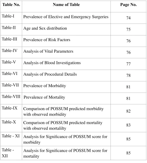
The next step is to open the peritoneum over the common hepatic artery as it leaves the celiac axis and to follow this to the origin of the gastroduodenal artery. Dissect the hepatic artery and sweep the adjacent lymphatic tissue towards the specimen. Divide the right gastric artery. Kocherize the duodenum and dissect it from the anterior surface of the pancreas for 4–6 cm. Excise any visible lymphatic tissue along the superior margin of the pancreas, the splenic artery, and the paraduodenal region. The duodenum is then divided with a surgical stapler. Examine the extent of the tumor and divide the stomach at least 8–10 cm proximal to the tumor. Restoration of gastrointestinal continuity can be achieved with either a Billroth II gastrojejunostomy or a Roux-en-Y gastrojejunostomy.
b) Small bowel resection
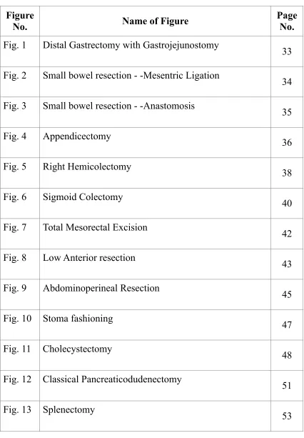
Once the diseased segment of bowel is identified, it is resected with its mesentery. This is of particular importance when dealing with neoplasms in order to get an adequate sampling of mesenteric lymph nodes. Identification and isolation of vascular arcades and their feeding vessels is facilitated by transillumination with the overhead lights. The arteries and veins are ligated with two ties and cut in between.
[image:43.612.133.513.318.601.2]
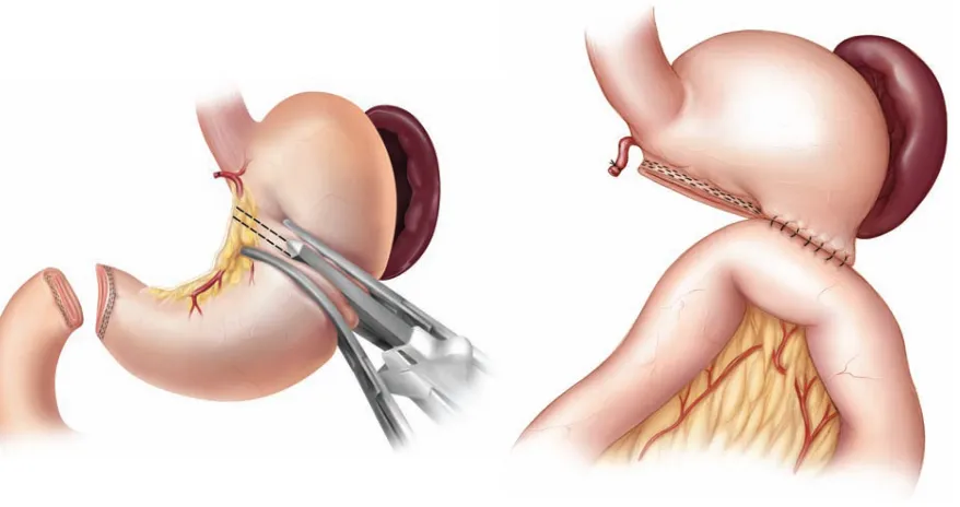
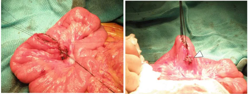
The anastomosis of the bowel can be performed in a hand-sewn fashion or with a stapling device. A hand-sewn anastomosis is usually performed end-to-end in two layers. The inner layer is done in running fashion with full-thickness bites using monofilament absorbable suture (It assures hemostasis and guards against anastomotic bleeding.) The outer layer of the anastomosis consists of interrupted sutures placed in Lembert fashion. Stapled anastomoses are usually performed in a side-to-side fashion but function end-to-end. Small enterotomies are made on the anti-mesenteric corner of the end or each segment of bowel. Each arm of the stapler is introduced into each limb of the intestine. The anti-mesenteric walls of the limbs are aligned and the stapler is fired creating a large enteroenterostomy. The anastomosis is completed by stapling across the now common enterotomy created by the first stapling manoeuvre.
c)Appendectomy
[image:45.612.72.327.372.734.2]Using the convergence of the tenia coli as a landmark, the base of the appendix is located and exposed. The mesoappendix and appendiceal artery are taken between clamps and ligated with absorbable ties. The base of the appendix is crushed with a clamp and the proximal edge of the crushed segment is doubly ligated with 2-0 silk or absorbable suture. The appendix is amputated and sent to pathology for analysis. The exposed mucosa of the appendiceal stump is cauterized. “Dunking” of the stump with a purse-string suture around the base of the appendix is considered optional.
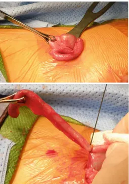
d) Right hemicolectomy
Upon entering the abdomen,the peritoneal reflection lateral to the cecum is incised and the terminal ileum is mobilized off the retroperitoneum. This dissection is continued distally along the lateral right colon and up to the hepatic flexure using electrocautery, making sure to avoid the right ureter as it passes anterior to the right common iliac bifurcation. It is necessary to identify the duodenum during mobilization of the hepatic flexure from the retroperitoneum, as the second and third portions may be injured by electrocautery during this step. The duodenum is kept posterior, mobilizing the colon anteriorly. The vessels contained within the hepatocolic ligament should be ligated and divided. This completes the mobilization around the hepatic flexure.
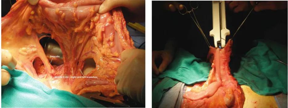
vessels, which are ligated with a heavy suture ligature and divided. The remaining small bowel mesentery is ligated and divided up to the terminal ileum. An area of the terminal ileum at least 5–10 cm proximal to the ileocecal valve is identified as the proximal margin. At this point the mesenteric resection is complete and preparation should be made for the ileocolic anastomosis.
[image:47.612.70.537.239.414.2]
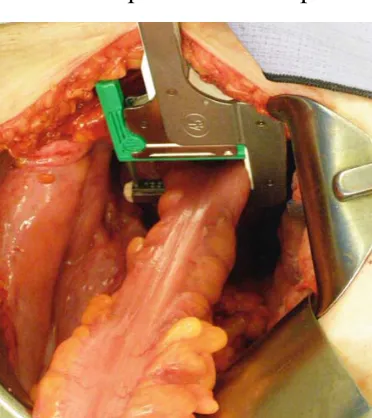
The ileocolic anastomosis can be created using a side-to-side stapled approach.The bowel is divided on the stapler with heavy scissors. The specimen is inspected on the back table and sent to surgical pathology for further evaluation.
e) Sigmoid colectomy
The mobilization of the sigmoid colon begins by incising the lateral peritoneal attachments, while staying anterior to the retroperitoneal fascia. The left ureter and gonadal vessels are identified and maintained posterolaterally to avoid injury to these structures during the medially directed sigmoid mobilization. Once the sigmoid colon is mobilized, dissection is continued proximally with electrocautery. The splenic flexure of the colon may be mobilized if it is clear that further length will be necessary in order to complete a “tension-free” anastomosis. First, the renocolic ligament is incised, allowing the splenic flexure to descend, increasing the distance between the colon and the spleen. This allows safer transection of the lienocolic ligament, which should be made along the colon wall to prevent splenic injury. The omentum is divided from the distal transverse colon as necessary.
Proximal ligation of the LCA helps to ensure adequate collateral blood flow to the proximal limb of the anastomosis from the left branch of the middle colic artery via the marginal artery of Drummond. The mesentery of the remaining colon is divided up to the bowel wall ensuring adequate mesenteric inclusion for cases of malignancy. The proximal and distal colon transections should be made in well-perfused bowel at least 5–10 cm from the tumor (in cases of malignancy) or proximally in an area of normal compliant bowel and distally at the rectosigmoid junction (in cases of diverticular disease). The proximal bowel is transected using a bowel clamp or a linear stapler
[image:49.612.73.259.337.546.2]
The anastomosis is begun by placing a purse-string suture at the proximal bowel clamp on the descending colon. The anvil shaft assembly of a circular stapler is then inserted into the lumen of the proximal bowel and the purse-string suture is tied to the groove on the shaft. The assistant then inserts the circular stapler transanally through the rectum and the center spike is delivered through the
rectal wall adjacent to the linear staple line. The anvil shaft is attached to the center spike and the stapler is closed and activated. The stapler is opened, removed, and inspected for two complete tissue rings, ensuring full circumferential tissue stapling.
f) Low anterior resection/ Abdominoperineal resection
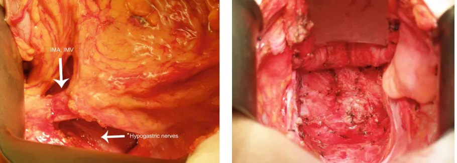
A total mesorectal excision is facilitated by retracting the divided colon anteriorly toward the pubis to identify the avascular plane posterior to the mesorectum. Incision of the areolar tissue posterior to the rectum along the endopelvic fascia is performed with electrocautery. This posterior dissection is continued down to the level of the pelvic floor. Care is taken to identify and preserve the hypogastric nerves, which are important for postoperative sexual and urinary function. These nerves can be seen and palpated at the sacral promontory, dividing bilaterally, and following the pelvic sidewalls. The peritoneal reflection is incised bilaterally as well as anteriorly and dissection in the mesorectal plane is continued circumferentially. Laterally, supporting ‘ligaments’ that may contain the middle rectal vessels and splanchnic nerve branches are carefully divided with cautery, maintaining the proper plane. In males, the seminal vesicles are visualized and kept anterior to the dissection. Similarly, in females, the posterior vaginal wall can be visualized and is carefully dissected within the rectovaginal septum.
Preservation of the fascia propria, which envelops the specimen, prevents tumor spillage and has been shown to reduce local recurrence.
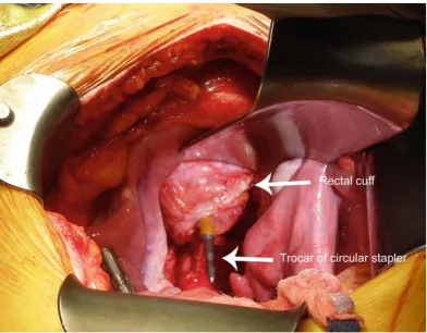
Low anterior resection
A distal margin of 2 cm is customary, though less may be acceptable if an adequate margin can be obtained without compromising the sphincter complex. An angled clamp is placed across the bowel distal to the tumor. A transverse, non-cutting stapler (30 or 45 mm) is placed at the distal most portion of this dissection and the rectum is divided. Alternatively, the anorectum is divided within the anal canal to obtain an adequate margin and facilitate a coloanal anastomosis. The specimen is sent to surgical pathology after confirming an adequate distal margin.
[image:52.612.84.280.412.565.2]
Several options for reconstruction exist. In tumors of the proximal rectum, an end-to-end anastomosis is appropriate. This anastomosis can be either handsewn or fashioned via a circular stapler.
Abdominoperineal resection
The abdominal phase of the APR procedure is identical to that of a LAR. After the complete abdominal mobilization of the rectum, the perineal phase begins. An elliptical incision is made around the anus including the entire sphincter mechanism. Dissection continues with cautery posteriorly until the coccyx is encountered.The anococcygeal ligament is then divided and the previous dissected presacral space is entered just anterior to the coccyx. The levator muscles are hooked with the surgeon’s finger and divided bilaterally with cautery. The dissection continues anterolaterally. In males, the anterior portion of the dissection is challenging due to the membranous urethra and prostate. In females, retraction of the vagina facilitates separation of anterior rectum and posterior vaginal wall. Eversion of the specimen through the perineal opening may help facilitate the remaining anterior dissection plane.
g) Stomas
Once an adequate length of bowel has been mobilized, the ostomy begins with a circular skin incision measuring 2–4 cm in diameter at the pre-marked site. Using electrocautery, a disk of skin is excised, leaving some of the subcutaneous fat behind, which will serve to support the bowel at the abdominal wall and prevent retraction. The rectus muscle is separated in the direction of its fibers with simple retraction and not divided. The posterior sheath is exposed. The posterior fascia and peritoneum are divided with electrocautery.
While keeping a finger through the opening to preserve the tract, a Babcock clamp is carefully placed through the hole in the skin into the peritoneal cavity. The clamp is placed onto the bowel segment and bowel is gently pushed through the fascial defect, with care to avoid pulling the intestine and tearing the mesentery. For loop stomas, a Penrose drain passed adjacent to the bowel wall allows gentle traction during placement. The bowel should protrude 2–4 cm from the skin. At this point, the abdominal portion of the procedure is completed and the abdominal incision is closed, to avoid contamination when the bowel is re-opened during stoma maturation.
are placed evenly around the lumen, starting inside-out through the entire thickness of the bowel and into the dermis. A clear stoma appliance is then placed over the everted bowel.
h) Cholecystectomy/ CBD exploration
[image:56.612.265.518.167.405.2]The peritoneum overlying the fundus of the gallbladder is incised. The peritoneum enveloping the gallbladder is incised along both sides of the gallbladder.The gallbladder is dissected out of the gallbladder fossa. The cystic artery is ligated and divided. The cystic duct is ligated and divided. The common bile duct is exposed in the porta hepatis. Stay sutures are placed on either side of
the planned choledochotomy and the common bile duct is opened. The common bile duct is explored, then closed over a T-tube.
[image:57.612.96.514.175.631.2]
i) Pancreaticoduodenectomy (Whipple’s)
Once disseminated disease has been ruled out, the surgeon proceeds with mobilization of the duodenum and head of the pancreas by the Kocher maneuver. Dissection of the lateral peritoneal attachments of the duodenum, which facilitates inspection of the duodenum, head of the pancreas, and periampullary tumor is usually bloodless; an avascular cleavage plane can be easily developed as the posterior wall of the pancreas is bluntly separated from the underlying vena cava and right kidney. Extensive kocherization should be performed to allow the surgeon to be comfortable that there is no extension of tumor beyond the uncinate process. Special care should be taken to identify and preserve the right gonadal vein, which often runs parallel to the inferior vena cava at this point in the retroperitoneal dissection. Further mobilization of the second and third portion of the duodenum is carried out to adequately determine resectability of the lesion.
involvement. The peritoneal attachments at the inferior border of the pancreas are incised and a cleavage plane over the superior mesenteric vein and behind the pancreas (the so-called “tunnel of love”) is developed.
The gallbladder is carefully dissected from the hepatic fossa. The cystic artery is identified, doubly clipped, and transected. Dissection should continue to the common bile duct where it is encircled with a vessel loop for subsequent transaction. The surgeon then proceeds to ligate the blood supply necessary for antrectomy. The right gastric artery is identified, ligated with 2-0 silk sutures, and subsequently transected. Next, the gastroduodenal artery (GDA), passing inferiorly from the hepatic artery at the point where the portal vein passes posterior to the pancreas, should be suture ligated with 4-0 Prolene sutures. Just before ligating and dividing the GDA, the vessel should be occluded with a vessel loop or bulldog clamp to ensure adequacy of the hepatic artery pulse. the right gastroepiploic vessels are ligated and tied.
directed toward mobilization of the upper jejunum. The transverse colon is flipped superiorly, allowing for adequate visualization of the jejunum and its mesentery. The upper jejunum may be grasped with Babcock forceps and the bowel held up in order to adequately visualize the vascular arcades supplying the jejunum. The ligament of Treitz, in its avascular plane, is taken down with cautery. Utilizing incisions made in the avascular portions of the mesentery, the jejunum is divided with a GIA stapler. The jejunal arcades are divided and ligated to facilitate mobilization of the upper jejunum. A small opening is made in the mesocolon underneath the SMV and the mobilized upper jejunum is passed through the retrocolic window.
Reconstruction is began with hepaticojejunostomy, followed by a duct-to-mucosa pancreaticojejunostomy and completed with an end to side gastrojejunostomy.
[image:60.612.72.511.498.652.2]
j) Procedures for chronic pancreatitis
The choice of operation is dependent on pancreatic ductal anatomy and the extent of disease throughout the gland. Operations to palliate abdominal pain either (1) drain a dilated pancreatic ductal system or (2) resect diseased pancreatic parenchyma in cases in which the duct is of normal diameter. The main pancreatic duct normally measures 4–5 mm in the head of the pancreas and gently tapers throughout the body (3–4 mm) and tail (2–3 mm).
!
k) Splenectomy
Open splenectomy is usually performed by a technique of medial mobilization of the spleen and dissection down to the pedicle of splenic artery and vein which is then finally divided. The procedure begins with mobilization of the spleen to the midline by division of the lateral and superior pole attachments. This includes division of the splenophrenic ligament superiorly, and the splenocolic and splenorenal ligaments at the lower pole. The short gastric vessels are then divided between ligatures or clips.
The spleen is medialized and hilar dissection performed carefully with isolation of the splenic vessels and gentle medial displacement of the tail of the pancreas to avoid pancreatic injury. In emergent cases, the splenic hilum may be clamped en bloc with three clamps in the manner of Federoff and divided and doubly ligated proximally and once distally.
[image:62.612.71.275.515.678.2]
2.4 POST OPERATIVE COMPLICATIONS
Classification of post operative complications
• Immediate - complications occurring at or immediately after surgery in the recovery room, e.g. postoperative airway obstruction.
• Early - complications occurring within 48 hours of surgery, e.g. reactive hemorrhage.
• Late - complications occurring 48 hours or more after surgery, e.g. pressure ulceration.
(i) Wound infection
The wound edges may begin to come apart and there may be a discharge from the wound (clear, bloody or purulent)
Mild wound infections may resolve spontaneously. More serious wound infections, with signs of inflammation, may require antibiotics. Initially, broad-spectrum antibiotics are administered; these can be changed later to targeted antibiotics based on sensitivities to cultured organisms.
The defect can then be allowed to heal by secondary intention or re-sutured when clean at a later date.
(ii) Wound dehiscence
Wound dehiscence is unplanned spontaneous re-opening of a wound following surgical closure. Partial dehiscence is the re-opening of the skin and superficial tissues; full dehiscence is total wound re-opening, so the floor of the wound or contents of the underlying cavity are exposed, e.g. burst abdomen.
Factors predisposing to wound dehiscence are
• Wound infection
• Poor surgical technique
• Poor blood supply
• Premature removal of sutures
• Chronic debilitated states.
(iii) Deep vein thrombosis
Thrombosis risks relate to Virchow’s triad:
Stasis - postoperative immobility allows venous pooling leading to stasis and DVT.
Vessel wall - extrinsic compression of deep veins, e.g. following orthopedic or abdominal surgery.
Blood constituents - blood may be hypercoagulable in postoperative patients, with dehydration or malignancy.
Many postoperative DVTs are asymptomatic. DVT can present with a painful, red, swollen, tender, slightly warm calf or leg and low grade systemic pyrexia. DVT can present as pulmonary embolism. This is initially a clinical diagnosis, based on symptoms and signs, and a high index of suspicion. The diagnosis can usually be confirmed on colour duplex Doppler ultrasonography. Occasionally, a venogram is required in difficult cases.
(iv) Pulmonary embolism
Large PE may present with sudden onset severe shortness of breath, collapse and sudden death.
A high index of suspicion is required in postoperative patients with respiratory problems. Investigation includes blood gas analysis confirming hypoxia, an ECG (unreliable) which may show evidence of right heart strain with classic S1 Q3 T3 changes and a ventilation/perfusion (VQ) scan. The most reliable method of diagnosis is CT pulmonary angiography.
Immediate management involves oxygen by mask, intravenous fluids and anticoagulation, initially with intravenous or subcutaneous heparin. After diagnostic confirmation full anticoagulation with warfarin is required. Indications for a vena caval filter include ongoing pulmonary emboli despite adequate anticoagulation and loose or free-floating thrombus in the leg or pelvic veins as diagnosed on Doppler ultrasound. Other treatments include thrombolysis.
(v) Myocardial infarction
The signs may include pallor, cold and clammy skin, a gallop rhythm, lung crepitations and occasionally pyrexia.
The risk of having another MI after elective surgery is about 35% in the first three months after the original MI, and 15% in the next three to six months. After six months it is about 4%. Therefore, elective surgery should be avoided where possible for at least the first six months. The mortality from re-infarction may be as high as 30-40%.
(vi) Post operative pyrexia
Low grade postoperative pyrexia can be part of the normal response to trauma. Persistent, relapsing, or high grade pyrexia can be due to:
• Wound infection.
• Chest infection / Urinary tract infection
• Abscess formation - usually a high grade swinging pyrexia.
• DVT or PE.
• Infected pressure area sores.
• Infected lines, drips or tubes.
(vii) Cerebrovascular accident
Strokes or cerebrovascular accidents (CVA) are due to an interruption to the blood supply to the brain or an intracerebral bleed. Many of the risk factors for CVA can occur around the time of surgery: hypertension as a response to pain; hypotension due to anesthesia or hypovolaemia; hypercoagulability due to dehydration; and hypocoagulability due to the use of heparin. Also, arrhythmias such as atrial fibrillation are common postoperatively, and can precipitate thromboembolic CVA. Some operations, such as carotid endarterectomy and neck dissections, can dislodge thrombus and cause a thromboembolic CVA. Other surgical procedures, often vascular, where the blood clotting time is iatrogenically prolonged, increase the risk of hemorrhagic CVA.
The basic work up should include full blood count, coagulation profile, ECG, blood glucose and lipids. Ultrasound imaging of the heart (ECHO) and the carotid arteries may be useful in selected cases. MRI or CT imaging of the brain is now advised in all cases where there are grounds to suspect a CVA.
(viii) Pneumonia
Hospital Acquired Pneumonia is a new onset pneumonia starting more than 48 hours after hospital admission. It may be due to aspiration of organisms from the nasopharynx, or due to nosocomial infection from equipment, especially ventilation equipment. It may also be due to infective emboli from distant sites. Hospitalised patients are particularly at risk if they have impaired consciousness, an inability to cough and clear secretions, are immunocompromised or have prolonged ventilation. The pathogens involved are a much wider group than community acquired pneumonia and include gram negative bacilli such as Enterobacter and E. coli and gram positive organisms such as Streptococcus pneumonia. Postoperative HAP is more likely in those at extremes of age, smokers and the obese.
ix) Atelectasis and respiratory failure
Atelectasis is collapse of portions of the lung tissue, due to inadequate ventilation of the alveoli and failure to clear pulmonary secretions. It is common following surgery, especially with upper abdominal and thoracic incisions. Other risk factors include immobility, poor postoperative analgesia, over-sedation, smoking, malnutrition, age, obesity and preexisting respiratory disease.
A high index of suspicion is required for this complication, especially in patients with the above risk factors. Ideally, patients should stop smoking pre-operatively for elective surgery, and supplementary oxygen and physiotherapy with adequate analgesia should be administered routinely in the early postoperative period. Early mobilisation and minimal postoperative sedation are also required.
Untreated atelectasis can lead to established chest infection requiring antibiotics and aggressive physiotherapy. Later sequelae may include bronchopneumonia and pleural effusions, and may require ventilator support.
Respiratory failure occurs when the pulmonary gas exchange is sufficiently impaired to cause hypoxia with or without hypercapnia. There are two types:
Type 1 - PaO2 <8kPa and PaCO2 normal or low. This is due to a diffusion defect, a ventilation-perfusion mismatch or a left to right shunt.
(x) Urinary retention/ infection
The inability to void in the postoperative period is most common in men with pre-existing prostatic hypertrophy. It occurs after lower abdominal surgery, e.g. inguinal hernia repair, or after removing the urethral catheter following other procedures. It may present with lower abdominal pain and distension, and the inability to pass urine. Occasionally it may present as urinary infection, postoperative distress or confusion.
(xi) Shock
Shock is an abnormality of the circulatory system that results in a situation where the body’s metabolic and oxygen requirements cannot be met. It is not defined solely by blood pressure criteria and there is no laboratory test for it. It is recognised by the clinical manifestations of inadequate organ perfusion and oxygenation, such as pallor, confusion, tachycardia, tachypnoea, and oliguria. The causes of shock can be classified as.
• Hypovolemic - haemorrhage, fistulae, vomiting, pancreatitis, burns.
• Distributive - septic, anaphylactic, neurogenic.
• Cardiac - cardiogenic.
Hypovolemic shock is treated by restoration of circulating blood volume and arresting ongoing bleeding. This may require surgery or interventional radiology
Septic shock is managed by resuscitation as outlined as above, plus large dose intravenous broad-spectrum antibiotics, invasive monitoring and vasopressors if necessary.
Anaphylactic shock requires exactly the same protocol as outlined above, plus intramuscular adrenaline 500 micrograms. This can be repeated as required; corticosteroids and bronchodilators are also given.
(xii) Acute respiratory distress syndrome
Acute respiratory distress syndrome (ARDS) indicates the acute diffuse pulmonary inflammatory response to either direct or indirect insults from extrapulmonary pathology
Direct—via airway or injury to chest (e.g. aspiration, toxic gases, pneumonia)
Indirect—blood-borne insults (e.g. sepsis, polytrauma, severe burns, drugs)
Can be diagnosed by the following criteria
Blood gas analysis (PaO2 / FiO2 of less than 200 mm Hg)
Chest X-ray shows bilateral diffuse infiltrates
Pulmonary artery wedge pressure (less than 15 mm Hg).
Management includes supportive measures and no specific therapy exists to modulate the sequence of events of ARDS:
✓
Monitoring of all vitals✓
Ventilatory management✓
Mechanical ventilation to permit adequate oxygen uptake✓
Nonventilatory management✓
Treatment of underlying risk factors✓
Enteral feeding(xiii) Systemic inflammatory response syndrome/ multi organ dysfunction syndrome
The response to injury that occurs in the body and leads to this hypermetabolic state is called systemic inflammatory response syndrome (SIRS) and the sequence of failing end-organs is referred to as multiple organ dysfunction syndrome (MODS).
The patients at risk are those who have sustained a major biological insult, such as:
• severe hemorrhage requiring massive blood transfusion (e.g. liver trauma);
• trauma resulting in major tissue injury (e.g. crush injury);
• large ischemia-reperfusion injury (e.g. reperfusion of a limb following )
• major burn;
• large inflammatory focus (e.g. peritonitis, pancreatitis);
• severe infection with bacteremia (e.g. ascending cholangitis).
The general aims of therapy are to:
• treat infection
• ensure adequate tissue oxygenation;
• maintain nutritional support; and
• minimize systemic inflammation.
2.5 SURGICAL AUDIT
Many governments and national organisations in developed countries have developed important strategies aimed at delivering safety and quality in healthcare. One such measure is instituting nationalized clinical audits.
Clinical audit is currently seen as the most effective way of assessing routine health care delivery and the basis of improving outcomes. Audit of outcome or process can be divided into five stages: each stage needs to be carefully planned to produce a clinically effective audit.
Preparing for audit Choose a topic and define the purpose of the audit. One option is to identify (by consulting patients and clinicians) a potential problem that may involve high costs or risks for which there is good evidence to inform standards and that may be amenable to change. NICE stresses the importance of identifying skills and resources to carry out the audit.
Selecting audit criteria Audit can assess process or outcome. • Define the patients to be included.
• Criteria to assess performance should be derived from the available evidence, e.g. trials, systematic reviews, society guidelines, or clinician consensus.
Measuring performance This is about collecting data. Identify patients or episodes from several sources (e.g. operating room logbooks and patient administration system (PAS)) to avoid missing patients because of incomplete data. Electronic information systems can improve data collection. Training dedicated audit personnel can improve the process further.
Making improvements Identify local barriers to change, develop a practical implementation plan, which should involve several interventions (practice guidelines, education, and training). Clinical governance programmes should provide the structure.
Chap
te
r 3
MATERIALS AND
MATERIALS AND METHODS
3.1
Type of study : Prospective and Descriptive Study 3.2 Study approval : Prior to commencement of this study -Ethical Committee of Stanley Medical College and Government General Hospital, Chennai
had approved the thesis protocol.
3.3 Place of study : Govt. Stanley Medical College and Hospital
3.4 Period of study : Duration starting from 01 Oct 2014 to 30 Aug 2015 3.5 Sample size : 154 cases
3.6 Selection of patients:
a) Sampling method- Purposive.
b) Inclusion criteria- Patients of age group 12 to 90 years undergoing midline laparotomy.
c)Exclusion criteria - -
1) Patients who underwent abdomen surgeries other than midline laparotomies
3.7 Study procedure:
Method of sampling was non-random, purposive. After admission short history
was taken and appropriate workup done on each patient admitted in surgery
department for laparotomy. Baseline investigations, as routinely required, were
done, followed by imaging studies. Patients were then explained about their
disease process and the possible line of management. All the necessary information
regarding the study was explained to the patients or their valid guardian. Informed
written consent was taken from the patients or their guardian willing to participate
in the study. Thorough physical examination was done in each case. Data
collection sheets were filled in by the investigator himself. All of the preoperative
factors related to the patient were noted down in the data sheet. After proper
evaluation and preparation, patients who required surgical management were taken
up for surgery. All patients were operated under general anesthesia. Strict aseptic
precautions were followed during the operation. Meticulous techniques were
practiced as far as possible. The operation procedure and related peroperative
factors were observed directly and recorded in the data collection sheet instantly.
3.8 Ethical consideration : All the patients/ legal guardians were given an explanation of the study and about the investigative and operative procedures with
their merits and demerits, expected results, and possible complications. If he/she
agreed then the case had been selected for this study. The study did not involve any
additional investigation or any significant risk. It did not cause economic burden to
the patients. The study was approved by the institutional review board prior to
commencement of data collection. Informed consent was taken from each patient/
guardian. Data were collected by approved data collection form.
3.9 Data collection : Data were collected by pre-tested structured questionnaire. Data were collected from all the respondents by direct interview after getting
informed written consent from them or from their legal guardian. The
physiological severity was scored on admission and operative severity at the end of
30 days
3.10 Data analysis : Data analysis was done both manually and by using computer. Calculated data were arranged in systemic manner, presented in various
table and figures and statistical analysis was made to evaluate the objectives of this
CHAPTER 4
Table 1 : Prevalence of Elective and Emergency surgeries in study group
Surgery Numbers Percentage
Elective 65 42.2
Emergency 89 57.8
Total 154 100
58%
42%