0022-538X/01/$04.00⫹0 DOI: 10.1128/JVI.75.21.10319–10325.2001 Copyright © 2001, American Society for Microbiology. All Rights Reserved.
Interleukin-7 in Plasma Correlates with CD4 T-Cell Depletion and
May Be Associated with Emergence of Syncytium-Inducing
Variants in Human Immunodeficiency Virus
Type 1-Positive Individuals
ANUSKA LLANO, JORDI BARRETINA, ARANTXA GUTIE´RREZ, JULIA` BLANCO, CECILIA CABRERA, BONAVENTURA CLOTET,ANDJOSE´ A. ESTE´*
Retrovirology Laboratory irsiCaixa, Hospital Universitari Germans Trias i Pujol, Universitat Auto`noma de Barcelona, 08916 Badalona, Spain
Received 11 January 2001/Accepted 26 July 2001
Human immunodeficiency virus type 1 (HIV-1) primary infection is characterized by the use of CCR5 as a coreceptor for viral entry, which is associated with the non-syncytium-inducing (NSI) phenotype in lymphoid cells. Syncytium-inducing (SI) variants of HIV-1 appear in advanced stages of HIV-1 infection and are characterized by the use of CXCR4 as a coreceptor. The emergence of SI variants is accompanied by a rapid decrease in the number of T cells. However, it is unclear why SI variants emerge and what factors trigger the evolution of HIV from R5 to X4 variants. Interleukin-7 (IL-7), a cytokine produced by stromal cells of the thymus and bone marrow and by keratin, is known to play a key role in T-cell development. We evaluated IL-7 levels in plasma of healthy donors and positive patients and found significantly higher levels in HIV-positive patients. There was a negative correlation between circulating IL-7 levels and CD4ⴙT-cell count in
HIV-positive patients (rⴝ ⴚ0.621;P< 0.001), suggesting that IL-7 may be involved in HIV-induced T-cell depletion and disease progression. IL-7 levels were higher in individuals who harbored SI variants and who had progressed to having CD4 cell counts of lower than 200 cells/l than in individuals with NSI variants at a similar stage of disease. IL-7 induced T-cell proliferation and up-regulated CXCR4 expression in peripheral blood mononuclear cells in vitro. Taken together, our results suggest a role for IL-7 in the maintenance of T-cell regeneration and depletion by HIV in infected individuals and a possible relationship between IL-7 levels and the emergence of SI variants.
Human immunodeficiency virus type 1 (HIV-1) primary in-fection is characterized by the presence of non-syncytium-in-ducing (NSI) variants with low replication kinetics, capable of infecting macrophages and CD4⫹memory T cells, that use the receptor CCR5 as a coreceptor for viral entry (21). Later, as the disease progresses, the syncytium-inducing (SI) variants emerge (2). SI variants are characterized by high replication kinetics in vitro and the capacity to infect naive CD4⫹T cells by using CXCR4 as a coreceptor (4, 26). The emergence of SI variants is accompanied by an accelerated decrease of CD4⫹ cell count, rapid disease progression, and the establishment of AIDS (10, 13). However, it remains unclear why SI variants emerge and how this relates to CXCR4 expression in vivo. It is probable that multiple host factors affect HIV-1 coreceptor levels or function; interleukin-4 (IL-4) has been shown to de-crease the expression of CCR5 and inde-crease CXCR4 expres-sion, favoring the propagation of X4 strains (42). Other factors that may induce overexpression of CXCR4 or block the repli-cation of R5-NSI variants may favor the selection of X4-SI HIV variants and could determine when SI variants will arise. During clinical latency, HIV-1 replicates, inducing the de-struction of CD4⫹ T cells and immature cells in the bone marrow, thymus, and lymph nodes, where T cells are produced
(25). The immune system responds by inducing the prolifera-tion of T cells, and hence the CD4⫹T-cell number is main-tained relatively constant during this stage of the infection (11, 20, 23, 43). The development of AIDS was thought to be caused by exhaustion of the immune system. That is, at a certain point, the immune system cannot maintain the high rate of T-cell production necessary to compensate for HIV-induced T-cell depletion (27). Nevertheless, HIV-1 infection destroys T-cell supplies in the periphery by direct infection and killing of cells and through hyperactivation of the immune system (15), suggesting that it may be not exhaustion but rather homeostatic inability, along with gradual wasting of T-cell sup-plies, that leads to T-lymphocyte depletion in HIV-1 infection (16, 17).
It is known that cytokines play an important role in HIV-1 infection. However, determination of their function in viral dynamics, replication, and disease progression is very complex, because different cytokines have opposite effects on viral rep-lication (19, 24, 32, 34, 42). IL-7 is a cytokine produced by stromal cells of the thymus and the bone marrow and by ker-atinocytes (18, 38, 39, 45). IL-7 has recognized functions in B-cell lymphopoiesis (30) and has been shown to take part in the differentiation of thymocytes into mature T cells that will leave the thymus and move to the periphery (7, 29). Similarly, IL-7 contributes to the development, proliferation, and ho-meostatic maintenance of T cells (12, 14, 33, 35, 40). IL-7 is known to enhance viral replication (6, 41) and may induce CXCR4 expression on resting CD4⫹memory T cells in vitro * Corresponding author. Mailing address: Fundacio´ irsiCaixa,
Ret-rovirology Laboratory, Hospital Universitari Germans Trias i Pujol, 08916 Badalona, Spain. Phone: 34-934656374. Fax: 34-934653968. E-mail: jaeste@ns.hugtip.scs.es.
10319
on November 9, 2019 by guest
http://jvi.asm.org/
to assess the effect of IL-7 on the evolution of HIV in vivo. We have evaluated IL-7 levels in plasma of healthy individ-uals and HIV-1-infected patients and correlated their expres-sion in HIV-positive individuals to CD4⫹ T-cell depletion, disease progression, and emergence of the SI phenotype.
MATERIALS AND METHODS
Patient and donor samples.Blood samples from healthy donors and from HIV-positive individuals were collected from our hospital blood blank and from the HIV unit, respectively. Samples were collected with informed consent and processed immediately after collection. Briefly, 10 to 20 ml of whole blood was collected in EDTA-Vacutainer tubes (Becton Dickinson [BD], Madrid, Spain). Plasma was isolated from each sample after centrifugation of blood samples at 400⫻gfor 10 min and was immediately cryopreserved and stored at⫺80°C until use. Peripheral blood mononuclear cells (PBMC) were obtained by separation on Ficoll-Hypaque density gradient and either used immediately in fractional studies or cryopreserved in liquid nitrogen for further determinations. Some patients were enrolled in clinical trials with monotherapy (zidovudine [AZT] or dideoxyinosine [ddI]) or dual therapy (AZT plus dideoxycytosine, AZT plus ddI, or AZT plus lamivudine [3TC]) and were later included in triple antiretroviral therapy. These patients usually initiated treatment at a late stage of disease and the treatment options were not efficacious, increasing the possibility selecting SI variants.
T-lymphocyte proliferation.Fresh PBMC (106) from two healthy donors were cultured with different antigens as follows: medium control, phytohemagglutinin (PHA) (4 ng/ml) plus IL-2 (4 ng/ml) as a positive control, PHA plus IL-2 plus IL-7 (10 ng/ml), and IL-7 alone (1 and 10 ng/ml). The cultures were maintained at 37°C in a 5% CO2incubator for 5 days. [3H]thymidine was then added to each well and incubated overnight. The cells were harvested, and the amount of incorporated [3H]thymidine was measured in a liquid scintillation counter (1450 Microbeta; Wallac, Turku, Finland). The stimulation index was calculated by dividing the counts per minute of PBMC after specific stimulation by the counts per minute of PBMC incubated with medium control. A stimulation index of⬎5 was considered to be a positive response in this assay.
CXCR4 expression.Fresh PBMC from healthy donors were cultured with IL-7 at different concentrations and with stromal cell-derived factor 1 (SDF-1) (500 ng/ml) or medium alone as controls. After 5 days of incubation, PBMC were collected and CXCR4 and CD4 expression was analyzed by flow cytometry as described below.
Detection of IL-7 and RANTES levels in plasma.Plasma IL-7 levels were determined by an ultrasensitive commercial enzyme-linked immunosorbent assay (ELISA) (Quantikine HS Human IL-7 Immunoassay; R&D Systems, Minneap-olis, Minn.) according to the manufacturer instructions. RANTES levels were measured by a commercial ELISA (Endogen, Barcelona, Spain).
Viral isolation and phenotype in MT-2 cells.PBMC (10⫻106) from HIV-infected individuals were cocultured with PBMC (5⫻106) from healthy donors stimulated with 3g of PHA per ml and 25 IU of IL-2 per ml. Viral replication was quantified by evaluation of antigen p24 production in coculture superna-tants, using a commercial ELISA (Innogenetics, Madrid, Spain). Coculture su-pernatants that were positive for p24 were collected after centrifugation at 400⫻ gfor 5 min, and the SI or NSI phenotype was determined in MT-2 cells as was previously described (9). For simplicity, individuals from whom SI or NSI vari-ants were isolated are referred to hereafter as SI or NSI individuals, respectively. Flow cytometry.CD4⫹and CXCR4⫹T-cell subpopulations were determined by flow cytometry analysis. Aliquots of 50l of whole-blood samples were stained with monoclonal antibodies CD4-PerCP and CXCR4-PE (BD) for 15 min, and then the samples were washed twice in phosphate-buffered saline, resuspended in phosphate-buffered saline containing 1% formaldehyde, and analyzed in a FACScalibur flow cytometer (BD).
Measurement of viral load.Plasma HIV RNA levels were determined using a commercial assay (Amplicor VIH-1 Monitor Assay; Roche Molecular Systems, Somerville, N.J.) according to the manufacturer’s instructions. Undetectable levels of RNA in plasma were considered equivalent to 200 copies/ml.
Statistical analysis.Statistical analysis was performed using parametric and nonparametric tests (Spearmanrand Mann-Whitney U tests).Pvalues of⬍0.05 were considered to have statistical significance. Data were analyzed using the SPSS version 9.0 software package.
RESULTS
T-lymphocyte proliferation. IL-7 has been described as an indispensable factor for T-cell development (33). As can be observed in Fig. 1, IL-7 induced the proliferation of T cells at 10 ng/ml (stimulation index ⫽ 11) and boosted PHA- and IL-2-induced cell proliferation activity in vitro, confirming a known property of this cytokine. Our data support previous studies in which IL-7 has been described as an important agent in T-cell proliferation (14).
IL-7 up-regulates CXCR4 expression in vitro.To determine if IL-7 modulates CXCR4 expression in T cells, we incubated PBMC of two uninfected donors for 5 days with different concentrations of this cytokine. IL-7 up-regulated the expres-sion of the CXCR4 receptor in a dose-dependent manner, whereas SDF-1, the natural ligand of CXCR4, down-regulated CXCR4 expression (Fig. 2A). Although IL-7 caused the great-est up-regulation of CXCR4 expression at 100 ng/ml (Fig. 2B), 0.1 ng/ml was sufficient to up-regulate CXCR4 expression in vitro. However, CD4 expression was not modified by IL-7 (Fig. 2B). This finding suggests a possible role of IL-7 in selection of SI variants in HIV-positive patients with high IL-7 levels in plasma through up-regulation of CXCR4.
IL-7 levels in HIV-1-infected patients and healthy donors.
[image:2.587.310.536.71.219.2]IL-7 levels in 49 plasma samples from healthy volunteers and in 131 plasma samples from HIV-positive patients were ana-lyzed in a cross-sectional study. The HIV-positive group had significantly (P⬍0.001) higher levels of IL-7 than the healthy donor group (Fig. 3). The IL-7 levels measured in plasma were 3.6⫾3.05 and 9.4⫾5.7 pg/ml (means and standard deviations [SD]) for the healthy donor and HIV-positive groups, respec-tively. Other immunological and virological characteristics FIG. 1. Lymphocyte proliferative response to PHA (4g/ml) plus IL-2 (4 ng/ml), PHA plus IL-2 plus IL-7 (10 ng/ml), and IL-7 alone (1 or 10 ng/ml). The stimulation index was calculated by dividing the counts per minute of PBMC in stimulated wells by the counts per minute of PBMC in medium alone. The results are from a represen-tative experiment of two performed.
on November 9, 2019 by guest
http://jvi.asm.org/
were evaluated for HIV-positive patients, with the findings that the means and SD of CD4 and CD8 T-cell counts and the log10 viral load were 243 ⫾ 221 cells/l, 796 ⫾ 586 cells/l, and 5.26⫾5.5 copies/ml, respectively. RANTES levels were eval-uated for healthy donors (12.7⫾16.1 ng/ml) and HIV-positive patients (27.7 ⫾ 21.2 ng/ml); the differences between the groups were significant (P⬍0.001), as previously described (1).
IL-7 as a marker of disease progression.It has been recently shown that increased production of IL-7 accompanies HIV-1-mediated T-cell depletion (31) Similarly, we have found a clear negative correlation between IL-7 levels in plasma and abso-lute CD4⫹ T-cell counts in HIV-positive individuals (r ⫽ ⫺0.621;P⬍0.001) (Fig. 4A). A similar but weaker correlation was found between IL-7 levels and CD8⫹ T-cell levels (r ⫽ ⫺0.406;P⬍0.001) (Fig. 4B). When we grouped HIV-positive individuals according to their immunological status (that is, stratifying HIV-positive patients according to their CD4⫹ T-cell count) (Fig. 4C), the subset with⬍200 CD4 cells/l had significantly (P ⬍ 0.001) higher levels of IL-7 in plasma (12.26⫾5.86 pg/ml) than the subsets with CD4 cell counts of
between 200 and 500 and above 500 cells/l (6.67⫾4.10 and 5.69⫾2.75 pg/ml, respectively). These data suggest that CD4⫹ T-cell depletion, caused by HIV-1 replication, may alter IL-7 levels in plasma as a means to regenerate T-cell numbers.
[image:3.587.309.534.72.214.2]FIG. 2. (A) Effect of IL-7 and SDF-1 on expression of CXCR4 in PBMC. IL-7 was evaluated at concentrations of 100, 10, 1, and 0.1 ng/ml. SDF-1 was evaluated at 500 ng/ml. CXCR4 expression is rep-resented as mean fluorescence intensity (MFI). (B) Comparison of CXCR4 and CD4 expression in PBMC from a healthy donor, either stimulated with 100 ng of IL-7 per ml (thick lines) or without stimu-lation (thin lines). The results are from a representative experiment of two performed.
FIG. 3. Expression of IL-7 levels in plasma of healthy donors and HIV-positive patients. Results are depicted in box plot diagrams, where the box represents the 25th and 75th quartiles and the line represents the median value. Bars indicate 5th and 95th percentiles, and circles indicate atypical values. The means⫾SD of IL-7 levels in plasma for the healthy donor group and the HIV-positive patient group were 3.6⫾3.05 and 9.4⫾5.7 pg/ml, respectively.
FIG. 4. (A and B) Correlation between IL-7 levels and CD4 T-cell count (r⫽ ⫺0.621) (A) and between IL-7 levels and CD8 T-cell count (r⫽ ⫺0.406) (B) in HIV-positive patients. (C) Levels of IL-7 in plasma of HIV-positive patients stratified according to CD4 T-cell count in three subsets, i.e., ⬍200, 200 to 500, and⬎500 CD4 cells/l. The means⫾SD of IL-7 levels in plasma for the three subsets were 12.26⫾
5.9, 6.67⫾4.1, and 5.69⫾2.7 pg/ml, respectively. Subsets are depicted in box plots as described for Fig. 3.
on November 9, 2019 by guest
http://jvi.asm.org/
[image:3.587.306.537.363.648.2]IL-7 as a marker for the emergence of the SI phenotype.
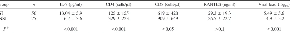
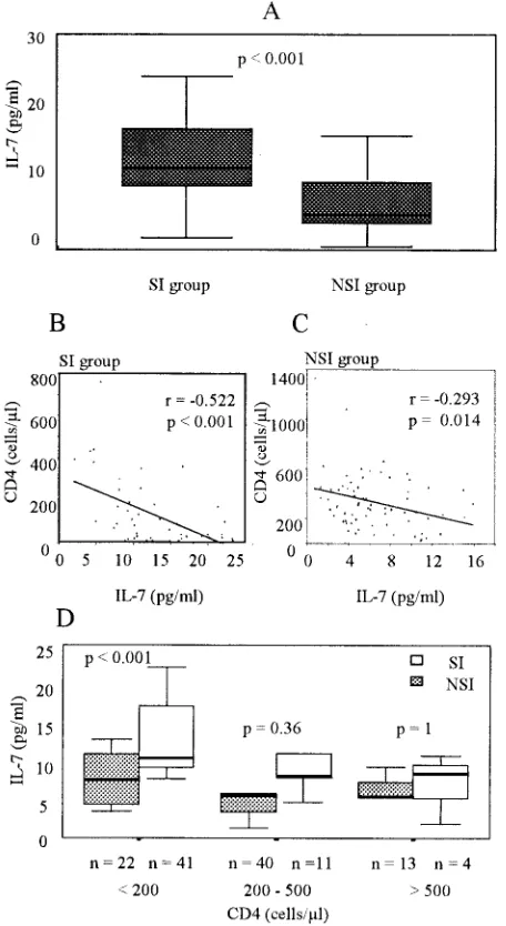
PBMC from HIV-positive individuals were cocultured with PBMC from healthy donors to isolate virus. HIV-1 p24 anti-gen-containing supernatant from each coculture was then used for evaluation of the SI phenotype in MT-2 cells. Fifty-six samples tested as SI, and 75 samples tested as NSI. When we analyzed IL-7 levels in the HIV-positive group separated into groups that harbored SI and NSI viruses, we found significantly (P⬍0.001) higher levels of IL-7 in the SI group (13⫾6 pg/ml) than in the NSI group (7 ⫾ 4 pg/ml) (Table 1; Fig. 5A). Significant differences were also found between the SI group and the NSI group in CD4⫹T-cell count, CD8⫹T-cell count, and viral load, suggesting a more advanced stage of disease in the SI group than in the NSI group. There were no significant differences in RANTES levels between the groups.
We correlated CD4⫹T-cell counts with IL-7 levels sepa-rately in the SI and NSI groups, finding a higher correlation between the two parameters in the SI group (r⫽ ⫺0.522;P⬍ 0.001) than in the NSI group (r⫽ ⫺0.293;P⫽0.014) (Fig. 5B and C).
Since HIV disease may progress in the absence of the SI phenotype, it was important to compare IL-7 levels between individuals in the NSI or SI group in a similar stage of disease. Thus, we stratified the individuals of both groups according to their CD4 T-cell count (Fig. 5D). The NSI group showed no significant differences in IL-7 levels in the three subsets of CD4 levels (7.3⫾3.2, 6.3⫾4, and 5.7⫾2.9 pg/ml). In contrast, in the SI group, we found significantly (P⬍0.001) higher levels of IL-7 (14.6⫾5.4 pg/ml) in the subset with the lower CD4 cell count (⬍200 cells/l) than in the two other subsets (7.8⫾4.5 and 5.6 ⫾ pg/ml, respectively). It is interesting that the SI subset with a CD4 cell level of⬍200 cells/l showed signifi-cantly (P⬍ 0.001) higher levels of IL-7 than the NSI subset with a similar CD4 cell count, whereas there were no signifi-cant differences in IL-7 levels between the NSI and the SI groups when the CD4 cell count was above 200 cells/l.
High IL-7 levels are associated with SI variants. To char-acterize the IL-7 level in plasma as a marker of the emergence of the SI phenotype, we stratified patients according to the IL-7 level in plasma (Fig. 6) The results showed that 89% of individuals with more than 13 pg of IL-7 per ml belonged to the SI group and that this decreased to 32 and 22% when IL-7 levels were below 13 and 2 pg/ml, respectively. The 78% of the individuals with IL-7 levels below 2 pg/ml belonged to the NSI group. As the IL-7 level increased (2 to 13 pg/ml and ⬎13 pg/ml), the proportion of NSI individuals was lower (68 and 11%, respectively). Thus, the IL-7 level in plasma may be a marker for the emergence of the SI phenotype.
Longitudinal study. A longitudinal study was designed to evaluate changes in IL-7 levels with respect to standard viro-logical and immunoviro-logical markers of HIV-1 disease (viral load and CD4 cell count) and to better characterize IL-7 as a marker of disease progression. Frozen plasma samples that were collected from five HIV-positive individuals for a period of 2 to 7 years were used to evaluate IL-7 levels in plasma. Patients were selected on the basis of availability of plasma samples taken at least every 3 months for the period of the study, in which IL-7 levels and viral load could be evaluated. All patients had received antiretroviral therapy during the course of the disease; therefore, the CD4⫹T-cell count, viral load, and IL-7 levels in plasma were expected to be influenced by drug treatment. In Fig. 7, we show IL-7, CD4⫹T-cell count, and viral load trends for two patients as a representative sam-ple of the longitudinal study. In patient A, a decrease in IL-7 level corresponded to an increase in CD4 T-cell count and to a decrease in viral load, indicating an effective response to the treatment with indinavir. The data for patient B revealed bi-phasic trends in the measured parameters; that is, this patient did not respond satisfactorily to the first treatment with double therapy (AZT plus 3TC), which caused a initial increase in IL-7 level and in viral load and a decrease in CD4 T-cell count. After 2 years the patient began receiving triple antiretroviral therapy, which caused a decrease in viral load and in IL-7 levels and an increase in CD4 T-cell count. Taken together, these data suggest that the plasma IL-7 level may be an effec-tive marker of disease progression in HIV-posieffec-tive patients.
DISCUSSION
In our cross-sectional study, we have found a significant difference in plasma IL-7 levels between HIV-negative donors and HIV-positive patients. Confirming recently published re-sults (12, 31), we have found a negative correlation between IL-7 levels in plasma and CD4⫹T-cell counts in HIV-positive patients (r⫽ ⫺0.621), suggesting that HIV infection may mask the proliferative effect of IL-7 (Fig. 1) (31).
In addition, a longitudinal study with HIV-positive patients showed that variations in CD4⫹T-cell counts caused by the response to treatment were accompanied by similar variations in plasma IL-7 levels. These data support the idea of IL-7 as an indicator of CD4⫹ T-cell depletion and consequently as a marker of disease progression.
There is controversy about the origin of the T-cell renewal that compensates for T-cell depletion in HIV infection. Some evidence points to a persistent immune activation induced by viral replication that causes proliferation of existing naive CD4⫹T cells in the periphery (16). Other evidence points to
on November 9, 2019 by guest
http://jvi.asm.org/
[image:4.587.49.543.84.142.2]thymic output of new naive T cells (8, 29) caused by a homeo-static response to T-cell depletion. Previous observations have associated abundant thymic tissue in HIV-positive individuals with increased numbers of naive T cells (8, 36). Since IL-7 is produced by stromal cells of the thymus and is implicated in thymocyte maturation, our data may indicate a homeostatic response that is mediated by IL-7. Alternatively, IL-7 produced by extrathymic tissue or induced by other factors (e.g., tumor necrosis factor alpha) could explain the observations made here. The fact that some individuals with⬍200 CD4 cells/l have low IL-7 levels in plasma may support the latter hypoth-esis.
Our results suggest a relationship between IL-7 levels in
plasma and HIV phenotype, since HIV-positive patients with high IL-7 levels had a high probability (0.89) of having the SI phenotype. It is unclear why individuals with low CD4 cell counts of the NSI phenotype have significantly lower IL-7 levels than those individuals with SI variants. If IL-7 increases in response to CD4 cell depletion, NSI individuals with CD4 cell counts of⬍200/l should have plasma IL-7 levels similar
[image:5.587.52.280.72.489.2]FIG. 5. (A) IL-7 levels in plasma in HIV-positive patients sepa-rated according to the viral phenotype (SI or NSI). IL-7 levels are depicted in a box plot as described for Fig. 3. (B and C) Linear regression between IL-7 levels and CD4 T-cell counts in patients of the SI group (r⫽ ⫺0.522) (B) and the NSI group (r⫽ ⫺0.293) (C). (D) Levels of IL-7 in plasma of HIV-positive individuals of the SI and NSI groups stratified according to CD4 T-cell count in three subsets as described for Fig. 4C.
[image:5.587.315.528.72.172.2]FIG. 6. Percentages of individuals belonging to the SI and NSI groups, stratified according to the levels of IL-7 in plasma in three subsets (⬍2, 2 to 13, and⬎13 pg/ml.
FIG. 7. IL-7 levels, CD4 T-cell counts, and log10viral load in two
patients along the course of the longitudinal study. In patient A, time zero corresponds to the initiation of treatment (AZT plus ddI plus 3TC plus indinavir). In patient B, time zero corresponds to the initi-ation of the first treatment (AZT plus 3TC plus d4T), which was continued for 2 years; the second treatment (AZT plus 3TC plus d4T plus indinavir) (time of initiation is shown by the arrowhead) covers the following 2 years. The lines represent the trends calculated by linear regression.
on November 9, 2019 by guest
http://jvi.asm.org/
[image:5.587.306.538.329.642.2]individuals with advanced disease progression.
IL-7 may be considered a causal factor for the emergence of the SI variants, together with other factors such as SDF-1. We have shown that individuals with high levels of SDF-1 were at a lower risk of developing HIV variants of the SI phenotype (28).
The immune system may respond to CD4 T-cell depletion caused by HIV replication by inducing the proliferation of circulating naive CD4 T cells and by producing homeostatic signals (such as IL-7) that induce production of new naive CD4 T cells. However, at a certain time during infection, the im-mune system may not be able to respond to the signals induced by decreased CD4 T-cell number, which in turn act on existing CD4 cells. Consequently, higher IL-7 levels may induce the overexpression of CXCR4 (Fig. 2A), allowing SI variants to grow. High SDF-1 levels could maintain lower expression of CXCR4 and keep SI variants at bay, and at the same time, high SDF-1 levels could block the effect of IL-7 and/or other factors that affect CXCR4 expression. This hypothesis could help to explain the correlation between the emergence of SI variants and the rapid CD4 T-cell decline and why SI variants appear late after infection. The effect of IL-7 on CXCR4 expression could be masked by the same principle governing IL-7 and CD4 cell count: it is possible that during HIV-1 infection, increased IL-7 could lead to the selective destruction of CXCR4-expressing cells. The regulation of CXCR4 expression appears to be governed by multiple factors that may be active at any given time and that require further study.
In conclusion, our observations on the increased production of IL-7 in HIV-positive individuals and its correlation to T-cell depletion suggest that IL-7 may have an important role in the maintenance of T-cell homeostasis in HIV infection. Intrapa-tient IL-7 production may be an effective marker of the disease progression and a causal factor for the emergence of SI HIV variants.
ACKNOWLEDGMENTS
This work was supported in part by Fundacio´n para la Investigacio´n y la Prevencio´n del SIDA en Espan˜a (FIPSE) project 3111/00, Minis-terio de Ciencia y Tecnologı´a project BFM2000-1382, and the Fun-dacio´ irsiCaixa. J. Blanco is an FIS researcher from the FunFun-dacio´ para la Recerca Biome´dica Hospital Germans Trias i Pujol. A. Llano and J. Barretina hold predoctoral scholarships from FIS.
REFERENCES
1.Aukrust, P., F. Muller, and S. S. Froland.1998. Circulating levels of RAN-TES in human immunodeficiency virus type 1 infection: effect of potent antiretroviral therapy. J. Infect. Dis.177:1091–1096.
2.Berger, E. A., P. M. Murphy, and J. M. Farber.1999. Chemokine receptors as HIV-1 coreceptors: roles in viral entry, tropism, and disease. Annu. Rev. Immunol.17:657–700.
3.Berkowitz, R. D., S. Alexander, and J. M. McCune.2000. Causal relation-ships between HIV-1 coreceptor utilization, tropism, and pathogenesis in human thymus. AIDS Res. Hum. Retroviruses16:1039–1045.
and N. Israel.1999. Thymocyte-thymic epithelial cell interaction leads to high-level replication of human immunodeficiency virus exclusively in ma-ture CD4⫹CD8⫺CD3⫹thymocytes: a critical role for tumor necrosis factor and interleukin-7. J. Virol.73:7533–7542.
8.Douek, D. C., R. D. McFarland, P. H. Keiser, E. A. Gage, J. M. Massey, B. F. Haynes, M. A. Polis, A. T. Haase, M. B. Feinberg, J. L. Sullivan, B. D. Jamieson, J. A. Zack, L. J. Picker, and R. A. Koup.1998. Changes in thymic function with age and during the treatment of HIV infection. Nature396: 690–695.
9.Este, J. A., C. Cabrera, J. Blanco, A. Gutierrez, G. Bridger, G. Henson, B. Clotet, D. Schols, and E. De Clercq.1999. Shift of clinical human immuno-deficiency virus type 1 isolates from X4 to R5 and prevention of emergence of the syncytium-inducing phenotype by blockade of CXCR4. J. Virol.73: 5577–5585.
10.Fauci, A. S.1996. Host factors and the pathogenesis of HIV-induced disease. Nature384:529–534.
11.Fauci, A. S.1993. Multifactorial nature of human immunodeficiency virus disease: implications for therapy. Science262:1011–1018.
12.Fry, T. J., E. Connick, J. Falloon, M. M. Lederman, D. J. Liewehr, J. Spritzler, S. M. Steinberg, L. V. Wood, R. Yarchoan, J. Zuckerman, A. Landay, and C. L. Mackall.2001. A potential role for interleukin-7 in T-cell homeostasis. Blood97:2983–2990.
13.Glushakova, S., J. C. Grivel, W. Fitzgerald, A. Sylwester, J. Zimmerberg, and L. B. Margolis.1998. Evidence for the HIV-1 phenotype switch as a causal factor in acquired immunodeficiency. Nat. Med.4:346–349.
14.Grabstein, K. H., A. E. Namen, K. Shanebeck, R. F. Voice, S. G. Reed, and M. B. Widmer.1990. Regulation of T cell proliferation by IL-7. J. Immunol. 144:3015–3020.
15.Hazenberg, M. D., D. Hamann, H. Schuitemaker, and F. Miedema.2000. T cell depletion in HIV-1 infection: how CD4⫹T cells go out of stock. Nat. Immunol.1:285–289.
16.Hazenberg, M. D., S. A. Otto, J. W. Stuart, M. C. Verschuren, J. C. Borleffs, C. A. Boucher, R. A. Coutinho, J. M. Lange, T. F. de Wit, A. Tsegaye, J. J. van Dongen, D. Hamann, R. J. de Boer, and F. Miedema.2000. Increased cell division but not thymic dysfunction rapidly affects the T-cell receptor excision circle content of the naive T cell population in HIV-1 infection. Nat. Med.6:1036–1042.
17.Hazenberg, M. D., J. W. Stuart, S. A. Otto, J. C. Borleffs, C. A. Boucher, R. J. de Boer, F. Miedema, and D. Hamann.2000. T-cell division in human immunodeficiency virus (HIV)-1 infection is mainly due to immune activa-tion: a longitudinal analysis in patients before and during highly active antiretroviral therapy (HAART). Blood95:249–255.
18.Heufler, C., G. Topar, A. Grasseger, U. Stanzl, F. Koch, N. Romani, A. E. Namen, and G. Schuler.1993. Interleukin 7 is produced by murine and human keratinocytes. J. Exp. Med.178:1109–1114.
19.Ho, D. D., K. L. Hartshorn, T. R. Rota, C. A. Andrews, J. C. Kaplan, R. T. Schooley, and M. S. Hirsch.1985. Recombinant human interferon alfa-A suppresses HTLV-III replication in vitro. Lanceti:602–604.
20.Ho, D. D., A. U. Neumann, A. S. Perelson, W. Chen, J. M. Leonard, and M. Markowitz.1995. Rapid turnover of plasma virions and CD4 lymphocytes in HIV-1 infection. Nature373:123–126.
21.Jansson, M., E. Backstrom, A. Bjorndal, V. Holmberg, P. Rossi, E. M. Fenyo, M. Popovic, J. Albert, and H. Wigzell.1999. Coreceptor usage and RANTES sensitivity of non-syncytium-inducing HIV-1 isolates obtained from patients with AIDS. J. Hum. Virol.2:325–338.
22.Jourdan, P., J. P. Vendrell, M. F. Huguet, M. Segondy, J. Bousquet, J. Pene, and H. Yssel. 2000. Cytokines and cell surface molecules independently induce CXCR4 expression on CD4⫹CCR7⫹human memory T cells. J. Im-munol.165:716–724.
23.Keet, I. P., P. Krijnen, M. Koot, J. M. Lange, F. Miedema, J. Goudsmit, and R. A. Coutinho.1993. Predictors of rapid progression to AIDS in HIV-1 seroconverters. AIDS7:51–57.
24.Kinter, A. L., M. Ostrowski, D. Goletti, A. Oliva, D. Weissman, K. Gantt, E. Hardy, R. Jackson, L. Ehler, and A. S. Fauci.1996. HIV replication in CD4⫹
T cells of HIV-infected individuals is regulated by a balance between the viral suppressive effects of endogenous beta-chemokines and the viral induc-tive effects of other endogenous cytokines. Proc. Natl. Acad. Sci. USA 93:14076–14081.
25.Kitchen, S. G., C. H. Uittenbogaart, and J. A. Zack.1997. Mechanism of
on November 9, 2019 by guest
http://jvi.asm.org/
human immunodeficiency virus type 1 localization in CD4-negative thymo-cytes: differentiation from a CD4-positive precursor allows productive infec-tion. J. Virol.71:5713–5722.
26.Koot, M., I. P. Keet, A. H. Vos, R. E. de Goede, M. T. Roos, R. A. Coutinho, F. Miedema, P. T. Schellekens, and M. Tersmette.1993. Prognostic value of HIV-1 syncytium-inducing phenotype for rate of CD4⫹cell depletion and progression to AIDS. Ann. Intern. Med.118:681–688.
27.Leonard, R., D. Zagury, I. Desportes, J. Bernard, J. F. Zagury, and R. C. Gallo.1988. Cytopathic effect of human immunodeficiency virus in T4 cells is linked to the last stage of virus infection. Proc. Natl. Acad. Sci. USA 85:3570–3574.
28.Llano, A., J. Barretina, A. Gutierrez, J. Blanco, B. Clotet, and J. A. Este´. 2001. SDF-1 prevents the emergence of the syncytium inducing phenotype of HIV-1 in vivo. AIDS15:1890–1892.
29.McCune, J. M., R. Loftus, D. K. Schmidt, P. Carroll, D. Webster, L. B. Swor-Yim, I. R. Francis, B. H. Gross, and R. M. Grant.1998. High preva-lence of thymic tissue in adults with human immunodeficiency virus-1 infec-tion. J. Clin. Invest.101:2301–2308.
30.Namen, A. E., S. Lupton, K. Hjerrild, J. Wignall, D. Y. Mochizuki, A. Schmierer, B. Mosley, C. J. March, D. Urdal, and S. Gillis.1988. Stimulation of B-cell progenitors by cloned murine interleukin-7. Nature333:571–573. 31.Napolitano, L. A., R. M. Grant, S. G. Deeks, D. Schmidt, S. C. De Rosa, L. A.
Herzenberg, B. G. Herndier, J. Andersson, and J. M. McCune.2001. In-creased production of IL-7 accompanies HIV-1-mediated T-cell depletion: implications for T-cell homeostasis. Nat. Med.7:73–79.
32.Osborn, L., S. Kunkel, and G. J. Nabel.1989. Tumor necrosis factor alpha and interleukin 1 stimulate the human immunodeficiency virus enhancer by activation of the nuclear factor kappa B. Proc. Natl. Acad. Sci. USA86: 2336–2340.
33.Plum, J., M. De Smedt, G. Leclercq, B. Verhasselt, and B. Vandekerckhove. 1996. Interleukin-7 is a critical growth factor in early human T-cell devel-opment. Blood88:4239–4245.
34.Saha, K., G. Bentsman, L. Chess, and D. J. Volsky. 1998. Endogenous production of beta-chemokines by CD4⫹, but not CD8⫹, T-cell clones cor-relates with the clinical state of human immunodeficiency virus type 1 (HIV-1)-infected individuals and may be responsible for blocking infection with non-syncytium-inducing HIV-1 in vitro. J. Virol.72:876–881.
35.Schluns, K. S., W. C. Kieper, S. C. Jameson, and L. Lefrancois.2000.
Interleukin-7 mediates the homeostasis of naive and memory CD8 T cells in vivo. Nat. Immunol.1:426–432.
36.Smith, K. Y., H. Valdez, A. Landay, J. Spritzler, H. A. Kessler, E. Connick, D. Kuritzkes, B. Gross, I. Francis, J. M. McCune, and M. M. Lederman. 2000. Thymic size and lymphocyte restoration in patients with human im-munodeficiency virus infection after 48 weeks of zidovudine, lamivudine, and ritonavir therapy. J. Infect. Dis.181:141–147.
37.Stanley, S. K., J. M. McCune, H. Kaneshima, J. S. Justement, M. Sullivan, E. Boone, M. Baseler, J. Adelsberger, M. Bonyhadi, J. Orenstein, et al.1993. Human immunodeficiency virus infection of the human thymus and disrup-tion of the thymic microenvironment in the SCID-hu mouse. J. Exp. Med. 178:1151–1163.
38.Sudo, T., M. Ito, Y. Ogawa, M. Iizuka, H. Kodama, T. Kunisada, S. Hayashi, M. Ogawa, K. Sakai, and S. Nishikawa.1989. Interleukin 7 production and function in stromal cell-dependent B cell development. J. Exp. Med.170: 333–338.
39.Sudo, T., S. Nishikawa, N. Ohno, N. Akiyama, M. Tamakoshi, and H. Yoshida.1993. Expression and function of the interleukin 7 receptor in murine lymphocytes. Proc. Natl. Acad. Sci. USA90:9125–9129.
40.Tan, J. T., E. Dudl, E. LeRoy, R. Murray, J. Sprent, K. I. Weinberg, and C. D. Surh.2001. IL-7 is critical for homeostatic proliferation and survival of naive T cells. Proc. Natl. Acad. Sci. USA10:10.
41.Uittenbogaart, C. H., W. J. Boscardin, D. J. Anisman-Posner, P. S. Koka, G. Bristol, and J. A. Zack.2000. Effect of cytokines on HIV-induced depletion of thymocytes in vivo. AIDS14:1317–1325.
42.Valentin, A., W. Lu, M. Rosati, R. Schneider, J. Albert, A. Karlsson, and G. N. Pavlakis.1998. Dual effect of interleukin 4 on HIV-1 expression: implications for viral phenotypic switch and disease progression. Proc. Natl. Acad. Sci. USA95:8886–8891.
43.Wei, X., S. K. Ghosh, M. E. Taylor, V. A. Johnson, E. A. Emini, P. Deutsch, J. D. Lifson, S. Bonhoeffer, M. A. Nowak, B. H. Hahn, et al.1995. Viral dynamics in human immunodeficiency virus type 1 infection. Nature373: 117–122.
44.Weitzmann, M. N., S. Cenci, L. Rifas, C. Brown, and R. Pacifici.2000. Interleukin-7 stimulates osteoclast formation by up-regulating the T-cell production of soluble osteoclastogenic cytokines. Blood96:1873–1878. 45.Wolf, S. S., and A. Cohen.1992. Expression of cytokines and their receptors
by human thymocytes and thymic stromal cells. Immunology77:362–368.