0022-538X/01/$04.00⫹0 DOI: 10.1128/JVI.75.20.9977–9982.2001
Copyright © 2001, American Society for Microbiology. All Rights Reserved.
Replacement of the Epstein-Barr Virus Plasmid with the EBER
Plasmid in Burkitt’s Lymphoma Cells
SEIJI MARUO, ASUKA NANBO,ANDKENZO TAKADA*
Department of Tumor Virology, Institute for Genetic Medicine, Hokkaido University, N15 W7, Kita-ku, Sapporo 060-0815, Japan
Received 26 March 2001/Accepted 11 July 2001
Transfection of an Epstein-Barr virus (EBV)-encoded plasmid containing EBER caused a substantial decrease in the level of plasmid containing EBV in Akata and Mutu Burkitt’s lymphoma (BL) lines, but failed to do so in other BL lines. The results suggest that EBER could replace the role of EBV, but other EBV products also play a role in the growth of BL.
Epstein-Barr virus (EBV) is present in tumor cells of more than 90% of cases of Burkitt’s lymphoma (BL) in areas of endemicity in equatorial Africa and New Guinea, as well as in tumor cells of about 10% of sporadic cases of BL occurring worldwide (9). However, the role of EBV in the genesis of BL is still not fully understood. To gain insight into this problem, we have investigated the pathogenic role of EBV in BL cell line Akata. The Akata cell line is an EBV-positive BL cell line derived from a Japanese patient (13). It retains BL-type EBV expression (termed type I latency) (12), which is characterized by expression of a restricted set of latent genes, including EBV nuclear antigen 1 (EBNA1), EBV-encoded RNAs (EBERs [specifically EBER-1 and -2]), transcripts from theBamHI-A region (BARF0), and latent membrane protein 2A (LMP2A). In most BL cell lines, latency is converted from type I to type III in serial cultures, in which cells express all the EBNAs (types 1, 2, 3A, 3B, 3C, and LP), LMPs (types 1, 2A, and 2B), EBERs, and BARF0 (9). Akata cells were originally 100% EBV positive, but after about 2 years of in vitro cultivation, a fraction of cells became EBV negative. We could successfully isolate EBV-positive and -negative subclones by the limiting dilution method (12). Comparison of EBV-positive and -neg-ative Akata cell clones with identical cellular backgrounds en-abled us to determine whether any phenotypic differences in cells were due to EBV. Using this system, we verified that, in Akata cells, EBV was necessary for the malignant phenotype, resistance to apoptosis, and upregulated expression of bcl-2 oncoprotein (7, 12). We also demonstrated that EBER was responsible for these phenotypes (6). Similar results have been reported by other groups (2, 10, 11, 14). We further demon-strated that EBER induces expression of cellular interleu-kin-10 (IL-10) in BL cells, including Akata and Mutu cells, and that the induced IL-10 acts as an autocrine growth factor for BL (5). These results clearly demonstrate that EBER is very important for the growth and malignant conversion of BL cells. EBV is maintained as an episome in EBV-infected cells. It has been reported that the system of plasmid maintenance operated by binding of EBNA1 to the replication origin of the
plasmid that contains EBV (oriP) (15) is not perfect, and 4% of cells per generation lose EBV plasmids (4). If so, theoretically, the EBV-negative population should increase during cultiva-tion, although we have not seen such a phenomenon in most EBV-infected cultures, with a few exceptions, including Akata cell culture. The most probable explanation is that EBV-in-fected cell lines depend on the presence of EBV for their survival. Accumulation of mutations of cellular genes during cultivation of Akata cells made some fraction of the cells in-dependent of EBV under ordinary culture conditions, and thus, we could isolate EBV-negative subclones.
Based on this background, in this study, we examined whether EBER could replace the role of EBV in Akata cells. As described above, EBV-negative cells appeared in an Akata cell culture that had been continuously cultivated in vitro for about 2 years. From that culture, we could isolate EBV-nega-tive subclones by the limiting dilution method. In contrast, an early culture of the Akata cell line, referred to as Akata-EC, maintained the EBV plasmid stably. Several attempts failed to isolate EBV-negative subclones from Akata-EC cells by limit-ing dilution. These results suggest that survival of Akata-EC cells depends on EBV infection. Therefore, we used Akata-EC cells to test whether EBER could be substituted for EBV.
EBER1 and -2 open reading frames are located at bp 6628 to 6796 and 6958 to 7129, respectively, on theEcoRI K frag-ment of Akata EBV DNA, which corresponds to theEcoRI-J fragment of B95–8 EBV DNA (1). Since the plasmid that contained a single copy of EBER could not induce levels of EBER expression in transfected cells equivalent to those in EBV-infected cells, we used the plasmid that contained 10 tandem repeats of the EBER1 and -2 subfragment (Sac
I-EcoRI fragment, bp 6297 to 7325) from theEcoRI-K fragment of Akata EBV DNA and a neomycin resistance (Neor) gene
driven by the simian virus 40 promoter (7). By electroporation, Akata-EC cells (5 ⫻ 106) were transfected with either the
EBER-containing plasmid or the control plasmid (pNeor),
which contained a neomycin resistance gene driven by the simian virus 40 promoter. After 2 days of transfection, cells were transferred to 96-well, flat-bottom plates at 5,000 cells per well in complete culture medium containing 1.5 mg of G418 per ml (GIBCO). Half of the medium was changed every 6 days until colonies emerged. Eight Neor-transfected clones and
* Corresponding author. Mailing Address: Department of Tumor Virology, Institute for Genetic Medicine, Hokkaido University, N15 W7, Kita-ku, Sapporo 060-0815, Japan. Phone: 81-11-706-5071. Fax: 81-11-717-1128. E-mail: [email protected].
9977
on November 9, 2019 by guest
http://jvi.asm.org/
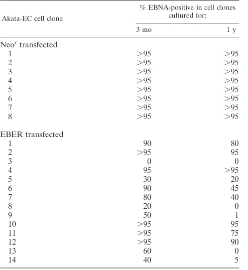
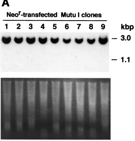
FIG. 1. (A) Detection of DNA from the EBV plasmid and transfected EBER DNA in Akata-EC cell clones. Akata-EC cells were transfected with the EBER plasmid carrying the neomycin resistance gene or a Neorplasmid (as a control) and cultured in selective medium containing 1.5 mg of G418 per ml. G418-resistant cell clones were isolated and continuously cultivated for 1 year. Cellular DNA (10g) was isolated from each cell clone at 3 months and 1 year of culture, digested withEcoRI restriction endonuclease, blotted onto a nylon membrane, and hybridized with a32P-labeled EBER probe. The 3.0-kbp band is DNA from the EBV plasmid, and the 1.1-kbp band is transfected EBER DNA. Photos of ethidium bromide-stained agarose gel are also shown. (B) EBNA expression in EBER- and Neor-transfected Akata EC cell clones. EBNA was stained in the anticomplement immunofluorescence assay.
on November 9, 2019 by guest
14 EBER-transfected clones were cultured in selective me-dium for 1 year.
These clones were examined by Southern blot analysis for the existence of EBV and EBER plasmids. Ten micrograms of cellular DNA was digested with theEcoRI restriction enzyme, separated in a 0.8% agarose gel, blotted onto Hybond N⫹
nylon membranes (Amersham), and hybridized with a 32
P-labeled EBER probe. As shown in Fig. 1A, all Neor
-trans-fected Akata-EC clones maintained the EBV-containing plas-mid, as illustrated by the presence of a 3.0-kb band. The
intensities of the bands were similar among the clones and did not change even after 1 year of culture, indicting that the EBV plasmid is stably maintained in Akata EC cells. In contrast, the intensity of the 3.0-kb band varied considerably among EBER-transfected clones. The 3.0-kb band was undetectable in one clone (clone 3) after 3 months of culture and in four clones (clones 3, 8, 9, and 13) after 1 year of culture.
Expression of EBNA in Neor- or EBER-transfected
Akata-EC clones was examined by immunofluorescence assay. Cell smears on glass slides were fixed in acetone-methanol (1:1) for
on November 9, 2019 by guest
http://jvi.asm.org/
2 min and sequentially incubated with a standard EBNA-pos-itive human serum containing complement and fluorescein iso-thiocyanate-conjugated antihuman C3c (Dako). The results are summarized in Table 1. All Neor-transfected clones were
virtually 100% positive for EBNA at any culture time. In con-trast, the frequency of EBNA-positive cells varied considerably from 0% to ⬎95% among EBER-transfected clones. There was a good correlation between the intensity of the band of the EBV plasmid in Southern blot analysis (Fig. 1A) and the fre-quency of EBNA-positive cells. Figure 1B shows the EBNA staining of representative clones. In summary, these results indicate that transfection of the EBER gene causes a loss or decrease of EBV plasmid in Akata-EC cells.
Although about half of EBER-transfected Akata-EC clones lost the EBV genome, the rest retained the virus like before after a year of culture. This might have been due to the low expression of EBER from the transfected EBER-containing plasmid. However, we cannot determine EBER expression from the transfected EBER plasmid in EBV-positive cells, because we cannot discriminate EBER RNA transcribed from the endogenous EBV genome from that transcribed from the transfected EBER plasmid. Therefore, we transfected EBER plasmid into EBV-negative Akata cells. After transfection, cells were transferred into 96-well plates and incubated in selective medium containing 1 mg of G418 per ml. Twenty-seven drug-resistant cell clones were randomly selected and examined for the expression of EBER by reverse transcription-PCR (RT-transcription-PCR), as described previously (6). As shown in Fig. 2, six clones expressed EBER at a level equivalent to or higher
[image:4.587.301.540.75.520.2]than that of EBV-positive Akata cells, but in nine clones, EBER was undetectable, and the remaining clones expressed it at a lower level. In contrast, four Akata-EC clones (clones 3, 8, 9, and 13), from which EBV plasmid was lost after EBER transfection (Table 1), expressed EBER at a level equivalent to that of EBV-positive Akata cells. These results suggested that clonal variation in EBER expression could explain why about FIG. 2. EBER expression in EBER-transfected Akata cell clones. EBER expression was determined by the RT-PCR method as de-scribed previously (6). (A) Determination of EBER expression by using different amounts of cDNA as templates. cDNA (100 ng) from EBV-positive Akata cells was diluted twofold and used as a template for PCR. (B) EBER expression in EBER-transfected EBV-negative Akata cell clones. One hundred nanograms of cDNA was used as a template for PCR. (C) EBER expression in EBER-transfected Akata-EC cell clones from which the EBV plasmid was lost. One hundred nanograms of cDNA was used as a template for PCR.
EBER transfected
1 90 80
2 ⬎95 95
3 0 0
4 95 ⬎95
5 30 20
6 90 45
7 80 40
8 20 0
9 50 1
10 ⬎95 95
11 ⬎95 75
12 ⬎95 90
13 60 0
14 40 5
aEBNA was stained by the anticomplement immunofluorescence assay. More
than 1,000 cells were examined to estimate the percentage of EBNA-positive cells.
on November 9, 2019 by guest
[image:4.587.43.292.93.364.2]half of EBER-transfected Akata-EC cell clones retained EBV plasmid.
Besides Akata cells, we examined other type I BL lines, such as Mutu I (3), Oku I, Sav I, and Kem I, to see whether EBER could replace the EBV plasmid in these cell lines. Southern blot analysis indicated that the level of the EBV plasmid de-creased in four of nine Mutu I cell clones that were transfected with the EBER plasmid (Fig. 3A). The immunofluorescence assay indicated that in at least one clone (clone 3), EBNA positivity decreased to 40%, while all Neor-transfected clones
retained 100% EBNA positivity (Fig. 3B). On the other hand, in the Oku I, Sav I, and Kem I cell lines, transfection of EBER plasmid did not influence the copy number of the EBV plasmid (data not shown).
Finally, we studied whether genome loss reflected some in-hibitory effect of EBER on oriP function. EBV-negative Akata cells were transfected with a plasmid, EBO, containingoriP, the EBNA1 gene, and the Neorgene (8) and maintained in
medium containing G418. G418-resistant cell clones were then isolated. These cell clones carrying the plasmid containingoriP
stably were transfected with the EBER plasmid or the Neor
[image:5.587.319.522.76.214.2]gene. Seven days after transfection, cells were harvested for detection oforiP plasmid DNA. Southern blot analysis indi-cated that there was no difference in the intensities of theoriP
[image:5.587.48.272.78.312.2]FIG. 4. Effect of EBER expression on retention oforiP-containing plasmid. EBV-negative Akata cell clones stably carrying the oriP-containing plasmid EBO were transfected with the Neor or EBER plasmid. Seven days after transfection, cells were harvested for detec-tion oforiPDNA. Cellular DNA (10g) was digested withHindIII restriction endonuclease, blotted onto a nylon membrane, and hybrid-ized with a32P-labeled EBO probe.
FIG. 3. Detection of DNA from the EBV plasmid and transfected EBER DNA in Mutu I cell clones. Akata-EC cells were transfected with EBER-containing plasmid carrying the neomycin resistance gene or a Neor plasmid (as a control) and cultured in selective medium containing 1.5 mg of G418 per ml. G418-resistant cell clones were isolated and cultivated for 1 year. Cellular DNA (10g) was isolated from each cell clone at 1 year of culture, digested withEcoRI restric-tion endonuclease, blotted onto a nylon membrane, and hybridized with32P-labeled EBER probe. The 3.0-kbp band is DNA from the EBV plasmid, and the 1.1-kbp band is transfected EBER DNA. Photos of ethidium bromide-stained agarose gel are also shown. (B) EBNA expression in EBER-transfected and Neor-transfected Mutu I cell clones. EBNA was stained in the anticomplement immunofluores-cence assay.
on November 9, 2019 by guest
http://jvi.asm.org/
We thank J. Sample for the Mutu I, Oku I, Kem I, and Sav I cell lines; K. Adachi for technical assistance; and K. Inoue for help with preparing the manuscript.
This work was supported by grants-in-aid from the Ministry of Ed-ucation, Science, Sports, and Culture, Japan.
REFERENCES
1.Baer, R., A. T. Bankier, M. D. Biggin, P. L. Deininger, P. J. Farrell, T. J. Gibson, C. Hutful, G. S. Hudson, S. C. Satchwell, C. Seguin, P. S. Tuffnell, and B. G. Barrell.1984. DNA sequence and expression of the B95-8 Epstein-Barr virus genome. Nature (London)310:207–211.
2.Chodosh, J., V. P. Holder, Y. Gan, A. Belgaumi, J. Sample, and J. W. Sixbey.
1998. Eradication of latent Epstein-Barr virus by hydroxyurea alters the growth-transformed cell phenotype. J. Infect. Dis.177:1194–1201. 3.Gregory, C. D., M. Rowe, and A. B. Rickinson.1990. Different Epstein-Barr
virus-B cell interactions in phenotypically distinct clones of a Burkitt’s lym-phoma cell line. J. Gen. Virol.71:1481–1495.
4.Kirchmaier, A. L., and B. Sugden.1995. Plasmid maintenance of derivatives oforiPof Epstein-Barr virus. J. Virol.69:1280–1283.
5.Kitagawa, N., M. Goto, K. Kurozumi, S. Maruo, M. Fukayama, T. Naoe, M.
apoptosis, and tumorigenicity in Burkitt lymphoma. Mol. Cell. Biol.19:1651– 1660.
11.Ruf, I. K., P. W. Rhyne, C. Yang, J. L. Cleveland, and J. T. Sample.2000. Epstein-Barr virus small RNAs potentiate tumorigenicity of Burkitt lym-phoma cells independently of an effect on apoptosis. J. Virol.74:10223– 10228.
12.Shimizu, N., A. Tanabe-Tochikura, Y. Kuroiwa, and K. Takada.1994. Iso-lation of Epstein-Barr virus (EBV)-negative cell clones from the EBV-positive Burkitt’s lymphoma (BL) line Akata: malignant phenotypes of BL cells are dependent on EBV. J. Virol.68:6069–6073.
13.Takada, K., K. Horinouchi, Y. Ono, T. Aya, T. Osato, M. Takahashi, and S. Hayasaka.1991. An Epstein-Barr virus-producer line Akata: establishment of the cell line and analysis of viral DNA. Virus Genes5:147–156. 14.Yamamoto, N., T. Takizawa, Y. Iwanaga, N. Shimizu, and N. Yamamoto.
2000. Malignant transformation of B lymphoma cell line BJAB by Epstein-Barr virus-encoded small RNAs. FEBS Lett.484:153–158.
15.Yates, J. L., N. Warren, D. Reisman, and B. Sugden.1984. Acis-acting element from the Epstein-Barr virus genome that permits stable replication of recombinant plasmids in latently infected cells. Proc. Natl. Acad. Sci. USA
81:3806–3810.