CLINICO ETIOLOGICAL STUDY OF ALLERGIC
CONTACT DERMATITIS WITH PATCH TESTING
Dissertation submitted in partial fulfilment of the Requirement for the award of the Degree of
M.D. DERMATO-VENEREO-LEPROLOGY
BRANCH XII-A
APRIL 2016
TIRUNELVELI MEDICAL COLLEGE
THE TAMILNADU DR.M.G.R. MEDICAL UNIVERSITY,
CHENNAI,
CERTIFICATE
This is to certify that the dissertation entitled “CLINICO ETIOLOGICAL STUDY OF ALLERGIC CONTACT DERMATITIS WITH PATCH TESTING” submitted by Dr. SANTHIYA VADHANA.A to the Tamilnadu Dr. M.G.R Medical University, Chennai, is an original work done in the Department of Venereo-Leprology, Tirunelveli Medical college for the award of the Degree of MD Dermato-Venereo-Leprology under our guidance and supervision during the academic period of 2013-2016.
Dr K. Punithavathi MD., (DERM) GUIDE
Associate Professor
Department of Dermato-Venereo-Leprology,
Tirunelveli Medical College, Tirunelveli
Dr P. Nirmaladevi MD., (DERM) Professor and
Head of the Department Department of
Dermato-Venereo-Leprology,
Tirunelveli Medical College, Tirunelveli
Dr K.Sithy Athiya Munavarah DCH., MD(PATHO) THE DEAN,
DECLARATION
I solemnly declare that the dissertation titled “CLINICO ETIOLOGICAL STUDY OF ALLERGIC CONTACT DERMATITIS WITH PATCH TESTING”
is done by me at the Department of Dermato-venereo-leprology, Tirunelveli Medical College, I also declare that this bonafide work or a part of this work was not submitted by me for any award, degree, or diploma to any other University, Board, either in India or abroad.
This is submitted to the Tamilnadu Dr. M.G.R. Medical University, Chennai in partial fulfilment of the requirements for the award of M.D Degree in Dermato-venereo-leprology.
Place: Tirunelveli.
Date:
Dr. SANTHIYA VADHANA.A Postgraduate Student,
M.D DVL,
Department of DVL
ACKNOWLEDGEMENT
I take immense pleasure to acknowledge all those who helped me to make this dissertation possible.
I am grateful to the Dean, Tirunelveli Medical College and Medical Superintendent of the Tirunelveli Medical College Hospital for permitting me undertake this study.
I express my profound sense of gratitude to Dr.P.Nirmaladevi MD, Professor and Head of the Department of DVL, Tirunelveli Medical College, for permitting and guiding me throughout the period of the study.
I whole heartedly thank Dr.K.Punithavathi M.D., Associate Professor, for being my guide and for her valuable suggestions and guidance throughout the period of the study.
My sincere thanks to Dr.M.Selvakumar M.D., Associate Professor, Venereology Department for his valuable suggestions and support.
I express my deep sense of gratitude to Dr.S.Judith Joy MD., for having guided me throughout the period of this study.
I immensely thank Dr.K.Dhanalakshmi MD., Dr.P.Kalyanakumar DDVL., Dr.P.Sivayadevi MD., Dr.R.Karthikeyan MD., my Assistant Professors for their constant support and encouragement.
I owe my sincere thanks to all those patients who participated in the study for their co-operation which made this study possible.
I thank my family and friends for their encouragement and support during this study.
CONTENTS
S.No. TITLE Page No.
1. INTRODUCTION 1
2. REVIEW OF LITERATURE 3
3. AIMS AND OBJECTIVES 60
4. MATERIALS AND METHODOLOGY 61
5. RESULTS 64
6. DISCUSSION 83
7. SUMMARY 92
8. CONCLUSION 94
9. BIBLIOGRAPHY
10. PROFORMA
11. MASTER CHART
ABBREVIATIONS
ACD - Allergic contact dermatitis DTH - Delayed type hypersensitivity
ICDRG - International Contact Dermatitis Research Group MHC - Major histocompatability complex
IL - Interleukin
TNF - Tumor necrosis factor
GM-CSF - Granulocyte-macrophage colony-stimulating factor APC - Antigen presenting cell
CAM - Cellular adhesion molecule LFA - Leukocyte functional antigen LC - Langerhans cell
CD - Cluster differentiation
CLA - Cutaneous lymphocyte-associated antigen CCL - Chemokine (C-C motif) ligand
HLA - Human leukocyte antigen NK cells - Natural killer cells
CHS - Contact hypersensitivity UVA - Ultraviolet A
EDTA - Ethylene diamine tetraacetic acid SQL - Sesquiterpene lactones
ABCD - Air-borne pattern contact dermatitis CAD - Chronic actinic dermatitis
AD - Atopic dermatitis
PABA - Para amino benzoic acid
PTBPFR - para-tert-butyl phenol formaldehyde resin MBT - Mercaptobenzothiazole
PEG - Polyethylene glycol
CLINICOETIOLOGICAL STUDY OF ALLERGIC CONTACT DERMATITIS WITH PATCH TESTING
ABSTRACT Background:
Patch testing is a scientific tool to make a diagnosis of allergic contact
dermatitis (ACD). It thus exposes the prevalence and current trends of allergic contact dermatitis in the community. As for now, there is no data on allergic contact
dermatitis with patch testing in our area Tirunelveli. Aim:
To study the incidence, clinical severity and morphological patterns of ACD in correlation with patch test results.
Methods:
This is an observational, prospective, single group, open labelled clinical study. 100 patients were recruited in the study duing the period of June 2014 to August 2015. The Indian Standard Series was used from Systopic pharmaceuticals pvt limited, Newdelhi. The patch test readings were interpreted according to ICDRG criteria. Results:
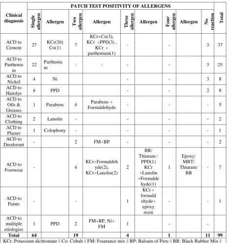
Of 100 patients(75 males ,25 females) , 89 patients showed one or more
Conclusion:
Our study revealed higher prevalence of cement and parthenium dermatitis in our area. Analysis with standard series is very significant to identify the cause of contact dermatitis and thus by avoiding the allergen, decrease the management cost and better quality of life.
1
INTRODUCTION
Allergic contact dermatitis(ACD) is an inflammatory disorder which
is T-cell mediated that occurs at the challenged site with a specific
substance of low molecular weight in an already sensitized individual.1
Contact dermatitis is one of the most common skin disorders all over the
world which accounts for 4-7% of all dermatological consultations.2
Substances responsible for contact dermatitis are haptens which are
capable of triggering the type IV hypersensitivity reaction after single or multiple
exposures. ACD occurs due to breakdown of cutaneous immune tolerance to
haptens. Sensitization phase is the prime event, which takes place before
elicitation phase occurs.
The clinical manifestations that occurs during the acute phase are
erythema, edema, papulovesicular eruptions and secondary skin lesions like
oozing and crusting. Lichenification, fissuring and pigmentation occurs in the
chronic phase. The common allergens vary from place to place and from time to
time. Parthenium dermatitis is common in India. And nickel in ornaments,
potassium di chromate and cobalt in cement, paraphenylenediamine in hairdye,
neomycin in topical medicaments, colophony in adhesive plaster, methyl
isothiazolinone a preservative in baby wipes, mercaptomix in rubber gloves are
few examples of contact sensitizers.
The gold standard method for identifying the causative allergen of ACD
2
there is high index of clinical suspicion and if tested with chemicals pertinent to
the clinical condition.
There are only few clinico etiological studies of ACD, which revealed
linear streaky pattern due to plant allergens, fingertip pattern in garlic users, eye
lid oedema due to dyes. In various Indian studies on patch testing done with
Indian standard series, the five maximum frequent allergens were potassium
dichromate, nickel, fragrance mix, cobalt chloride, mercaptobenzthiazole, even
though percentage varies with different studies.
Through a prospective study we are focussing on determining the
incidence of ACD and the causative allergen of ACD by patch testing and
analysing the morphological patterns of presentation of various allergens. We did
an analysis of clinicoetiologic correlation of ACD with Patch testing and the
3
REVIEW OF LITERATURE
DEFINITION:
Allergic Contact Dermatitis (ACD) is a, type IV, T cell mediated, delayed
type hypersensitivity reaction (DTH) (i.e) an inflammatory reaction triggered by
contact with specific exogenous allergen to which a person has established
allergic sensitization. It is characterized in early stages by erythema, papules and
vesicles, followed in late stages by lichenification, scaling, fissuring and xerotic
skin.7
HISTORY:
Allergic contact dermatitis is most likely documented even in antiquity,
because it has complemented menfolk all the way through the past.
In 1906 , the Scientist Von Pirquet coined from Greek words the term
‘allergie’, allos & ergon meaning other or different work.8 In 1840, Fuchs
suggested that ‘dermatitis venenata’ was a manifestation of constitutional
idiosyncrasy. Neisser used the word ‘Idiosyncrasy’ to describe iodoform
dermatitis in 1884. Bloch and Steiner-Woerlich proved contact allergy of the
skin, by using Primula extract on humans. Landsteiner and Jacobs substantiated
that hapten molecules must combine with skin cells proteins to cause
sensitization.
A standardized technique called ‘Patch test’ is used for the confirmation
of the role of suspected causative agents in producing an allergic contact
dermatitis. Patch test signifies a valuable diagnostic tool that unravels the
4
Historical aspects of ACD in 20th century, is inseparable from patch
testing, which is the diagnostic tool that unmasks the relevant allergens of ACD
and the patch test is inseparable from the pioneer in the field, Josef Jadassohn
(1860-1936).
In 1895, Josef Jadassohn introduced the patch test technique, while
working at Breslau University, when he described the patch testing role in
Dermatitis medicamentosa and He is considered the father of Patch testing.
During the 17th, 18th, 19th centuries, some researchers made a replica of contact
dermatitis, by applying the suspected allergen.
In 1847, Stadeler described the blotting paper strip technique. In 1889,
Collins who was an ophthalmologist, tried atropine patches to the patients who
manifested adverse reactions after atropine eyedrops instillation.
Bruno Bloch, a dermatological pioneer, upgraded Jadassohn’s technique,
and gave the grading system for patch testing, and introduced the concept of
standard series of allergens, cross sensitization and systemic ACD.9,10
Marion Sulzberger introduced the patch test technique in New World.
Paul Bonnevie, Professor of Occupational Medicine in Copenhagen, expanded
the standard series of allergens, the archetype of our current series.
In 1986, Fisher stated that ‘Patch tests’ are the only scientific proof of
Allergic Contact Dermatitis, when properly applied and correctly interpreted. He
also emphasized that learning the art of patch testing technique is as important as
5
Scandinavian dermatologists and other European members formed the
International Contact Dermatitis Research Group (ICDRG) to formulate a
standard protocol for patch testing and for international research in this field.11
EPIDEMIOLOGY:
In India, allergic contact dermatitis has an incidence of 4-7%, which is
one of the major occupational health problems.12 The socio economic impact is
also significant. 40-60% of industrial non-attendance is ascribed to some form of
contact dermatitis.Incidence can vary depending on the degree of socioeconomic
and industrial development in the area as well as the interest of the dermatologist
in allergic contact dermatitis. The common allergens implicated to cause ACD
varies from place to place and time to time.
Total population research works and scrutiny of random samples of
people have revealed the incidence of contact dermatitis to be 1.5% to 6%.
In definite professions like construction work and in biochemical and metallic
industries, the frequency is predominantly increased.13
ETIOPATHOGENESIS:
Allergic Contact dermatitis is an inflammatory skin condition that is
hapten specific. Haptens are substances of low molecular weight which is less
than 500 Daltons. These haptens penetrate the stratum corneum to the nucleated
layers of epidermis to induce and elicit the contact sensitization. After single or
multiple exposures, non-protein chemicals, i.e. haptens, induce ACD. ACD is
well thought-out as an interruption of cutaneous immune tolerance to haptens.14
6
Phase 1 - Sensitization phase (also referred to as afferent phase or
induction phase)
Phase 2 - Elicitation phase (also known as efferent or challenge phase)
I) Sensitization phase:
The prime events of this phase are
The Allergen binding to components of skin
The ‘complete’ or conjugated antigen recognition
Sensitized T lymphocytes - Proliferation and dissemination
The Allergen binding to components of skin
Allergens that penetrate the skin bind covalently with skin peptides
directly or alternatively to form a reaction product that binds with major
7
surface of dendritic cells and Langerhans cells. Epicutaneously applied allergen
allies with these antigen-presenting cells in 6 hours.
The ‘complete’ or conjugated antigen recognition
The APCs undergo a series of events activation, maturation and migration
for which co-stimulatory factors like IL-1β, TNFα and GM-CSF are required. In
the absence of these co-factors, tolerance develops.
Within 24 hours of antigen exposure, APCs travel via the afferent
lymphatics to the paracortical areas of the regional lymph nodes, where they are
presented to T lymphocytes. This binding is strenghtened by physical factors, the
ruffled membrane and dendritic nature of the Langerhans’ cells and the intricate
structure of the paracortical areas and also by specialist cellular adhesion
molecules (CAMs). For example, leukocyte functional antigen-1 (LFA-1) on
CD4 T helper cells interacts with intercellular adhesion molecule-1 (ICAM-1) on
Langerhans’ cells. Subsequently, cytokines are released, IL-1 by LCs and IL-2
by T lymphocytes. An intact draining lymphatic system is required to induce a
contact hypersensitivity reaction.15
Sensitized T lymphocytes- Proliferation and dissemination:
The blast formation in the lymph nodes and the multiplying of
antigen-specific cytotoxic CD8+ (Tc1) and also CD4+ (Th1) lymphocytes is caused by
the cytokines.16 The T cells disseminate into the blood stream and throughout the
body via the efferent lymphatics vessel and thoracic duct and interact with
Langerhans’ cells and residual antigen in the skin. Contact sensitization is
8
antigen (CLA). Production of the chemokine CCL27 by basal keratinocytes is
responsible for the localization of inflammation and binds to dermal
glycoprotein; CLA-positive lymphocytes also express CCR10, the receptor for
CCL27.17 CD8+ T cells induce apoptosis in these Keratinocytes and the skin is
damaged which drives the inflammatory response. CD4+ Th1 & CD8+ T cells
act as effectors on target cells. Sensitization phase lasts for 10 to 15 days.
II) Elicitation phase:
After sensitization has occurred, re exposure to the specific allergen
causes eczematous dermatitis. On re-contact to the similar allergen, a clinically
visible reaction occurs within 24–48 h, which is mediated via activated
keratinocytes that express HLA-DR on their surface and can release IL-1, thus
amplifying the function of LCs. Both types of cells present the antigen to specific
T cells that are already present in the epidermis in small numbers, inducing a
quick inflammatory response. This is responsible for the recruitment of
leukocytes (including regulatory T cells) from the blood to the skin leading
to the development of skin lesions.
The role of skin memory:
The mechanism for site specific allergen skin memory is related to
chemokine CCL27 that causes retention of CCR10+ CD4+T cells perivascularly
in the dermis at the site of Patch testing.
The role of Keratinocytes in all phases of ACD:
In initiation phase- it secretes TNF alpha
9
In peak of inflammatory phase - interacts directly with epidermotropic T
cells
Resolution of ACD- produce anti-inflammatory cytokines IL10 & IL16 –
recruits T Reg cells
The cytokines produced by keratinocytes are
1) IL 1: Enhances activation of accessory dendritic cells, which in
turn activates T cells
2) IL 5: Stimulates T cell proliferation
3) IL 8: Has a strong chemotactic effect of T cells
Recent concepts in ACD:18
Innate immune cells such as Natural Killer(NK) cells play a significant
role in ACD
NK T cell are necessary for initiation of ACD & it also presents in
elicitation phase of ACD
Studies from mice lacking Langerhans Cells also shows contact
hypersensitivity , hence cells other than LCs also play a prime role in
CHS
Dermal Dendritic Cell also acts as Antigen Presenting Cell that
complements the function of epidermal Langerhans Cell
T regulatory cells (T- Reg) cells plays a critical role in control of ACD
(i.e) resolution of T cell inflammation
Loss of T- Reg cells cause chronic inflammation
10
PREDISPOSING FACTORS FOR ACD:
I) INDIVIDUAL VARIATIONS
i) CONSTITUTION:
Sensitization depends on individual susceptibility.19 The role of atopy in
ACD is a matter of debate. One study reported high prevalence of contact allergy
in atopic individual but another study showed same prevalence and others
reported decrease in the prevalence of contact allergy.20
ii) ROLE OF SEX:
Women are supposed to have stronger cell mediated immunity responses
than men.21 The reason for female preponderance is due to prior ‘conditioning’
exposure and subclinical sensitization to large number of metals, exposure to
fragrances, cosmetics and hair dyes.22
iii) HORMONES:
Pregnancy, menstrual cycle, use of gestagens either exacerbate or
attenuate the readings of patch tests.23,24 Exacerbation has been reported during
the premenstrual phase of menstrual cycle.25
iv) RACE:
Racial differences exist but it is a reflection of exposure rather than
tendency.26,27 Afro–Caribbeans are less susceptible than white people due to
decreased exposure.28 In another study black men were more sensitive than white
11
v) AGE:
Age factor plays less significant role on ability for sensitization.30 But
positivity of patch test reactions increase with age due to allergen exposure that
have acquired over a lifetime.31 Nickel, Fragrance, Thiomerosal, Medicaments,
Rubber chemicals, Chromate are common allergens in children.32
vi) MEDICATION:
Medication will affect the patch test results. Prednisolone (>15mg/day)
and potent topical steroids will subdue the patch test reactions.
Immunomodulators such as ciclosporin and azathioprine may reduce the
intensity of patch test reactions.33
vii) COINCIDENTAL DISEASES:
Patients with acute or debilitating diseases such as cancer, Hodgkin's
disease and those with impaired T-lymphocyte function have impaired capacity
for contact sensitisation.34,35
viii) LOCAL FACTORS:
Preexisting or concomitant allergic or irritant dermatitis harms the skin,
upsetting its barrier function and producing increased opportunities for allergen
absorption. Occlusion promotes percutaneous absorption and contributes to the
high incidence of medicament dermatitis in stasis dermatitis, leg ulcers and
12
II) ENVIRONMENTAL FACTORS:
Certain important environmental factors predisposing to ACD are
1. Climate
2. Flora and fauna
3. Socio-economic and cultural factors
i) CLIMATE:
UV exposure, heat and relative humidity can influence the burden of
contact allergy. UVB exposure shall diminish the skin’s immune response to
contact allergens, however decline in immune responsiveness by UVA exposure
is transient due to adaptive mechanism.36
ii) FLORA AND FAUNA:
Seasonal variations are most common in plant dermatitis. Many allergic
plants especially compositae family plants are shattered by cold and frosty
weather but reappearance occurs during spring and summer season.
Geographical location plays an important influence. In India parthenium contact
dermatitis is more common. Allergenicity of Primula obconica change with
weather and sunlight. Fauna has only little influence.
iii) SOCIOECONOMIC AND CULTURAL FACTORS:
Exposure to cheap metals, various cosmetics and perfumes shall vary
according to social class. In the Middle and Far East, the traditional herbal
medicines and balms are commonly used to treat skin disorders. Hair dyes,
13
III) CHEMICAL COMPOUNDS AND THEIR SENSITIVITY:
Skin cells are composed of molecules that contain nucleophilic atoms
whereas allergens contain electrophilic atoms. Interaction between these two
result in strong covalent bonding to form a “Complete antigen”.
Skin cellmolecules: Contain nucleophilic atoms
Hapten molecules: Contain electrophilic atoms (positively charged, electron
deficit) covalent bonding
[image:25.595.116.505.438.624.2]+ Hapten (<500 Da)
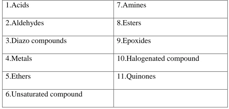
Table: Classification of haptens based on functional grouping38
1.Acids 7.Amines
2.Aldehydes 8.Esters
3.Diazo compounds 9.Epoxides
4.Metals 10.Halogenated compound
5.Ethers 11.Quinones
6.Unsaturated compound
Skin cell
14
Sensitization Potential
It is the capability of a given allergen to induce sensitization in a group of
humans.
Various test procedures to assess the sensitization39
1. Maximization test(described by Kligman and Epstein)
2. Buehler test
3. Open epicutaneous test
4. The Draize test
5. Freud’s complete adjuvant test
6. The local lymph node assay
7. The mouse ear swelling test40
Factors that can enhance the risk of sensitization:
1. Increased allergen absorption due to barrier disrupted skin.
2. Recruitment of immune competent cells and cytokines which leads to
priming of immunological response
3. Accumulation of mononuclear cells.
Matzinger’s ‘danger model’ concept for sensitization41
Contact allergy may develop in the presence of cytokine release from the
keratinocytes which is provoked by a coexisting irritant or trauma. If there is no
15
HISTOPATHOLOGY OF ACD
`Histopathologic assessment of ACD is mainly helpful to eliminate other
conditions that clinically simulates ACD. But other types of spongiotic
dermatitis cannot be differentiated.
Cutaneous changes seen by light microscopy depends on two factors42 Severity of response to allergen
Time of biopsy taken after exposure to allergen
Early lesions of ACD are acute spongiotic dermatitis. If vesicles develop,
they may contain clusters of Langerhans cells. There is superficial dermal
infiltrate of lymphocytes, macrophages and Langerhans cells with
accentuation around the small vessels. Eosinophils may be present in the dermal
infiltrate as well as within areas of spongiosis. In patients with continued
exposure to the antigen, the biopsy may show a subacute or later a chronic
spongiotic dermatitis.
HISTOPATHOLOGY OF ICD:
The histopathologic picture differs from widespread ulceration, to
simply diffuse hyperkeratosis or parakeratosis with congestion and ectasia, to
a spongiotic pattern essentially identical to allergic contact dermatitis. In some
instances, there is significant necrosis with nuclear karyorrhexis and cytoplasmic
pallor (Bandmann's achromia). In severe reactions, the necrosis may extend into
16
Clinical approach to a case of ACD:
The Key symptom of ACD is Pruritus. The morphological pattern and
distribution of dermatitis must raise the index of suspicion of ACD. One must
consider contact allergy in patients of any types of dermatitis (eg. Atopy).
Patients with stasis dermatitis have increased risk of ACD to topical
medicaments. ACD is not always B/L even though Ag exposure is B/L
(glove/shoe). Even when exposure to allergen is uniform, eczematous lesions are
often patchy. ACD does affects palms and soles
In acute phase, ACD is characterized by erythema, oedema, followed by
appearance of papules, vesicles, oozing & crusting.
In chronic stages, skin becomes lichenified, fissured, pigmented and
scaling.
ACD can be classified as
1) Eczematous CD
a. Primary pattern
b. Secondary pattern
2) Non-eczematous CD
3) Photo allergic CD
I) ECZEMATOUS CONTACT DERMATITIS
PRIMARY PATTERNS:
17
Hands & arms:
There are multifactorial reasons for hand eczema. Housewives dermatitis
and many of the occupational dermatitis mostly are confined to the hands.
Chromate in cement, N-isopropyl-N′-phenyl-p-phenylenediamine (IPPD) and 1,
2-benzisothiazolin-3-one are the most common allergens that causes palmar
pattern of allergic dermatitis. Discoid patterns of eczema occurs with chromate
allergy. Allergy to nickel, chromate and p-tertiary-butyl phenol formaldehyde
resin also develop at the wrists because of sensitivity to the metal, leather and
glue, respectively, in watchstraps. Rubber gloves cause a clear pattern of
dermatitis at the site of contact. Streaky dermatitis on the fingers and dorsa of the
hands is caused by plants. Dust (exotic woods, cement), nickel and textiles
induces dermatitis in the flexural aspect of elbows.
The morphological patterns of hand eczema described are pompholyx,
recurrent focal palmar peeling, ring eczema, fingertip eczema, hyperkeratotic
eczema, apron eczema, chronic acral dermatitis, and gut eczema.
Face:
Facial allergic contact dermatitis occurs due to fragrances, hair dyes,
preservatives and other ingredients of cosmetics and skincare products, including
nail varnish. Dermatitis caused by a cosmetic presents with dryness, tightness
and itching45. ‘Hair dyes’ might be a reason for acute oedema and intense
pruritus. Spectacle frames containing nickel or plastics may be the reason for
18
‘‘Status cosmeticus’’ due to ‘‘stinging’’ compounds in cosmetics
The recently available cosmetics are free of compounds that cause allergic
hypersensitivity. But, nonspecific irritation from cosmetics occurs. Thus, some
persons appear to be in a condition of ‘‘status cosmeticus’’ in which every
cosmetic or soap applied to the face produces itching, burning, or stinging
sensations.43
Eyelids:
The skin of the eyelids is thin, sensitive and often fiddled by the fingers
causing eyelid dermatitis due to airborne droplets (e.g. fragrance sprays) or
volatile substances (e.g. epoxy resin). Nickel and/or rubber Eye creams present
in eye shadows, mascara and makeup applicators cause contact allergy at the site
of contact.44,45 Eye drops and contact lens solutions also contain preservatives
(benzalkonium chloride, EDTA, mercurials), which sensitizes. Common
sensitizers in eye drops and ointments are neomycin, framycetin, gentamycin,
tobramycin, local anaesthetics, β-blockers and sympathomimetics.
Lips and perioral area:
ACD can occur due to lipsticks, nickel, medicaments, flavourings, garlic,
shellac and cosmetic excipients.46,47 Lipstick dermatitis does not extend beyond
the vermilion border and manifests as dry, scaling or cracked lip. Cheilitis and
perioral eczema occurs due to allergy to toothpaste and due to flavours like
cinnamic aldehyde, spearmint oil and l-carvone.48,49 Allergic contact cheilitis
19
additives such as sodium metabisulphite, preservatives, colours and
antioxidants.50
Ears:
External otitis always have a chronic relapsing course. Earrings cause
dermatitis on the ear lobes which is mostly due nickel and gold. Piercing of the
ear lobe may be the sensitizing event in nickel dermatitis.
Earplugs for noise protection contain antiseptics, dyes, rubber and plastic
chemicals, Hearing-aids contain acrylates and stabilizing chemicals, Headsets
contain urea and phenol-formaldehyde resins, Earphones has rubber, plastic
components, including epoxy resins and acrylates, all these cause ear
dermatitis.51
Spectacle-frame dermatitis may occur due to metals, particularly nickel
and palladium, in some frames.52,53 Granulomatous contact allergy after ear
piercing can occur due to nickel, palladium and gold.54
‘‘Sugarcane’’ ears, resembling cauliflower ears, occur in workers who
carry bundles of burned stalks of sugar and the lesions are unilateral, depending
on whether the worker is right or left-handed. This was described by Arnold.55
Scalp:
Scalp dermatitis is caused by Hair-styling products such as mousses, gels
and holding sprays, fragrances and preservatives p-phenylenediamine and related
semi-permanent dyes and amphoteric detergents in hair cosmetics and manifests
as persistent itching of the scalp and gradually spread to the ears, neck and face.
20
isothiazolinones added as preservative cause scalp dermatitis. Topical minoxidil
lotion applied over scalp to promote hair growth is also a sensitizer.56
The Hindu practice of wearing a central forehead dot of color known as
a bindi cause leukoderma associated with paratertiary butylphenol resin in the
adhesive, with or without a related dermatitis or positive patch test.57,58
Neck:
Nickel in the clasps of necklaces or zip fasteners cause dermatitis on the
nape of the neck. Nail varnish from fingertips cause patchy allergic dermatitis.
Textiles (finishes in collars, dyes) and necklaces (nickel, exotic wood) produce
collar like dermatitis, or eruptions on the neck. Dermatitis from photosensitizers
and airborne allergens is sharply limited by the collar to the ‘V’ of the neck if
blouses or open-necked shirts are worn.
Axillae:
Dermatitis in axillary region occurs due to sweating, occlusion and the use
of antiperspirants which contain aluminium salts. Allergic sensitivity occurs due
to fragrances that are used to mask odour. Textiles cause periaxillary dermatitis.
Dermatitis from dresses, blouses and sweaters affects folds of the axilla and the
allergens are usually textile dyes.
Trunk / Torso:
Nickel buttons and zip fasteners are the reason for the contact dermatitis.
Truncal eczema is due to chromate sensitivity from leather and rubber.
21
workers sensitized to Compositae plants manifests as dermatitis which affects the
sun exposed areas. Diffuse papular eczema occurs due to medicament sensitivity.
Anogenital region:
Medicament sensitization is the most common reason for ACD in
anogenital region and the common medications implicated are neomycin,
hydroxyquinolones, ethylenediamine and topical antifungals. Over-the-counter
medicaments causes dermatitis in anogenital region. Ectopic contact dermatitis
occurs due to nail varnish.60 Moist toilet tissues contain preservatives that cause
allergic hyper sensitivity.61 Cashew nut oil in butter causes perianal dermatitis.62
Rubber accelerators in condoms cause genital eczema or pruritus vulvae.
Delayed hypersensitivity can occur to semen.63
Gluteal region:
A follicular-type of dermatitis occur on the buttocks due to long time
contact with wet bathing suits. By swimmers it is commonly called as ‘‘bikini
bottom’’ and scientifically called as occlusive folliculitis characterized by
annoying wet blisters all over the buttocks which is due to wearing swimsuit all
day.
Thighs:
Textile dermatitis occurs at the contact site of the underclothing worn.
Nickel coins and keys or boxes of matches may cause dermatitis on the
22
Lower legs:
Allergic contact dermatitis of the legs occurs due to application of
sensitizing medications and dressings to stasis eczemas and ulcers. In a study of
venous leg ulcer patients, the sensitizers implicated were fragrances in 30%,
antimicrobials in 20%, vehicle ingredients in 20%, rubber accelerators in 13%,
and topical corticosteroids in 8%.64 Rubber allergy occurs due to compression
bandaging.
Feet:
Dermatitis may occur due to shoe materials including leather, rubber,
glues and nickel, stockings, topical medicaments, antiseptics and antiperspirants.
Nails:
Allergens produce onychia and nail discoloration. Thinness, fragility,
splitting, separation into layers, detachment from the nail bed and long-standing
infections are various manifestations of ACD that occurs to jewelers, weavers,
metal platers, and printers. Occupational chemicals and trauma are common
causes of onychia, koilonychia, nail dystrophy and discoloration of the nail.
Generalized:
Generalized erythroderma occurs due to chronic contact dermatitis
because of continued exposure to many allergens.
Mucous membranes:
The constitutional make up of skin and mucous membranes have many
23
membranes is because of absent stratum corneum layer, absent lipid secretion
and washaway of substances by saliva.65
ACD in mucous membranes is uncommon and is often secondary to skin
sensitization. Intraoral blistering occurs from cinnamon allergy.66 Orofacial
granulomatosis occurs with contact allergy to food additives.67 Lichenoid
reactions occur due to mercury from amalgam fillings.68,69 Generalized skin
eruptions and perioral dermatitis may occur after dental filling.70 Gingivitis
occurs due to eugenol in dental cement.71
Secondary pattern:
The primary site pattern determines the secondary pattern. The dermatitis
of the hands have a tendency to spread to forearms, arms and face. Similarly,
dermatitis of feet spreads to legs and hands. Sensitization presenting as ‘id’
reaction occurs in stasis eczema.
II) NON ECZEMATOUS CONTACT DERMATITIS
Non-eczematous responses in ACD includes
i) Contact urticarial eruptions
ii) Lichen planus like lesions
iii) Lichenoid eruptions
iv) Lymphomatoid eruptions
v) Erythema multiforme-like reactions
vi) Purpuric lesions
vii) Pigmented lesions
24
ix) Granulomatous reactions
x) Systemic non eczematous reactions
xi) Onycholysis
III) PHOTO ALLERGIC CONTACT DERMATITIS
Photosensitizers are the allergens which are transformed into irritants or
sensitizers after irradiation with UV or short-wave visible radiation (280–600
nm). Photoactivated molecules are haptens. The etiopathogenesis is same as
contact allergic reactions. The action spectrum for photoallergy is generally in
the UVA range.
Photoallergens:
The most common photo allergens are UV filters including p
-aminobenzoic acid and its derivatives, cinnamates, benzophenones and
dibenzoylmethanes. Benzophenone 3 (oxybenzone) seems to be the most
commonly identified photoallergen.
Other photocontact allergens are topical non-steroidal anti-inflammatory
agents, especially Ketoprofen, Phenothiazines, Sulphonamides, Quinines,
Perfumes and Halogenated salicylanilides in soaps and detergents.
Clinical features of photoallergic contact dermatitis
The sun exposed areas like the face, ‘V’ of the neck, back of the hands,
dorsal forearms are the most common sites. The scalp, periorbital areas and the
skin immediately under the chin are relatively spared. Sharply delineated areas
25
sign is the exempt ‘Wilkinson’s triangle’ behind the earlobe. Some spread to
covered sites.
ACD TO SPECIFIC ALLERGENS
PARTHENIUM ANTIGEN
Parthenium dermatitis is caused by Parthenium hysterophorus.72 It is
caused by airborne dry and friable plant particles, and the most important
allergens responsible for allergic contact dermatitis are sesquiterpene
lactones(SQLs).73 Among the SQLs, parthenin was found to be the major
allergen, others being coronophillin, tetraneurin A, hymenin etc.
Patterns of parthenium dermatitis
Clinical features
Air-borne pattern contact dermatitis (ABCD): Classical pattern affects
eyelids and neck, V area of the chest and the cubital and popliteal fossae.
Chronic actinic dermatitis (CAD): Lichenified lesions over the exposed
areas
Mixed pattern dermatitis: (Combination of air-borne and CAD)
Exfoliative dermatitis
Hand and feet dermatitis
Atopic dermatitis
Rare patterns like photosensitive lichenoid eruption, prurigo
nodularis-like, perianal dermatitis, vesicular hand eczema and dermatitis simulating
26
Management includes avoiding contact with allergen, managing
dermatitis with topical corticosteroids/tacrolimus, and other immunosupressives
like azathioprine.
POTASSIUM DI CHROMATE:
In Construction workers, potassium dichromate (hexavalent chromium)
was the commonest allergen with the prevalence of sensitivity being more in
men.
Levels of chromate in cement should be restricted to 2 ppm hexavalent
chromium. The metal itself, if not dissolved in oil or acids or as a salt, seems to
be non-sensitizing, unlike nickel and cobalt due to the insoluble monomolecular
layer of chromium (III) oxide (Cr2O3) on the surface.
Source of potassium dichromate:
Cement, antirust paints (lead chromate and zinc chromate) painted metals,
alloys, lithography/offset printing materials, anticorrosive oil, cutting oils,
matches, photographic chemicals, chemicals for fat determination in milk,
welding fumes , plating salts, wood preservatives and ashes, wood pulp, glazing
enamels, catgut.
Clinical patterns caused by Potassium di chromate:
i) Acral dermatitis
ii) Hand dermatitis
iii) Airborne contact dermatitis
iv) Acro-facial dermatitis
27
vi) Atopic eczema like
vii) Discoid pattern like
viii) Mixed pattern
a. Acrofacial and trunk dermatitis
b. Acrofacial and scalp dermatitis
Management:
Advised to avoid contact with sources of chromate
Ferrous sulphate added to cement changes soluble hexavalent chromate
to insoluble trivalent chromate, and thereby preventing sensitization.
Chelating compounds and ion exchangers.74
Dapsone tried, but studies are lacking.75
NICKEL SULPHATE:
Nickel is the most common contact allergen of metal allergy and contact
sensitization is more in females than males. Nickel allergy is a chronic and
recurring skin problem. The nickel salts, nickel chloride (NiCl2) and nickel
sulphate (NiSO4), are freely soluble in water and sweat and have strong
sensitizing character.
The most common sources of metallic nickel are fashion jewellery, coins,
machinery parts, utensils, stainless steel items etc.
Role of diet in causing dermatitis:
Nickel dermatitis can occur in a nickel sensitized person if the diet
contains excess amount of nickel. And the foods that have increased nickel
28
Pattern of nickel allergy:
Dermatitis at the site of contact
A “secondary rash” due to spread of dermatitis to distant regions is rarely
observed.76
Hand eczema pattern ; Vesicular type after consumption of nickel in
diet.77
As baboon syndrome - a generalized rash involving gluteal region,
anogenital area, flexural areas and eyelids.78
Erythema multiforme and vasculitis – rare patterns.79,80
Chronic urticaria81
Nickel is patch tested at 5% conc in aqueous form
Therapy
Barrier creams and cleansers can be tried
Combination creams containing clioquinol and steroids
Low-nickel diet in recurrent palmar vesicular eczema can be advised
Disulfiram ( Antabuse), which has nickel chelating property
Prevention
The most effective means of preventing nickel sensitization would be to
reduce exposure to nickel from costume jewelry, particularly earrings. The
European Union has banned nickel objects, that release nickel in excess of 0.5
29
Spot test:
The dimethylglyoxime test is an easy technique to find nickel release
from metal objects. A cotton swab is dipped in two drops each of a 1% solution
of dimethylglyoxime in alcohol and a 10% solution of ammonium hydroxide in
water, and is wiped regularly over test item for 30 seconds. If the cotton swab
turns light pink to red, it confirms the release of nickel.
COBALT:
Cobalt metal, its oxides and salts (e.g. CoCl2 and CoSO4) are sensitizers.
Cobalt is tested at 1% concentration in petrolatum.
Sources:
Cobalt is present in magnets and jewellery, as a contaminant in nickel, in
alloys, in dentures and in nails for pinning fractures, glass and ceramics, crayons,
multivitamin pills, textile dyes, tattoos, soaps, dyes and detergents.
PARAPHENYLENEDIAMINE (PPD):
PPD is used for permanent hair coloring. It is patch tested at a 1%
concentration in petrolatum. In ACD due to cosmetics, the allergen implicated
commonly are first fragrances, 2nd preservatives and 3rd is PPD.
Clinical aspects:
PPD causes weeping dermatitis of scalp, eyelids, face, hairline and spread
to involve the neck, upper portion of the trunk and arms, hands with
generalization.
30
Sources of PPD:
PPD is present in permanent hair dyes, cosmetics, leather dyes, rubber and
plastics industry, lithography, oils, greases, epoxy resin hardeners and temporary
tattoo, photographic developers etc
COLOPHONY:
Colophony (Rosin) is a yellow, complex, natural residue left after
distilling off the volatile oil from oleoresin obtained from the coniferous trees
Pinus palustris.
Colophony is patch tested at 20% concentration in petrolatum. The most
potent allergen has been shown to be 15-hydroperoxyabietic acid.82
Source of colophony:
It is used in a wide range of cosmetics, topical medications, industrial
products like paper and paper products, printing inks, adhesives, tapes, bandages,
waxes, varnishes, polishes, paints, dental cements.
BLACK RUBBER MIX :
It is composed of the following:
N-isopropyl-N-phenyl-4-phenylenediamine - 0.1 %
N-cyclohexyl-N-phenyl-4-phenylenediamine - 0.25%
N-N-diphenyl-4-phenylenediamine - 0.25%
The preceding amines are used as antioxidants and antiozonants in the
production of rubber and are the most effective and commonly used of available
agents. The compounds prevent drying and cracking of the final rubber products.
31
The most potent sensitizer in the mix has been shown to be IPPD. The
mix ingredients are chemically related to the hair dye base p-phenylenediamine,
and cross-reactivity can occur.
Sources :
The three p-phenylenediamine compounds are extensively used in rubber
manufacture. Since these agents discolour the final product, most finished
products are dark, either gray or black.
These include tires, heavy black rubber gloves and boots, shoes
(especially soles), cushions, earphones, and walking-stick handles.
CAINE MIX (BENZOCAINE):
Caines are local anesthetics that are used primarily in non-prescription
topical medicaments, which are designed to ease pain and pruritus.
Benzocaine is tested at 5%concentration in petrolatum.
Sources:
Sources include over-the-counter medicines used to treat sunburns,
dermatitis, athlete's foot and calluses, otic preparations for earaches, enemas and
anal suppositories for hemorrhoidal discomfort, oral mucosal products for teeth
pain and canker sores
Benzocaine- and tetracaine-sensitive individuals may also have to avoid
PABA and PABA esters containing sunscreens, permanent hair dye
(p-phenylenediamine, certain diuretics or fluid pills (hydrochlorothiazide), oral
32
(and PASl, azo and aniline dyes, and an important cardiac medication,
procainamide.
EPOXY RESIN:
Epoxy resins are plastics that were synthesized for industrial purposes.
They have been used extensively because of their versatility, chemical and
electrical resistance, excellent adhesion, toughness, low shrinkage, and ability to
be cured rapidly or slowly at various temperatures. Cured epoxy resin is
nonsensitizing. Allergic contact dermatitis occurs with exposure to uncured
resin.
The epoxy resin is a bisphenol A-based resin patch tested in a
1%concentration in petrolatum.
Sources:
Epoxy resins are used primarily in adhesives and glues, laminates,
electrical encapsulators, surface coatings, paints and inks, eyeglass frames and
vinyl gloves.
FORMALDEHYDE:
Formaldehyde (methanal) is a colorless gas that is readily soluble in
water, alcohol, ether, and other polar solvents. It is the simplest member of the
aldehyde series and is generally sold commercially as an aqueous solution,
formalin.
Formaldehyde is patch tested as 1%in water. Formaldehyde was first
used as a biologic preservative in 1868, and by 1889 it was being manufactured
33
Clinical aspects:
The adverse effects of this chemical, include mucous membrane and
respiratory tract irritation, allergic and irritant contact dermatitis of the skin,
contact urticaria, and potential carcinogenicity.
Textile dermatitis typically involves the peripheral parts of the axillae, the
antecubital fossae, the neck, and upper parts of the trunk.
Because of partial combustion seen in cigarette smoke, automobile
exhaust, and incineration products, formaldehyde is produced and released in the
general environment
Sources:
Formaldehyde is a common chemical that is found in cosmetics,
household products (disinfectants, Cleaners), medicated creams, leather tanning
agent, photography, textiles, paper manufacturing, pathology fixative, rubber
industry preservative, fertilizers, insulation and renal dialysis.
Clothing or avoidance measures such as a change in jobs to prevent
dermatitis.
p-tert-BUTYLPHENOL FORMALDEHYDE RESIN:
p-tert-Butylphenol formaldehyde resin (PTBP formaldehyde resin) is one
of a large group of synthetic polymers made by reacting formaldehyde with
phenol or related alcohols to form network polymers. They are used primarily as
adhesives and were the first synthetic polymers to be used commercially. The
34
Clinical Aspects:
PTBP formaldehyde resin is a formaldehyde-based phenol resins. It is
used exclusively as a glue or an adhesive. This usage depends on its superior
qualities of rapid adhesion, durability, and pliability. It cures slowly without
additional hardeners at room temperature. The pliability and flexibility make it
particularly useful in the bonding of shoe components and parts of watch straps,
handbags, hats, and belts. For this purpose it is frequently combined with natural
or synthetic rubber.
Sources :
p-tert-Butylphenol (PTBP) formaldehyde resin is used primarily a
component glue in leather shoes, handbags, and watch straps, plywood, boxes,
insulation and automobiles.
PARABEN MIX:
The parabens are alkyl esters of p-hydroxybenzoic acid. They are the most
commonly used preservatives in cosmetics and are usually patch tested as a
paraben mix (16% in petrolatum) containing 4% each of methyl, ethyl, propyl,
and butyl parabens.
Sources:
The parabens are the most frequently used preservative in cosmetics,
35
MERCAPTO MIX:
Mercapto mix is composed of the following thiazoles:
N-Cyclohexyl-2-benzothiazole-sulfenamide (CBS)
2,2 1-Benzothiazyl disulfide (MBTS)
4-Morpholinyl-2-benzothiazyl disulfide (MMBn)
Each thiazole is present at a 0.333% concentration in petrolatum (1%total)
in the mercapto mix. The thiazoles are the most commonly used rubber
accelerators in the world.
Clinical Aspects:
The thiazoles are frequently reported sensitizers in shoe and glove allergy
but may also be responsible for dermatitis due to contact with rubberized fabric
in undergarments, swimwear, and elastic bandages. Thiazole sensitivity is
possible in the workplace in many industries involved in rubber manufacturing
and the use of rubber in manufacturing other products.
Thiazoles may also be used in nonrubber products, including veterinary
and pet products, cutting oils, antifreeze, disinfectants, adhesives, cements,
greases, and photographic emulsion.
Sources:
Mercapto mix thiazoles are used primarily in the production of rubber or
latex products. They are found in gloves, rubber shoes, leather shoes, rubber in
elasticized clothings and other nonrubber sources like disinfectants, repellents,
36
MERCAPTOBENZOTHIAZOLE:
Mercaptobenzothiazole (MBT) is a thiazole rubber accelerator.
Clinical Aspects:
MBT and other thiazoles are the frequently used accelerators in the
production of rubber. Shoe contact dermatitis is mostly due to a rubber
component allergy, usually MBT and next thiurams. Usually the dermatitis is
limited to the area of contact. This may be primarily the soles of the feet
bilaterally, but patients with such an allergy may also have unilateral
involvement. MBT is second to the thiurams as the etiologic agent in allergic
contact dermatitis to gloves.
Sources:
Used in cutting oils, antifreeze, industrial greases, anticorrosive agents,
cements and adhesives, detergents, and fungicides. The most common sources
are gloves and shoes.
THIURAM MIX:
Thiuram mix is composed of equal quantities of the following four
chemicals:
Tetra methyl thiuram disulfide (TMTD)
Di penta methylene thiuram disulfide (PTD)
Tetra methyl thiuram monosulfide (TMTM)
37
Sources:
These four chemicals are used primarily as accelerators in the production
of rubber and as disinfectants, germicides, and insecticides in agriculture; in
adhesives; in soaps and shampoos etc.
Thiuram mix is patch tested at a total concentration of 1% (0.25% of each
component) in petrolatum .
Clinical Aspects
The most common sources of thiuram exposure leading to the
development of sensitivity appears to be in rubber gloves and shoes. In allergic
contact dermatitis due to gloves, thiurams are found to be the most common
sensitizer, whereas in shoe allergy, thiurams are found to be the second most
common allergen following mercaptobenzothiazole.
Glove dermatitis is a particularly vexing problem, since gloves are
frequently used as protection during wet work by people with hand dermatitis of
various types.
Glove-induced rubber component allergy is likely to persist as health care
workers continue their usage of gloves as a part of "universal precautions" for
prevention of the transmission of human immunodeficiency virus (HIV) and
hepatitis infections.
BALSAM OF PERU:
Balsam of Peru is a natural, viscous, dark brown, liquid mixture from
Myroxylon pereirae (Toluifera pereirae), a tree that grows in Central America. It
38
Clinical Aspects:
Myroxylon pereirae (Balsam of Peru) is a naturally occurring mixture of
resins
(20% to 40%) in the essential oil called cinnamein. It is an aromatic compound
used in pharmaceuticals, fragrances, and flavourings and has antifungal,
antibacterial and scabicidal activities.
Balsam of Peru is incorporated in the standard tray as a screen for
fragrance sensitivity.
The International Fragrance Association endorses that Balsam of Peru
must not be used as an ingredient in fragrances. Flare-ups of dermatitis in balsam
of Peru-sensitive patients have occasionally occurred after the ingestion of
spices.
Sources:
Used in cosmetics, pharmaceuticals, tobacco and food industries, baby
products, flavours, spices and medicated substances.
FRAGRANCE MIX:
The fragrance mix (8%) includes common fragrance allergens like
Cinnamic Alcohol, Cinnamic aldehyde, Hydroxycitronellal,
Amylcinnamaldehyde, Geraniol, Eugenol, Isoeugenol, Oakmoss absolute each
constituting 1%.
39
Sources:
Fragrances are found in a wide variety of products to enhance odor or
mask undesirable odours in cosmetics, household products, industrial exposure
and medicated creams, ointment and traditional Chinese medicaments
LANOLIN:
Lanolin (Wool alcohols) is a complex, natural substance got from the
sebum of sheep that constitutes 5%to 25% of the weight of sheared raw wool.
Patch testing is done with lanolin alcohol (wool alcohols) at a 30%
concentration in petrolatum.
Sources:
Lanolin is predominantly found in cosmetics, medicated creams, polishes
and waxes, paper and cutting oil emulsions.
NEOMYCIN SULFATE:
Neomycin is the most common sensitizer in topical antibacterial
preparations. The patch test concentration is 20% in petrolatum. It is the active
agent in creams and ointments designed for skin use as well as otic and
ophthalmologic preparations. Neomycin is frequently used in combination with
other antibacterials like polymyxin and bacitracin, antifungals, and
corticosteroids. It is also infrequently used in deodorants, cosmetics, soaps, pet
foods, and veterinary products.
Many reports document higher levels of sensitivity in individuals with
atopic eczema, stasis dermatitis, and external otitis. In addition to acute localized
40
urticaria with anaphylaxis; as "dermal" papular dermatitis, especially in atopic
persons; and as a systemic eczematous dermatitis in sensitized patients receiving
oral neomycin.
NITROFURAZONE:
Nitrofurazone (Furacin) is a topical antimicrobial agent that is used
primarily to treat skin disease, burns, and injuries and is a potent sensitizer. It is
tested at a concentration of 1% in petrolatum.
Clinical Aspects
Nitrofurazone is used as a topical antibiotic and available as ointment, cream and
powder medications.
p-CHLORO-m-CRESOL:
p-Chloro-m-cresol is a substituted phenol that is used more commonly in
medicaments than in cosmetics because of its bad smell. It is patch tested as a
1% concentration in petrolatum.
Clinical Uses
p-Chloro-m-cresol is a preservative that is widely used in medicated
products, cosmetics, adhesives and glues.
POLYETHYLENE GLYCOLS:
Polyethylene glycols (PEGs) are clear, viscous liquids and white, solid
polymers of ethylene oxide. They are used extensively in cosmetics and topical
41
Clinical Uses
Polyethylene glycol is used as a solvent in cosmetics, medicines and
industry and is in cosmetics, topical medicines, detergents, toothpaste,
contraceptives, insect repellents, paper coating and polishes.
PATCH TESTING
Introduction
Patch testing is the gold standard method of choice in the diagnosis of
ACD. It is a proof of hypersensitivity. It is used both as a Screening test &
provocative test. Fisher stated that correctly applied and properly interpreted
patch tests are, the only scientific ‘proof’ of allergic contact dermatitis.83
The patch test is used to detect hypersensitivity to a substance that is in
contact with the skin so that the allergen may be determined and corrective
measures taken. So many allergens can cause allergic contact dermatitis that it is
impossible to test a person for all of them. In addition, a good history and
observation of the pattern of the dermatitis, its localization on the body, and its
state of activity are all helpful in determining the cause. The patch test is
confirmatory and diagnostic, but only within the background of the history and
physical findings.
Indications of patch testing84-89
Allergic contact dermatitis syndrome
Highly suggestive history or distribution
Specific antigen or substance suspected
42
Dermatitis that flare or do not respond to treatment
o Highly suspected
Atopic dermatitis
Stasis dermatitis
Hand dermatitis
Irritant contact dermatitis
Dyshidrotic eczema or pompholyx
Pustulosis palmaris et plantaris
Psoriasis of palms and soles
o Less likely
Seborrheic dermatitis
Chronic tinea pedis or manum
Nummular eczema
Occupationally related dermatitis
Undiagnosed cutaneous problem
Erythroderma
Urticaria
Photodermatoses
Systemic contact dermatitis
Contraindications
• Patients with Immune deficiencies
• Patients on Immuno suppressive treatment
43
Principles of patch testing
It is based on provoking inflammation on a limited skin area < 1 cm^2.
Only known substances in “standard concentration” must be used. For
unknown substances open or “use” tests with controls done.
If the dermatitis is acute, test must not be done
The patient is informed to leave the patches on for 48 hours
Initial reading must be taken at 48 hours and next readings are taken
between 72 and 120 hours.
The patient is informed not to shower, get the back wet, or engage in
sports. Heavywork have to be avoided.
It is difficult to distinguish irritant reaction from allergic reaction. Itching
is more common in allergic reaction
Methodology
The principle of patch testing is to induce a delayed type of
hypersensitivity response by stimulating previously sensitized person to specific
amount and concentration of allergen and the response is measured. For patch
testing, chambers or discs are used. Chambers are aluminium chambered. A
non-irritant, non-allergenic fixing tape is used. The test is repeated if the fixing tape is
peeled off. A well informed consent must be obtained from the patient.
Patch testing is not done in patients with active dermatitis. The patch
testing must be delayed for at least two weeks until the test site has been clear.
Corticosteroids and other immunosuppressive drugs like methotrexate and
44
reduces the positive patch test reaction. But prednisolone less than 15 mg will
not reduce the positive patch test reaction.
Patch testing could be delayed for 28 days following sun bathing. The
patches should not be exposed to UV light including sun light. Patch tests can be
done in infants, young children when indicated, but the number of allergens
tested can be decreased. Pregnant patients should not be patch tested because of
adverse effects.
Instructions to the patient
1. Patch should be left in place for two days and two nights
2. Patient should not take bath or wash or wet the back during this period
3. Patient should be instructed to avoid tight underclothes
4. To avoid exercise or any heavy physical activity which causes
excessive sweating
5. To avoid friction or rubbing and lying on the back because patches
will become loose
6. To avoid scratching the patch test site. Report immediately if there is
severe itching or irritation
7. To avoid exposure to sunlight/UV light
8. To come after 48 hours and 72/96 hours for patch test reading.
Patch test vehicles
Certain allergens may be applied to the skin as they are. They are mixed or
dissolved in a vehicle to avoid an irritant effect. The test substances should be
45
Petrolatum is the vehicle, most commonly used , because it is occlusive and
it prevents oxidation. The shelf-life of the allergen is prolonged . Water, olive
oil, methl ethyl ketone, alcohol acetone are the other vehicles used. Irritants like
chloroform and benzene must be avoided. Petrolatum may not be ideal in hot
climates. Petrolatum allergic reactions are rare.90 Recently, Modified Plastibase
has been tried.
Test material
The list of CODFI antigens used are from Indian Standard battery Series.
Finn chambers on Scanpor tape is commonly used to apply patch test allergens.
46
LIST OF CODFI ANTIGENS
(INDIAN STANDARD BATTERY)
S.NO Compound Conc.% Veh
1 Control 100.0 pet
2 Potassium Dichromate 0.5 Pet
3 Neomycin Sulphate 20.0 pet
4 Cobalt Chloride 1.0 pet
5 Benzocaine 5.0 pet
6 4-Phenylenediamine base (PPD) 1.0 pet
7 Parabens 15.0
_Methyl-4-hydroxybenzoate 3.0
_Ethyl-4-Hydroxybenzoate 3.0
_Propyl-4--hydroxybenzoate 3.0
_Butyl-4-Hydroxybenzoate 3.0
_Benzyl-4-Hydroxybenzoate 3.0
8 Nickle Sulphate 5.0
9 Colophony 20.0 pet
10 Gentamicin 20.0 pet
11 Mercapto Mix 2.0 pet
_N-cyclohexylbenzothiazyl sulfenamide 0.5
_Dibenzothiazyl disulfide 0.5
47
12 Epoxy resin 1.0 pet
13 Fragrance mix 8.0 pet
_Cinnamic Alcohol 1.0
_Cinnamic aldehyde 1.0
_Hydroxycitronellal 1.0
_Amylcinnamaldehyde 1.0
_Geraniol 1.0
_Eugenol 1.0
__Isoeugenol 1.0
_Oakmoss absolute 1.0
14 Mercaptobenzothiazole(MBT) 2.0 pet
15 Nitrofurazone 1.0 pet
16 Chlorocresol 1.0 pet
17 Wool Alcohols 30.0 pet
18 Balsam of Peru 25.0 pet
19 Thiuram Mix 1.0 pet
_Tetramethylthiuram monosulfide (TMTM) 0.25
_Tetramethylthiuram disulfide (TMTD) 0.25
_Tetraethylthiuram disulfide(TETD) 0.25
_Dipentamethylenethiuram disulfide (DPTD) 0.25
20 Chinoform 3.0 pet
48
Allergens Storage
Allergens are kept in the dark, at 4 degree C, because certain allergens on
exposure to sunlight lose their stability. Expiry date is labelled in commercial
preparations. If they are not refrigerated properly, homogeneity of allergens may
be lost.
Patch test concentrations
Choice of the allergen is of fundamental importance because it is
selected by exhaustive experience. The concentration of the allergen used for
patch testing is always greater than the concentration that caused dermatitis.
Patch test dose
• Allergens are kept in a vehicle in disposable syringes of length 5 mm
• Finn chamber is of standard size
_N_isopropyl-N-phenyl-4-phenylenediamine 0.1
_N--cyclohexyl-N-phenyl-4-phenylenediamine 0.25
_N-N-diphenyl-4-phenylenediamine 0.25
22 P-tert Butylphenol formaldehyde resin 1.0 pet
23 Formaldehyde 1.1 aq.
24 Polyethylene Glycol 400(PEG 400) 100.0 aq.
25 Plant Antigens