1
A Study of
CLINICAL STUDY ON ETIOLOGY AND MANAGEMENT OF
OBSTRUCTIVE JAUNDICE DUE TO EXTRAHEPATIC BILIARY
OBSTRUCTION
Dissertation submitted to
THE TAMIL NADU
DR. M.G.R. MEDICAL UNIVERSITY CHENNAI 600 032
With fulfillment of the Regulations For the Award of the Degree of
M.S. GENERAL SURGERY
(BRANCH I)
April 2016
DEPARTMENT OF GENERAL SURGERY
2
CERTIFICATE
This is to certify that the dissertation entitled “ CLINICAL STUDY ON ETIOLOGY AND MANAGEMENT OBSTRUCTIVE JAUNDICE DUE TO EXTRA HEPATIC BILIARY OBSTRUCTION” submitted by Dr. A.SARAVANA KUMAR to the Tamil Nadu Dr. M.G.R. Medical
UniversityChennai in partial fulfillment of the requirement for the award of M.SDegree Branch – I (General Surgery) is a bonafide research work and was carried out by him under direct supervision & guidance.
Prof. Dr. K.KUBERAN Bsc,M.S.,
Prof.Dr.S.VISWANATHAN.M.S
Unit Chief,
HOD,
Dept. of General Surgery Dept. of General Surgery
Govt Stanley medical college Govt.Stanley medical college
Chennai01 and hospital ,
Chennai 01
Dr.ISAAC CHRISTIAN MOSES ,MD,FICP,FACP
The Dean,
3
DECLARATION
I, Dr. SARAVANA KUMAR A, hereby declare that I carried out this
work on,“ CLINICAL STUDY ON ETIOLOGY AND
MANAGEMENT OF OBSTRUCTIVE JAUNDICE DUE TO
EXTRAHEPATIC BILIARY OBSTRUCTION ”at the Department
of general Surgery, Govt. Stanley medical college andHospital,
chennai during the periodof aNOVEMBER2013 to OCTOBER
2015.
under the guidance and supervision of my unitchief,
Prof.Dr.K.KUBERAN,Bsc.,MS., Professor of SurgeryI also
declare that this bonafide work or a partof this work was not
submitted by me or any others for any award, degree ordiploma to any
other University, Board either in India or abroad.This is submitted to
The Tamilnadu Dr. M.G.R. Medical University, Chennai inpartial
fulfillment of the rules and regulations for the M.S degree
examination inGeneral Surgery.
Place : CHENNAI Dr.A.SARAVANAKUMAR
4
ACKNOWLEDGEMENT
My sincere thanks to
PROF. Dr.ISAAC CHRISTIAN MOSES, MD.,FICP.,FACP.,
The Dean, Govt. Stanley Medical College for permitting me to conduct the study and use the resources of the College.I consider it a privilege to have done this study under the supervision of my beloved Professor and Head of the Department Prof.Dr.S.VISWNATHAN, who has been a source of constant inspiration and encouragement to accomplish this work.
I am highly indebted to my guide and Mentor, Prof. Dr.K.KUBERAN, Professor of Surgery for his constant help, inspiration and valuable advice in preparing this dissertation.I express my deepest sense of thankfulness to my Assistant Professors Dr.R.VIJAYALAKSHMI, Dr.G.CHANDRASEKAR for their valuable inputs and constant encouragement without which this dissertation could not have been completed. I express my sincere gratitude to Prof.Dr.J.Vijayan, former Head of Department of General Surgery and my former Professor. I would like to thank my former Assistant Professor Dr.Thirumuruganand, for his valuable suggestions and help in completing this dissertation.
5
CONTENTS
Page No
1. INTRODUCTION
1
2.REVIEW OF LITERATURE I
7
3,REVIEW OF LITERATURE II
31
4. AIMS OF STUDY
71
5.MATERIALS AND METHODS
72
6
.TABLES AND CHARTS
75
7.RESULTS & OBSERVATION
84
8.DISCUSSIONS OF ANALYSIS
88
9. SUMMARY AND CONCLUSIONS
91
PROFORMA
MASTER CHART
6
1.INTRODUCTION
Jaundice or icterus a generic term used for yellowishdiscoloration of the skin, mucous membrane or sclera caused by a heterogeneous group of disorders. It is useful to divide the causes of obstructive jaundice into two categories, cholestasis from
parenchymal liver disease and mechanical obstruction from a block of the intrahepatic or extrahepatic biliary tract.
Surgical jaundice or Obstructive jaundice occurs due to the intra or extra hepatic obstruction to the biliary flow.
It can present as a problem in diagnosisand management because there is a group of jaundiced patients in whom it is very difficult to distinguish between organic / Structural obstruction and a medical cause of jaundice particularly intrahepatic cholestasis.
Biliary obstruction produces local effects on the bile ducts that lead to derangements of hepatic function and ultimately to widespread
systemic effects. •
.Francis Glisson (1640), Abrahmson Vater (1720) and Ruggero Oddi (1887) refined anatomy with description of sphincteric mechanics.
7
the passage of CBD stones which were jaundice, pain abdomen and fever (Charcot triad).
• Telfer Reynold added hypotension and altered mental status to Charcot’s triad (Reynolds’s pented) related to sepsis and cholangitis.
• Langenbach performed first cholecystectomy in the year 1882.
• Robert Abbe (1889) was the first to performed choledochotomy.
• Lawson Trait performer Choledocholithotomy.
• Ludwig Courvoisier (1843-1918) states Courvoisier’s law.
Courvoisier Law:
In obstruction of the common bile duct due to a stone,
8
common. If there is no disease of gall bladder and the obstruction is due to a cancer of ampulla, pancreas and bile duct, then the gall bladder well may well distended.
• William Stewart Halstead performed
Choledochoduodenal anastomosis for ampullaryCarcinoma.
• Emil Theodor Kocher’s introduced Kocher incision and Kochermaneuverer.
• Charles Mcburney- Tran’s duodenal Choledochotomy.
• Hans Kehr – Invented T-tube
• John B murphy – Cholecystoenterostomy avoiding choledochotomy
• The first mention of carcinoma gall bladder was published in 1777 in Ratio Mendendi of maximillian stall.
9
• Graham Cole (1925)- Oral cholecystography.
• Mirrizzi (1931) – Intra operative cholangiography.
• Okuda (1973) – CHIBA needle for percutaneous Transhepatic Cholangiography.
• Wildegans of Germany (1953) introduced modern choledochoscope.
Patients with complete biliary obstruction have clinical
jaundice, whereas patients with intermittent biliary obstruction may present with pain, pruritus, fevers and biochemical changes without developing clinical jaundice. Patients with chronic incomplete
obstruction eventually can develop hepatic fibrosis and biliarycirrhosis.
Two third of casesof obstructive jaundice are caused by
10
Malignant diseases like carcinoma head of pancreas,
Periampullary carcinoma, and cholangiocarcinoma and gall bladder malignancies are responsible for the rest.
Surgery as the modality of treatment for jaundice is not fully acceptable to a large majority ofpopulation in our part of the country. May be because of high belief in ayurvedic medicine,which is accepted as the best remedy for jaundice, and probably due to lower incidence ofobstructive jaundice in our population in the past. Anyhow there is an increasing evidence ofobstructive jaundice
especially malignant obstructive jaundice. Surgeons thus face anincreasing number of patients with obstructive jaundice reaching them in a fairly advancestage.
The fundamental problem met with in dealing with a patient with prolonged jaundice is theaccurate diagnosis of its cause whether obstructive or not and if obstructive what exactly itscause.
11
The accurate diagnosis of mechanical obstruction to CBD becomes difficult at times becausethe clinical features and biochemical investigation may be atypical. Intrahepatic cholestasisgives rise to clinical features and laboratory data similar to mechanical block of common bile
duct. Many times hepatocellular damage and mechanical obstruction coexist making thediagnosis much more difficult. Treatment of malignant obstructive jaundice is challenging. Surgical treatment ranges from
12
2.REVIEW OF LITERATURE – I
EMBRYOLOGY OF LIVER AND BILIARY TRACT
Liver develops from an endodermal bud that arises from the ventral part of the junction between foregut and midgut. This bud grows into the ventral mesogastrium and passes into the septum transversum. This bud enlarges and divides into larger pars hepatica, and a smaller pars cystica. The pars hepatica divides into right and left parts and forms each lobe of liver. Sinusoids are formed from the mesenchyme of the septum transversum.
Bile formation begins in third month of gestation. The bile is responsible for the black colour of the first stools (meconium).
Gall bladder and Biliary passages:
The Gall Bladder and cystic duct develops from the pars cystica which divides from pars hepatica. The bile duct develops from the proximal part of the hepatic bud. The bile duct opens into ventral aspect of the developing duodenum. As a result of differential growth
of the duodenal wall, and as a result of the rotation of the duodenalloop, the bile duct opens on the dorso-medial aspect of the duodenum
13
ANATOMY OF BILIARY TREE
The anatomy of the bile duct follows that of the portal system and segmentation of the liver. A bile duct is part of the portal triad, which enters the liver through invagination of Glisson's capsule at the hilum. According to the vascular
anatomy, the right and left hemiliver are drained by a right and a left hepatic duct, respectively. Segment 1 is drained by several ducts joining both the right
and left ducts close to the biliary confluence at the hilum. This
anatomical knowledge is essential for hilar cholangiocarcinoma
surgery. These hepatic ductsunite to form the common hepatic duct,
which receives the cystic duct to become the common
bile duct. The common bile ducts in most of the cases before entering
the duodenum receivesthe pancreatic duct and the two share a final
channel for about 7 mm in length within the
pancreas. Thus the biliary tree has
1. Hepatic component
2. Extrahepatic component
14
Defects in the form of blockade anywhere in the hepatobiliary
pancreatic system thus results
in cholestatic jaundice.
GROSS ANATOMY
LIVER
Liver is the largest gland in the body. It is almost completely covered
with peritoneum, whichis reflected on to the adjacent structures
forming the so called falciform ligament of the liver.
The region devoid of peritoneum is the bare area of the liver. The
liver has got diaphragmatic and visceral surfaces. The diaphragmatic
surface has anterior,posterior, superior and lateral surface or parts.
The bare area of liver comes in the posterior
surface of the liver. The porta hepatis and the gall bladder are related
to the visceral or inferiorsurface.
Grossly the liver is divided into a larger right and a smaller left lobe
by the attachment of thefalciform ligament on the superior and
15
ligamentum venosum on the posterior surface. Thus the anatomical
right lobe includes thequadrate lobe on the inferior surface and
[image:15.612.106.397.193.367.2]caudate lobe on the posterior
figure;1
From the surgical point of view the more important one is the
functional right and left lobesof the liver, the dividing line pass
through the gall bladder bed anteriorly and inferiorly and
through the groove for the inferior vena cava posteriorly. This
functional division is based onthe territory of arterial supply, venous
16
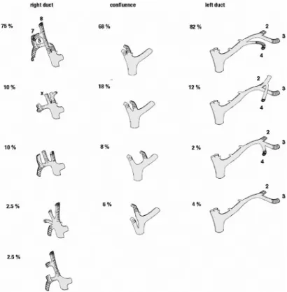
lobes belong to the functional left lobe of the liver. The left hepatic
duct drains segments 2, 3, and 4 of the left hemiliver. The “normal”
confluence comprises a duct formed from ducts of segments 2 and 3
and one or more ducts from segment 4. The segment 3 duct follows
the left horn of the Rex recessus and joins the segment 2 duct above
the segment 2 portal branch (at the level of the curve of the hilar part
in the posterior-anterior portion of the portal branch). This duct is 2.5
cm long, from 2 to 5 cm, depending on the size of the posterior
margin of the quadrate lobe. Being extrahepatic in this portion, it runs
transversely in the hilum, from left to right. Running first above and
behind the left portal branch, it crosses the superior edge and joins the
right hepatic duct to form the biliary confluence. For the left hepatic
duct
1, this normal anatomy is reported in 82%. In 4% of patients, a
right sectoral duct can join the left hepatic duct (3% posterior and 1%
anterior).
The right hepatic duct drains all segments of the right hemiliver
(segments 5, 6, 7, and 8). The ducts of segments 6 and 7 form the
posterior right hepatic duct, and those of segments 5 and 8 form the
17
located to the left of the anterior branch of the portal vein. The
direction of the posterior duct is more horizontal, running superior
(being epiportal in the Hjortsjö crook)to the anterior portal branch and
joins the anterior duct. In approximately 20% of instances, the right
duct runs inferiorly (being hypoportal) to the anterior portal branch. A
complete anterior duct was present in 35% and a complete posterior
duct in 61%. The “normal” confluence
1of these two ducts forms the
right hepatic duct, above the right portal branch, in an extra-hepatic
position. The right hepatic duct can be absent, the anterior and
posterior ducts joining directly to the left hepatic duct, forming a
triple confluence (12%). The right hepatic duct may join the main
hepatic duct below the normal confluence in 25% of cases (9% the
anterior and 16% the posterior). This anatomical variation is known as
“convergence étagée” or selved confluence. The normal right duct is
18
[image:18.612.95.300.110.319.2]Biliary confluence
figure;2
The main biliary confluence is formed outside the liver parenchyma,
before becoming distal to the common hepatic duct. It runs along and
anterior to the origin of the right branch of the portal vein The duct is
displaced superiorly and medially to the left of the main portal vein.
This classic junction occurs in 61% of instances. During a right
hepatectomy, the anatomical situation of the main biliary confluence
explains the risk of ligating the confluence or the left duct. The
Bismuth–Corlette classification
5is valid only for a “normal”
19
into account not only the type of confluence, but also its height in
relation to the portal vein.
At the level of the hilum, Glisson's capsule is both thicker and denser,
forming the connective tissue of the hilar plate. The biliary ducts are
enclosed within this tissue. Adhesions between this capsule and
arterial and portal branches are less important. It is therefore easy to
dissect the portal branches at the hilum, but more difficult for the
arterial branches and almost impossible to separate the bile duct of the
hilar plate
.
In the case of hilar cholangiocarcinoma, the proximity of
the portal triad explains the frequent tumor invasion of portal
branches. A lobar atrophy may result from the vascular invasion
and/or from a biliary obstruction. Owing to the absence of vascular
interposition at the anterior part of the hilar plate, it is also possible to
20
Gall Bladder
:
The gallbladder is 7-10 cm long and has a capacity of 30-50 ml.
It is located on the visceral surface of the liver in a shallow fossa at
the plane dividing the right lobe from the medial segment of the left
lobe (the GB-IVC line). In other words, the gallbladder fossa is found
at the junction of the quadrate lobe (segment IV) and the right lobe of
the liver along the line of Rex. The gallbladder is separated from the
liver by the connective tissue of Glisson's capsule. Anteriorly, the
peritoneum of the gallbladder is continuous with that of the liver.
The gallbladder can be divided into fundus, body, infundibulum,
neck, and cystic duct.
The fundus projects beyond the inferior margin of the liver, in
the angle between the lateral border of the right rectus abdominis and
the ninth costal cartilage. It is entirely surrounded by peritoneum
21
upper narrow part of the body is continuous with the neck at the right
end of the porta hepatis. The superior surface of the body is devoid of
peritoneum, and is adherent to the liver. The inferior surface is
covered with peritoneum, and is related to proximal part of transverse
colon and first part of the duodenum.
The neck is the narrow upper end of the gall bladder. It first
curves anterosuperiorly and then posteroinferiorly to become
continuous with the cystic duct. Its junction with the cystic duct is
marked by a constriction. The posteromedial wall of the neck is
dilated to form a pouch called the Hartmann’s pouch which is directed
downwards and backwards.
Cystic Duct:
Cystic duct is about 3 to 4 cm in length. It ends by joining with
the common hepatic duct at an acute angle to form the common bile
duct. The mucous membrane of the cystic duct forms a series of 5 to
10 crescentic folds, arranged spirally to form the so-called “
spiral
22
Common Bile Duct:
The common bile duct begins at the union of the cystic and
common hepatic ducts and ends at the papilla of Vater in the second
part of the duodenum. It varies in length from 5 cm to 15 cm,
depending on the actual position of the ductal union. In 22%, the
common hepatic and cystic ducts, on average, run parallel for 17 mm
before the ducts actually unite. The average diameter is about 6 mm
The common bile duct can be divided into four portions or
segments: supraduodenal, retroduodenal, pancreatic, and intramural.
The supraduodenal portion of the common bile duct lies between the
layers of the hepatoduodenal ligament in front of the epiploic foramen
of Winslow, to the right or left of the hepatic artery, and anterior to
the portal vein. Its length is 2-5 cm.
The distal part of the supraduodenal portion is related to the
posterior superior pancreaticoduodenal (PSPD) artery, which has a
retroduodenal location and which crosses the duct first anteriorly and
23
supraduodenal artery, which also may pass anterior to the common
bile duct. In the majority of cases the retroportal artery joins the PSPD
artery, but it may join the right hepatic artery directly and send
branches to the common duct en route. The PSPD artery is easily
injured while exploring the common duct.
If the junction of the cystic and common hepatic ducts is low,
the supraduodenal segment is short or even absent. Large lymph
nodes may be fixed to the right side of the supraduodenal segment.
The retroduodenal portion of the common bile duct is between
the superior margin of the first portion of the duodenum and the
superior margin of the head of the pancreas. It is 1-3.5 cm long. The
duct may be free or partially fixed to the duodenum11.
The pancreatic portion of the common bile duct extends from
the upper margin of the head of the pancreas to the point of entrance
into the duodenum. It passes downward to the right, posterior to the
pancreas or within the pancreatic parenchyma.
The intramural portion of the common bile duct takes an oblique
24
receives the main pancreatic duct inferiorly. The two ducts usually lie
side-by-side with a common adventitia for several millimetres. The
diameter of both ducts decreases within the duodenal wall. The
septum between the ducts is reduced to a thin mucosal membrane
before the ducts become confluent3 11.
The terminal part of the bile duct is surrounded just above its
junction with the pancreatic duct by a ring of smooth muscle that
forms the sphincter choledochus. This sphincter is always present. It
keeps the lower end of the bile duct in closed status. As a result, bile
formed in the liver keeps accumulating in the gall bladder and also
undergoes considerable concentration. When food enters the
duodenum, especially a fatty meal, the sphincter opens and the bile
stored in the gall bladder is poured into the duodenum.
Another less developed sphincter, which is usually but not
always present around the terminal part of the pancreatic duct, is
called sphincter pancreaticus. A third sphincter surrounds
thehepatopancreatic ampulla and is called the sphincter ampullae. The
sphincter ampullae may extend proximally to enclose the lower parts
25
The sphincters named above are often referred to collectively as
the sphincter of Oddi
.
PANCREAS
Pancreas is a retroperitoneal elongated gland lying between the C
loop of duodenum and
splenic hilum. It has a head, body and tail with a small constricted
part between head andbody called the neck and another small
[image:25.612.97.396.345.590.2]downward projection from the head called the
26
Uncinate Process. The main ductal system of the pancreas, the Duct
of Wirsung, starts fromthe tail lying near the posterior than the
anterior surface with small ducts of the lobes draining
to it at right angles forming a herring bone pattern. It traverses the
body and on reaching theneck it binds down posteriorly to join with
the common bile duct to form a common dilatedhepatico pancreatic
Ampulla of Vater surrounded by the Sphincter of Oddi which
prevents thereflux of bile into the pancreas and vice versa.
Blood supply:
The cystic artery arises from the right hepatic artery as it crosses
the calot’s triangle to the right of the common hepatic duct. The
lymph node of Lund usually lies just superficial to the position of the
cystic artery in the cystic triangle, and can be a good guide to finding
and ligating it. Reaching the gallbladder behind the common hepatic
duct, the cystic artery usually branches into an anterior superficial
branch and a posterior deep branch. These branches anastomose and
send arterial twigs to the adjacent liver. The cystic artery may arise
from the left hepatic artery or the gastroduodenal artery
27
from the cystic artery above and from the posterior superior
pancreaticoduodenal artery below.
The epicholedochal arterial plexus of the CBD is derived from
the retroduodenal or pancreaticoduodenal arteries. The collateral
circulation is enhanced by two intramural plexuses. These may be
compressed between the oedematous mucosa and the external tough
fibrous coat in pathologic conditions such as cholangitis or common
bile duct obstruction secondary to choledocholithiasis.
Veins:
The superior surface of the gallbladder is drained by multiple
small veins passing through the gallbladder bed that breaks up into
capillaries within the liver. They do not form a single "cystic vein."
Veins from the hepatic surface drain directly into the liver. Veins on
the inferior surface open directly or follow the hepatic ducts into the
liver. From the peritoneal surface, one vein usually drains the fundus
and body and other veins drain the neck and upper portions of the
cystic duct as well as the hepatic ducts. These small veins enter the
liver together with ascending veins from the common bile duct. These
28
Nerve Supply:
• Coeliac plexus
• Seven to nine thoracic sympathetic fibres
Pain from the gall bladder may travel along the vagus, the
sympathetic nerves, or along the phrenic nerves. It may be referred to
different parts through these nerves as follows.
1. Through the vagus to the stomach
2. Through the sympathetic nerves to the lower pole of the
scapula
Calot Triangle:
It is bounded, right side by the upper part of the gallbladder
and cystic duct, left side by the common hepatic duct and superiorly
29
Histology:
The bile ducts are composed of an external fibrous layer of
connective tissue, a few thin smooth muscle layers (longitudinal,
oblique, and circular), and an internal layer of mucosa of columnar
epithelium. The gallbladder wall is formed, from external to internal,
by the following layers:
Serosa
Adventitia
Fibro muscular layers
Mucosa
Serosa is the typical visceral peritoneum formed by
mesothelium on the surface with loose connective tissue directly
beneath. Adventitia is a layer of dense connective tissue that is found
external to the muscularis externa where the gallbladder is attached to
the surface of the liver. The adventitia contains large blood vessels,
autonomic fibres for innervation of muscularis externa and blood
vessels, a rich lymphatic network, and a plethora of elastic fibre’s and
adipose tissue.
30
fibres among bundles of smooth muscle cells. No muscularis mucosa
or submucosa is found in the gallbladder. Mucosa is distinguished by
having very tall, slender columnar epithelial cells. While no glands
are found in the mucosa, this layer is thrown into elaborate folds
which on first inspection give the impression of glands. These folds
form deep diverticula of the mucosa and have been identified as
"Rokitansky-Aschoff sinuses"; in some cases, these extend through
the muscularis externa. Bacteria have been known to accumulate in
these folds, and chronic inflammation may develop.
Physiology:
Bile produced by hepatocytes, drains into the hepatic canaliculi.
It travels from the terminal bile ducts to the right and left hepatic
ducts. Then it moves to the common hepatic duct. The majority of the
bile goes from the common hepatic duct through the cystic duct to the
gallbladder, drains to the common bile duct, and then to the
duodenum. The remainder of the bile goes to the common bile duct,
then to the duodenum, bypassing the gallbladder.
31
the duodenum each day. The gallbladder has a capacity ranging from
15 to 60 ml (average approximately 35 ml). The gallbladder
concentrates bile by absorbing sodium, chloride, and bicarbonate ions
and water such that bile salts can be concentrated 5 to 250 times.
Potassium ions are concentrated as the water is absorbed; further
concentration results from simple diffusion. Bile contains significant
amounts of carbonate and calcium ions. The epithelium secretes
hydrogen ions, and the carbonate ions are converted to bicarbonate.
Calcium and bicarbonate ions are absorbed by the epithelial cells and,
thus, calcium carbonate precipitation in the gallbladder is avoided9.
The hormone cholecystokinin causes contraction of the
gallbladder muscle, forcing bile out. Stimulation from the vagus nerve
also causes the gallbladder to contract. The sphincteric apparatus of
Oddi becomes inhibited in the presence of cholecystokinin and
relaxes as a reaction to gallbladder contraction. All of these actions
32
Physiology of the Gallbladder and Bile Ducts
The anatomy of the biliary tree is a little complicated, but it is
important to understand. The liver's cells (hepatocytes) excrete bile
[image:32.612.81.356.240.529.2]into canaliculi, which are intercellular spaces between the liver cells.
33
These drain into the right and left hepatic ducts, after which bile
travels via the common hepatic and cystic ducts to the gallbladder.
The gallbladder, which has a capacity of 50 milliliters (about 5
tablespoons), concentrates the bile 10 fold by removing water and
stores it until a person eats. At this time, bile is discharged from the
gallbladder via the cystic duct into the common bile duct and then
into the duodenum (the first part of the small intestine), where it
begins to dissolve the fat in ingested food.
The liver excretes approximately 500 to 1000 milliliters (50 to 100
tablespoons) of bile each day. Most (95%) of the bile that has entered
the intestines is resorbed in the last part of the small intestine (known
as the terminal ileum), and returned to the liver for reuse.
The many functions of bile are best understood by knowing the
composition of bile:
Bile Salts (cholates, chenodeoxycholate, deoxycholate): these are
produced by the liver's breakdown of cholesterol. They function in
bile as detergents that dissolve dietary fat and allow it to be absorbed.
Hence, disruption of bile excretion disrupts the normal absorption of
34
the fat is not absorbed (steatorrhea) , and develop deficiencies of the
fat-soluble vitamins (A, D, E, and K).
Cholesterol and phospholipids-while only 4% of bile is cholesterol,
the secretion of cholesterol and its metabolites (bile salts) into bile is
the body's major route of elimination of cholesterol. Phospholipids,
which are components of cell membranes, enhance the cholesterol
solubilizing properties of bile salts. Inefficient excretion of cholesterol
can cause an increased serum cholesterol. This predisposes to
vascular disease (heart attacks, strokes, etc.)
Bilirubin-while this comprises only 0.3% of bile, it is responsible for
bile's yellow color. Bilirubin is a product of the body's metabolism of
hemoglobin, the carrier of oxygen in red blood cells. Disruption of the
excretion of this component of bile leads to a yellow discoloration of
the eyes and skin (jaundice).
Protein and miscellaneous components
Bile production and recirculation is the main excretory function of
the liver. Tumors that obstruct the flow of bile from the liver can also
35
these other functions to understand the symptoms that these tumors
can cause. These include:
Metabolic functions, such as the maintenance of glucose (blood
sugar) levels
Synthetic functions, such as the synthesis of serum proteins such as
albumin, blood clotting (coagulation) factors, and complement (a
mediator of inflammatory responses)
Storage functions, such as the storage of sugar (glycogen), fat
(triglycerides), iron, copper, and fat soluble vitamins (A, D, E, and K)
36
CHAPTER III
3.REVIEW OF LITERATURE – II
JAUNDICE
The term ‘Jaundice’ is derived from the French word meaning ‘Yellow’ and refers to the presence of an excess of bile pigments in the tissues and the serum. It is a presenting sign of anumber of hepatic and non-hepatic diseases. The differential diagnosis and management aredependent upon a appreciation of normal and abnormal variants of bile pigment metabolism.
PATHOPHYSIOLOGICAL CLASSIFICATION OF JAUNDICE
I. PREDOMINANTLY UNCONJUGATED HYPERBILIRUBINEMIA
A. Excess production of bilirubin
1. Hemolytic anaemia
2. Resorption of blood from large internal hemorrhages
3. Ineffective erythropoiesis
B. Reduced hepatic uptake
1. Drug induced
2. Prolonged fasting
3. Sepsis
C. Impaired bilirubin conjugation
1. Gilbert’s Syndrome
37
4. Diffuse hepatocellular disease (hepatitis, cirrhosis)
II. PREDOMINANTLY CONJUGATED HYPERBILIRUBINEMIA (CHOLESTATIC
JAUNDICE)
increase in serum bilirubin is in the unocnjugated indirect reacting bilirubin, no bilirubinappears in the urine but there is an increase in the fecal and urinary urobilinogen. An excessof bilirubin production also occurs in shunt
hyperbilirubinemia in which indirect bilirubinaccumulates in the absence of any reduction in red cell life span.Constitutional defects of liver function may also cause hyperbilirubinemia withoutimpairment of bile flow. In Gilbert’s disease there is defect in the bilirubin transport into theliver cell, while in Criggler-Najjar syndrome the defect is an inability of liver to conjugate the
38
A. Decreased intrahepatic excretion of bilirubin
1. Dubin Johnson Syndrome 2. Rotor’s Syndrome
3. Drug induced
4. Hepatocellular disease (viral hepatitis) 5. Primary biliary cirrhosis
6. Sclerosing Cholangitis
B. Extrahepatic biliary obstruction
1. CBD stones
2. Carcinoma of the head of pancreas, extrahepatic hile ducts and ampulla of Vater
3. Extrahepatic biliary atresia
NORMAL BILE PIGMENT METABOLISM
The bile pigment – bilirubin is a tetra pyrrole, which is formed to the greatest extent fromhemoglobin and to a lesser extent from myoglobin breakdown and hepatic synthesis itself.
39
removed and the heme ring is opened and transformed into biliverdin, which is green. Thelater is reduced to become bilirubin, which is yellow. The bilirubin combines with albumin toform a relatively stable protein-pigment complex and is transported as such to the hepaticparenchymal cell. This complex which is referred to as indirect reacting bilirubin, since itgives the Vanderbergh diazo reaction only after treatment with alcohol and other substance
that split the protein, is poorly soluble in water and is not excreted in the urine. In the hepatic parenchymal cell the albumin is removed and the bilirubin is conjugated withglucuronic acid to form diglucuronide, which is water soluble and is excreted into the biliarycanaliculi. This substance gives an immediate diazo reaction and hence termed as directreacting.
This is passed into urine. Normally there is less than 1.2 mg of direct reacting serumbilirubin and less than 0.3 mg of indirect reacting bilirubin per 100 ml of serum.
40
accompanies the use of intestinal antibiotics. Some of the urobilinogen is reabsorbed by wayof portal venous system and returns to the liver, where it is either removed or to a small extentexcreted in urine.
ABNORMAL BILE PIGMENT METABOLISM
No classification is totally satisfactory. The classification most widely used distinguishesbetween hemolytic, obstructive and hepatocellular jaundice. However, it is most reasonable to
categorize as
1. Those disease states in which the bile flow is unimpeded.
2. Those types that are associated with an impairment of the bile flow.
NORMAL BILE EXCRETION
The overproduction of bile pigment from excessive hemolysis creates a situation in which
normal liver is confronted with more pigment than it is able to remove. This occurs inphysiological jaundice of infancy and all pathological hemolytic states. However the reservecapacity of the liver is great and even when the bilirubin production is increased six times
41
albumin and cannot be excreted by the kidney, thus prompting the term acholuric jaundice.
IMPAIRED BILE EXCRETION
All other diseases are associated with an accumulation of conjugated bilirubin in the bloodand impaired excretion. The bilirubin pigment which is water soluble, is readily excreted into the urine, which becomes brown. The obstruction may be intrahepatic or extrahepatic.
INTRAHEPATIC OBSTRUCTIVE JAUNDICE
In the Dubin-Johnson Syndrome, which is associated with the appearance of iron free pigmentin the hepatic cells and normal liver function, the hepatic excretion of conjugated bilirubin isimpaired. Intrahepatic cholestasis has also been related to a variety of drughepatocellular disease. Methyltestosterone and norethiandrolene damage the microvilli of the
bile canaliculi and may cause jaundice. The phenothiazine drugs such as chlorpromazine mayevoke a hypersensitivity reaction in a small percentage of patients and result in cholangitichepatitis and intrahepatic cholestasis. A lesion along the excretory pathway within the liver is
42 EXTRAHEPATIC CHOLESTASIS
This is caused by anatomical obstruction to flow of bile from liver to the intestine. Theobstacle may be situated anywhere from the junction of right and left hepatic ducts to thetermination of common bile duct in the duodenum. Atresia, stricture, choledocholithiasis,tumours of bile duct and pancreas, choledochal cysts and parasites have been implicated.
Obstruction of extrahepatic duct results in an increase in serum bilirubin particularly thedirect reacting type, the appearance of bile in the urine and passage of clay coloured stools.
43
EFFECTS OF BILIARY TRACT OBSTRUCTION
PHYSICAL EFFECTS
The normal secretary pressure of bile is 120-250 mm of water. Following total bile ductobstruction, bile secretion will continue until CBD pressure rises to 170-220 mm of waterafter which secretion decreases. Cholesterol and
phopholipid secretion is more readilyreduced by high pressure than bile salt secretion making bile less lithogenic.
Complete obstruction of main extrahepatic bile duct of major segmental duct will normallylead to proximal dilatation. The lack of intrahepatic dilatation may be due to seconday hepaticfibrosis of co-existing alcoholic cirrhosis.
PAIN
Painless progressive jaundice is the classical hallmark of malignant biliary tract obstruction.But it is not uncommon to elicit a history of abdominal pain in these patients,the cause ofpain being distention of gall bladder and bile duct or
associated stretching of liver capsule inrapidly progressive obstruction. PATHOLOGICAL CHANGES IN BILE DUCTS AND CANALICULI In biliary obstruction the canaliculi become dilated and microvilli distorted and swollen. Bilepigment thrombi may be seen in canaliculi and adjacent
44
leads to marked inflammatory reaction in the portal tracts with
polymorphonuclear leucocyteinfiltrate. The hepatocyte of periportal zone shows disruption and eventually leading on topiecemeal necrosis. Experimental
evidence shows that if obstruction is relieved within weeks morphological changes are reversible.
CHOLANGITIS
Although the neutrophil associated with cholangitis is a chemical reaction associated withbiliary obstruction and does not imply bacterial inflammation, in presence of biliary stasis,secondary bacterial colonization may produce the additional element of infective cholangitisalthough classically referred to as ascending cholangitis the actual mechanism for entry ofbacteria into the unoperated biliary tract may not always be clear. Studies by McPherson et al (1982) showed that organisms are found in bile in approximately 1/3rd of
patients withmalignant biliary enteric anastamosis this rate may be higher. In another study Jackaman et al(1980) found that highest rate of biliary
colonization were found in patients withcholedocholithiasis and benign bile duct strictures where as may as 80% of patients hadpositive cultures.
ATROPHY
45
presence of normal contralateral lobe.The practical importance of lobar atrophy in a surgical context lies in the fact that an atrophicliver lobe may be inadequate to support life following the resection of normal or hyperplastic
liver tissue and biliary drainage of such an obstructed lobe may also fail to produce resolutionof jaundice.
BIOCHEMICAL EFFECTS
BILIRUBIN
Conjugated hyperbilirubinemia is the classical biochemical feature of
obstructive jaundice.But prolonged partial obstruction with functional effects on hepatocytes may produce a mixedbiochemical picture with elevated circulating unconjugated bilirubin.
ALKALINE PHOSPHATASE
Elevation of this enzyme is the most widely used and probably the most sensitive indicator. Itmay be the only biochemical indicator of incomplete of segmental obstruction. Acuteobstruction of bile duct causes regurgitation of enzyme from biliary compartment andincrease in hepatic synthesis.
PROTEIN SYNTHESIS
46
frequent association of biliaryobstruction with malignancy and malnutrition causes hypoalbuminemia.An active marker of hepatic protein synthesis, serum prealbumin is more valuable since it hashalf life of only 1.9 days. The most important aspect of protein synthesis relates to synthesis
of coagulation factors II, VII, IX & X and its failure is due to failure to absorb vit K due toabsence of bile salts from intestine.
LIPIDS
Cholesterol level may be elevated in biliary tract obstruction. A number of alterations in lowdensity lipoproteins have been observed which are of no major functional importance.
CARBOHYDRATE METABOLISM
Abnormal glucose tolerance may be seen in patients with impaired liver
function. But themalignant disease causing obstruction might be primary cause. BILE SALT CIRCULATION
47
ENDOTOXEMIA & RETICULOENDOTHELIAL FUNCTION
Endotoxin is a lipopolysaccharide derived from the cell walls of gram negative bacteriapresent in the gut. Normally only minute quantities of endotoxin enters the portal circulationand these traces are cleared by hepatic reticuloendothelial system. In obstructive jaundice theabsence of bile salts from intestine causes increased formation of endotoxin by alteredmicroflora and decreased clearance of absorbed endotoxin due to depressedreticuloendothelial cell function
resulting in endotoxemia in more than 50% of patients. The
bile salt absorption is also quiet fast in biliary obstruction probably due to increased vascularpermeability. The circulating endotoxin causes pathological effects like renalvasoconstriction, redistribution of intrarenal blood flow and activation of complement,leukocytes and platelets.
CHANGES AFTER RELIEF OF OBSTRUCTION BILE SECRETION
Postoperative study of biliary secretion is done by the insertion of external percutaneoustranshepatic drain. There is frequently a prompt and major
cholestasis and bile volumes mayexceed 4 liters per day. Failure to replace large volumes of fluid and electrolyte losses mayresult in dehydration and electrolyte depletion with a metabolic acidosis. The replacement of
48
During the first few days of biliary drainage the bile produced is of low
bilirubin and bile saltconcentration. This may be partly due to a slow return of impaired liver to normal functionand also to loss of enterohepatic circulation of bile salts.
RECOVERY OF FUNCTION
In majority of case plasma bilirubin begins to fall promptly after insertion of a drainageIn majority of case plasma bilirubin begins to fall promptly after
insertion of a drainagecatheter or an internal biliary bypass procedure and this is accompanied by clinical . However, return of hepatocyte function is not
instantaneous.Assessment of liver function by serial antipyrene clearance
measurement after relief ofobstruction has shown that it takes about 6 weeks for it to return to normal values.
STRUCTURAL CHANGES
The reversal of structural changes in liver and biliary tract following
decompression ofobstruction is variable. Bile ducts which have beeen subjected to edema, inflammatoryinfiltration, cholangitis, and fibrotic changes are likely to retain some rigidity for considerabletime after decompression.
As regards reversal of intra hepatic fibrotic changes following drainage it is difficult to obtainclear evidence since this would rely upon serial liver biopsies in asymptomatic patients solong as fibrotic changes remain short of true
49 with such drainage.
CLINICAL FEATURES SYMPTOMS
Typically a patient with obstructive jaundice presents with dark urine, pale stools and pruritusof varying severity. Information regarding initial onset and whether clinical course isintermittent and associated with pain, fever and rigors must be short. Attack precipitated byfat intake can be relevant. An episode of cholangitis is recognized if jaundice is associatedwith pain, rigor and pyrexia. Jaundice without significant pain or pain radiating to back may
indicate pancreatic pathology. However this is not certain and patients with gallstones anpresent with back pain whereas patients with extensive carcinoma of head of pancreas may present with typical history of biliary colic.
A fluctuating depth of jaundice is suggestive of intermittent obstruction as in periampullarycarcinoma or temporary alteration of stone in the ampulla of vater. It is very rare in pancreaticcancer and cholangiocarcinoma. Weight loss, anorexia and pallor suggest malignancy of shortduration. When these symptoms occur with painless jaundice, neoplasm of head of pancreas
50 PHYSICAL EXAMINATION
GENERAL INSPECTION
The common stigmata of liver disease should be looked for – they are all indications of liverdysfunction. Jaundice is due to staining of tissues with bilirubin and possibly other pigmentssuch as biliverdin. It is initially noticed in sclera. As jaundice progresses the skin becomesprogressively more pigmented, spider naevi, which are vascular skin lesions supplied bycentral arteriole is occluded with a pinhead. Spider naevi usually occur in the region of
superior vena cava – chest above the level of nipple, face, neck and arms. Palmar erythema isobvious and pronounced reddish flushing of palms. It particularly affects the thenar and hypothenar eminece and bases of fingers. Spontaneous bruising, echymosis and bleedingaround venipuncture sites are well recognized signs of liver disease occurring due toabnormality in
coagulation mechanisms. Long standing pruritis causing scratch marks all over the body can also be noted.
EXAMINATION OF LIVER
Palpation of liver should be combined with percussion to determine the upper and lowerborders. The upper border of liver normally extends upto 5th
intercostals space. Auscultationover the liver may give some evidence of
51 SPLENIC ENLARGEMENT
Splenomegaly can be detected by palpation commencing in the right iliac fossa andprogressing towards the left hypochondrium. Splenic notch can sometimes be recognized onthe anterior border of grossly enlarged spleen.
ASCITIS
Clinical confirmation of ascitis is achieved by eliciting shifting dullness on percussion or fluidthrill on palpating the flanks. Ascitis could be due to
hypoalbuminemia of liver dysfunction,portal hypertension or manifestation of advanced malignancy either of liver or pancreas.
GALL BLADDER SIGNS
The finding of a palpable gall bladder in the presence of features of obstructive jaundicesuggests malignant obstruction of the biliary tree (Courvoisier’s Law). However failure topalpate gall bladder does not exclude the presence of
malignant biliary obstruction. On theother hand it is possible to have a palpable gall bladder in the presence of gallstones whereone stone obstructs the common bile duct and another is impacted in the Hartmann’s pouch or
cystic duct resulting in an empyema or mucocele of the gall bladder. An intermittentlypalpable gall bladder is suggestive of periampullary carcinoma. EVIDENCE OF PORTAL HYPERTENSION
52
DIFFERENTIAL DIAGNOSIS IN CHOLESTASIS I. EXTRAHEPATIC CAUSES
1. STONES
a) gallstones slipping into CBD
b) gallstone in cystic duct and getting impacted onto CBD (Mirrizi syndrome) c) pancreatic calculus obstructing at the ampulla of Vater
2. STRICTURES
a) malignant carcinoma of CBD b) Benign – surgical trauma c) primary sclerosing cholangitis
3. TUMOURS OF THE BILIARY TREE a) periampullary carcinoma
b) carcinoma of head of pancreas c) cholangiocarcinoma
4. EXTRINSIC PRESSURE ON EXTRAHEPATIC BILIARY TRACT a) Metastatic lymphnodes near the biliary tract by pressure and later by infiltration
produce obstruction to biliary passages
b) primary lymphnodular disease involving the lymphnodes near biliary pathways –
53
c) Metastatic involvement of the connective tissue of hepatic hilum causing extrinsic
compression on bile ducts
5. MISCELLANEOUS CAUSES
a) parasitic occlusion of CBD – Schistosomiasis b) Mycotic condition
c) Choledochal cysts
d) Hepatic artery aneurysm II. INTRAHEPATIC CAUSES 1. INTRAHEPATIC STONE
2. INTRAHEPATIC BILIARY STRICTURES 3. KLATSKIN’S TUMOUR
4. BILIARY DYSPLASIA a) Congenital hepatic fibrosis b) Cystic disease of the liver c) Caroli’s disease
5. CONGENITAL AND INFANTILE ATRESIA OF BILE DUCTS 6. ANEURYSM OF BRANCHES OF HEPATIC ARTERY
7. CYSTS OF THE LIVER a) congenital
b) parasitic
54
CLINICAL CLASSIFICATION OF OBSTRUCTIVE BILIARY TRACT DISEASE
Classification proposed by Benjamin (1983) has proved useful in clinical practice. It
recognizes four types of biliary obstruction. They are: TYPE I: COMPLETE
Obstructive – producing progressive jaundice Eg.:
a) Tumours of head of pancreas b) Cholangiocarcinoma
c) Ligation of CBD
d) Parenchymal damage to liver TYPE II: INTERMITTENT
Obstruction which produces symptoms and biochemical changes with or without jaundice
Eg.:
55 f) polycystic liver disease
g) intrabiliary parasite
TYPE III: CHRONIC INCOMPLETE
Obstruction with or without symptoms and biochemical changes eventually producing
pathological changes in bile ducts of liver Eg.:
a) strictues of CBD 1) congenital 2) traumatic 3) post irradiation
b) stenosed biliary enteric anastamosis c) stenosis of sphincter of Oddi
d) chronic pancreatitis e) cystic fibrosis
TYPE IV: SEGMENTAL
Obstruction in which one or more anatomical segments of biliary tree may be obstructed. This
in turn may be complete, intermittent or chronic incomplete. Eg.:
56 c) sclerosing cholangitis
d) cholangio carcinoma INVESTIGATIONS BIOCHEMISTRY
Biochemical features of cholestasis are: 1. Conjugated hyperbilirubinemia
2. Elevation of alkaline phosphatase, 5’ nucleotidase, gamma glutamyl
transpeptidase. Theenzyme 5’ nucleotidase is the most reliable since its level is not influenced bybone disease or alcoholism.
3. Minimal or no elevation of serum transaminases
4. Presence of bilirubin in the urine as conjugated bilirubin, which is water soluble andhence filtered by glomeruli
5. Elevation in serum cholesterol and bile acid levels although these are not routinelymeasured inpatients with cholestasis jaundice.
IMAGING TECHNIQUES
PLAIN ABDOMINAL AND CHEST SKIAGRAM
57 ULTRASONOGRAM
This is non-invasive and quick to perform, but requires experience in technique andinterpretation. Extrahepatic biliary obstruction can be diagnosed by
demonstration of dilatedbiliary radicals. In experienced hands the accuracy in diagnosing ductal dilatation is over95%. In most iof the cases the cause of biliary obstruction can be traced by ultrasonogram.
Enlargement of head of pancreas is suggestive of carcinoma. Difficulties in achieving adefinite diagnosis arises principally with small lesions at the lower end of common bile ductand which is often obscured by gas in the duodenum or colon. As it does not involveradiation, it can be used safely in pregnancy.
ENDOSCOPIC RETROGRADE CHOLANGIO-PANCREATICOGRAPHY (ERCP)
Upper GI endoscopy with a forward or oblique viewing pan endoscope should be performedin jaundiced patients as significant gastrointestinal pathology is encountered in 25% ofjaundiced patients. This is indicated when obstructing agent is lower down in the CBD. Thisis also ideal when ducts are not dilated or visualization of pancreatic duct or ampulla isrequired. It permits concomitant endoscopic examination and biopsy of lesions encountered
58
ERCP has very low morbidity due topancreatitis (1%) and very low mortality (0.1%).
PERCUTANEOUS TRANSHEPATIC CHOLANGIOGRAM (PTC) This is done by injecting the contrast material into the dilated biliary radicles through acannula. This more useful when the obstruction is higher up in the bile or hepatic ducts.
CT findings in pancreatic carcinoma are: a) Focal mass
b) Pancreatic atrophy/pancreatitis c) CBD/PD dilatation
d) Vascular enhancement or displacement e) Regional lymphadenopathy
MAGNETIC RESONANCE CHOLANGIO-PANCREATICOGRAPHY (MRCP)
It is a newer development in MRI. It provides multiplanar, cross-sectional, reconstructiveimage of pancreaticobiliary tree. It is purely diagnostic with no therapeutic interventionpossible. It offers advantage over dynamic CT in
59 ENDOSCOPIC ULTRASONOGRAPHY
It is in the early stage of development. A useful modality for tumour exclusion whentransabdominal utrasonography or CT has failed and high index of suspicion of carcinomaexists due to elevated tumour markers, prior to elevated tumour markers or ERCP. It canconfirm tumour of 1.2 cm in head of pancreas. LAPAROSCOPY
Should be routinely used by surgeons in all patients with jaundice. It gives direct visualizationof underlying pathology and is valuable in staging hepatobiliary and pancreatic tumours. Itavoids unnecessary laparotomy for patients with inoperable diseases.
ANGIOGRAPHY
Preoperative angiography is indicated in
1. history of previous major upper abdominal surgeries 2. doubtful resection on clinical and CT appearances
3. when it is anticipated to remove major vascular structures Angiographic findings in pancreatic carcinoma are:
1. parenchymal hypovascularity 2. angulation of vessels
3. encasement of vessels (arterial/venous) 4. displacement of vessels
60
FINE NEEDLE ASPIRATION CYTOLOGY
61 CARCINOMA OF PANCREAS
It includes carcinoma of the head proper and periampullary region. Almost all carcinoma ofpancreas arise from the ductal epithelium. Only 1% arises from acini. The average age ofpatient is about 60 years, but carcinoma of ampulla the average age is about 5 years less.Males are more affected.
PATHOGENESIS
Incidence of carcinoma of the pancreas has risen steadily over the past 10 years. There aresome factors, which can be considered as initiating or provoking carcinoma of pancreas. Theyare:
1. Cigarette smoking 2. Consumption of coffee 3. Diet rich in fat
4. Chemicals such as beta napthylamine and benzidine 5. Diabetes
6. Carcinogens in duodenal contents refluxing into the pancreatic duct 7. Alcohol consumption
PATHOLOGY
62
Carcinomas of the ampulla of Vater are columnar cell adenocarcinoma. This neoplasm arisesin duodenal papilla, in the ampulla of Vater or in the duodenal mucosa adjacent to the papillathere may be an area of pancreatitis in the head of pancreas. The primary lesion is so smallthat it is difficult to palpate. In such carcinoma, jaundice may not be progressive as recurrentsloughing of the central portion of the tumour will relieve obstruction of bile duct and
jaundice becomes intermittent. CLINICAL FEATURES
Carcinoma of the head of the pancreas usually presents with painless
progressive obstructivejaundice. Progressive jaundice is usually associated with pruritis due to the presence of bilesalts in blood. The jaundice usually
progresses steadily until the patient is almost green incolour. In case of periampullary carcinoma, the jaundice may be intermittent. Pain is not a marked feature. Patient may complain of dull and aching pain in the epigastrium. Pain is oftenrelieved by sitting in hunched position and is aggravated by supine position. Eating mayaggravate pain. Weight loss is the single most common symptom of carcinoma of thepancreas irrespective of the position of the tumour. Diarrhea with pale and foul smelling stool
63
due to biliary obstruction.Carcinoma of ampulla of Vater shows a few peculiar symptoms and signs. Pain is lessfrequent in this condition but when present is apt to be more colicky in nature. Jaundice isintermittent. Chills and fever are not uncommon due to associated cholangitis. Hematemesisand melena occasionally occurs in late cases as a result of direct invasion of duodenal or
gastric mucosa by tumour and portal hypertension secondary to splenic or portal veincompression by the tumour.
CHOLANGIOCARCINOMA
The reported autopsy incidence of malignant bile duct tumour ranges from 0.01-0.5%. Thereis slight preponderance of male (1.5:1). The age at presentation varies but the peak incidenceis in sixth decade. The etiology of bile duct cancer is unknown. Cholangiocarcinoma is seenwith increasing frequency in parasitic infestation of biliary tree, cystic disease of biliary tract,
chronic typhoid carriers, and ulcerative colitis and sclerosing cholangitis. PATHOLOGY
Tumours are best classified into the anatomical site of origin 1. Intrahepatic tumour from minor hepatic ducts
2. Proximal from right or left hepatic ducts, cystic duct and it confluence with CBD.
64
4. Distal from the distal common bile duct and periampullary region.
Tumours of the minor hepatic ducts are often diffuse (multicentric) and difficult todifferentiate from primary hepatocellular carcinoma. The gross appearance of cholangiocarcinoma assumes one of the three forms. They are:
1. Strictures 2. Nodular 3. Papillary
The majority of tumours are adenocarcinoma of varying origin. All
cholangiocarcinomas havea special predilection for perineural spread and do not metastasise beyond the liver. The bestprognosis is encountered after resection especially of distal and periampullary lesions.
CLINICAL FEATURES
The main presentation (90%) is with obstructive jaundice which is progressive andaccompanied by itching and anorexia. Dull upper abdominal pain is a frequent symptom.
Some patients present acutely with cholangitis. Physical examination reveals hepatomegaly.Anemia is present in patients with papillary tumours especially at the lower end of bile ductand periampullary region. It is caused by chronic blood loss. The feces of these patients havea characteristic silvery appearance due to combination of steatorrhoea and altered blood. A
65 PRE-OPERATIVE PREPARATION
1. All jaundiced patients must be kept in a good state of nutrition and hydration withsupplemental intravenous fluids, elemental diet and multivitamins as deemednecessary. Renal failure due to hypovolemia is a tremendous hazard post-operativelyand a continuous diuresis is maintained at all times. If the patient is grosslymalnourished, a period of parenteral hyperalimentation both before and after operationmay be of additional benefit.
2. Blood clotting deficiencies must be corrected. Anaemia is corrected by blood transfusions. Daily injection of Vit K is administered, preferably 4-5 days prior tooperation. Six units of fresh frozen plasma, six units of platelets and atleast six units ofblood should be made available in operating room.
3. Cardiopulmonary function should be assessed by pulmonary function tests, chest Xray and ECG. smoking is prohibited. Intensive pulmonary
physiotherapy, activemobilization and leg exercises are strongly encouraged post operatively.
4. Antibiotic prophylaxis should be given since there is impaired wound healing due todepressed immune function.
5. Nutritional status to be assessed and supported as there is impaired wound healing dueto decreased fibroblastic activity and general protein and calorie malnutrition.
66 b) sepsis
c) hepatorenal failure
d) severe cardiopulmonary disease e) malnutrition
a percutaneous transhepatic biliary drainage or endoscopic decompression should beattempted to tide over the patient for 2-3 weeks before major surgery. If the techniqueof percutaneous biliary drainage or endoscopic stenting is not available, a simplecholecystectomy or T-tube drainage of CBD may be undertaken.
TREATMENT OF MALIGNANT OBSTRUCTIVE JAUNDICE Treatment can be either
1. Curative 2. Palliative
CURATIVE TREATMENT
Surgery is now considered as the gold standard for treatment of malignant obstructivejaundice against which all other new modalities are considered. Halsted performed firstcurative and successful resection of periampullary carcinoma at John Hopkins Hospital in1898. He performed local resection of ampullary tumour. Presently standard resection for
periampullary carcinoma and head of pancreas tumours involves a
67
popularized by Whipple 1935. The gallbladder, CBD, entire duodenum, head of pancreas, pancreas upto the level of superiormesenteric vein, pylorus and distal stomach are resected. Restoration of gastrointestinal
continuity utilizes the proximal jejunum, brought out through the transverse mesocolon forpancreaticojejunostomy, hepaticojejunostomy and
gastrojejunostomy. The standard Whippleresection remains the classic thrrapy for these tumours and can be successfully performed inexperienced hand swith mortality less than 5%. A modification of standard Whipple resection,
the pylorus preserving pancreaticoduodenectomy has gained popularity in recent years. Thismodification eliminates gastric resection and leaves a 2cm cuff of duodenum for entericreconstruction as duodenojejunostomy.
PALLIATIVE SURGERY
Palliative surgery for periampullary carcinoma is performed in patients with unresectabledisease discovered at the time of laparatomy or in patients with prohibitive risk for resectionaltherapy (advanced age, limited cardiopulmonary reserve and also poorly alleviated nonoperatively).
1. Relief of jaundice, pruritis and impending cholangitis: Biliary tract decompression canbe done either by cholecystojejunostomy or by hepaticojejunostomy (each with
68
2. Relief of duodenal obstruction: If the patient lives for more than few months, duodenalobstruction usually occurs. It is therefore advisable to perform a
gastrojejunostomy atthe primary operation. NON OPERATIVE MANAGEMENT
When a patient is unfit or refuses surgery an alternative method of palliation of the jaundice isby endoscopic sphincterotomy and placement of biliary stent. This approach does not relieveany additional obstruction, which may be present. If patient survives for more than a fewmonths, recurrent cholangitis associated with stent blockage is a problem that necessitates
regular endoscopic removal and replacement of the stent.
Percutaneous transhepatic placement of internal expandable metal stent is being tried byinteventional radiologist and offers yet another option for palliation of the jaundiced patientwith malignant biliary tract obstruction.
TREATMENT OF CHOLANGIOCARCINOMA
Resection is the best method of treatment and is indicated for all operative tumours in fitindividuals. The reported respectable rate varies but averages 20%. The benefits of resectionare:
1. The possibility of cure or long term survival especially for distal bile duct tumours
69
segmentectomy IV provides good access to confluence, allowing good
clearance proximal tothe tumour and facilitates hepaticojejunostomy. When the tumour extends along the right orleft duct with extension to respective lobe, the resection includes a lobectomy in continuation
with main tumour mass. Middle tumours are excised from just below the confluence down tothe duodenum together with associated pericholedochal lymphnodes. The surgical treatmentof periampullary tumours is
pancreaticoduodenectomy. The results of hepatic transplantation
for cholangiocarcinoma (diffuse intrahepatic type) have been disappointing.
PALLIATIVE SURGERY
If tumour is inoperable, a bilio-enteric bypass is performed. Anastamosis of Roux loop tosegment III duct using the round ligament approach gives the best results for inoperable hilarlesions. Longmire operation in which anastamosis of the segment III duct to Roux loop ofjejunum after left lateral segmentectomy and Smith operation used to be done earlier, butthere is no added advantage to these procedures. A cholecystojejunostomy is performed forinoperable distal tumours. A gastrojejunostomy is added if duodenal obstruction is present or considered imminent in patient with periampullary tumour.
NON OPERATIVE MANAGEMENT
70
old and frail, palliation of jaundice is achieved by percutaneous transhepatic or endoscopicstenting. The endoprosthesis has to be large 8-10 FG and may require replacement if itbecomes blocked. Recently self-expandable stainless steel wire endoprosthesis have beenintroduced in management of patients with malignant biliary strictures. Other causes ofmalignant obstructive jaundice are due to extrinsic compression on the biliary tract bytumours, Metastatic
lymphnodes near the biliary tract by pressure and later by infiltration andprimary lymphonodular disease involving lymph nodes near biliary pathways. Treatment isprimarily to relieve obstructive jaundice and troublesome pruritis and steatorrhoea
Choledocholithiasis
1. Endoscopic sphincterotomy, stone extraction/CBD stenting followed by Lap/Open cholecystectomy
2. Lap/Open Cholecystectomy followed by Lap/Open CBD Exploration
CBD Exploration
First surgical exploration of the CBD was done in 1980 byLudwig Courvoisier. Indications
1. PREV.HISTORY OF JAUNDICE / CHOLANGITIS / PANCREATITIS
71 3.DILATED CBD
4. MULTIPLE SMALL STONE
Lap CBD exploration most commonly done Either trancystic or transductal
Transductal
• Stones >6mm • Intrahepatic stones
• Cystic duct diameter<4mm
• Cystic duct entrance either posterior
or distal
T-Tube
•
For decompression if CBD not cleared
• Later study of biliary system
• Access to biliary system for recurrent stones
Placing ‘T’ Tube
:
• Shorten limbs & remove part of wall
• Allows sphincter edema to settle
• 14 F size
• Tract for future intervention if retained stones are
detected
72
• If normal – remove > 12 days
• Retained stone – keep ‘T’ tube