EVALUATION OF OCCLUSAL HARMONY OF FIXED PARTIAL PROSTHESIS FABRICATED BY FUNCTIONALLY GENERATED PATH TECHNIQUES USING T-SCAN – AN IN VIVO STUDY
A Dissertation submitted to the
THE TAMILNADU Dr. MGR MEDICAL UNIVERSITY
In partial fulfillment of the requirements for the degree of
MASTER OF DENTAL SURGERY
(BRANCH – I)
(PROSTHODONTICS AND CROWN & BRIDGE)
Certificate
This is to certify that Dr.T.MARIA SINGAM, Post Graduate student (2012 - 2015) in the Department of Prosthodontics and Crown and Bridge, has done this dissertation titled “EVALUATION OF OCCLUSAL HARMONY OF FIXED PARTIAL PROSTHESIS FABRICATED BY FUNCTIONALLY GENERATED PATH TECHNIQUES USING T-SCAN - AN IN VIVO STUDY” under my direct guidance and supervision in partial fulfillment of the regulations laid down by The Tamil Nadu Dr. M.G.R. Medical University, Guindy, Chennai – 32 for M.D.S. in Prosthodontics and Crown & Bridge (Branch I) Degree Examination.
Guided by Head of the department
Dr.A.MEENAKSHI,M.D.S., Dr. C.SABARIGIRINATHAN,M.D.S.,
Professor, Department Of Prosthodontics, Professor and Head of the Department, Tamil Nadu Govt. Dental College &Hospital, Department Of Prosthodontics, Chennai-600 003. Tamil Nadu Govt. Dental College &
Hospital, Chennai -600 003. .
Head of the institution
Dr.SRIDHAR PREM KUMAR,M.D.S., PRINCIPALTRIPARTITE AGREEMENT
This agreement herein after the “Agreement” is entered into on this day, December 23,
2014 between the Tamil Nadu Government Dental College and Hospital represented by its
Principal having address at Tamil Nadu Government Dental College and Hospital, Chennai-3, (hereafter referred to as, „the College‟)
And
Dr.A.MEENAKSHI,M.D.S., aged 48 years working as professor in the Department of Prosthodontics at the college, having residence address at No. 137, fifth street, secretariat colony, Kelleys,Chennai-10 (herein after referred to as the „Principal Investigator‟)
And
Dr.T.MARIA SINGAM aged 32 years currently studying as Post Graduate student in
the Department of Prosthodontics and Crown & Bridge, Tamil Nadu Government Dental College and Hospital, Chennai-3 (herein after referred to as the „PG/Research student and Co- investigator‟).
Whereas the „PG/Research student as part of his curriculum undertakes to research on the study titled “EVALUATION OF OCCLUSAL HARMONY OF FIXED PARTIAL
PROSTHESIS FABRICATED BY FUNCTIONALLY GENERATED PATH TECHNIQUES
Whereas the parties, by this agreement have mutually agreed to the various issues including in particular the copyright and confidentiality issues that arise in this regard. Now this agreement witnesseth as follows:
1. The parties agree that all the research material and ownership therein shall become the vested right of the college, including in particular all the copyright in the literature including the study, research and all other related papers.
2. To the extent that the College has legal right to do go, shall grant to licence or assign the copyright do vested with it for medical and/or commercial usage of interested persons/entities subject to a reasonable terms/conditions including royalty as deemed by the college.
3. The royalty so received by the college shall be equally by all the parties.
4. The PG/Research student and PG/Principal Investigator shall under no circumstances deal with the copyright, confidential information and know how generated during the course of research/study in any manner whatsoever, while shall sole vest with the manner whatsoever and for any purpose without the express written consent of the college.
5. All expenses pertaining to the research shall be decided upon by the Principal investigator/Co-investigator or borne sole by the PG/Research student (Co-investigator).
6. The College shall provide all infrastructure and access facilities within and in other institutes to the extent possible. This includes patient interactions, introductory letters, recommendation letters and such other acts required in this regard.
research, topic and area research by the student researcher under guidance from the principal investigator shall be subject to the prior approval, recommendations and comments of the Ethical Committee of the college constituted for this purpose.
8. It is agreed that as regards other aspects not covered under this agreement, but which pertain to the research undertaken by the Student Researcher, under guidance from the Principal Investigator, the decision of the college shall be binding and final.
9. If any dispute arises as to the matters related or connected to this agreement herein, it shall be referred to arbitration in accordance with the provisions of the Arbitration and Conciliation Act, 1996.
In witness whereof the parties herein above mentioned have on this the day month and year herein above mentioned set their hands to this agreement in the presence of the following two witnesses.
College represented by its Principal Student Guide
Witnesses PG Student
DECLARATION
I, Dr.T.MARIA SINGAM, do hereby declare that the dissertation titled
“EVALUATION OF OCCLUSAL HARMONY OF FIXED PARTIAL PROSTHESIS
FABRICATED BY FUNCTIONALLY GENERATED PATH TECHNIQUES USING
T-SCAN – AN IN VIVO STUDY” was done in the Department Of Prosthodontics, Tamil Nadu Government Dental College & Hospital, Chennai 600 003. I have utilized the facilities provided in the Government Dental College for the study in partial fulfilment of the requirements for the degree of Master of Dental Surgery in the speciality of
Prosthodontics and Crown & Bridge (Branch I) during the course period 2012-2015
under the conceptualization and guidance of my dissertation guide, and professor
Dr.A.MEENAKSHI,M.D.S.,
I declare that no part of the dissertation will be utilized for gaining financial assistance for research or other promotions without obtaining prior permission from the Tamil Nadu Government Dental College & Hospital.
I also declare that no part of this work will be published either in the print or electronic media except with those who have been actively involved in this dissertation work and I firmly affirm that the right to preserve or publish this work rests solely with the prior permission of the Principal, Tamil Nadu Government Dental College & Hospital, Chennai 600 003, but with the vested right that I shall be cited as the author(s).
Signature of the PG student Signature of the HOD
ACKNOWLEDGEMENT
I am extremely thankful to Dr.C.SABARIGIRINATHAN,M.D.S., Professor and Head of the Department, Department of prosthodontics, Tamil Nadu Government Dental College and Hospital, Chennai, I consider it my utmost privilege and honour to express my most sincere and heartfelt gratitude to my esteemed for his wholehearted support, constant guidance, help, encouragement, valuable suggestions and support he has rendered at various stages of the dissertation. It has been a proud privilege for me to work under his able guidance.
My sincere thanks to Prof. Dr.SRIDHAR PREMKUMAR, M.D.S., Principal, Tamil Nadu Government Dental College and Hospital for his kind help, valuable suggestions in this study and permitting me to use all the facilities in the institution. I also thank him for the valuable guidance he has given throughout the period of my post graduate course.
I am extremely thankful and I consider it my utmost privilege to express my sincere and heartfelt gratitude to my former Head of Department Dr.C.THULASINGAM, M.D.S.,
Professor, Department of Prosthodontics, Tamil Nadu Government Dental College and Hospital for his able guidance, valuable suggestions, encouragement, monitoring and support he has rendered at various stages of the dissertation. With all the gratitude that I feel and warm regards that I can muster, I thank you Sir for imbibing the precious seeds of knowledge, patience, discipline and duty that is a treasure for a lifetime.
I extend my immense gratitude and thanks to Associate Professors,
Dr.R.Rupkumar, M.D.S., Dr.Sriramprabhu, M.D.S., Dr.M.Rajakumar, M.D.S., for their
constant encouragement and support, which made me go through some of the unnerving moments with ease and Senior Assistant professor Dr.T.Jeyanthikumari, M.D.S., for guiding and helping me at different stages of this study.
I am thankful to Assistant Professors,Dr.S.Vinayagam,M.D.S., Dr.Gandhimathi,M.D.S., Dr.V.Parimala, M.D.S., Dr.M.Kanmani, M.D.S., Dr.V.Harishnath, M.D.S., Dr.Preethi chandran, M.D.S., and Dr.SivaSakthiKumar, M.D.S., for helping me at different stages of this study.
I particularly would like to thank Dr.Murugavel, M.D.S., Best laser dental clinic valasarawalkam, by helping me with his infrastructure in T-scan. I am thankful to
Dr.Kathakaran, B.D.S., Six dental ceramics, Chennai, for assisting me in the laboratory works.
I thank Mr.S.Venkatesan, Statistical analyst, Zigmaa, Chennai for helping me, to carry out the statistical analysis of the various test results.
My special thanks to my Parents, my Family members and my close Friends for their constant support and motivation.
My heart fills with fond gratitude as I reminisce the cherishable moments of benevolence and selfless co-operation I received from my batch mates, Dr.V.Akalya subramanian,
Dr.K.Venkata Seethalakshmi, Dr.Nusrath Fatima, Dr.K.Hema, Dr.Bhabagrahi Sahu,
Dr.Suresh kumar(OMR), senior postgraduates and junior postgraduates in the department
for their constant encouragement and timely help.
ABSTRACT
Introduction: This study was performed to analyze methods to fabricate the restorations in harmony with both static and dynamic positions of mandible. This was attempted by using two different methods to incorporate functionally generated path by double casting technique and provisional restoration technique. By these methods the occlusal discrepancies encountered during fabrication of conventional restorations were eliminated.
Aim: Evaluation of occlusal discrepancy of cast metal fixed partial restoration by using three different fabrication techniques.
Keywords: Functionally generated path, Double casting, Provisional restoration, Pattern resin, Aluwax.
Materials and methods: The occlusal harmonies of the restorations fabricated by the three different methods were evaluated by T-scan using clusion and disclusion time. The readings were recorded and subjected to statistical analysis.
Results: The parameter of clusion time and disclusion time selected in the study has very little flexibilities, that is the time period between 0.1-0.3 secs was taken as the clusion time in centric position and the time period of less than 0.5 sec was set as the standard disclusion time
for eccentric positions. It was found that the occlusal discrepancy was very minimal when the clusion and disclusion time was closer to these values.
CONTENTS
SL NO. TITLE PAGE NO.
1. INTRODUCTION 1
2. AIM AND OBJECTIVES 5
3. REVIEW OF LITERATURE 6
4. MATERIALS AND METHODS 22
5. RESULTS 37
6. DISCUSSION 46
7. SUMMARY & CONCLUSION 57
8. BIBLIOGRAPHY 59
LIST OF ABBREVIATIONS
Sl no.
ABBREVIATION
EXPANSION
1
FPD
Fixed partial denture
2
FGP
Functionally generated pathway
3
CR
Centric relation
4
MIP
Maximum intercuspal position
5
RL
Right lateral
6
LL
Left lateral
7
RS
Restorative side
9
NS
Normal side
10
P
Protrusion
11
PRE
Pre-existing
12
PRERS
Pre-existing restorative side
13
PRENS
Pre-existing normal side
14
PREP
Pre-existing protrusion
16
CTRS
Conventional technique restorative side
17
CTNS
Conventional technique normal side
18
CTP
Conventional technique protrusion
19
DCT
Double casting technique
20
DCTRS
Double casting technique restorative side
21
DCTNS
Double casting technique normal side
22
DCTP
Double casting technique protrusion
23
FGPRS
Functionally generated provisional
restorative side
24
FGPNS
Functionally generated provisional
normal side
LIST OF PHOTOGRAPHS
S. No
PHOTOGRAPHS
1
Material and armamentarium used during diagnostic stage
2
Material and armamentarium used during diagnostic
mounting and face bow transfer
3
Material and armamentarium used during tooth
preparation, impression and temporization
4
Material and armamentarium used during die preparation,
Wax and resin pattern fabrication and casting
5
Pre operative intra oral view - frontal
6
Pre operative intra oral view - occlusal
7
Diagnostic articulation
8
Occlusal view after tooth prepation
9
Placement of retraction cord
10
Final impression
11
Face bow transfer
12
Articulation done for fabrication of restoration
14
Artificial tooth placed for fabrication of provisional
restoration
15
Making putty index
16,17
Wax patterns before casting
18,19
Base crowns for FGP
20,21,22
Steps in fabrication of FGP by double casting
23,24,25
Fabrication of provisional restoration by FGP
26,27,28
Sprue placement
29
Cetrifugal casting machine used in the study
30,31,32,33
Restorations trial in the mouth
34
Sensor foil
35
T-Scan
LIST OF FIGURES
SL NO FIGURES
1 CENTRIC RELATION - PREEXISTING
2 CENTRIC RELATION - CONVENTIONAL
3 CENTRIC RELATION - DOUBLE CASTING
4 CENTRIC RELATION - PROVISIONAL RESTORATION
5 RIGHT LATERAL - PREEXISTING
6 RIGHT LATERAL - CONVENTIONAL
7 RIGHT LATERAL - DOUBLE CASTING
8 RIGHT LATERAL - PROVISIONAL RESTORATION
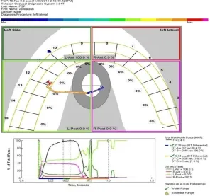
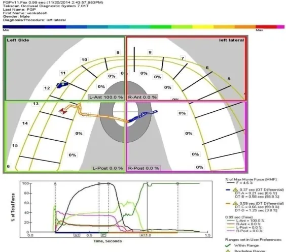
9 LEFT LATERAL - PREEXISTING
10 LEFT LATERAL - CONVENTIONAL
11 LEFT LATERAL - DOUBLE CASTING
12 LEFT LATERAL - PROVISIONAL RESTORATION
13 PROTRUSION - PRE EXISTING
14 PROTRUSION - CONVENTIONAL
15 PROTRUSION - DOUBLE CASTING
LIST OF TABLES
S.NO TABLES PAGE NO
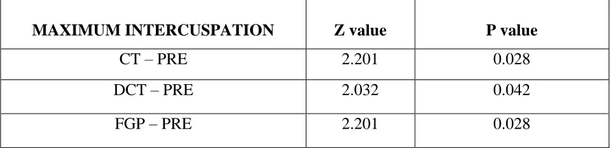
1 ON MAXIMUM INTERCUSPATION WITH PREEXISTING 37
2 ON MAXIMUM INTERCUSPATION BETWEEN
RESTORATIONS
37
3 LATERAL EXCURSION ON RESTORATIVE SIDE WITH
PEREXISTING
38
4 LATERAL EXCURSION ON NORMAL SIDE WITH
PEREXISTING
38
5 ON PROTRUSIVE EXCURSION WITH PEREXISTING 38
6 LATERAL EXCURSION ON RESTORATIVE SIDE
BETWEEN RESTORATIONS
39
7 LATERAL EXCURSION ON NORMAL SIDE BETWEEN
RESTORATIONS
39
8 ON PROTRUSIVE EXCURSION BETWEEN
RESTORATIONS
39
9 MULTIPLE COMPARISON USING FRIEDMAN TEST ON
MAXIMUM INTERCUSPATION
40
10 MULTIPLE COMPARISON USING FRIEDMAN TEST
FOR LATERAL EXCURSION ON RESTORATIVE SIDE
40
11 MULTIPLE COMPARISON USING FRIEDMAN TEST
FOR LATERAL EXCURSION ON RESTORATIVE SIDE
40
12 MULTIPLE COMPARISON USING FRIEDMAN TEST ON
PROTRUSION
LIST OF BARDIAGRAMS
S.NO BARDIAGRAMS
1 COMPARISON OF CLUSION TIME BETWEEN THE
THREE RESTORATIONS WITH THE PRE EXISTING
2 COMPARISON OF CLUSION TIME BETWEEN THE
THREE RESTORATIONS WITH THE PRE EXISTING
3 COMPARISON OF DISCLUSION TIME BETWEEN THE
THREE RESTORATIONS WITH THE PRE EXISTING
4 COMPARISON OF DISCLUSION TIME BETWEEN THE
INTRODUCTION
The best thing a man can achieve is to resemble nature because he cannot replace it.
So it is necessary first to understand the natural stomatognathic system composed of teeth, muscles and TMJ. Nature has build the occlusal surfaces and incisal edges of the teeth to have certain curved pathways which balance and function harmoniously with the movements of the condyle in the glenoid fossa1. The human jaw with all its interferences and limitations makes the best articulator. Even the cusp, fossa or the inclination of the teeth is in harmony with this dictum of nature. So when we attempt to replace a single or many teeth especially in the form of a fixed partial denture, care is taken to replace these teeth closer to the position occupied by them naturally.
During chewing, as teeth are said to be extremely sensitive organs of the body any interferences in the restoration would be transmitted by proprioceptive mechanisms to the muscles thereby creating tension and compromising their function.
So this study to analyze methods to fabricate the restorations in harmony both during static and dynamic positions. This was attempted by using two different methods to incorporate functionally generated path by double casting technique2 and provisional restoration technique3,4,5. By these methods the stumbling blocks encountered during fabrication of conventional restorations may be overcome. This makes the interdependent trio of occlusal harmony, muscular harmony and joint harmony to be successful.
This procedure sometimes becomes cumbersome and laborious in a routine clinical practice. So an alternative method to reproduce a most precise occlusion, which was developed way back in 1930‟s has become increasingly popular in the present days, is the FGP2
.
The original technique described by Meyer for obtaining the “functional occlusal path” for complete dentures1 and fixed partial dentures6 fabricated by a direct or indirect technique. Later the frontiers of this technique was expanded to be used in complete occlusal rehabilitation by Mann and Pankey.7,8,9,10. Recently FGP is becoming increasingly popular for the fabrication of implant retained FPD‟s11
.
This technique may be completed in two steps, either during the actual fabrication of restoration or as a three dimensional check bite technique to correct the completed restorations12.
For overcoming some of these shortcomings of conventional casting technique, functionally generated path technique was implemented by two methods. First method is the double casting technique where the patient functional movements were recorded over the metal copings with retention beads attached to the occlusal surfaces. This technique of using pattern resin over the stable bases (metal copings) results in less dimensional inaccuracies during casting2.
silicone pastes do not accurately reproduce the accurate occlusal contacts. However the reliability of these techniques is highly susceptible to inaccuracies due to the thickness, strength and elasticity of these materials in the oral environment20. The accuracy of these techniques is highly dependent on the clinician‟s discretion and it is not accurate13,14,15
. Dental fraternity widely accepts articulating paper and shim stock as a standard for occlusal analysis. In 1987 the world health organization emphasized the importance of reliability in clinical measurement regarding oral health care.
An ideal occlusal indicator should exclude positional errors influenced by tooth displacement and extended mandibular movements21. Recently reports have come supporting the fact that T- Scan produces clinically better and reliable results when compared with conventional method of occlusal analysis. The digital occlusal analyzer system known as T-Scan was first introduced by the Chairman of Prosthodontics of Boston University, Professor William L. Maness in partnership with M.I.T.5423. Lorreta et al gave the confidence level of using T-Scan for craniomandibular practice.
AIM AND OBJECTIVES
AIM
Evaluation of occlusal discrepancy of cast metal fixed partial restoration by using three
different fabrication techniques.
OBJECTIVES OF THE STUDY
1. To find out the occlusal discrepancy in fixed partial restorations fabricated using functionally generated double casting technique with the help of clusion and disclusion time. 2. To find out the occlusal discrepancy in fixed partial restorations fabricated using functionally generated provisional restoration technique with the help of clusion and disclusion time.
3. To find the occlusal discrepancy in fixed partial restorations fabricated using conventional technique with the help of clusion and disclusion time.
REVIEW OF LITERATURE
Meyer FS (1959)1 Discussed the principles and procedures involved in the functional generated path technique for complete dentures. According to the author the two main hurdles in complete dentures are (1) absence of functional and balanced occlusion in centric relation as well as in all the excursions of the mandible. (2) Processing errors. The later can be minimized to a great extent, where as the former must be taken care of for fabricating a successful prosthesis.
The author discussed various most difficult and fundamental principles based on (1) functional occlusal path,
(2) centric occlusion,
(3) cusps and sulci analysis,
(4) development of cuspal paths and occlusal surfaces.
The author concluded that occlusal paths and cuspal paths generated in the mouth create records, which are in complete harmony with the condylar path and the neuromuscular system.
Meyer FS (1959)6 Made a discussion on the use of the generated path technique in fixed partial denture fabrication. The author divided the procedure into various steps.
Mann AW, Pankey LD (1960)7 The authors recommended the use of P-M instrument for restoring lower posterior teeth. The authors listed the purposes of the P-M instrument as follows; a) To evaluate the entire oral rehabilitation before a single tooth preparation is made. b) To decide the occlusal plane on the lower cast. c) To study and plan the preparations of lower and upper teeth. d) To orient the relationship of both the arches in centric position at the same time providing maximum esthetics and conservation of tooth structure. e) The guide‟s planes are to be prepared and buccal contours are to be waxed on the teeth of the mounted study casts, exactly as they will be in the finished restorations. f) To establish and carve the occlusal plane and curvature in the wax patterns and g) To check the finished restorations.
The authors concluded that the reconstruction of the maxillary posterior teeth and cuspids should be accomplished after permanently arranging the lower posteriors. The maxillary incisors may be built either before or after these procedures are completed.
Zimmermann EM (1966)24 In this article made a review of the principles of the functionally
generated path technique. The author pointed out certain hazards and suggested modifications of the functionally generated path technique. The author described the functionally generated path record as a “three dimensional static expression of dynamic tooth movement‟‟
According to the author, when the functionally generated path procedures have been 1) The opposing anterior teeth will contact in both centric and eccentric positions.
2) The posterior centric holding studs will clearly show through and be flush with the functional wax.
Edalat MP, Khadjavi K (1973)5 Described a simple, accurate and time saving technique for the fabrication of a fixed partial denture in which both a chew in technique and a one piece casting were assimilated.
The authors suggested the use of acrylic crowns as a base to carry the FGP recording material, to eliminate any dimensional changes occurring due to soldering and double casting.
Azarmehr P, Azarmehr HY, Javdan B (1974)32Described a technique to fabricate functional
occlusion for porcelain fused to gold restoration using the Meyer‟s chew–in technique. The authors used metal copings as a foundation to develop the occlusal form in blue inlay wax. The occlusal morphology was developed initially in the articulator and later improved in the patient‟s mouth. A negative key was fabricated in stone against which the porcelain occlusal surfaces could be fired.
The authors came to a conclusion that this technique allows the dentists to incorporate the functional occlusion in porcelain fused to Gold restorations without the use of any complicated instruments.
Melvin A.Engelman, Curtis L.Engelman, (1983)4 Conducted a study where it is said that FGP harmoniously reproduces the occlusal surfaces with minimal chair side
adjustments and avoids the need for counter models and adjustable articulators for construction of inlays, crowns and short span fixed partial dentures. Here a simple technique is implemented making the use of FGP tray, FGP fast set stone and FGP wax to establish the occlusal surfaces of the restorations.
Michele Cacciali, Massimo Fuzzi, Alessandro Treccani, P.L.Negri, (1984)30 accordingly to
this study, FGP facilitates the registration of kinematic relation of different occlusal positions only when the anterior guidance is coincident with the present occlusal situation in the absence of interferences. In this procedure, tacky wax is opted for as it has sufficient working time and reproduction of the functional pathways is better in comparison with other materials. Simple articulators like twin stage occluder, verticulator and Denar‟s correlator are preferable in this technique for having a good control of centric occlusion.
Lacy AM, Fukui H, Jendresen MD (1983)44 Conducted a study to investigate the effects of
Balshi TJ (1986)36 Proposed a method to resolve the esthetic complications that occur with osseointegration using a double casting technique. The author states that this technique maintains the esthetic integrity of the facial surfaces of the prosthetic teeth and easy access for future maintenance.
William L.Maness, Micbael Benjamin (1987)23According to them, T-Scan is a computerised
device used to diagnostically quantify occlusal contacts in three new ways: balance plot, time display and comparison screen. It provides the dentist with greater ability to visualise, diagnose and treat complex occlusal problems. The display compares the occlusal contact patterns prior to and after treatment and assessing the similarity and reproducibility of closure patterns and at the same time recording it.
Eeckman J, De Boever JA (1988)31 In their study investigated the accuracy of three different
types of waxes used for functional interocclusal registrations under conditions comparable to the clinical situation. The authors used three waxes specifically recommended for functionally generated path (FGP): Ash bite registration wax, Tacky Synthetic Wax and HifiJelenko functional bite material. The authors finally came to a conclusion that, complete reproducibility of centric and eccentric contact areas could not be found in all these situations. They further emphasized the need for a plastic material, which could sustain the mouth temperature and register the static centric and eccentric registrations more reliably.
Dawson PE (1989)12 Described the functionally generated path for recording of precise border
pathways. The pre-requisites for the use of this technique were described and the steps for bilateral recording of the functionally generated path for both the maxillary and mandibular teeth were described in detail. Use of FGP for quadrant dentistry, for a single tooth and for cross bite was described. The author also illustrated the various difficulties
M.Reza Moini, Peter A. Neff (1991)20 According to him T-scan is an accurate device as it reproduces actual occlusal contacts, timing and force of each contact and records these datas for research and analytical purposes.
It overcomes the shortcomings of conventional methods like effect of saliva, inability to store the data , non standardisation in thickness, strength, marking substance etc., in this study T-scan reproduces the same contacts recorded by the silk ribbon in 100% of the trials.
Hansen CA, Clear K, Wright P (1994)33 Described a procedure to reproduce the occlusal
morphology of complete denture and removable partial denture teeth or those of an implant supported prosthesis in gold.
The authors used resin teeth in the prostheses after reducing the occlusal surfaces by approximately 1.5 mm. A wide strip of Almore bite registration wax was utilized to create occlusal morphologies in the patient‟s mouth. The authors suggested this technique as a simple and accurate method, which could be used for routine dental practice.
Kerstein (1994)47 In his study found that combined right and left disclusion time were comparatively greater in cases of MPDS, Open occlusion, Orthodontic treatment. Pre-treatment disclusion time analysis aids the clinician to find out whether the elevated levels of contractile muscle activity in masseter and temporalis muscles was created by the existing occlusal scheme.
Hammad IA, Nourallah H (1996)25 Described a procedure to enable the clinician to record occlusal and border anatomy at the correct vertical dimension. The authors suggested the use of a Vaccu-Press machine to make a plastic coping of a clear acetate sheet, which was 1.5 to 2 mm short of the margins. Duralay acrylic resin was used to contour the axial portion of the pattern keeping it 1 mm short of the margin, and the occlusal surfaces were made out of contact with the opposing cast or the opposing teeth in the mouth. Functional wax was used to create a functionally generated path in this technique. The authors concluded that this technique was a simple and accurate method to develop a functional occlusion.
A.Garcia Cartagena, O. Gonzalez Sequeros ( 1997)52 Made a study to analyse the occlusal
contact registration with the T-Scan using two methods like time and force analysis modes. It is found that the number of contacts differ both between patients and between four mandibular positions like maximum inter cuspation, edge to edge protrusion , right and left laterality. Time mode registers most contacts, force records least variability, but in either case dispersion of data is small and finally T- scan is found to be reliable.
V.C Garrido Garcia, A. Garcia Cartagena,O. Gonzalez Sequeros ( 1997)51 Made a study
to evaluate the occlusal contacts in maximum intercuspation using T-Scan. The variation within subjects is very less when compared to between subjects and this is used as a new identification method like that of genetic markers, sweep microscopy. It is identified based on the number and distribution of contacts, even with the data as little as seven teeth.
the opposing arch will not be able to provide the occlusal pathways needed for shaping of the occlusal surfaces.
By using tacky wax functional tracing was made immediately following occlusal reduction and the obtained functionally generated path was used to develop the functional core, which was mounted onto the twin stage occluder. The occlusal morphology was first developed against the anatomic cast and later verified against the functional core obtained . The authors concluded that the FGP technique is a simple procedure and produce excellent results.
Minagi S, Tanaka T, Sato T, Matusuaga T (1998)2 Presented an innovative technique for
fabrication of fixed prosthesis that requires precise occlusion. Their investigation revealed experimental data supporting the fact that the double casting technique was a viable approach in accurately reproducing a difficult occlusal topography for a cast restoration.
A master abutment was made of silver gold palladium alloy with a 4-mm height and 6°
of taper. 2 wax patterns were fabricated on the master abutment, one for the conventional casting and the other for the double casting technique. The latter had an occlusal clearance on to which retentive beads were attached. The wax patterns were cast, using Ag-Au-Pd alloy and the castings of the base crown were fitted to the master abutment.
Autocuring resin was added to the occlusal surfaces of the base crown (made for double casting technique) to create the experimental occlusal surfaces. After analyzing the results, the following conclusions were drawn;
1. The clinical error for a double casting method was significantly to a lesser degree than the conventional casting method.
Karne M, Patyk A, Kobes LWR (1998) 42 The purpose of this study was to determine the surface structure of 16 residue-free burning resins and to know which resins could replace the waxes used in the double casting technique.
The authors came to a conclusion that the surface structure of the residue free resins (Palvit G, Pattern Resin, Visio Form, Novolen Hostalen, Lupolen) were within an acceptable range for the dental casting technique. An increased application of these residue free resins in dental casting technique is therefore being recommended. These resins could not only complement waxes or wax / resin compositions, but could even, replace them.
Curtis SR (1999)26 The author made a review and emphasized the drawbacks of using the
previous techniques and use of a soft functional wax for functionally generated path for fabrication of ceramometal restorations. The prosthesis was casted and later prepared for porcelain addition. A full contour wax-up was made with inlay wax and patient was instructed to perform the various mandibular excursions. A stone index /matrix of the occlusal path obtained in wax and porcelain was veneered on to the framework using the stone matrix to guide its placement.
The author concluded that the advantages of this technique, over the conventional method are that; 1) A functional path tray is not required, 2) Inlay wax which is harder and more resistant to distortion is utilised, 3) Laboratory procedures are made simple with the help of the stone matrix.
Davies SJ, Gray RMJ, Smith PW (2001)34Developed guidelines for a good occlusal practice
patient‟s occlusion. The authors stated that the confirmative approach is the safest way of ensuring harmless post restoration occlusion and that finally the post treatment occlusion should be a product of examination, design, execution and checking (EDEC).
The authors recommended various other techniques for occlusal analysis, which included the photographic method and three-dimensional bite registration methods.
Rosenstiel SF, Land MF, Fujimoto J. (2001)43 These authors described the various
procedures involved in the tissue management and impression making for fixed partial dentures. The authors also discussed many factors regarding the fabrication and material science of wax patterns.
In-Sung Yeo and Jae-HoYang (2001)29 Made a study where it is found that incorporation of
group function occlusion in fabrication of fixed partial dentures is not easy when compared to mutually protected occlusion as it is difficult to achieve it by gnathologic instruments. Functionally generated path concept solves this problem easily where occlusal restoration of the prosthesis is customised to the patient‟s own occlusal patterns.
Hajime Shirai, Jun-ichiSejima, Yuka Mantani, (2002)38 Conducted a study were patients with highly keen oral sensory complaints are restored with fixed partial dentures using double casting method. It not only provides functionally generated occlusal path but also precise outline form adapting to the surrounding soft tissues. The advantages are, it avoids the technical errors in casting, error in the distortion of opposing cast, occlusal registration, mounting and errors caused by proximal contact with adjacent tooth as the fitting is checked prior to molding of occlusal surfaces.
Sutton AJ, Sheets DW Jr, Ford DE (2003)11 This article described a functionally generated path technique to obtain optimal articulation between an implant-retained fixed partial denture and the patient‟s natural dentition. Single tooth provisional crown copings were attached to the implant replicas and pattern resin was placed all around the copings. The pattern resin copings were lubricated, and a functional impression wax was added onto the occlusal surface of the recording table and the functionally generated occlusal path was generated by guiding the patient to perform the MIP and eccentric movements. A dental stone core was poured into the wax recordings obtained, which was subsequently used for fabrication of final restorations.
Luk HWK, PowEHN, McMillan AS, Hui CF (2004)37 Presented a simple, two stage casting
Peter S. (2004)45 Discussed the epidemiology, etiology and prevention of dental caries. The author evaluated the caries susceptibility of individual teeth. According to those epidemiological surveys the upper and lower first molars are 95% susceptible to caries, hence the most common missing teeth in the whole dentition.
Kerstein, John Radke (2006)48 Clinical observations of 62 patient‟s precision measurements
obtained with the simultaneous recording of excursive function and muscle activity levels demonstrated that reduction in prolonged disclusion time from an average of 1.4 seconds per excursion to less than 0.41 seconds per excursion, created a therapeutic effect. Within one month time following treatment, increases in the treated subject's maximal clenching capacity in the masseter and temporalis muscles were noted. This treatment effect appears to be the result of decreased ischemia in these same muscles resulted from decreased compression time of the posterior teeth into their periodontal ligament fibers during excursive function. This increase in maximal clenching capacity provides additional evidence supporting the previously described explanations for the reported MPDS symptom reductions resulting from disclusion time reduction therapy.
Jorge A. Learreta, Jorge Beas, Andrea E. Bono, Andreas Durst (2007)55 The purpose of
Pokorny PH, Wiens JP, Litvak H. (2008)28 Gnathological concepts offer a systematic methodology for prosthodontic treatment in the presence of a disorganized or dysfunctional occlusion requiring fixed prosthodontics. Gnathology will be judged as a significant stimulus to relate the physiology of occlusion to biomedical concepts in complex restorative treatment. The lack of an evidence-based model does not diminish the goal of precision and excellence in the clinical management of fixed prosthodontics. Ultimately, the clinician must evaluate and assimilate the available literature and research evidence along with individual clinical experiences and accepted parameters of care. Occlusal factors have different effects in different individuals. So the guideline is developed based on consensus, clinical research outcome studies.
E Prashanti, Suresh Sajjan, Jagan Mohan Reddy (2009)35 Accordingly, double casting technique is basically an error compensation step as it eliminates the inherent dimensional errors of indirect method. The possible errors are only related to investing, casting and polishing procedures and it avoids the tedious job of metal trimming where the occlusal morphology may be lost in an attempt to correct the interferences. Thus the occlusal morphology becomes closer to normal anatomy of teeth definitely leading to improved patient satisfaction and confidence.
Bernd Koos, Arnim Godt, Christine Schille, GernotGoz (2010)49 Made a study to evaluate
The T-scan systems measures the distribution of forces per tooth, both the halves of jaw and the center of force each time, thus the premature contacts and interferences in dynamic occlusion are identified easily.
R. B. Helms, T. R. Katona & G. J. Eckert (2011)57 The aim of the study is to find out whether the products like Accu film I&II, articulating silk (thick and thin), T-scan alter the occlusion during the detection of occlusal contacts. It is found that the flexibility of the testing device influences the measured load and thin, plastically deformable detection products are preferred because the stiffer products create a negative neuromuscular response. The effects of these products are multifactorial like mechanical properties, thickness and surface friction. So more research is needed to overcome the disadvantages and help in the better performance. Sarah Qadeer, Robert Kerstein, Ryan Jin Yung Kim, Jung-Bo Huh, Sang-Wan Shin (2012)18 Made a study to determine the relationship between the size of the articulating paper marks and the percentage of force applied to the same tooth. There was a low positive correlation of 38.3% even with the largest paper mark. It is evident that tooth morphology is the important factor deciding the actual paper mark surface area. Sometimes a large mark can have a low force and a large mark can have a higher force associated with it. So this is not an accurate indicator and employment of non subjective quantifying occlusal indicator becomes essential.
handed tendency of anterior temporalis muscle may be corrected by left handed asymmetry of the masticatory muscles. To conclude the symmetry of EMG activity in asymtomatic young adults has no correlation with symmetry of occlusal contacts.
Nicholas B. DuVall & Paul M. Rogers (2013)27 Conducted a study where FGP technique was used in the fabrication of mandibular posterior restorations in a patient with Bilateral Group Function Occlusion in order to eliminate the interferences. Here a stone crib is used to capture FGP recording at the same time indexing it to the contralateral and ipsilateral mandibular dentition. This procedure provides stability to the stone core and reduces the error during mounting.
Satheesh B. Haralur (2013)46 This study that the functional dynamic occlusal contacts were evaluated by conventional method and T Scan analysis for subjects with TMD and normal joints. Within the limitations of the study, it can be concluded that the balancing side interferences and centric slide was more than 2 mm found to have a strong association with TMD. The study indicates that the susceptibility to temporomandibular disorders were more prevalent in group function occlusal scheme. The T scan III results concluded that both occlusion time and disclusion time in the patients with TMD disorders were significantly extended than the normal subjects.
assess the patient‟s occlusal strength and acclimatize the patient to intercuspate well for future recordings.
So the gold standard for diagnosing occlusal interferences and prematurities involves a combination of patient self report opinion and occlusal examination.
MATERIALS AND METHODS
The subjects for this study were selected from the OPD, Department of Prosthodontics and Crown and Bridge, Tamil nadu government dental college and hospital, Chennai - 600003. This present study was performed to evaluate the occlusal discrepancies found in the fixed partial restorations both in pre-insertion and post-insertion stages. The functionally generated pathway technique is selected to fabricate the restorations and they were compared with restorations made by conventional casting methods.
ARMAMENTARIUM
INSTRUMENTS FOR EXAMINATION
1. Mouth mirror 2. Explorer
3. Periodontal probe 4. Kidney tray 5. Gloves 6. Mask 7. IOPA 8. OPG
FOR MAKING DIAGNOSTIC MODEL
1. Rubber Bowl and Alginate Spatula 2. Alginate
4. Stainless steel perforated dentulous rim lock trays 5. Dental stone
6. Dental plaster
FOR TOOTH PREPARATION
1. Local Anaesthesia (lignocaine) 2. Diamond burs
3. Retraction cord
4. Putty elastomeric impression material 5. Light body elastomeric impression material 6. Tray adhesives
7. Spacer (cellophane Sheet) 8. Metal Stock trays
9. Bite registration wax
10. Autopolymerising resin (tooth coloured)
LAB PROCEDURE
1. Die stone 2. Die pin
3. Die cutting saw
4. Semi adjustable articulator 5. Die spacer
6. Inlay wax 7. Debubblizer 8. Casting ring
13. Sand blasting machine 14. Metal trimmers
15. Micro motor
16. Pattern resin (GC Corp) 17. Cold mold seal
18. T-Scan armamentarium
SELECTION CRITERIA: INCLUSION CRITERIA:
1. Patients requiring 3 unit posterior maxillary or mandibular fixed partial dentures for replacement of missing 1st molar (Unilateral or Bilateral).
2. Intact dentition opposing the edentulous space (any restoration if required was completed before the study was undertaken).
3. The incisal guidance should be acceptable, if not it should be corrected either by occlusal equilibration or by restorative procedures.
4. Elimination of posterior interferences and finally achieving good occlusal harmony 5. Presence of good periodontal health.
6. Both males and females selected were of age between 25 and 45 years.
EXCLUSION CRITERIA :
1. Loss of anterior guidance, which cannot be corrected without extensive restorative procedures
2. Multiple tooth missing (long span) 3. Poor periodontal health
4. Missing opposing teeth.
6. Pregnant mothers
7. Mentally challenged patients 8. Rotated, supra erupted teeth
9. Recently extracted /unhealed edentulous space 10. Uncorrectable occlusal discrepancies
11. Teeth in cross bite
12. Patients having severely attrited teeth.
13. Patients lacking proper neuromuscular control 14. Patients having deep bite
15. Patients with history of orthodontic treatment
16. Patients having disharmony in occlusion and TMJ dysfunction
SAMPLE SIZE
STUDY DESIGN
GROUP C (FGP by provisional
restoration) GROUP B (FGP by
double casting)
GROUP A (conventional technique)
6 samples 6 samples
SL NO MATERIAL NAME MANUFACTURER’S NAME
1 Alginate (Vignette, Dentsply, India )
2 Polyvinyl Siloxane Putty
Impression Material
(Aquasil, Dentsply, India )
3 Polyvinyl Siloxane Light Body
Impression Material
(Aquasil, Dentsply, India )
4 Intermediate Restorative Material (IRM, Dentsply, India )
5 Die Stone (Kalrock®,KhalabhaiKarson Pvt
Ltd, Mumbai, India).
6 Inlay Wax (Uni wax, Normal,India)
7 Pattern Resin (GC Corporation, Tokyo, Japan)
9 Retraction Cord Ultrapack ( #00) Displacement Cord Ultradent Products, USA)
10 Tooth Preparation Kit (Shofu IAC Kyoto, Japan)
11 Airotor Hand Piece (NSK, Nakanishi Inc, Japan)
12 Casting Machine (Bego)
13 Micromotor (Marathon, Japan )
14 Autopolymerising Resin (Tooth
Coloured)
(Dpi, India )
15 T-Scan III Computerized Occlusal
Analysis System
(Tekscan Inc., South Boston, MA USA)
PLACE OF STUDY
METHODOLOGY STUDY DESIGN
Six patients, who required 3 unit fixed partial dentures for the replacement of their maxillary or mandibular posterior teeth were selected for the study. In each patient three methods of generating the occlusal morphology and two methods of castings were employed. Grouping was done based on the technique used for generating occlusal anatomy and casting.
Group A -- for evaluating the conventional wax pattern and casting technique,
Group B -- for evaluating the functionally generated occlusal morphology and a double casting technique.
Group C -- for evaluating the functionally generated occlusal morphology with provisional restoration technique and conventional casting
In the initial appointment, a preliminary impression was made using irreversible hydrocolloid impression material (Vignette, Dentsply, India ) and the diagnostic casts were mounted on a semi adjustable articulator (C.S.A 600 Articulator,Corident Co., Ltd) using a face-bow transfer (Cori Facebow, Corident Co., Ltd.). Centric and Protrusive records were made using Aluwax. The patient was trained to close in maximum intercuspation (MIP) and perform various other eccentric movements. [Right lateral (RL) left lateral (LL) and protrusive (P)].
METHODS
TOOTH PREPARATION
Two putty index of the diagnostic cast were made involving the missing tooth to be replaced and the adjacent two teeth on either side. One for verification of abutment tooth reduction and another one for fabrication of provisional restoration. Under local anaesthesia, tooth preparation was done with diamond burs and airotor hand piece after depth orientation grooves were made. Reductions of the occlusal and facial surfaces were done to 1.5mm and 1mm on the lingual and proximal surfaces. Equigingival margins of shoulder with bevel is made in the facial aspect, chamfer margins in proximal and lingual aspects were made. Twisted Retraction cord was ( Ultrapack ( #00) Displacement Cord Ultradent Products, USA) placed and subsequently 2 stage putty and light body impression technique was followed. Two such impressions were taken, one for the fabrication of the provisional restoration and one for the fabrication of wax pattern and castings. Casts were poured with die stone and later die pins were placed, base were made and die preparation was done.
FABRICATION OF RESTORATIONS CONVENTIONAL METHOD
Face bow transfer was done and maxillary cast mounted in the articulator, mandibular cast was articulated in the semi adjustable articulator according to centric interocclusal record using Aluwax. Programming of articulator was done using centric and protrusive interocclusal records.
trimmers and polishing done with the polishing kit and rouge. The finished castings were inserted in the patient's mouth for fit and accuracy.
ESTABLISHMENT OF FUNCTIONALLY GENERATED PATH USING DOUBLE CASTING METHOD
Wax patterns were fabricated in infraocclusion of 0.5 to 1 mm and retention beads were attached on the occlusal surfaces of wax pattern for aiding in the retention of the pattern resin during functional generation of the occlusal morphology.
Wax Pattern was sprued, invested and casting was done. Later it was trimmed and finished. The base crowns were verified for accuracy of fit and proximal contacts on the models and in the mouth. Any adjustments needed to ensure the fit were made.
The pattern along with the base casting was invested and doubles casting done. The double cast specimens were placed on abutment after trimming, finishing and polishing.
ESTABLISHMENT OF FUNCTIONALLY GENERATED PATH USING
PROVISIONAL RESTORATION
In the study model the denture tooth was placed in wax in the edentulous area and putty index was made. The index was used to fabricate the provisional restoration on the model after the preparation of teeth with tooth coloured autopolymerising resin. The occlusal surface of the provisional restoration was made short of contacts with the opposing tooth and verified in the patient‟s mouth. After addition of autopolymerising resin in the dough stage onto the occlusal surface of the restoration, the patient was asked to perform movements starting from maximum intercuspation, then right and left lateral, protrusive movements and finally ending in maximum intercuspation until the materials sets. Trimming, polishing was done after verification of occlusal morphology, the provisional restoration was cemented with intermediate restorative material. After 2 weeks, a second provisional restoration was made ready and the previous one was removed from the patient‟s mouth and second provisional was cemented.
The removed provisional restoration was examined and cement was removed and cleaned with ultrasonic cleaner. It was subsequently sprued, invested and cast with non precious Ni-Cr alloy. The casting was divested, trimmed and polished and placed in patient‟s mouth.
The occlusal harmonies of the restorations fabricated by the three different methods were evaluated by T-Scan using clusion and disclusion time. The readings were recorded and subjected to statistical analysis.
Statistical analyses used are
Wilcoxon Signed Ranks Test
USE OF T-SCAN
For properly applying this technique we must first ask the patient to relax and then educate them to bite in the most repeatable centric and eccentric positions. As an initial step, the occlusal discrepancy in the existing normal dentition is evaluated after thorough diagnosis and treatment planning and it is recorded with the help of T-Scan. This gives us the picture of premature contacts or interferences which must be removed to establish a harmonious occlusion. Next the patient is asked to bite on the sensor and verified for any remaining occlusal problems. Now it is found that it is almost fully eliminated on both the sides of the natural dentition and clusion and disclusion time were evaluated and it was brought to reach almost the normal values. These readings were saved in the computer which will be used as a future reference for analysing occlusal discrepancies after the restorative phase.
The restorations fabricated using the conventional technique, functionally generated path using double casting technique and the restorations made using provisional restoration technique were placed in the patient‟s mouth and the clusion and disclusion time values were obtained. These values were compared with that of the pre-existing occlusion and that between the three restorations.
The sensor used here helps to obtain reliable measurements of occlusal biting forces on individual tooth by recording the sequence force, and timing of contact quantitatively. The sensor device is licensed as a “medical attachment device– contact sensor system”. The sensor foils used have a layer thickness of 100 μm and are hence within the range of commercially available articulating foils, papers and silk (8–200μm).
2-D Columns: displays the pressures as a two-dimensional, contoured image, with differences in occlusal force represented by colors ranging from red (greatest) to blue (lowest)
2-D COLUMNS
According to Qudeer et al multiple numbers of readings can be taken to get a correct value of bite force of the individual during the preliminary investigations.
CLUSION TIME
It is found that the time elapsed from the 1st contact to the complete occlusal interdigitation should be ideally zero or as minimum as possible, but achieving this clinically is impossible without the use of T-scan which gives us the sequence, time and duration of individual tooth contacts. So we keep the time frame of 0.1- 0.3 sec as standard in our study as it is also easier to achieve clinically.
DISCLUSION TIME
RECORDING OF CLUSION AND DISCLUSION TIME
The patient is asked to relax and then bite in the most repeatable centric and eccentric positions. The time elapsed from the 1st contact to the complete occlusal interdigitation is the clusion time and is shown in the graph as converging force lines show a static, non changing force represented by horizontal lines. The amount of Clusion time starts from the start of the "A" Line ends at "B" Line and is recorded in seconds. Now the patient is asked to perform right lateral, left lateral and protrusive movements and the force lines start diverging as the patient makes an excursive movement. C-D Increment lines can be used to denote the start and end of the Disclusion Time.
INTERPRETATION OF FULL CLOSURE GRAPH
This graph illustrates the converging force changes occurring as the patient closes in the MIP or CR. These converging force lines show a static, non changing force represented by horizontal lines. This is a representation when the mandible remains static and fixed against the maxilla during the time of static interdigitation.
The 2 quadrant force plot describes the force changes that occur during the force movie in the 2 halves of the dental arch
The amount of Clusion time starts from the start of the "A" Line (OT-A) ends at "B" Line (OT-B), in seconds. The A-B Increment/Differential lines can be used to denote the start and end of the clusion Time (OT-A and OT-B). As the operator moves the A-B lines to their chosen locations, the software simultaneously computes the elapsed time and displays it in the graph box to aid in easy interpretation.
INTERPRETATION OF AN EXCURSIVE GRAPH
In this graph also the converging force changes occurring as the patient closes in the MIP or CR are seen but they start diverging as the patient makes an excursive movement. Here the mandibular closure precedes the start of excursion. After recording the Force movie, a Force Vs Time graph gets displayed and the C-D Increment/Differential lines can be used to denote the start and end of the Disclusion Time (DT-C and DT-D).
FORCE Vs TIME GRAPH IN DISCLUSION TIME RECORDING
SIGNIFICANCE OF A-B LINES
The time elapsed from the 1st contact to the complete occlusal interdigitation should be within the range of 0.1 - 0.3 sec which illustrates that both the halves of the dental arch function simultaneously and harmoniously.
SIGNIFICANCE OF C-D LINES
Aid to determine the effectiveness of anterior guidance in posterior disclusion by calculating the posterior disclusion time.
Photo 1: Materials and armamentarium used during diagnostic stage
Photo 3: Materials and armamentarium used during tooth preparation,
impression and temporization
Photo 5: Pre operative intra oral view - frontal
Photo 8:
Occlusal view after tooth prepation
Photo 10: Final impression
Photo 12: Articulation done for fabrication of restoration
Photo 14:
Artificial tooth placed for fabrication of provisional restoration
Photo 16 Photo 17
Wax patterns before casting
Photo 18 Photo 19
Photo 20
Photo 21 Photo 22
Photo 23
Photo 24 Photo 25
Photo 26
Photo 27
Photo 28
Photo 29:
Cetrifugal casting machine used in the study
Photo 30 Photo 31
Photo 32 Photo 33
Photo 34:
Sensor foil
Photo 35: T-Scan
Photo 36: Obtaining readings in T-Scan
FIG.1
.
CENTRIC RELATION - PREEXISTING
FIG.2
.
CENTRIC RELATION - CONVENTIONAL
FIG.3
.
CENTRIC RELATION - DOUBLE CASTING
FIG.4
.
CENTRIC RELATION - PROVISIONAL
RESTORATION
FIG.5
.
RIGHT LATERAL – PREEXISTING
FIG.6
.
RIGHT LATERAL - CONVENTIONAL
FIG.7
.
RIGHT LATERAL - DOUBLE CASTING
FIG.8
.
RIGHT LATERAL - PROVISIONAL
RESTORATION
FIG.9
.
LEFT LATERAL - PREEXISTING
FIG.10.LEFT LATERAL - CONVENTIONAL
[image:72.612.196.492.109.386.2]
FIG.11
.
LEFT LATERAL - DOUBLE CASTING
FIG.12
.
LEFT LATERAL - PROVISIONAL
RESTORATION
[image:73.612.181.463.114.362.2]FIG.13
.
PROTRUSION - PRE EXISTING
FIG.14
.
PROTRUSION - CONVENTIONAL
FIG.15
.
PROTRUSION - DOUBLE CASTING
FIG.16
.
PROTRUSION - PROVISIONAL
RESTORATION
RESULTS
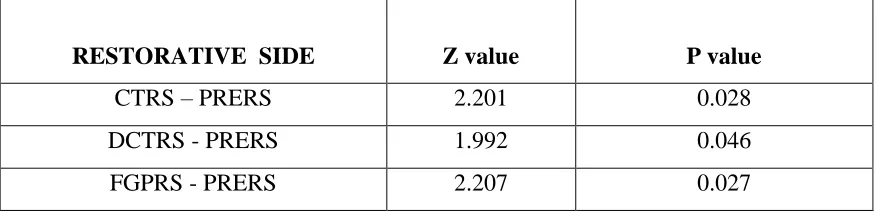
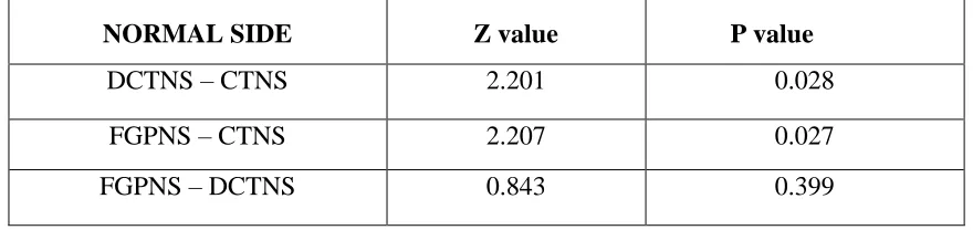
The collected data was analysed with SPSS 16.0 version. To describe about the data descriptive statistics mean and S.D were used. To find the significant difference between the bivariate samples in Paired groups (PRE with CT, DCT & FGP) Wilcoxon signed rank test was used and for repeated measures (CT, DCT & FGP) the Friedman test was used. In both the above statistical tools the probability value 0.05 is considered as significant level. Z value obtained in this study is the absolute value obtained during comparison between the values in Wilcoxon signed rank test. It is found that as Z value increases, P value decreases and the level of significance increases.
[image:76.612.86.530.429.540.2]TEST STATISTICS
TABLE 1 - ON MAXIMUM INTERCUSPATION WITH PREEXISTING
Wilcoxon Signed Ranks Test
TABLE 2 - ON MAXIMUM INTERCUSPATION BETWEEN RESTORATIONS
MAXIMUM INTERCUSPATION Z value P value
CT – PRE 2.201 0.028
DCT – PRE 2.032 0.042
FGP – PRE 2.201 0.028
MAXIMUM INTERCUSPATION Z value P value
DCT – CT 2.201 0.028
FGP – CT 2.207 0.027
[image:76.612.87.534.582.698.2]TABLE 3 - LATERAL EXCURSION ON RESTORATIVE SIDE WITH PREEXISTING
RESTORATIVE SIDE Z value P value
CTRS – PRERS 2.201 0.028
DCTRS - PRERS 1.992 0.046
FGPRS - PRERS 2.207 0.027
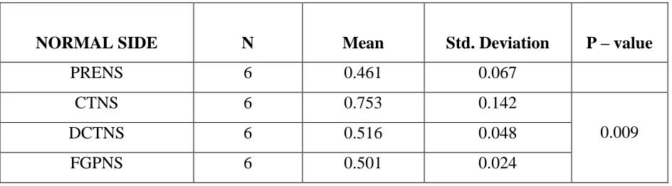
TABLE 4 - LATERAL EXCURSION ON NORMAL SIDE WITH PREEXISTING
NORMAL SIDE Z value P value
CTNS – PRENS 2.207 0.027
DCTNS – PRENS 1.572 0.116
FGPNS – PRENS 1.363 0.173
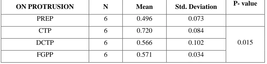
TABLE 5 - ON PROTRUSIVE EXCURSION WITH PREEXISTING
ON PROTRUSION Z value P value
CTP – PREP 2.207 0.027
DCTP – PREP 1.992 0.046
Figure
Related documents