1
DETECTION OF VANCOMYCIN RESISTANT ENTEROCOCCI WITH VAN A GENOTYPE IN CLINICAL ISOLATES FROM A TERTIARY
CARE CENTRE
DISSERTATION SUBMITTED TO
In partial fulfillment of the requirement for the degree of DOCTOR OF MEDICINE IN MICROBIOLOGY
(Branch IV) M. D. (MICROBIOLOGY) of
THE TAMIL NADU DR. M. G. R MEDICAL UNIVERSITY CHENNAI- 600032
DEPARTMENT OF MICROBIOLOGY TIRUNELVELI MEDICAL COLLEGE
TIRUNELVELI- 11
2
BONAFIDE CERTIFICATE
This is to certify that the dissertation entitled “Detection of Vancomycin
resistant Enterococci with Van A genotype in clinical isolates from a tertiary care centre” submitted by Dr. E. Manimala to the Tamilnadu Dr. M.G.R Medical University, Chennai, in partial fulfillment of the requirement for the award of M.D. Degree Branch – IV (Microbiology) is a bonafide research work carried out by her under direct supervision & guidance.
Head of the Department, Department of Microbiology Tirunelveli Medical College, Tirunelveli.
3
CERTIFICATE
This is to certify that the Dissertation “DETECTION OF VANCOMYCIN
RESISTANT ENTEROCOCCI WITH VAN A GENOTYPE IN
CLINICAL ISOLATES FROM A TERTIARY CARE CENTRE” presented
herein by Dr. E.Manimala is an original work done in the Department of Microbiology, Tirunelveli Medical College Hospital, Tirunelveli for the award of Degree of M.D. (Branch IV) Microbiology under my guidance and supervision during the academic period of 2016 -2019.
The DEAN
Tirunelveli Medical College, Tirunelveli - 627011
4
DECLARATION
I solemnly declare that the dissertation titled “DETECTION OF
VANCOMYCIN RESISTANT ENTEROCOCCI WITH VAN A
GENOTYPE IN CLINICAL ISOLATES FROM A TERTIARY CARE CENTRE” is done by me at Department of Microbiology, Tirunelveli Medical College hospital, Tirunelveli. I also declare that this bonafide work or a part of this work was not submitted by me or any others for any award, degree, or diploma to any other University, Board, either in India or abroad.
The dissertation is submitted to The Tamilnadu Dr. M.G.R. Medical University towards the partial fulfilment of requirements for the award of M.D. Degree (Branch IV) in Microbiology.
Place: Tirunelveli Dr. E. ManiMala
Date: Postgraduate Student, M.D Microbiology,
5
ACKNOWLEDGEMENT
My Research project is made possible with the support of many people. I take this opportunity to express my gratitude towards them.
I am grateful to The Dean, Dr. Dr.S.M Kannan M.Ch, Tirunelveli Medical College and, Tirunelveli Medical College hospital Tirunelveli for permitting me to carry out this study.
It is with great previlage and respect that I express my cordial and humble thanks to Dr.C.Revathy,M.D., Professor and Head, Department of Microbiology, Tirunelveli Medical College, whose kindness, guidance and constant encouragement enabled me to complete this study.
I wish to thank Dr. V.Ramesh Babu, M.D., Professor ,Department of Microbiology, Tirunelveli Medical College, for his valuable guidance for the study.
I am deeply indebted to Dr.S.Poongodi@ Lakshmi,M.D., Professor, Department of Microbiology, Tirunelveli Medical College, who helped me offering most helpful suggestions and corrective comments.
I am very grateful to Dr.P .Sorna jeyanthi,M.D., Professor, Department of Microbiology, Tirunelveli Medical College, for the constant support rendered throughout the period of study and encouragement in every stage of this work. I am highly obliged to Senior Assistant Professors Dr.B.Cinthujah,M.D., Dr. G.Velvizhi, M.D., Dr. G.Sucila Thangam, M.D, Dr. V.P.Amudha M.D, Dr.
I.M Regitha M.D., Dr.S.Gowri, M.D ,Dr.Kanagapriya, M.D.,
6
Special thanks are due to my co-postgraduate colleagues Dr.Saishruti , Dr. L.Gracia Paul , Dr.Mayakumar and Dr.R.Uma Maheswari for never
hesitating to lend a helping hand throughout the study.
I would also wish to thank my seniors Dr. D.Jeya Ganguli ,Dr. S. Punitha ranjitham, Dr.R.P.R.Suyambu Meenakshi, Dr.V.Uma Maheswari and
Dr.Ambuja Sekhar and my juniors Dr.Roohee Zubaidha, Dr. S.K. Jayaswarya, Dr. V.Aswini , Dr. A. Sangeetha, Dr G. Malathi , Dr. V. Thanalingam, Dr. S. I. Saheed Askar, Dr. M. Srividya, Dr.R. Priyadharshini and Dr. Cini B Fernz for their help, motivation and support. Thanks are due to the, Messer V.Parthasarathy, V.Chandran, S.Pannerselvam, Murugesan , S.Santhi, S.Venkateshwari, S.Arifal Beevi,
S.Abul Kalam, A.Kavitha, ,T.Jeya, K.Sindhu,
K.Mangai,Manivannan,K.Umayavel, Sreelakshmi and other supporting staffs for their servicesrendered.
I thank my parents Mr.G.Esakkiappa and Mrs.E.Rajalakshmi for being my backbone and not only giving me moral support but tolerating my dereliction during the study.
I am indebted to my husband Mr.G.Madasamy, my daughter M.M.Ishaa Gopika, my brother Mr.E.Muthu Nagararajan,Uncle, Aunty,Sister,Brother in law and all the family members for not only their moral support but also for tolerating my dereliction of duty during the period of my study.
7
CERTIFICATE – II
This is certify that this dissertation work title “DETECTION OF
VANCOMYCIN RESISTANT ENTEROCOCCI WITH VAN A
GENOTYPE IN CLINICAL ISOLATES FROM A TERTIARY CARE
CENTRE” of the candidate Dr.E. Manimala with registration Number
201614302 for the award of M.D. Degree in the branch of
MICROBIOLOGY(IV). I personally verified the urkund.com website for the purpose of plagiarism check. I found that the uploaded thesis file contains from introduction to conclusion page and result shows 13 percentage of plagiarism in the dissertation.
9
10
CONTENTS
Sl. S.No Title Page No.
1 INTRODUCTION 1-3
2 REVIEW OF LITERATURE 5-45
3 AIMS & OBJECTIVES 47
4 MATERIALS AND METHODS 49-62
5 RESULTS 64-86
6 DISCUSSION 88-94
7 SUMMARY 96-97
8 CONCLUSION 99-100
9 BIBLIOGRAPHY
10 ANNEXURE
Data Collection Proforma Preparation of Media Colour plates
Master Chart Abbreviations
11
1
The genus Enterococcus consists of Gram-positive, facultatively anaerobic organisms that are spectacle shaped and may appear on smear in short chains, in pairs or as single cells. Enterococci, though commensals in adult feces are essential nosocomial pathogens.
Enterococcal infections may of at least 12 species
including Enterococcus faecalis, E. faecium,E. durans, E. avium, E. casseliflavu s, E. gallinarum, E. hirae, E.malodoratus, E. mundtii, E. pseudoavium, E. raffin osus, and E. solitarius. Among enterococcal species, E.faecalis and E. faecium are the two major human pathogens accounting for 85-89% and 10-15% of all enterococcal infections, respectively.
Prior to the 1990s also, enterococci have been recognized as an important cause of bacterial endocarditis for almost a century. However, recently they are recognized as a cause of nosocomial infection and "superinfection" in patients receiving antimicrobial agents. The most common Enterococci-associated nosocomial infections are of the urinary tract, followed by surgical site infections and bacteremia.
2
aminoglycoside (streptomycin/gentamicin), More ever, emergence of high level aminoglycoside resistance (HLAR), 𝛽 lactam antibiotics and to vancomycin by some strains has led to failure of synergistic effects of combination therapy.
Vancomycin is an effective antimicrobial for treating infection caused by gram positive organisms. Gram positive isolates are often routinely tested for vancomycin susceptibility. In the 1970s, hospital-associated enterococcal infections in the United States were mainly due to E.faecalis. More recently, E.faecium has emerged as therapeutically challenging organism because of its resistanceto vancomycin and pencillin. These VRE isolates also have a high level of resistance to aminoglycosides Resistance to glycopeptides is mediated by alteration of the drug target from alanine to D-alanine-D-lactate. So far, eight genotypes of glycopeptide resistance, which are different in the level and range of resistance and in transfrability of glycopeptides, have been described for enterococci. Five of the van genes are acquired (van A , B , D , E , G) and three (vanC1,C2,C3) are intrinsic. Multiple epidemics have been predominantly reported with vanA type . vanA gene cluster is located within transposon Tn1546 and can be transferred through acquired resistance.
3
spread has caused the occurrence of many hospital Out breaks worldwide. In the United States, vancomycin-resistance Enterococcus faecium accounted for 4 per cent of healthcare-asssociated infections. It is the second most common pathogen causing mortality and morbidity and the 3rd leading cause of hospital acquired blood stream infection.
The prevalence in Asian countries is decreased and probably due to recent emergence of this resistance in this continent and only a handful of studies to document. In India, the prevalence of VRE has been reported as 8%, 5.5% and 23% in New Delhi, Chandigarh, and Mumbai, respectively, all of vanB phenotype.
4
5
DESCRIPTION OF GENUS:
Enterococcus was historically termed as a diverse genus identified as being 'faecal streptococci', associated with the gastrointestinal tract of human (Giraffa 2002). Thiercelin in 1899 first coined the term 'enterocoque' to describe a newly found Gram-positive diplococcus species.
Andrews and Horder in 1906, isolated the same organism from an endocarditis patient and named it 'Streptococcus faecalis' (Murray 1990). Based on antigens identified as being group-specific, enterococci were placed in Streptococcus group D, while pyogenic streptococci belong to groups A, B, C, E, F or G using antisera. Enterococci were thus classified as group D streptococci because of their morphology and Lancefield antigenicity.
The antigenicity of the carbohydrate moiety of the cell wall is designated according to a system devised by Lancefield in the 1930s (Smith, Niven et al. 1938). The established lance field antigen of Streptococcus is a virulence determinant. For example, in group A streptococci it plays a significant role in resistance to platelet-derived antimicrobials in serum, neutrophil killing and the cathelicidin antimicrobial peptide LL-37 (van Sorge, Cole et al. 2014).
6
pyogenic, viridans, lactic streptococci and enterococci. In 1984 research carried out using nucleic acid hybridization revealed the latter
group showed only meagre association to streptococci (Sherman, Mauer et al.1937).
Subsequently based on molecular techniques, DNA hybridisation,DNA: rRNA hybridisation and 16S rRNA sequencing revealed that S.faecalis and S. faecium were only distantly related to other streptococci.
7
The genus of Enterococcus is composed of more than forty species (The National Center for Biotechnology Information, NCBI), classified on the basis of pigment production, motility and ability to generate acids from a range of carbohydrates (Fischetti, Novick et al. 2006).
Based on the chemotaxonomic and phylogenetic studies, the establishment of 16S rRNA sequences led to the description of seven clonal complexes within the genus namely (i) E. faecalis, E. haemoperoxidus and E. moraviensis; (ii) E.faecium, E. durans, E. hirae, E. mundtii, E. pocinus, and E. villorum; (iii) E.avium, E. pseudoavium, E. malodoratus, and E. raffinosus; (iv) E.casseliflavus, E. gallinarum and E. flavescens; (v) E. cecorum and E.columbae; (vi) E. dispar and E. asini; (vii) E. saccharolyticus and
Domain: Bacteria
Division: Firmicutes
Class: Bacilli
Order: Lactobacillales
Family: Enterococcaceae
8
E.sulfureus. Other species are E. gilvus, E. pallens and E. ratti (Klein 2003) While there are multiple species in the genus Enterococcus, two are associated with the majority of human infections, E. faecalis and E. Faecium (Magi, Capretti et al. 2003).
Characteristics of Enterococci:
The enterococci are gram-positive cocci typically arranged in pairs and short chains, non-motile and non-capsulate. The cocci are facultative anaerobes and grow optimally at 35°C, although most isolates can grow in the temperature range l0ºC to 45°C. They grow readily on blood agar , with large, white colonies appearing after 24 hours of incubation; the colonies are typically non-hemolytic but can be ∝-non-hemolytic or 𝛽 -non-hemolytic. It grows readily on ordinary nutrient media and on MacConkey agar, on which it forms small (0.5-1 mm), usually magenta-colored colonies.
Distinctive Features of Enterococci:
9
medium they produce deep pink colonies. Enterococci are PYRase test positive. They do not hydrolyze hippurate.
Enterococcal Species:
The genus was established in 1984 with the characterization of Enterococcus faecalis and Enterococcus faecium; however, a further 32 species have now been added to the genus on the basis of chemotaxonomic and phylogenetic studies. These additions were based on evidence provided by 16S rRNA sequencing studies.
E. faecalis Schleifer and Kilpper-Balz 1984
E. faecium Schleifer and Kilpper-Balz 1984
E. avium Collins et al. 1984
E. casseliflavus Collins et al. 1984
E. durans Collins et al. 1984
E. gallinarum Collins et al. 1984
E. malodoratus Collins et al. 1984
10
E. mundtii Collins et al. 1986
E. raffinosus Collins et al. 1989
E. solitariusa Collins et al. 1989
E. pseudoavium Collins et al. 1989
E. cecorum Devriese et al. 1983; Williams et al.1989
E. columbae Devriese et al. 1990
E. saccharolyticus Farrow et al. 1984; Rodrı´gues
andCollins1990
E. dispar Collins et al. 1991
E. sulfureus Martinez-Murcia and Collins 1991
E. seriolicidaa Kusuda et al. 1991
E. flavescensb Pompei et al. 1992
E. asini De Vaux et al. 1998
E. villorumc Vancanneyt et al. 2001
E. haemoperoxidus Svec et al. 2001
11
E. ratti Teixeira et al. 2001
E. porcinusc Teixeira et al. 2001
E. gilvus Tyrrell et al. 2002
E. pallens Tyrrell et al. 2002
E. phoeniculicola Law-Brown and Meyers 2003
E. canis De Graef et al. 2003
GENOME:
12
HABITAT:
As outlined in Habitat, the enterococci are primarily members of the gastrointestinal microflora of humans, occurring in numbers as high as 108 colony forming units (CFU) per gram of feces of adult individuals (Noble 1978; Huycke et al. 1998). Enterococcal populations in the intestinal tract fluctuate in size according to the age and physiological condition of the human host, being more numerous during early life (Tannock and Cook 2002). Diet also seems to affect the numbers of enterococci in fecal samples. Although E.faecalis appears to be the enterococcal species most commonly detected in human feces, in all likelihood most Enterococcus species are normal inhabitants of the gastrointestinal tract of humans. Since the enterococci are opportunistic pathogens, the incidence of each species found in human infections probably reflects the distribution of the different Enterococcus species in the human gastrointestinal tract.
This site is believed to represent an important reservoir for strains associated with disease; from this location they may migrate to cause infections and can also disseminate to other hosts and environmental surfaces. E. faecalis is usually the most frequent enterococcal species isolated from human clinical specimens, representing 80–90 percent of the isolates, followed by E. faecium
13
infections with E. casseliflavus and E. raffinosus have been reported. Therefore, the distribution of species varies with each clinical setting. Although less frequently or even rarely, several of the other enterococcal species, including E. avium, E. cecorum, E. dispar, E. durans, E. gallinarum, E. gilvus, E. hirae, E. mundtii, E. pallens, and E. faecalis variant strains, have also been
isolated from human sources. E. columbae, E. haemoperoxidus, E. malodoratus,
E. moraviensis, E. porcinus, E. , E. ratti, E. saccharolyticus, and E.sulfureus
have not been isolated from human sources.
The comparison of data from different publications and the evaluation of the real incidence of the different species of enterococci as members of the intestinal microflora or as members of the microflora in other body sites have been impaired due to differences in the methodology used and the changes in the taxonomy of the genus.
Enterococcus as a commensal:
14
immunocompromised patients are more susceptible to infection even with commensal strains
Enterococcal Infections :
The enterococci inhabit the gastrointestinal tract and the genitourinary tract in humans and other animals. Enterococci are frequent causes of nosocomial infections and may cause urinary tract infection, bacteremia, infective endocarditis, biliary tract infection, intra-abdominal abscess complicating diverticulitis, peritonitis and wound infection.
PATHOGENESIS OF ENTEROCOCCAL INFECTIONS:
15
Whereas enterococci are thought to account for no more than 1% of the adult intestinal microflora, the medical importance of enterococci outweighs their relative abundance . Of the 26 species that have been proposed to belong to the genus, only 11 (E. avium, E. casseliflavus, E. durans, E. dispar, E. faecalis, E. faecium, E. gallinarum, E. hirae, E. mundtii, E. pseudoavium, and E. raffinosus) have been described as associated with human disease . E. faecalis accounts for 80-90% of enterococcal isolates of clinical origin, with E. faecium the second most prevalent enterococcal species. Despite the lower frequency of isolation from clinical settings, E. faecium isolates are disparately resistant to treatment with antimicrobial chemotherapy.
Although normally commensal in nature, enterococci are responsible for approximately 10% of urinary tract infections and 16% of nosocomial urinary tract infections . They are also commonly isolated from wound infections of the abdominal area as well as those from crushing injuries . Enterococcal bacteremia is the third leading cause of nosocomial bacteremia with an estimated fatality rate of 28 to 58% . Enterococci are also responsible for between 5 and 20% of cases of bacterial endocarditis . Enterococci have been described as one of the most destructive agents that cause postoperative complications of cataract surgery .
16
the hospital environment. This is a significant observation given the ability of enterococci to colonize surfaces of the hospital environment and persist on fingertips and dry surfaces. As a result, enterococci seeding the clinical environment may be more easily spread if infection control measures are poorly implemented.
CLINICAL SYNDROMES:
Urinary tract infections:
Enterococci have become the second most common agent recovered from nosocomial urinary tract infection (UTI). UTIs are the most common of the enterococcal infections: enterococci have been implicated in approximately 10% of all UTIs and in 16% of nosocomial UTIs. Enterococcal bacteriuria usually occurs in patients with underlying structural abnormalities and/or in those have undergone urologic manipulations.
Bacteremia and Endocarditis:
17
diseases and conditions (e.g., prematurity, diabetes, malignancy, congestive heart failure, renalinsufficiency, deep-seated infections, prior gastrointestinal, genitourinary, or respiratory tractinstrumentation, long-term hospitalization, indwelling devices, and the use of broad-spectrum antibioticshaving little or no anti-enterococcal activity (e.g., cephalosporins). Bacteremias caused by E. Faecium are associated with a poorer prognosis than those due to E. faecalis primarily because of increasedantimicrobial resistance among the former species and the inherent difficulties of adequately treating moreresistant isolates.
Enterococci, particularly E. faecalis, are also a common cause of prosthetic valve endocarditis. Endocarditis usually occurs in older male patients with underlying valvular disease orwith prosthetic valves and is generally subacute in clinical presentation, with patients having fevers, weight loss, malaise, and other vague constitutional symptoms. Endocarditis in these patients often follows procedures involving the gastrointestinal (e.g., transrectal prostatic biopsies, colonoscopy, fiberoptic sigmoidoscopy) or genitourinary (e.g., cystoscopy, prostatectomy) tracts. Complications of this infection include embolic phenomena frequently involving the CNS. In up to half of patients, enterococcal endocarditis results in acute heart failure that requires valve replacement
18
valves affected commonly. E.faecalis is isolated more frequently than E.faecium and other species. Malignant and inflammatory conditions and procedures involving genitor urinary or gartro intestinal tracts is the source of origin.
Typical presentation involves a sub acute course with fever, malaise, weight loss, cardiac murmur and less than frequent peripheral signs. Heart failure is the common complication followed by embolic penomina, the most important end organ being brain. Mortality is mainly due to heart failure or embolization and the overall mortality rate ranges from 11% to 35%.
Meningitis:
19
high white blood cell counts, elevatedprotein, and low or normal glucose. Postsurgical enterococcal meningitis may or may not be associated with enterococcal infections at other sites. In a review of enterococcal meningitis, E. faecalis accounted for 76% of isolates, and 15 of the 25 cases that were due to E. faecium were caused by vancomycin-resistant strains.
Intraabdominal,pelvic and soft tissue infections:
Intra-abdominal and pelvic infections are the next most commonly encountered infections. However,cultures from patients with peritonitis, intra-abdominal or pelvic abscesses, biliary tract infections, surgical-site infections, and endomyometritis are frequently polymicrobial, and the role of enterococci in this setting remains controversial. Enterococci are being recovered from wound infections at an increasing rate, which likely results from increased antibiotic usage and emerging resistance among these organisms.
20
Enterococci ranks Third amongst the causative agents of hospital acquired surgical-site infections. E.faecalis is the common species isolated Enterococci often colonize the decubitus ulcer and diabetic foot and can be a source of bone infections.
Other infections:
Enterococcal infections of the respiratory tract or the central nervous system, as well as otitis,
sinusitis, septic arthritis, and endophthalmitis may occur but are rare. Enterococci are often found in wound andsoft tissue infections (e.g., burns, decubitus ulcers) with other facultative and anaerobic bacteria, andcomplications associated with such infections (e.g., enterococcal osteomyelitis) are rare
LABORATORY DIAGNOSIS:
Collection, transport and storage of specimens:
21
acid, phenulethyl alcohol, chromogenic subrates, or cephalexin-aztreonam-arabinose agar should be used for isolation of enterococci.
Direct examination:
The direct microscopic examination of gram stained smears of normally sterile clinical specimens like blood is useful in diagnosing Enterococcal infections. However, only a presumptive report of “presence of Gram positive cocci” can be given in case of nonsterile specimens. Direct detection of Enterococci especially VRE from clinical specimens and surveillance specimens (feces, rectal swab) by using conventional and real-time PCR based methods have been developed and evaluated.
A multiplex real-time PCR assay (Light cycler septifasttest) for rapid detection and identification of major pathogens of nosocomial bacteremia in whole blood is available for use in the US.
Isolation procedures:
Clinical specimens from normally sterile body sites, can be plated onto tryptic soy agar, brain heart infusion agar or blood agar base containing either 5% sheep, horse or rabbit blood for primary isolation of Enterococci. Samples for blood culture are inoculated into conventional
22
azide, bile salts, antibiotics and esculin , tetrazolium can be used for primary isolation. However not all Enterococci grow on selective media. Use of enrichment broth ( Enterococcosel broth- BEA medium with 6µg vancomycin) increases the recovery rate of Enterococci especially VRE from feces and rectal samples especially surveillance specimens. Various chromogenic media from different manufacturers also have been evaluated for the primary isolation.
Identification of Enterococcus species:
24
Various commercial identification systems – manual, semi automated and
automated systems like API 20S, API Rapid ID32STREP, Crystal gram positive ID system, Gram positive identification card of Vitek system etc..are available. These are reliable for the detection of most common species E.faecalis and to a lesser extent E.faecium.
Molecular methods
The introduction of various molecular techniques has substantially improved the ability to discriminate enterococcal isolates and has provided critical insights into
epidemiological aspects of enterococcal infections. As a result of the use of more discriminatory typing methods, it has been possible to demonstrate that strains can be exogenously acquired by direct and indirect contact among patients. The first molecular techniques developed for typing of enterococci were the analysis of plasmid profiles and the restriction enzyme analysis (REA) of genomic DNA by conventional electrophoresis.
25
Multi locus enzyme electrophoresis (MLEE) , Ribotyping and the polymerase chain reaction (PCR)-based typing methods, such as the random amplified polymorphic DNA (RAPD)-PCR assay and the repetitive element sequence (REP)-PCR have also been used to investigate the genetic relationship among enterococcal strains. Sequencing of PCR products and restriction fragment length polymorphism (RFLP) analysis of PCR products have been used to trace and to determine differences among specific resistance genes in enterococci, therefore representing additional tools for typing resistant strains. Analysis of SmaI restriction digests of genomic DNA by PFGE is widely useful for studying enterococcal species.
Antimicrobial Resistance:
Resistance to several commonly used antimicrobial agents is a remarkable characteristic of most of the enterococcal species. Moreover, the majority of the information available is based on studies with E. Faecalis and E. faecium, the two species that are more frequently associated with human infections. Antimicrobial resistance can be classified as either intrinsic or acquired.
26
from either mutation in existing DNA or acquisition of new genetic determinants found in plasmids or transposons.
Intrinsic resistance of enterococci:
Enterococcal intrinsic resistance involves two major groups of antimicrobial therapeutic drugs: the aminoglycosides and the b-lactams. Because of the poor activity of several antimicrobial agents against enterococci due to intrinsic resistance, the recommended therapy for serious infections (i.e. endocarditis, meningitis, and other systemic infections, especially in immunocompromised patients) includes a combination of a cell-wall-active agent, such as a B-lactam (usually penicillin) or vancomycin, combined with an aminoglycoside (usually gentamicin or streptomycin).
These combinations overcome the intrinsic resistance exhibited by the enterococci and a synergistic bactericidal effect is generally achieved since the intracellular penetration of the aminoglycoside is facilitated by the cell-wall-active agent.
27
Acquired resistance of enterococci to ß –Lactams and aminoglycosides
antibiotics:
Aminoglycoside resistance:
Over the past decades, the occurrence of acquired antimicrobial resistance among enterococci, especially high-level resistance (HLR) to aminoglycosides, 𝛽 -lactams, and resistance to glycopeptides (especially vancomycin), has been
increasingly reported. These resistance traits are of particular clinical relevance as they confer resistance to agents used in the treatment of serious enterococcal infections and can abolish the activity of the therapeutic regimens with proven bactericidal activity against enterococci. Isolates that are resistant to the cell-wall-active agent or have HLR to aminoglycosides are resistant to the synergistic effects of combination therapy and constitute an even more serious problem concerning the effective management of enterococcal infections. Therefore, the detection of resistance to these groups of antimicrobial agents is important to predict the likelihood of synergy by using antimicrobial association as a therapeutic strategy.
28
concentrations (MIC) >2000 lg/ml and cannot be detected by diffusion tests with conventional disks.
Special tests using high-content gentamicin and streptomycin disks (Sahm and Torres 1988), as well as a single dilution method, were developed to screen for this type of resistance (Swenson et al. 1995). Strains exhibiting HLR to penicillin and ampicillin due to altered penicillin-binding proteins have also disseminated widely in the past several years (Murray 1990, 1998; Boyce et al. 1992; Huycke et al. 1998; Strausbaugh and Gilmore 2000), and strains producing β-lactamase have been identified (Murray 1990; Gordonet al. 1992).
ß -Lactam antibiotic:
29
Vancomycin:
Definition:
Vancomycin is a glycopeptide antimicrobial produced by the soil bacteria Streptomyces orientalis. It was developed and introduced in the 1950s. Another glycopeptide authorised for use in humans in Sweden is teicoplanin. Glycopeptides interfere with the cell wall production resulting in a destabilized cell wall and lysis of the bacteria. When the bacterial cell wall is synthesized, polysaccharide-pentapeptide complexes are linked together via a transpeptidation reaction in which the end amino acid of the pentapeptide is removed. Glycopeptides interferewith this process by binding tightly to the D-Alanyl-D-Alanin (D-Ala-DAla) end of the pentapeptide and hiding it from the transpeptidase that is to catalyse the cross-linking in the peptidoglycan synthesis.
30
Mode of action:
Like many antibiotics (including the penicillins), vancomycin acts by interfering with the construction of cell walls in bacteria, blocking the enzymes necessary for bacterial cell wall assembly.
Resistance to vancomycin:
History:
Scientists introduced Vancomycin into hospitals more than forty years ago in response to new strains of Staphylococci developing resistance to penicillin. The introduction of methicillin decreased the use and importance of Vancomycin for a few years; however, when methicillin-resistant S. aureus strains appeared in the past two decades, the glycopeptide antibiotic was reinstated as a therapeutic agent.
Vancomycin is now seen as the last-resort drug because it is often the last opportunity that a physician may have to eliminate infections caused by multi-drug resistant bacteria.
Genes of vancomycin resistance:
31
types with E. faecium carrying the vanA genotype as the most common combination (Fisher & Phillips, 2009; Werner et al., 2008). An additional variant (vanF) has also been described but thus far only in Paenibacillus popilliae (Patel et al., 2000). Since the vanF variant has a high similarity in amino acid sequences to the vanA variant, P. popilliae has been suggested as a possible origin for vancomycin resistance in enterococci (Patel et al., 2000). Other plausible sources are various glycopeptide producing organisms, even if genetic differences make an older common source more likely (Patel, 2003). Common to all variants of Vancomycin resistance in enterococci is the ability to cause a change in the structure of the pentapeptide incorporated in the 3 dimensional web of peptidoglycans composing the bacterial cell wall: from the original Ala to either Lactate (D-Ala-DLac) or D-Ala-D-Serine (D-Ala-D-Ser) (Courvalin, 2006). This shift results in a reduced affinity for Vancomycin by 1000 and seven times respectively (Fisher & Phillips, 2009).
In all different variants of vancomycin resistance are several genes involved in the alteration of the cell wall structure which results in the resistance. The number and organisation of these genes are somewhat similar among the different variants. For the vanA variant, the genes are organized as in Figure4. VanS is a sensor gene which in the presence of a glycopeptide
32
After activation of the gene complex, VanH mediates production of lactate from pyruvate which vanA uses to synthesize the alternative D-Ala-D-Lac end of the pentapetide (French, 1998). It is essential for resistance that production of the normal D-Ala-D-Ala end of the pentapetide does not continue. This is resolved by the vanX and vanY genes where vanX hydrolyzes and thereby interrupts the production of the pentapeptides, and vanY cleaves the pentapeptides that might still be produced (French, 1998; Arthur et al., 1996). In the absence of a glycopeptide, vanS initiates dephosphorylation of vanR resulting in deactivation of the gene (Courvalin, 2006).The function of the vanZ gene is not understood (Courvalin, 2006).
33
34
DIFFERENT TYPES OF VANCOMYCIN RESISTANCE FOUND IN
ENTEROCOCCUS SPP.
Phenotypic description:
VRE strains have been classified by phenotypes and genotypes. Six types of glycopeptide
35
high) levels of inducible resistance to vancomycin only, encoded by the vanB gene; and the VanC phenol type, with non inducible low-level resistance to vancomycin.
The VanA and VanB phenotypes are considered the most clinically relevant and are usually associated with E. faecium and E. faecalis strains while the VanC resistance is a intrinsic characteristic of E. gallinarum (vanC1 genotype) and E. casseliflavus (vanC2 and vanC3 genotypes) strains (Clark et al. 1998; Huycke et al. 1998; Murray 1998; Cetinkaya et al. 2000).
The remaining three types of enterococcal glycopeptides resistance seem to occur rarely and are encoded by genetic determinants that were recently recognized, named vanD (Perichon et al. et al. 2000).
The details of vancomycin resistance have been best documented with the Van A gene cluster found on the transposon, Tn1546 (Arther and Courvalin, 1993; Arther et al., 1993). Van B isolates were initially believed to be inducibly resistant to more modest levels of vancomycin but are susceptible to teicoplanin. VanB resistance determinants also reside on large mobile elements that can be transferred from 1 strain of enterococcus to another (Quintiliani et al., 1993, 1994).
36
Certain limitations of this classification method have become evident over time. For example, the genetic determinants of the VanA Phenotype have now appeared in E. gallinarum and other enterococcal species (Dutka Malen et al., 1994 ).
Nevertheless, this phenotypic classification is still useful, because it usually corresponds well to the genetic classification and utilizes information that can be derived simply and inexpensively in laboratory (Elipoulos, 1997).
Genotypic classification of VRE:
VanA glycopeptide resistance:
37
vancomycinandteicoplanin-inducible, transposon-mediated, high-level resistance to both vancomycin (MIC, 64 to1,000 µg/mL) and teicoplanin (MIC, 16 to 512 µg/mL).1147
VanB glycopeptide resistance:
The vanB cluster genes is often located on the host chromosome and initially was thought not transferable to other bacteria. However, it can also occur on plasmids, and, even when it is chromosomal, this gene cluster has been transferable as part of large mobile elements, perhaps related to large conjugative transposons (Quintiliani et al., 1994). Strains with the vanB genotype (VanBphenotype) have acquired vancomycin-inducible resistance to various concentrations of vancomycin(MIC, 4 to 1,000 µg/mL) but remain susceptible to teicoplanin (MIC, 0.5 to 1 µg/mL), although rare vanB strains may also be resistant to the latter antibiotic.
VanC glycopeptide:
The genes encoding the VanC type of vancomyin resistance are endogenous, species-specific components of E. gallinarum (vanC-1)and E. casseliflavus/E. flavescens (vanC-2/vanC-3 (Navarro and Courvalin , 1994).
VanE glycopeptide resistance:
38
16µg/ml ( Butz et al. 1990). Isolates that have the vanC genotype display intrinsic,constitutive, low-level resistance to vancomycin (MIC, 2 to 32µg/mL) and are susceptible to teicoplamin(MIC, 0.5 to 1 µg/mL). The vanC genotype corresponds to the intrinsic glycopeptide resistance seen in E.gallinarum, E. casseliflavus, and E. flavescens. This vanC gene cluster is not transferred by conjugation to other organisms, is generally constitutively expressed, and is chromosomal in origin.
VanD glycopeptide resistance:
39
Organisation of VanA-type glyc opeptide resistance operon. The arrows show regulatory and resistance and the accessory coding sequences reproduced from Courvalin 2006.
Epidemiology and control of VRE:
Geographic distribution and spread within hospitals:
40
Hospital outbreaks of infection or colonization have been reported with both VanA and VanB isolates (Boyce et al., 1995). Patients may be colonized simultaneously with more than one strain of VRE (Mato et al., 1996; Wade, 1995). Stool isolates of VRE have included a
number of different species such as E. faecalis, E. faecium, E.gallinarum, and E. avium (Bates et al., 1995). Fortunately, rates of stool colonization with VRE among hospitalized patients by far exceeds infection rates with these organisms (Lam et al., 1995;Montecalvo et al.,1995). Gastrointestinal tract colonizatin with VRE may persist for weeks or monthes, and single negative cultures may be intermixed with positive culters over time (Montecalvo et al., 1995). During outbreaks,enviromental cultures in hospital rooms have yielded VRE (Boyce et al., 1995; Mato et al., 1996 ; Slaughter et al., 1996).
VRE in the community:
In the USA, attention has focused on the epidemiology of VRE mainly in hospitals, and there is little evidence to suggest that transmission of VRE to healthy adults occurs to any significant extent in the community (Murray, 1995). In a study in Texas, investigators failed
41
The situation in Europe is quite different from that in the United States. In Europe, VRE have been isolated from sewage and various animal soureces (Bates, et al., 1995; Klare, et al., 1995). It has been suggested that the use of glycopeptide-containing animal feeds in some reigons of Europe may have contributed to such differences . In one study, VanA resistant E. faecium was isolated from frozen poultry and pork and from the feces of 12 of 100 non hospitalized inhabitants in a rural area ( Klare et al., 1995) VanA VRE have also be found in the feces or intestines of other farm animals (Devriese et al., 1996). These observations suggest a potential for VRE or the resistance genes of VRE to reach humans through the food chain or through contact with domesticated animals (Gordst et al., 1995) .
Reservoirs:
42
to chickens, swine, and cattle. In the US, avopracin has not been used as a feed additive for animals, and culture surveys of a limited
number of chickens in several cities have failed to detect VRE (Harrison et al., 1995). Further studies of animal-based food products are needed to determine if food items represent a community reservoir for VRE in that country ( Boyce, 1997).
Mode of transmission:
Transmission of VRE by hospital workers whose hands become contaminated with the organism while caring for patients is probably the most common mode of hospital acquired infection transmission (Tornieporth et al., 1996; Zervos et al., 1987). Transmission of VRE may also occur by way of contaminated medical,surgical equipment, although this is probably much less important than transmission by the hands of personnel. Electronic thermometers contaminated with the outbreak strain were epidemiologically implicated in an outbreak described by Livornese et al., (1992).
43
Prevention and control:
The epidemiology of VRE has not been completely elucidated; certain patient populations are at high risk for VRE infection or colonization . These include critically ill patients or those with severe underlying disease or immunosuppression, such as intensive care unit (ICU) patients or patients in oncology or transplantation. Those who have had an intra-abdominal or cardiothoracic surgical procedure, and those who have had prolonged hospital stay or received multiple antimicrobial agents (Boyce et al., 1994; Boyle et al., 1993; Centers for Disease Control and prevention. 1993; Friden et al., 1993; Handwerger et al.,
1993; Karanfil et al., 1992 and Montecalvo et al., 1994). Because enterococci are part of the normal flora of the gastrointestinal tract and the female genital tract, most infections with those organisms have been attributed to the patient’s endogenous flora (Murray, 1990). However, recent reports have demonstrated that enterococci, including VRE, can be spread by direct patient-to patient contact or indirectly via transient carriage on the hands of personnel (Boyce et al., 1994), Contaminated environmental surfaces (Boyce et al., 1994; Karanfil et al., 1992), or patient care equipment (Livornese et al., 1992).
44
vancomycin, education of hospital staff, effective use of the microbiology laboratory, implementation of infection control measures (including the use of gloves and gowns) and isolation of patients, as appropriate to specific conditions ( Boyce, 1997; Centers for Disease Control and prevention. 1995).
Education programs:
Continuing educational programs for hospital staff (including students, pharmacy personnel, nurses, laboratory personnel) should include information about the epidemiology of VRE (Centers for Disease Control and prevention, 1995). Because detection and containment of VRE require high performance standards for hospital personnel, special awareness and educational sessions may be indicated (Boyce, 1997).
Role of the microbiology laboratory in the detection of VRE:
Early detection of patients colonized or infected with VRE is an essential component of any hospital program designed to prevent nosocomial transmission of VRE (Boyce, 1997 ). Once the prevalence of VRE reaches high levels within an institution, prevention of transmission is more difficult. The microbiology laboratory is the 1st line of defense against the spread of VRE in
45
when recognition of the problem is delayed ( Centers for Disease Control and Prevention,1995).
In addition, cooperation and communication between the laboratory and the infection control program would facilitate control efforts (Friden, et al., 1993; Karanfil, et al., 1992).
46
47
1. To isolate and speciate enterococci from cases of nosocomial infection.
2. To determine the antibiotic susceptibility pattern of the isolates and the pattern of vancomycin resistance.
48
49
This study was conducted during the period from April 2017 to May 2018 at the Department of Microbiology, Tirunelveli Medical College , Tirunelveli.
INCLUSION CRITERIA:
During the study a total of about 100 non duplicate clinical isolates of Enterococci were collected from different clinical samples like clinical specimens such as urine, blood,
pus, tissue fluids obtained from both in-patient and out-patient departments of Tirunelveli Medical College. The Enterococcal species were identified by standard biochemical techniques.
EXCLUSION CRITERIA:
Patients who were already on treatment with vancomycin .
A detailed history regarding previous hospital admission within two years, antibiotic intake in previous six months was elicited from every patient.
Ethical clearance:
50
Informed consent:
Informed consent was obtained from all persons involved in this study.
Proforma:
A filled in proforma was obtained from the patients with details like name, age, sex, ward, clinical diagnosis, risk factors, surgical intervention, hospital stay, previous use of Vancomycin or any other antibiotics and other parameters relevant to the study.
Sample storage:
The Enterococcal isolates were sub-cultured on to nutrient agar slope and stored at 2 to 80C. The Enterococcal isolates were sub-cultured every fortnight.
Primary isolation and identification of Enterococci :
Samples:
51
Microscopy:
A primary smear is made from the sample and stained with Gram stain, gram positive cocci which appear mainly in pairs slightly ovoid in shape and may appear in short chains, or as single cells were suspected of being enterococcus.
Biochemical reactions:
Catalase test:
It was done by slide test or tube test.
Slide test-
A single colony taken from nutrient agar plate was placed over the clean glass slide, to this one drop of 3% H2O2(hydrogen peroxide) was added, effervescence was not observed. When effervescence appeared it was a negative test.
Tube test-
52
Bile esculin agar:
The suspected Enterococcal isolates were then inoculated onto Bile esculin agar ( containing 40% bile) , incubated aerobically at 370C overnight. The next
day the isolates showing black discoloration of the medium due to hydrolyse of esculin to esculetin and dextrose which reacts with ferric citrate were identified as BEA positive.
Aesculin agar plates were inoculated with the suspected isolates and incubated at 370C for 48 hr. aerobically. Isolates that produced blackening of the medium were tentatively identified as
enterococci.
Growth in 6.5% NaCl:
Two or three colonies of the suspected enterococci were inoculated into nutrient broth containing 6.5% NaCl. The inoculated broth was incubated at 370C for 3
days aerobically. Growth was indicated by development of turbidity.
Heat tolerance test:
The suspected Enterococcal isolates along with the control strains were tested for heat tolerance by inoculating them into BHI broth and incubating them along at 600C for 30 minutes in a water bath. Subcultures from the broth were
53
The growth of the positive control was checked before reading other isolates. The ATCC control strain growth both before and after heating the broth at 600C
for 30 minutes was noted. The isolates showing growth before and after 30min of incubation at 600C were taken as heat tolerant Enterococcal isolates .
Salt tolerance:
Salt tolerant property of the suspected Enterococcal isolates were tested by inoculating 2 to 3 identical colonies of suspected isolates along with control strains into a tube containing nutrient broth with 6.5% sodium chloride and incubated at 370C for 24-72 hours. 1% bromo cresol purple was added as an
indicator to detect yellow discoloration on growth. The broth showing turbidity with or without yellow discoloration was taken as positive reaction and was confirmed by subculturing the broth on blood agar / Mac Conkey agar. The salt tolerant isolates grew well even in the presence of 6.5% NaCl. Salt tolerant , BEA positive isolates, which were able to grow on MacConkey agar and at temperatures of > 45ºC were identified as Enterococci and selected for further speciation.
54
Further speciation was based on acid production from specific carbohydrates and motility and pigment production.
Arginine dihydrolysis:
Arginine dihydrolysis was tested by inoculating the isolate into a tube of Moeller’ s decarboxylase broth containing arginine and a control tube ( without arginine), overlaid with sterile liquid paraffin and incubated for seven days at 370C . Control strains were also included in the test. Development of deep purple colour due to alkalinisation after an initial change to yellow colour was read as positive reaction.
Persistant yellow color indicates negative reaction.
Identification of Enterococcus species
Identification E.faecalis E.faecium
Gram Stain Cocci in pairs & short chain
Cocci in pairs & short chain
Catalase Negative Negative
Motility Nonmotile Nonmotile
Blood Agar α – or non hemolytic,
small, cream
α – or non hemolytic,
55
colored,smooth colonies with entire edge
colored,smooth colonies with entire edge
MacConkey agar Lactose fermenting, majenta coloured colonies
lactose fermenting, majenta coloured colonies
Bile esculin agar Positive Positive
Heat tolerance survives a temperature of 60ºc for 30 minutes.
survives a temperature of 60ºc for 30 minutes.
Arginine dihydrolysis Positive Negative
ANTIBIOTIC SUSCEPTIBILITY TESTING :
56
ANTIBIOGRAM BY KIRBY-BAUER DISC DIFFUSION
METHOD :
The antibiotic susceptibility pattern of the isolates was determined by on Mueller –Hinton agar (MHA) .The bacterial inoculums was prepared by inoculating few identical colonies in a Nutrient broth and incubated for 3-6hrs. It was standardized with 0.5 Mc Farland turbidity
standard (1.5×10⁸ CFU/ml) before inoculation, if the bacterial suspension is too thick it should be diluted to match the standard and if it is less turbid, it has to incubated further.
After standardization, a sterile swab is dipped in that broth and the excess fluid is squeezed out by pressing on the side of the test tube, and it was streaked on the surface of the agar three times, turning the plate at 60º each time to produce a lawn culture of the organism. Then it is allowed to dry and the antibiotic discs were placed over the lawn culture within 15 minutes of inoculation.
57
All the materials and antibiotic disc were procured from Himedia laboratories Pvt.Ltd. Mumbai. The inoculated plates were incubated aerobically at 37ºC overnight. Next day the zone of inhibition of the bacterial growth around each disc was measured using ruler under reflected light except for Vancomycin and read through transmitted light. The interpretation as susceptible, intermediate and resistant were done according to the CLSI guidelines.(Appendix)
DETECTION OF VANCOMYCIN RESISTANCE BY VANCOMYCIN
SCREEN AGAR :
Presumptive identification of Vancomycin resistance was done by Vancomycin screen agar (i.e) brain heart infusion (BHI) agar containing 6 µg /ml Vancomycin. 10µl of 0.5 McFarland suspension of the isolate ,along with positive and negative control strains, was spot inoculated onto the agar surface and incubated aerobically for 24hrs at 35±2ºC.Growth of > 1 colony indicated presumptive Vancomycin resistance which was confirmed by determining the Minimum inhibitory concentration (MIC) for vancomycin.
58
MIC for vancomycin- resistant Enterococcus by E strip method :
Minimum inhibitory concentration (MIC) value for Vancomycin were determined using Hi Comb MIC Strip (Hi-media, Mumbai). Any Enterococcus was considered VRE if the MIc was ≥16microgram/ml.
E-test was done to determined the minimum inhibitory concentration of Vancomycin for all the clinical isolates of enterococci. The E-test is comprised of two strips: Strip A: 240-0.01 µg and Strip B: 4-0.001 µg (Hi Comb, MIC test, HIMEDIA laboratories).The results were interpreted as per CLSI guidelines.
MIC MINIMUM INHIBITORY CONCENTRATION FOR
TEICOPLANIN:
E-test was done to determined the minimum inhibitory concentration of Teicoplanin for the VRE isolates .The glycopeptide teicoplanin MIC was also tested in the same method as described above for Vancomycin using Hi Comb, MIC test, The E-test is comprised of two strips: Strip A: 240-0.01 µg and Strip B: 4-0.001 µg HIMEDIA laboratories. Any Enterococcus was considered Teicoplanin resistant if the MIC was ≥32 microgram/ml.
MOLECULAR METHOD FOR THE DETECTION OF VRE:
59
E.faecalis by the PCR Kit procured from Helini Biomolecules,Chennai. The DNA was extracted from the Enterococcal
isolates by using Helini Pure Fast Bacterial Genomic DNA Mini Spin Purification Kit and subjected to PCR and the gene product viewed by gel electrophoresis.
Extraction of DNA from the Enterococcal isolates :
1. 1ml of overnight bacterial culture centrifuged at 6000rpm for 5 min. 2. Supernatant discarded
3. Pellet is suspended in 0.2ml PBS.
4. 180µl of Lysozyme digestion buffer and 20µl of Protinase K added, 5. Incubated at 37ºC for 15 min.
6. 400µl of Binding buffer, 5µl of control template and 20µl Protinase K added, Mixed well by inverting several time.
7. Incubate at 56ºC for 15 min.
8. Added 300µl of Ethanol and mixed well.
9. Transferred entire sample into the PureFast spin column. Centrifuged for 1 min. Discard the flow-through and place the column back into the same collection tube.
60
11. Added 500µl wash buffer-2 to the PureFast spin column. Centrifuge for 30-60 seconds and discarded the flow-through. Place the column back in to the same collection tube.
12. Discard the flow-through and centrifuge for an additional 1 min. This step is essential to avoid residual ethanol.
13.Transferred the PureFast spin into a fresh 1.5ml micro-centrifuge tube.
14.Added 100µl of Elution Buffer to the centre of PureFast spin column membrane.
15.Incubate for 1 min at room temperature and centrifuge for 2 min.
16.Discard the column and store the purified DNA at -20ºc. Quality and Quantity of extracted DNA is checked by loading in 1% agrose gel and 5µl of extracted DNA is used for PCR amplification.
PCR Procedure:
1. Reactions set up as follows:
Components Quantity
HELINI RedDye PCR Master mix 10µl
HELINI RedDye PCR -Primer mix 5µl
Purified bacterial DNA 5µl
61 2.Mixed gently and spin down briefly
3. place into PCR machine and program it as follows;
Initial Denaturation: 95ºC for 5 min
Denaturation : 94ºC for 30 sec
Annealing : 58ºC for 30 sec 35 cycles
Extension : 72ºC for 30 sec
Final extension : 72ºC for 5 min
Loading:
1. Prepared 2% agarose gel. [ 2gm of agarose in 100ml of 1X TAE buffer] 2. Run electrophoresis at 50V till the dye reaches three fourth distance and observe the bands in UV Transilluminator.
Agarose gel electrophoresis:
1. Prepared 2% agarose. (2gm agarose in 100ml of 1X TAE buffer and melted using micro oven)
2. When the agarose gel temperature was around 60ºC, added 5µl of Ethidium bromide.
3. Poured warm agarose solution slowly into the gel platform. 4. Kept the gel set undisturbed till the agarose solidifies. 5. Poured 1XTAE buffer into submarine gel tank.
62
7. PCR samples are loaded after mixed with gel loading dye along with 10µl HELINI 100 bp DNA Ladder.
[100bp, 200bp, 300bp, 400bp, 500bp, 600bp, 700bp, 800bp, 900bp, 1000bp and 1500bp]
8. Run electrophoresis at 50V till the dye reaches three fourth distance of the gel.
63
64
5.1. STUDY DESCRIPTION
This study was conducted at the Department of Microbiology, Tirunelveli Medical College, over a period of one year from April 2017 to March 2018. Out of 996 culture positive samples received in the Microbiology laboratory , a total of 100 enterococcal species were isolated.
These enterococcal isolates were subjected to antimicrobial susceptibility testing by disc diffusion method for the following antibiotics: Penicillin, Tetracycline, Linezolid, Ciprofloxacin, Chloramphenicol, Teicoplanin, Doxycycline, High level Gentamycin and Vancomycin. Susceptibility of the isolates to vancomycin was tested by vancomycin screen agar and E strip method. Those isolates found resistant to vancomycin was screened for MIC of Teicoplanin by E strip and Van A gene using PCR.
5.2. STATISTICAL ANALYSIS
Data collected were entered in Microsoft Excel and analysed using statistical analysis software Statistical Package for Social Services (SPSS) v.16. Appropriate descriptive and inferential statistics were calculated. p values <0.05 were statistically significant.
5.3. ISOLATION OF ENTEROCOCCI
65
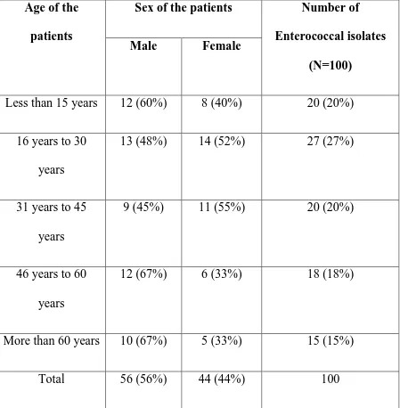
[image:76.595.75.524.221.679.2]years with a SD of 21.5 years. The age of the patients ranged from a minimum of 2 years to a maximum age of 84 years. The table below shows the age and sex distribution of the patients.
Table 1 Age and sex distribution of the patients
Age of the
patients
Sex of the patients Number of
Enterococcal isolates
(N=100)
Male Female
Less than 15 years 12 (60%) 8 (40%) 20 (20%)
16 years to 30 years
13 (48%) 14 (52%) 27 (27%)
31 years to 45 years
9 (45%) 11 (55%) 20 (20%)
46 years to 60 years
12 (67%) 6 (33%) 18 (18%)
More than 60 years 10 (67%) 5 (33%) 15 (15%)
66
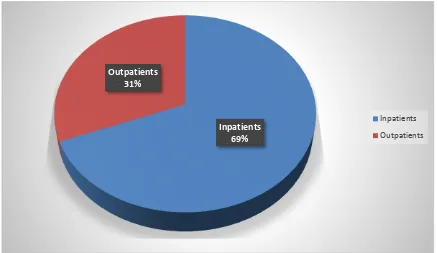
Table 2 Distribution of isolates among Inpatients and Outpatients
Type of patients Percentage
Out patients 31
Inpatients 69
Out of the 100 samples, 69 samples were collected from inpatients and remaining 31 from outpatients.
Figure 1 Distribution of isolates among inpatients and outpatients
Inpatients 69% Outpatients
31%
Inpatients
[image:77.595.106.543.400.653.2]67
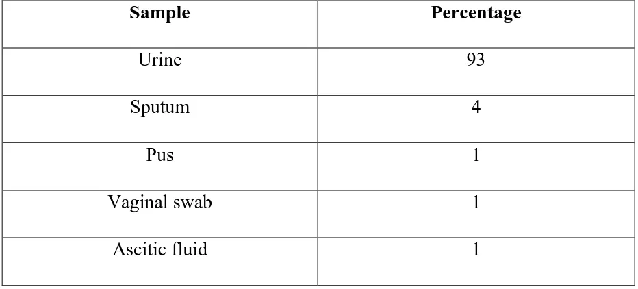
Table 3 Distribution of the Enterococcal isolates in different clinical samples
Sample Percentage
Urine 93
Sputum 4
Pus 1
Vaginal swab 1
Ascitic fluid 1
Enterococci were commonly isolated from urine (93%), followed by sputum (4%), ascitic fluid (1%), pus from leg ulcer (1%) and vaginal swab (1%).
Figure 2 Distribution of the Enterococcal isolates in different clinical samples
1%
4%
93% 1%
1%
[image:78.595.108.497.469.715.2]68
Table 4 Distribution of Enterococcal isolates in different wards
Ward Number of isolates
(N=100) (%)
Medicine 22 (22%) Paediatrics 21 (21%) Urology 17 (17%) Surgery 13 (13%) Maternity 9 (9%)
IMCU 8 (8%)
Nephrology 3 (3%)
CMCHIS 2 (2%)
Gynaecology 2 (2%) Thoracic 2 (2%) Trauma 1 (1%)
Most of the urinary isolates (43%) were from Medicine and Pediatric ward followed by Urology ward (17%), surgery ward (13%), maternity ward (9%) and IMCU (8%).
Figure 3 Distribution of Enterococcal isolates in different wards
[image:79.595.72.505.438.698.2]69
Table 5 Species distribution among isolates
Species isolated Percentage
E.faecalis 90
E.faecium 10
[image:80.595.76.508.367.667.2]The isolates were identified to be Enterococcus faecalis (90%) and Enterococcus faecium (10%).
Figure 4 Species distribution among isolates
E.fecalis 90% E. faecium
10%
70
Table 6 Species distribution in different clinical samples
Sample No.of Isolates E.faecalis E.faecium
Urine 93 85 8
Sputum 4 4 -
Pus 1 1 -
Ascitic fluid 1 - 1
Vaginal swab 1 - 1
Total 100 90 10
The above table shows the species of the Enterococcus in different clinical samples. All the four sputum isolates and pus isolates were found to be E.faecalis whereas the ascitic fluid isolate and vaginal swab isolate was found to be E.faecium.
71
Figure 5 Species distribution in different clinical samples
0 20 40 60 80 100 120 140 160 180 200
Urine Sputum Pus Ascitic fluid Vaginal swab
72
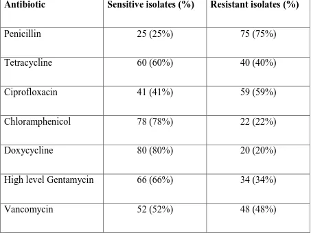
5.4. ANTIBIOTIC SUSCEPTIBILITY PATTERN OF THE ISOLATES
5.4.1. Disc diffusion method
[image:83.595.66.519.418.758.2]The table below shows the antibiotic susceptibility pattern of the enterococcal isolates by Kirby bauer disc diffusion method on Mueller Hinton agar according to CLSI guideline Highest prevalence of resistance was observed against Penicillin (75%), followed by Ciprofloxacin (59%) and Vancomycin (48%). On the other hand, Doxycycline was found to be sensitive for 80% of isolates, followed by Chloramphenicol (78%) and High level Gentamicin (66%).
Table 7 Antimicrobial sensitivity pattern of the Enterococcal isolates by disc diffusion method (N=100)
Antibiotic Sensitive isolates (%) Resistant isolates (%)
Penicillin 25 (25%) 75 (75%)
Tetracycline 60 (60%) 40 (40%)
Ciprofloxacin 41 (41%) 59 (59%)
Chloramphenicol 78 (78%) 22 (22%)
Doxycycline 80 (80%) 20 (20%)
High level Gentamycin 66 (66%) 34 (34%)
73
[image:84.595.71.506.228.481.2]It could be seen from the below figure that only 52% of the isolates were sensitive Vancomycin by disc diffusion method.
Figure 6 Antimicrobial susceptibility pattern of the isolates
0 10 20 30 40 50 60 70 80 90
74
[image:85.595.66.533.173.291.2]5.4.2. Multi drug resistant isolates
Table 8: Multi drug resistant isolates
Total no of isolates MDR isolates
100 66
Out of the 100 enterococcal isolates, 66 isolates were found to be multi drug resistant (resistant to three or more antibiotics).
Figure 7 Proportion of Multi drug resistant isolates
MDR 66%
[image:85.595.72.506.473.727.2]75
Table 9 Distribution of MDR isolates from various clinical samples
Sample No of isolates No. of MDR isolates
Urine 93 59 (63.4%)
Sputum 4 4(100 %)
Vaginal swab 1 1(100 %)
Ascitic fluid 1 1(100 %)
Pus 1 1(100 %)
Total 100 66