Sally Anne McFadden
A thesis submitted for the degree of
Doctor of Philosophy
of the
I
1o
~haxJ
Jlan~
jr
6Ver~115
I To
my
parentsI
I 'i,,
I ' , "' !~Declaration
Acknowledgements
Publications
Abstract
Plate captions
CONTENTS
Table captions
Figure captions
Common abbreviations
CHAPTER ONE
GENERAL INTRODUCTION
1.1 THE NATURE/NURTURE ISSUE
1.2 SECONDARY MONOCULAR CUES TO DEPTH
1.3 MOTION PARALLAX
1.4 OCULOMOTOR CUES
1.5 STEREOPSIS
1.9 THE EVOLUTION OF BINOCULAR VISION
CHAPTER TWO
2.1
INTRODUCTION
2.2 METHODS
2.3 RESULTS
2.4 DISCUSSION
DEMONSTRATION OF BINOCULAR DEPTH
PERCEPTION IN THE PIGEON
PAGE NOe
!1 " ~ I 11 ;
,,
11 II
' I 1, 11CHAPTER THREE
THE ROLE OF THE VISUAL WULST IN
BINOCULAR DEPTH PERCEPTION AND DEPTH
ACUITY IN THE PIGEON
3.1 INTRODUCTION
3.2
A.
PIGEON METHODS
3.2
B.
HUMAN METHODS
3.3 RESULTS
3.4 DISCUSSION
CHAPTER FOUR
THE ROLE OF THE INTERHEMISPHERIC
CONNECTIONS IN THE BINOCULAR
PERCEPTION OF DEPTH
4.1
INTRODUCTION
4.2 METHODS
4.3 RESULTS
4.4 DISCUSSION
CHAPTER FIVE
5.1
SYNOPSIS
SA.
THE EXTENT
BINOCULAR FIELD
5.2 INTRODUCTION
5.3 METHODS
5.4 RESULTS
5.5 DISCUSSION
OF THE RETINAL
OF THE PIGEON
5.6 INTRODUCTION
5.7 METHODS
5.8 RESULTS
5.9 DISCUSSION
DURING DISCRIMINATION PERFORMANCE
CHAPTER SIX
CONCLUSIONS
6.1
GENERAL DISCUSSION
Bibliography
239
242
246
255
263
I •
DECLARATION
This thesis describes original research carried out by
the author during the tenure of an Australian Government
Postgraduate Research Award in the Department of Behavioural
Biology, Research School of Biological Sciences and the
Department of Psychology, School of General Studies. The
ophthalmoscopic experimental measurements in Chapter 5
were carried out jointly with Liz Reymond. The work
described in this thesis has not previously been presented
for a degree at this or any other university and to the best
of my knowledge and belief contains no work previously
written or published by another person unless due reference
is made in the text. The animal experiments were carried out
according to the guidelines set by the Committee on
Animal Experimentation, Proposal Number R. BB. 20. 83.
ACKNOWLEDGEMENTS
I wish to sincerely thank my supervisors, Martin Wild for his
unerring readiness to help with context and content at all times, Bill
Bellingham for his consistent financial and motivational support, and
Richard Mark for his imaginative and clear points of view. I am also
extremely grateful for the delightful hours spent learning with
Professor Bishop in the latter stages of this thesis.
I'm not sure that I should thank Mark Woolston for his 'insight'
in suggesting I do a PhD in the first place, but he taught me all I
know about computer machine coding, without which 'Sally's boxes'
would never have run. Thanks are also due to Greg Fetingis who,
through his ingenuity kept me on the road after the consistent and
inevitable equipment and corresponding emotional breakdowns.
In looking after my pigeons, I would never have survived without
my lab coat pockets and Mandy Devlin, who mothered my birds in
sickness and in health.
I would especially like to thank Barb Piper for help where it was
needed most and in the typing of this thesis.
Thanks also to all the troops at M Block including Liz, Faulksie
and Lidia for their companionship and Morgan, Reiner, Margaret,
Lauren, Maggie, Bridget, Bob and Gert for both technical assistance
and making the Department a home away from home with all its ups and
downs.
Finally, I am deeply appreciative of all my family support,
I .
I
I
. I
,,
I
'
I
I I
1
II;
PUBLICATIONS
Some of the work in this thesis has been presented at the following
scientific meetings.
McFadden, S.
&
Wild, J.M. (1982) Stereopsis in the pigeon?PPoc.
Aust. Exp. PsychoZ. Soc.
P27.
McFadden, S.
&
Wild, J.M. (1982) The pigeon's perception of depthunder binocular and monocular conditions.
PPoc. Aust.
PhysioZ. PhaPmacoZ. Soc.
13:141P.McFadden, S.
&
Wild, J.M. (1982) Stereopsis as a primary cue fordepth perception in the pigeon.
Soc. NeuPosc. Abs.
8:270.11.McFadden, S.A.
&
Wild, J.M. (1984) Role of the posterior commissurein binocular depth perception in the pigeon
(CoZumba Livia).
ABSTRACT
Stereopsis depends on both stimulation of corresponding retinal
points of each eye and a neural site in which integration of the two
monocular visual signals combine. Stereopsis has only been shown to
be present in frontally eyed animals that have a predatory life
style. This thesis presents the first evidence for the existence of a
binocular depth mechanism in a granivorous bird
(Columba livia)
thatdoes not hunt and catch prey and possesses a panoramic field of view
by the lateral placement of the two eyes. The pigeon has a small
region of binocular overlap which was found in this thesis to be
symmetrical about the eye-beak axis.
The behavioural procedure used was a discrete trial simultaneous
operant conditioning paradigm. Pigeons were found to be able to
discriminate depth in 'Frisby' stimuli that were based on a clinical
test of stereopsis used with young children (see Hinchliffe, 1978).
The birds' performance on the depth task was superior under binocular
rather than monocular viewin g conditions. In addition, partial
disconnection of the two half brains severely impaired binocular depth
perception.
The smallest depth difference discriminable at threshold in terms
of retinal disparity, was found to be 1 minute of arc. This depth
acuity is better than the spatial frequency acuity of the pigeon and
surpasses that predicted by the sampling mosaic of the retina.
the depth acuity is likely to reflect stereoacuity.
Thus
In all birds, there is almost complete decussation at the optic
chiasm. \
Thus, binocular convergence must occur at a subsequent stage
in the visual system. It was found that bilateral lesions of the
effects on the behavioural depth discriminations, even when tested at
the psychophysical limits of binocular depth perception. In contrast,
interruption of binocular neural integration by separation of the two
half brains at the level of the tectal and posterior commissural
systems resulted in behavioural deficits on the depth tasks.
Taken together, the results give support for the presence of
stereopsis in a lateral eyed, non-predacious bird. Hence stereopsis
is not an emergent capacity bestowed on a relatively small number of
elite animals, but may be a fundamental attribute of vertebrate
J [I I I I I 1! I j ,
Ii
,1::
I I!I
I'
• I IPlate 1.1
Plate 1.2
Plate 1.3
Plate 1.4
PLATE CAPTIONS
Camouflage I: Short horned grasshopper
Camouflage II: Huntsman Spider
Camouflage III: Map Butterfly
Camouflage IV: Lantern Fly
Plate 2.1 Position of the velcro ring during monocular occlusion
Plate 2.2 Goggle in position during monocular occlusion
Plates 4.lA-F Example photographs of the supraoptic decussation lesion sites
Plates 4.2A-F Example photographs of the tectal and posterior commissure lesion sites
Table 2.1
Table 2.2
Table 2.3
Table 2.4
Table 2.5
Table 2.6
Table 2.7
Table 2.8
Table 3.1
Table 3.2
TABLE CAPTIONS
Assignment of subjects to groups
Parameters of the initial learning curves for the SDT
Parameters of the initial acquisition curves for all subjects in group 2 tabulated according to month
ANOVA table for analysis of the standard depth task
ANOVA table for analysis of the monocular performance on the SDT and Pattern 1 discriminations
Parameters of the initial acquisition of the SDT, Pattern 1 and LDT
ANOVA table for monocular compared with binocular performance on the Less Dense Task
ANOVA table for binocular and monocular performance on the SDT and LDT
Nuclei which receive afferent projections from the optic tectum
Assignment of subjects to groups
Table 3.3
Table 3.4
Table 3.5
Table 3.6
Table 3.7
Table 3.8
Table 4.1 Table 4.2
Table 4.3
Table 4.4
Table 5.1 Table 5.2
Table 5.3
Table 5.4
Table 5.5
Table 5.6
Table 5.7
Table 5.8
Percentage area of missing tissue and severe cell loss for all subjects subjected to Wulst lesion surgery
Percentage area of damage for all subjects that had undergone sham operations
Parameters of the acquisition curves after Wulst lesions
The number of sessions taken to reach criterion on the Pattern 2 and the Standard Depth Task
The mean threshold ( Yt) in terms of retinal disparity for both Wulst lesioned and normal subjects
Range of thresholds for each single criterion subject in the two groups 4A and 4B
Assignment of subjects to conditions
Parameters of the initial acquisition curves for the SDT, Pattern 2 and LACE task
Estimate of deficits in performance after lesions to DSo+CA or to CA alone
Estimates of the deficits in performance after the second surgical session
Parameters of the binocular field
Identification numbers of subjects filmed during various behavioural tasks
Distance and visual angles during head fixations for Pattern 2 subjects
Distance and visual angles during head fixations for SDT subjects
Distance and viewing angles for four depth differences during threshold testing
Distance and viewing angle during head fixations for subjects trained on the LACE task
Distance and viewing angles during head fixations for monocular subjects performing the LACE task
' I I ' i I I, :
.
I.
' 1, '1 In. I IFigure
1.1
Figure
1.2
Figure 1.3 Figure
1.4
Figure
2.1
Figure 2.2 Figure 2.3
Figure
2.4
Figur~
2.5
Figure
2.6
Figure
2.7
Figure
2.8
Figure 2.9
Figure
2.10
Figure
2.11
Figure
2.12
Figure 2.13 Figure
2.14
Figure
2.15
Figure 3.1
Figure 3.2
Figure 3.3
FIGURE CAPTIONS
Demonstration of the influence of past experience on the secondary monocular cue of interposition
The Vieth-Muller horopter circle Physiologic diplopia
Illustration of ambiguity in global stereopsis
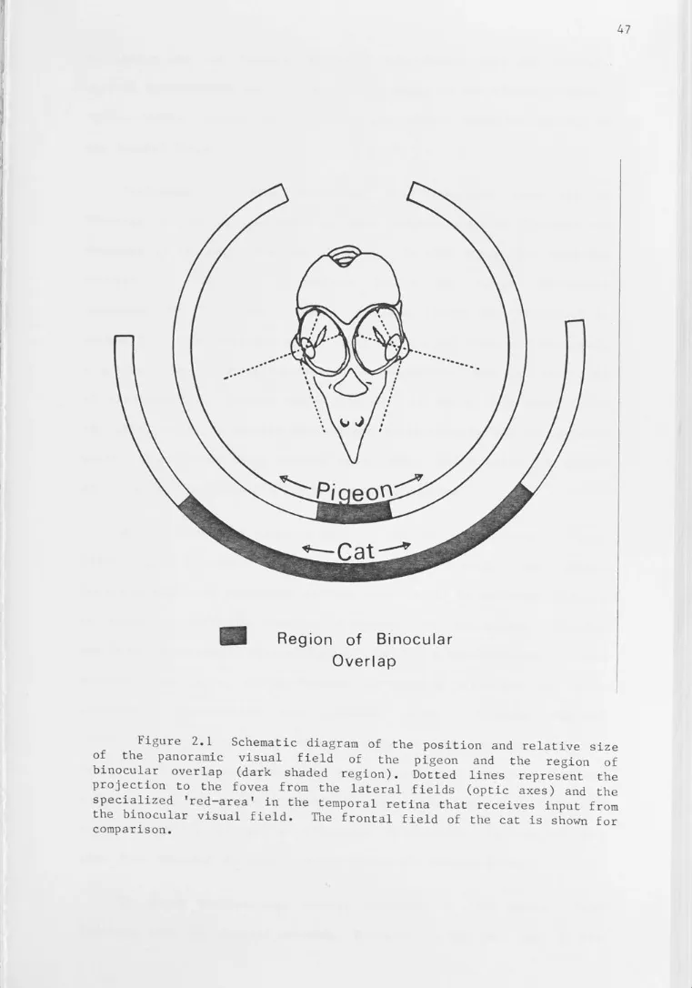
Schematic diagram of the position and relative size of the panoramic visual field of the pigeon and the region of binocular overlap
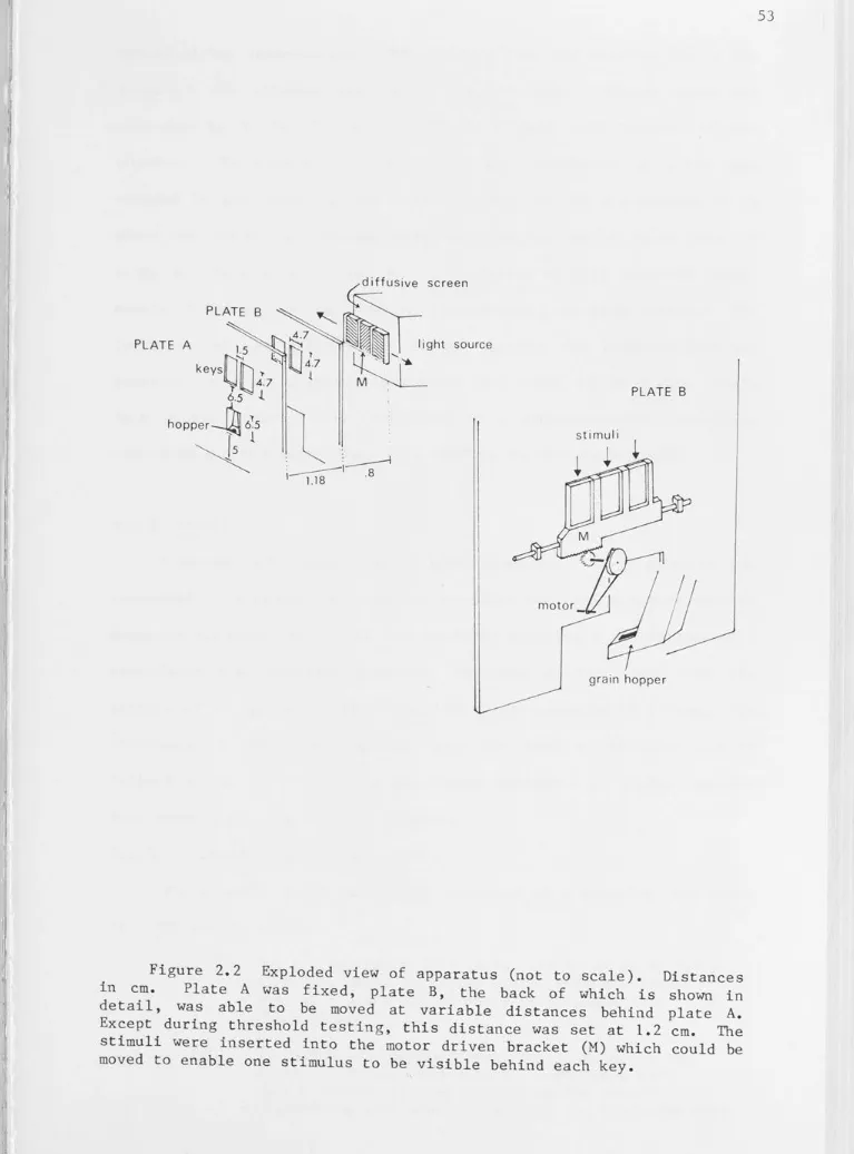
Exploded view of apparatus
Stimulus configurations for the standard depth task, less dense task and Pattern 1 task
Flow chart of the behavioural discrimination Attachment of goggle to velcro ring
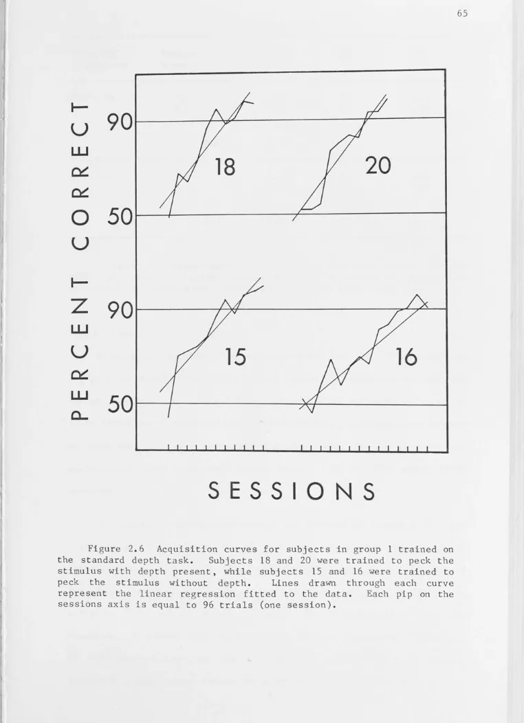
Acquisition curves for subjects in group 1 trained on the standard depth task
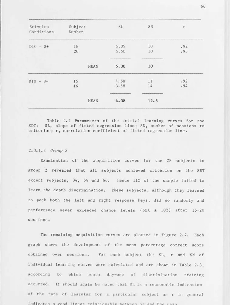
Acquisition curves of the standard depth task for all subjects in group 2
The mean (SN) and the mean slope plotted as a function of the month
Performance both before and during monocular testing on the standard depth task
Performance both before and during monocular testing on the Pattern 1 task
Performance both before and during monocular testing on the less dense task
Scattergram showing the independence of learning the SDT and the observed monocular deficit
Transfer to the relative depth task Illustration of stimulus variables
Demonstration of the binocular cues in the SDT and RDT
Diagrammatic representation of the efferent projections of the optic tectum
'
Diagrammatic representation of a possible pathway involved in binocular visual processing
I I Ii I, I 1, Ii I , ; ,, I. .1 '1 I I. I I I ! 1
Figure 3.5 Figure 3.6
Figure 3.7 Figure 3.8
Figure 3.9
Figure 3.10 Figure 3.11
Figure 3.12
Figure 3.13
Figure 3.14
Figure 4.1 Figure 4.2 Figure 4.3
Figure 4.4
Figure 4.5
Figure 4.6A
Figure 4.6B
Figure 4.7
Figure 4.8
Figure 4.9
Reconstructions of the Wulst lesions
Acquisition curves for birds trained on the SDT after aspiration of the visual Wulst
Performance on the SDT after a bilateral Wulst lesion Performance on the Pattern 2 discrimination after a bilateral Wulst lesion
Depth thresholds for the cases with lesions of the Wulst
Depth thresholds for all normal birds
Psychometric threshold functions for normal and Wulst lesioned birds
The development of binocular and monocular depth thresholds
Pyschometric functions of the threshold depth differences for five human subjects
Performance curves generated by a monocular human subject on a series of depth differences
Stimuli for the LACE task
Midsaggital section showing approach for commissurotomy The positive stimulus used during the monocular cue simulation
Reconstructions of the supraoptic decussation and the anterior commissure after the Ll transection
Reconstructions of the transection of the posterior and tectal commissures after the L3 lesion
Diagrammatic histogram showing the extent of the transection of the DSOd, DSOv and the CA for all experimental subjects in the SDT, SDT Threshold and Pattern 2 groups
Diagrammatic histogram showing the extent of the
transection of the DSO and CA for all subjects in the LACE group
Diagrammatic histogram showing the extent of the transection of the posterior and tectal commissures
The initial acquisition and post-operative performance for the SDT experimental birds
Post-operative performance after commissurotomy or sham lesions
Figure 4.10
Figure 4.11
Figure 4.12
Figure 4.13 Figure 4. 14 Figure 4.15
Figure 5.1 Figure 5. 2 Figure 5.3
Figure 5.4
Figure 5.5
Figure 5.6 Figure 5.8
Figure 5.9 Figure 5.10
Post-operative performance for all birds trained on the LACE task
The development of the depth threshold measured on the SDT after either DSO and CA or CA alone were transected
The difference between the binocular and monocular percentage correct scores obtained on the LACE task Monocular performance after commissurotomy
Pecking errors after commissurotomy
Absolute convergence and retinal disparity as cues in the SDT and LACE discriminations
Alignment of the bird's head to the tangent screen The mean retinal binocular field
Retinal binocular fields plotted as a function of the width of the field and the elevation angle
Comparative equatorial orthographic projections of the mean retinal binocular fields
Comparison of the binocular fields found in various studies
Typical image and viewing angles found in each frame Projection of the binocular field onto the positive stimulus in the Standard Depth Task
Position of head fixations found in various situations Geometrical determination of possible overlap of
~ If
'
II I I ' I1; I l I 11 Ii ,, Ii I I! l'I ,, ,, I AP CA CP criterion CT ~ C
d DSO DSOd DSOv DlO DV EW Fl F2 Frisby HA HD HIS
IHA
IOT stimuliLACE task
LACE 6
Ll
L2
COMMON ABBREVIATIONS
anterio-posterior anterior commissure posterior commissure
three successive sessions in which performance exceeds 90% correct
tectal commissure
change in initial post-operative performance relative to criterion
viewing distance
supraoptic decussation
dorsal supraoptic decussation ventral supraoptic decussation depth 10 stimulus
dorso-ventral
Edinger-Westphal nucleus first head fixation
last head fixation
depth stimuli based on a clinical test for stereopsis using real depth targets
hyperstriatum accessorium hyperstriatum dorsal
hyperstriatum intercalatus suprema
nucleus intercalatus hyperstriati accessorii interocular transfer
task in which depth is present over the entire positive stimulus giving a 'lacy' two-dimensional appearance
LACE 6 stimulus
transection of supraoptic decussation and
.
\
.
anterior commissure
CONTENTS
1.1 THE NATURE/NURTURE ISSUE
1.2 SECONDARY MONOCULAR CUES TO DEPTH
1.2.l Light and Shade
1.2.1 Relative Size
1.2.3 Secondary Cues as Higher Order Stimulus
Variables
1.3 MOTION PARALLAX
1.4 OCULOMOTOR CUES
1.4.1 Accommodation
1.4.2 The Relationship between Accommodation
and Convergence
1.4.3 Accommodation and Depth Perception in
Animals
1.5 STEREOPSIS
1.5.1 The Horopter
1.5.2 Spatial range of stereopsis
1.5.4 Local and Global Stereopsis
1.5.5 Models of Global Stereopsis
1.5.6 Neural Mechanisms
1.5.7 Behavioural Demonstrations of
Stereopsis in Animals
1.5.7.1 Cats
1.5.7.2 Meerkat
1.5.7.3 Primates
1.5.8 Stereopsis in Non-mammals
1.5.8.1
1.5.8.2
Reptiles and Insects
Birds
1.9 THE EVOLUTION OF BINOCULAR VISION
PAGE NO.
This thesis is an investigation into the pigeons' perception of
the relative distance (depth) between objects in space. Location of
objects in space with respect to oneself (egocentric) or other objects
(exocentric) is a necessary and often highly refined function of many
animals. In the bird, the location of objects is necessary for
survival and is implicit in a range of activities from the accurate
location of food with respect to the beak, to the estimation of the
position of obstacles while flying and the detection of predators.
One may argue that detection is not synonymous with the location of an
object. With respect to a predator it may be enough to be aware of
existence and direction alone. Yet, in detecting an object, an animal
often has to contend with breaking camouflage (see plates 1.1
1.4). An effective way to do so is to locate the desired object or
animal with respect to the background - that is - perceive the object
stereoscopically. Such depth perception is clearly an advantage, not
only for detection of predators, but also for the detection of food
sources.
Indeed, one may again use the camouflage example to point out the
universal effectiveness of the few principles of camouflage employed
throughout the animal kingdom (Cott, 1966). Hence, although obvious
differences exist between the optic arrays of various environments,
the underlying spatial metric and optical principles are common to all
species. Thus, in the literature review which follows, I have
described the perception of depth in animals in terms of what is known
about our own abilities to perceive the location of objects in
space. Classically, the perception of depth, intimately related to
egocentric absolute distance estimation, has been considered as the
perceptual integration of a number of cues. These cues may be
elicited on the basis of vision with a single eye, or may require the
Bay, Western Australia (Photograph courtesy of D. Knowles).
Plate 1.2 Camouflage II: Huntsman Spider·
(SpaPPassidae),
Yuin(Nymphalidae),
Kutai Reserve,Knowles). Kalimanton (Photograph courtesy of D.
Plate 1. 4 Camouflage IV: Lantern Fly
(Fulgor>idae),
Eunung Reserve,North Sumatra. Did you notice a similar species to the left of the
Many of the monocular cues for the perception of depth are
considered 'higher order' and are referred to as secondary cues, while
the binocular and oculomotor cues are considered as primary cues to
three dimensional space. This distinction between primary
(accommodation, convergence, motion parallax and stereopsis) and
secondary cues (relative size, interposition, linear and aerial
perspectives, and light and shade) is reflected in the earliest modern
observations of depth perception, which were guided by the question of
whether appreciation of space was learned or innate.
In the following review, some early studies on the nature/nurture
issue are examined. Some examples of secondary monocular cues to
depth are then presented, particularly as they relate to the birds'
visual capabilities. However, most of the review is dedicated to the
primary cues to depth, as these have received by far the most
attention in recent years.
binocular depth perception.
In particular, emphasis is placed on
As little work has been done in this
field on birds, much of the discussion centres on the current state of
knowledge of binocular depth perception in our own visual system and
in other animals.
1.1 THE NATURE/NURTURE ISSUE
In 1873, Spalding commented on the behaviour of young dark-reared
chicks on their first exposure to visual space:
I
A number of investigators, notably Grinnell (1921), Benner (1938) and
Pumphrey (1948) (see Shinkman 's review, 1962), Bird (1926) and Hess
(1950, 1956, 1961) have subsequently examined the question of the
nature/nurture issue. They have used the response of young chicks
pecking at grain as an indicator of depth discrimination.
Shepard and Breed (1913) concluded that practice effects in
pecking were complete after two days and that any subsequent
improvement was due to maturation. Bird (1926) divided the pecking
response into three components and noted the proportion of misses,
hitting but not seizing the grain and seizing but failing to
swallow. He found that increased accuracy, mostly that seen in the
missing error, was due to maturation. Reduction in the number of
missing reactions also occurred in the absence of practice, although
practice aided in reducing the error (Cruze, 1935).
These findings confound the effects of sensory and motor
development, so it is not clear what role maturation plays in the
discrimination of depth. The perception of depth is not a sufficient
condition of the peck response. Furthermore, there was no attempt to
break depth perception into the component cues.
1.2 SECONDARY MONOCULAR CUES TO DEPTH
1.2.1 Light and Shade
The use of light and shade as a cue to depth was examined by
Benner (1938, cited in Walk, 1965). He noticed that chicks would peck
at a pictorial representation of grain with shadows and ignore real
grain illuminated in such a way that shadows were eliminated. Hess
(1950) found that chicks could be reared to peck at pictures of grain
gained in an environment which was illuminated from below. Normally
reared chicks always pecked at grain with downward directed shadows.
Hess concluded that the chick not only makes use of the cues of light
and shade for depth perception, but it is an acquired response
dependent upon previous experience.
This conclusion presupposes that a chicken's preference for a
particular photograph varies as a function of perceived depth. Twenty
years later Hershberger (1970) trained chicks to discriminate convex
from concave dents on the basis of primary cues alone. No
illumination cues were present. When tested in extinction for
preference to photographs in which illumination was the only cue,
subjects always interpreted attached shadow orientation in terms of
depth as though there were an overhead source of illumination. This
occurred despite varied conditions of illumination during rearing.
Hence, antithetical to the empiricism espoused by Hess (1950),
Hershberger concluded that there appears to be an innate perceptual
parameter corresponding to an 'overhead source of illumination' in
terms of which orientation of attached shadow is interpreted as depth.
1.2.1
Relative SizeAs an alternative to using instinctive motor responses, depth
perception was also examined in terms of size constancy tests.
Discrimination of distance is dependent on the size of the retinal
image provided by an object, combined with past and present experience
with objects of the same class. The reliance upon memory processes
clearly demonstrates the higher order nature of this secondary cue to
depth. Here retinal image size out of context is an unreliable cue to
depth. It was generally regarded that an animal used some other cue
subject that had been trained to chose the larger of two identical
stimuli, still continued to do so when the positive stimulus was
displaced in depth such that the retinal image size of the two stimuli
was equal.
Shinkman (1962) reported that both the carp (Herter, 1930, 1953)
and the three-spined stickleback (Meesters, 1940) exhibit normal size
constancy under binocular conditions, but failed monocularly. Size
constancy has also been found in chicks (Gatz, 1926: see Gunter,1951),
the bluejay (Hertz, 1928: see Shinkman, 1962) and chimpanzees (Kohler,
1915; Kluver, 1933: see Shinkman, 1962). Gatz comments:
in spite of the inability to see
stereognostically, hens reveal behaviour which
allows us to conclude that they have an
amazingly highly developed size constancy •••
(see Gunter, 1951).
These studies indicate that binocular cues to depth are necessary for
size constancy. Implicit in this statement is the corollary that
depth perception and hence size constancy is impossible under
monocular conditions. However the cat, which also exhibited the size
constancy phenomena with binocular vision, only failed to discriminate
when made monocular for the test, while cats with 8 months of
monocular experience learned to abstract the retinal image space
appropriately (Gunter, 1951).
A problem in relating the size constancy studies to depth
perception is that the animal is trained to pick the larger object
rather than the nearer. A more direct approach came with the use of
the visual cliff (Gibson & Walk, 1960). This apparatus evolved from
studies using a jumping stand combined with a physical cliff. One of
measured the time taken by different species of turtles to step off a
platform placed at various heights. He found that land turtles
remained on the platform longer than water turtles while an amphi bious
species fell neatly in between! He interpreted this result as
reflecting the evolutionary importance of depth discrimination for
species of the same family which live in different environments.
However, the amount of prior experience would have been a confounding
factor. Kurke (1955) found that chicks jumped from a greater height
than controls, if they wer e forcibly given experience of heights.
Jumping stand studies have been done primarily with rats.
Russell (1932) found that the horizontal force exerted by an animal
leaving a stand was a function of distance to a nearby platform. He
observed that rats eyes seemed to converge before jumping, suggesting
they were utilizing binocular convergence. However, with a full array
of cues, monocular performance was almost as good as binocular. If
all cues except retinal size were held constant, rats were unable to
discriminate distance. In general, discrimination deteriorated the
greater the number of cues that were eliminated. Greenhut and Young
( 1953) proposed that some of Russell's results were caused by serial
order effects as they found that the force-distance relationship was
not correlated if the distance was varied randomly rather than
symmetrically as done in Russell's studies. However, this does not
explain why the serial order effect was abolished if retinal size was
the only available cue. Furthermore, in order to induce the animal to
leap, Greenhut and Young administered electric shock and commented
the animals were emotionally disturbed throughout the experiment, as indicated by
This alone was likely to destroy the correlation between force and
distance.
The confounding of visual with non-visual stimuli was partly
avoided with the visual cliff. This apparatus consisted of shallow
and deep patterned sides divided centrally by a runway. Both sides
were covered by clear glass at the level of the runway. · Gibson and
Walk (1960), and Walk and Gibson (1961) found that chicks, turtles,
rats, lambs, goats, pigs, kittens, dogs and human infants all reliably
chose the shallow side of the visual cliff after being placed on the
runway. Tallarico (1961) observed a tendency for chicks to choose the
shallow side as early as 3 hours after hatchin g . This was also the
case with dark-reared chicks (Shinkman, 1962, 1963) or rats (Nealey
&
Edwards, 1960), indicating innate depth discrimination capabilities.
Interestingly, if dark rearing was extended to 10 months of a g e,
temporary deficits occurred (Nealey & Edwards, 1960).
The pattern on either side of the runway was usually checked
squares. Wben the retinal image size of the checked pattern was made
constant, both day-old chicks and adult rats still invariably chose
the shallow side (Walk, 1965). If relative size was the only depth
cue, day-old chicks were unable to choose one side over the other.
This was not so with adult rats, who tended to choose the side with
the larger pattern elements. An animal will descend from the centre
board to a pattern in preference to a lack of pattern. Furthermore,
if the visual cliff is made with a non-textured grey instead of the
usual checked pattern, rats show no tendency to choose the shallow
side (Gibson & Walk, 1960). Clearly the visual cliff discrimination
is based on visual cues. It is also clear that relative size is a
Insects too appear unable to judge distanc e on the basis of
retinal image size ( Wallace , 1959; Via, 1 977). Gogel, Hartman and
Harke r (1957) have shown that in humans the retinal size of a familiar
object is not an important cue to absolute distance . However under
reduced cue conditionsj the size constancy effect can be diminished to
conditions closer to laws of visual angle alone (Holway & Boring,
1941). Hence, monocular human subjects show less inclination to abide
by the laws of size constancy (Taylor
&
Boring, 1942).Much of the data cited above implicates innate processes in depth
perception. Combined with the apparent lack of use of the secondary
monocular cues, particularly in young animals , one could conclude that
to be effective they must be acquired through learning and experience.
1.2.3
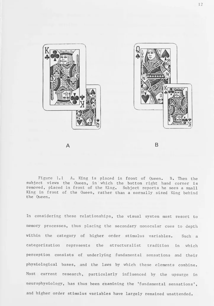
Secondary Cues as Higher Order Stimulus VariablesAmes (see Ittelson, 1968) formulated a striking demonstration of
the influence of past experience in humans on the secondary monocular
cue of interposition (the cue of interposition occurs when an
overlapping object is said to be nearer than an overlapped object).
Ames demonstrated that a false interposition cue could over-rule
spatial information derived from other cues and rever se the real depth
relationship based on the subject 's past experience (see Figure
1. 1). Processes of this sort are of the kind that von Helmholtz
(1867) described as unconscious inferen ce.
The secondary monocular cues to depth are by definition, two
dimensional cues to three dimensional space. Thus each pictorial cue
produces an optic array that is shared by an entire family of
three-dimensi onal arrangements. In order to resolve the ambiguity implicit
in each cue alone, the spatial attributes of form perception must
co nside r the orientations and spatial relationships between the
A
B
Figure 1.1 A. King is placed in front of Queen. B. Then the
subject views the Queen, in · which the bottom right hand corner is
removed, placed in front of the King. Subject reports he sees a small
King in front of the Queen, rather than a normally sized King behind the Queen.
In considering these relationships, the visual system must resort to
memory processes, thus placing the secondary monocular cues to depth
within the category of higher order stimulus variables. Such a
categorization represents the structuralist tradition in which
perception consists of underlying fundamental sensations and their
physiological bases, and the laws by which these elements combine.
Most current research, particularly influenced by the upsur g e in
neurophysiology, has thus been examining the 'fundamental sensations',
The three dynamic areas of depth perception revolve around the
cues motion parallax, accommodation, convergence and stereopsis. The
latter two have been accompanied by a growing interest in the
mechanisms and evolution of binocular vision.
1.3 MOTION PARALLAX
The head, body, and in most species, the eyes, continually move
with respect to the visual environment which consists of both
stationary and moving objects. Hence the visual system may sample the
same point in space at different points in time. The differential
angular velocity of objects moving with respect to the retina or
vice-versa, thus provides motion parallax as a dynamic monocular cue to
depth.
Von Helmholtz (1867) emphasized the importance and effectiveness
of motion parallax in comparison with other depth cues:
Suppose for instance, that a person is standing still in a thick wood, where it is impossible
for him to distinguish, except vaguely and
roughly, in the mass of foliage and branches all around him what belongs to one tree and what to another •••• But the moment he be g ins to
move forward, everything disentangles
itself (p 296).
Yet most early human studies have failed to show that motion parallax
in isolation is an effective cue to depth. If one simulates the
transformations that would be produced by a surface tilted in depth,
subjects either do not report a consistent impression of relative
depth or slant (Gibson
&
Carel, 1952), or they produce estimates whichvary widely (Gibson
et al.,
1959). Epstein and Park (1964) and GogelI:
effective in isolation as an exocentric due to depth. Gogel & Tietz
(1973) in examining the specific distance tendency, found that head
motion only provides cues to distance by allowing perceptual learning.
In contrast to these studies, Eriksson (197 4) studied successive
discrimination on the basis of motion parallax where the observer
moved his head, and found that perceived distance increased with
target distance in the range of 2-4 metres. Johansson (1972: see
Foley, 1978) also found that accurate discrimination - based upon motion
parallax co-varied with accommodation over distances of 30-240 cm.
Perhaps the clearest evidence for the effectiveness of motion
parallax comes from studies which have used complex or
information-rich stimulus displays. Rogers and Graham (1979) used a random dot
display that could be transformed by each movement of the observer or
the di splay oscilloscope. As soon as the observer's head was moved,
relative movement was generated between the elements (dots) which
matched the motion parallax produced by a real three dimensional
surface. This produced consistent and unambi guous impressions of
relative depth not unlike that found with stereopsis. The relative
motion of the rows of dots was not perceived. Thus, Ro gers and Graham
conclude that motion parallax provides an accurate and unambiguous
source of information about the depth of objects. Other authors,
notably Braunstein (1976) have come to similar conclusions. Ullman
(1979) has shown mathematically that there is sufficient information
in a sequence of only 3 discrete views of 4 points to uniquely specify
the three dimensional surface structure of any rigidly moving surface.
Unlike stereoscoic vision, monocular movement parallax provides
conditions for depth perception with movement along any axis, not only
the horizontal axes. J.J. Gibson (1950) pointed out that motion
transformations. Other retinal transformations include the optical
expansion patterns produced by movement through the visual world and
the patterns of retinal change produced by object rotation (Gibson,
1966; Braunstein, 1976). Hence for motion parallax to be effective in
a complex visual world, either the objects or the observer may move in
any direction. However, the threshold sensitivity for motion parallax
in humans is not equal for all directions. It is best for horizontal
and worst for vertical axes of movement (Graham, 1965). Nor is this
effect due to binocular mot:i.on in depth cells tuned in the horizontal
plane (Cynader
&
Regan, 1978) as the thresholds were measuredmonocularly. It may reflect differences in retinal gradients along
the various foveal-peripheral axes.
Only a handful of studies have directly examined motion parallax
as a cue to depth in animals, although it has frequently been
implicated by observations such as head bobbing in birds (Grinnel,
1921; Pumphrey, 1948: see
insects (Wallace, 1959).
Shinkman, 1962) and peering behaviour in
Walk (1965) describes experiments of
Schiffman (1961) and Trychin (1962) who found that both chicks and
rats chose the shallow side of the visual cliff under monocular
conditions with no relative size cues present. Furthermore this
discrimination fails if only one pattern element is present,
indicating that accommodation is not operative. Addition of elements
increased the probability of discrimination, and peering movements
implied the possible use of motion parallax information.
A more direct study of motion parallax in relation to peering
behaviour was done with the locust (Collett, 1978). This study
revealed that the angular orientation of the locust head remained
constant, and amplitude of pee,ring increased with greater object
decline in accuracy with distance (Horridge, 1977). Collett concludes
that, provided the locust knows how far (or fast) it moves its head,
it can measure image displacement (or velocity) and thus compute the
distance of the object. This, however, is not synonymous with motion
parallax seen in the human situation as it simply allows egocentric
distance estimation, rather than relative distances (i.e. depth)
between objects to be measured. Discrimination of movement is,
however, a necessary prerequisite for detection of the differential
angular velocity.
The importance of movement as a visual stimulus is apparent
across species. An early review of moti-0n sensitivity can be found in
Kennedy (1936). Not only amphibians, but most snakes, lizards, and
many carnivorous turtles do not respond to motionless prey. Walls
(1942) recognized that the successful use of the habit of 'freezing'
is in itself an evidence that the predator does not identify them as
well visually.
Birds too seem to easily detect movement. An eagle can see
moving prey at great distances. It is often reported that a homing
pigeon can detect a moving hawk well in advance of any human
observer. It has even been postulated that the avian optical
structure, the pecten, accentuates this motion sensitivity by the
creation of a stroboscopic shadow on the retina (Menner, 1938, see
Walls, 1942, p.365).
Physiological evidence has shown that neurones respond
preferentially to moving contours at almost every level of the pi geon
visual system (Maturana & Frenk, 1963; Jassik-Gerschenfeld
et al.,
1970), yet the least detectable stimulus velocities from
psychophysical studies in the pigeon are surprisingly high and readily
Accurate apparent movement detection has been shown in the pigeon
(Siegel, 1970), although stimulus control may have been achieved by
illumination change on a local area of the rotating disc, rather than
by the apparent movement of an entire pattern. A thorough study by
Hodos, Smith and Bonbright (1975) measured absolute thresholds for
detecting movement of a grating stimulus. The threshold was found to
be 4-6° / sec. and is well above the human threshold which is of the
order of 0.4-6.0 min/sec (Brown, 1931; Leibowitz, 1955).
Neither absolute nor apparent movement detection implies that the
pigeon uses motion parallax as a cue to depth. Perhaps closer to the
mechanism underlying motion parallax is differential motion
sensitivity. Mulvanny (1978) reports that the thresholds for
differential motion sensitivity are only 10-12°/sec. Whether relative
velocity detection is used to estimate the depth of objects, however,
is open to debate. It remains clear, that in the frontal visual
field, motion sensitivity measured by various means, appears
relatively poor.
Detection of movement is not confined to central vision. The
extreme periphery will discern a wriggling finger without knowledge of
the brightness, colour or form of the finger. Clearly, peripheral
movements have a saliency and attention value across species, quite
out of proportion to the clarity with which they are actually
discriminated.
Of interest is a study by Martinoya
et al.
(1983) who examinedabsolute motion sensitivity in both the frontal and lateral visual
fields of the pigeon. The pigeon has often been described as myopic
in the frontal field, and hyperoptic in the lateral fields (Catania,
'
1964; Millodot
&
Blough, 1971), thus allowing observation of food11
I '
I
,,
'
'
I
1,
I ,
I!
I,
predators with lateral regard (Catania, 1964; Goodale, 1983b; Bloch
&
Martinoya, 1983). Martinoya
et aZ.
(1983) trained pigeons todiscriminate the direction of moving square wave gratings presented
tachistoscopically. The stimuli were presented at 40cm, in either the
frontal binocular or lateral visuals fields. For frontal viewing the
threshold was similar to that found by Hodos, Smith and Bon bright
(1975), but the lateral thresholds were in the range 1.4 - 2.5°/sec.
Hence, the lateral sensitivity to motion was some three times better
than that seen in the frontal field.
It could be argued that the difference between the two visual
fields was due to the fact that the moving gratings presented in the
frontal field were horizontal and moved downwards, while for the
lateral presentations, the grid was projected vertically and moved
backwards. Although consistent with the optic flow while flying, the
patterns of movement were stimulating the retina in orthogonal
directions. Recall that motion parallax in humans varies as a
function of the axis of movement. The superiority of lateral motion
sensitivity may arise at the level of the retina. It is known that in
the cat, Y-cells are prevalent in the peripheral retina. These cells
are responsive to fast target movements, and may well analyse temporal
patterns (Sherman, 1982). Similar mechanisms may exist in the pigeon,
although the lateral superiority to motion detection may arise at
subsequent stages in the visual system.
It has been found very difficult (Bloch & Maturana, 1971) if not
impossible (Catania, 1963) to train pigeons with a lateral
presentation of a form discrimination. Nye (197 3) reports that the
pigeons' ability to discriminate colour, form, brightness and moving
patterns diminished as the stimuli progressed in the lateral
develop a direct associative pecking response to laterally located
stimuli. This clearly contradicts the results from Martinoya
et al.
(1983). Furthermore, Nye's hypothesis cannot be entirely correct as
laterally presented colour stimuli can be readily associated with
pecking in the pigeon (Bloch
&
Maturana, 1971).1.4 OCULOMOTOR CUES
1.4.1
AccommodationThe process by which the refractive power of the eyes is altered
to ensure a clear retinal image is known as accommodation. Points
nearer or further than the fixated point produce blur circles which
vary as a function of distance between the point and the fixated point
(Southall, 1937). Discrimination of depth based on accommodation
alone was first reported by Wundt (1862, see Foley, 1978) and later by
Le Grand (1967). Their subjects viewed through a tube a thread
against a bright background, but possible artifacts such as would have
arisen if the eye, tube and thread positions were not in alignment
were never eliminated. Subsequent investigators have found no
relationship between perceived distance and accommodation (Heinemann
et al.,
1959), ordinal (Woodworth, 1938; Biersdorf, 1966) or anon-linear relationship (Gogel
&
Tietz, 1973; Foley, 1977). Hence, whileaccommodation may serve as a cue to depth in humans, it is severely
limited in its capacity to do this. It is neither precise nor
accurate over distances greater than a metre or so (Baird, 1903;
1.4.2 The Relationship between Accommodation and Convergence
The change in the relative positions of the visual axes is called
convergence when the angle formed by the axes increases, and
divergence when this angle decreases. The synkinesis between
accommodation and convergence plays an important physiological role in
binocular vision in near fixation. Whenever a person exerts a certain
amount of accommodation, a determined amount of convergence is
elicited. The reverse also seems to hold (Finchan & Walton, 1957).
Convergence so elicited is expressed clinically as the
accommodation/convergence ratio (Fry, 1939; Fry
&
Haines, 1940).Accommodative convergence provides the gross adjustment for the
position of the eyes, but acting alone it rarely, if ever, provides
binocular single
convergence (see
retinal stimuli.
vision. Fine
Von Noorden,
adjustment is
1980) and is
obtained
elicited
by
by
fusional
disparate
Owens (1974, quoted in Foley, 1978) has shown that the resting
state of convergence is more closely related to perceived distance in
the absence of information, than is the state of accommodation. Here
there is an interdependence between the depth signal in the absence of
distance information, and the oculomotor state. Discrimination based
on convergence alone appears to fail when the stimuli are such as to
create the impression that they are physically identical (Foley,
1978). This is due, perhaps, to the conflict between size constancy
and convergence. Other studies have implicated convergence as an
1.4.3 Accommodation and Depth Perception in Animals
Homatropine has been used to abolish accommodation in kittens
(Walk, 1965) and curare has been used for the same purpose in chicks
(Shinkman, 1963), but there is no resultant impairment in performance
on the visual cliff. Nealey (in work discussed by Shinkman, 1962)
projected a blurred or a clear pattern of equal size onto both sides
of the visual cliff at equal depth and found that rats showed no
preference for either side. Due to non specific drug effects and
indirect modification of accommodative depth cues, these studies can
hardly be proclaimed to support the notion that accommodation was not
used for the perception of depth.
Ingle (1968, 1972) measured the snapping response of frogs and
toads. He found that estimation of distance to prey was done as well
with one eye as with two, and proposed that the animals do this by
monitoring the accommodative state of their eyes when the prey is in
optimum focus. The chameleon has also been proposed to use a similar
mechanism for depth localization (Harkness, 1977). This reptile has a
central fovea with extensive independent voluntary eye movements.
Walls (1962) describes:
their total lack of binocular conjugation, are all made conveniently conspicuous by the fact that the creature's high eye bulges prominently and has the lids permanently fused with its
surface... ( p. 75).
Harkness found that binocular and monocular chameleons perceived prey
distance as some function of the plane of focus, and accommodation was
not just used as a back-up cue when binocular information was
1.5 STEREOPSIS
Wheatstone's invention of the stereoscope in 1838 revealed that
the basis of stereopsis is binocular disparity due to the horizontal
parallax shift between corresponding points on the left and right
retinas.
1.5.1 The Horopter
Vieth (1818) first geometrically specified that points seen
binocularly as single in the horizontal plane of the fixation point
should lie on a circle that passes through the fixation point and the
optical centres of the two eyes. The geometrical construction became
known as the Vieth-Muller circle (Muller, 1826, see Boring, 1942).
Thus in Figure 1.2, points X, Y and Z are on the horopter and are said
to stimulate corresponding retinal points.
Zr
Yr
Xr
X
Figure 1. 2 The Vieth-Muller horopter circle. Xr and X1 are
corresponding points. Similarly for Yr and y
1 and Zr and z1• O,
Von Helmholtz (186 7, translated 1925) and Hering (1879,
translated 1942) realised that the Vieth-Muller circle was an
oversimplification, and worked out forms of a general horopter based
mainly on the determination of the apparent frontoparallel plane. The
variety of criteria that have subsequently been proposed as a basis
for the horopter (Ogle, 1962; Shipley
&
Rawlings, 1970) testify to thefact that the concept of the horopter has yet to find a clearly
defined place in the theory of binocular vision. Bishop (1981)
suggests that the horopter may best be considered as the
zero-disparity reference plane that contains the fixation point and
relative to which stereoscopic depth estimates are made.
Q
F
a.
b ..
Figure 1. 3 Physiologic diplopia. a. Crossed disparity of the
object P, closer than the fixation point F. b. Uncrossed disparity of
the object P, more distant than ·the fixation point. O, optical
An object nearer (P) or further (Q) (within limits) from the
fixation point (F) in Figure 1. 3, will produce crossed or uncrossed
disparity,
disparity
respectively,
between the
by virtue of the relative horizontal
retinal points which do not exactly
correspond. Thus, the difference between the visual angles
e
2
and 8 1 defines the relative disparity between points F and P, or F
and
Q,
and leads to the stereoscopic perception of P orQ
appearing tolie in depth relative to F. Notice that only horizontal, not vertical
disparities give rise to stereopsis.
1.5.2 Spatial range of stereopsis and the distinction between fine
and course mechanisms.
Binocular vision is not limited to points on the horopter but
extends to include what is known as Panum's fusional area, defined by
the limiting values of disparity. Ogle in
1952
described two distinctkinds of stereoscopic perception. Patent stereopsis occurs within
Panum's fusional area where the magnitude of perceived depth co-varies
with the magnitude of disparity (Richards,
1971).
The depthexperience is evident and compelling and relies upon a centrally
mediated fine vergence reflex mechanism.
When stimuli exceed Panum' s area, they are no longer fused but
are diplopic, and continuous variation in disparity no longer produces
concomitant variations in perceived depth. Despite the fact that the
disparity-depth relationship is lost, the stimuli can still be
qualitatively localized
(Westheimer
&
Mitchell,as nearer or further than the fixation point
1969;
Mitchell & O'Hagen,1972).
Og lereferred to the stereoscopic perception of diplopic stimuli as
quantitative stereopsis. Similarity of the two retinal images is not
Ii
Ii
.
,,II
I
f
I
I I
D
stereopsis has associated with it a peripheral coarse stereopsis and
fusional mechanism. Fine single vision is always accompanied by
coarse fusion.
Tests for Stereopsis
Wheatstone's stereoscope is an example of a haploscopic device in
which separate two-dimensional targets (in this case by way of
mirrors) are presented to each of the two eyes.
Subsequent investigators examining stereoscopic acuity and
factors influencing stereopsis have used real depth instruments. The
classic example is the Howard-Dolman apparatus (Howard, 1919) in which
two vertical rods, one stationary and the other moveable, are viewed
through an aperture against a luminated background. The subject can
then adjust the moveable rod until aligned and side by side with the
stationary rod. Although it is conceivable that retinal image size
may mediate the discrimination, Howard showed that monocular
thresholds were 20 times as great as thresholds obtained on the basis
of binocular vision. As measurements were made outside the range of
effective convergence, this apparatus relies on the cue of retinal
disparity for successful discrimination. Our own observations on a
group of 30 students revealed that motion parallax obtained by lateral
head movement could aid monocular discrimination such that error of
alignment was reduced by half (from a mean of 63. 6cm to 30. 4cm).
Binocular performance, however, was always superior to monocular
performance (McFadden
&
Gillette, 1982, unpublished observations). Avariation of the Howard-Dolman apparatus is the Verhoeff stereopter
where vertical wires replace the rods.
contamination by retinal size cues~
In clinical testing of stereoacuity haploscopic devices are
normally used. Examples are the Titmus, Wirt, T.N.O. and Reinecke E
Test. Separate images are presented to each eye by the use of red and
green filters worn over each eye and matched to each of the two
coloured images, or polarized goggles in which the vectographs are so
imprinted that each target is polarized at 90 degrees with respect to
the other (see Hinchliffe, 1978).
1.5.4
Local and Global StereopsisJulesz (1960) was the first to consider the use of
computer-generated random dot arrays to produce a stereogram in which each
target appears as a homogeneous random array of small squares or
elements (dots) without any global shape or contour visible. Retinal
disparity, defining the configuration of a particular form, is
produced by laterally displacing a subset of dots in one matrix
relative to corresponding dots in the other matrix. In such random
dot stereograms (RDS), if the horizontal shift is always kept an
integral multiple of the cell size, then no cell of the background
will ever be partly covered by the shifted areas, and thus no
monocular cue will be present.
When the target components of the RDS are binocularly fused, the
particular form that is displaced 'jumps out' in vivid depth above the
background. In order for this to occur retinal corresponding points
must be identified. Since in a RDS any local dot in one field can be
matched by many neighbouring identical dots in the other field, many
false localizations may occur (Figure
1.4) • .
The ambiguity can besolved by pooling several adjacent dots that have similar binocular
disparities and perhaps some other feature in common. The search for
certain areas is the essence of global stereopsis. Since the visual
system manages to overcome this ambiguity and arrive at the correct
global solution in a RDS, Julesz has effectively shown that global
stereopsis can occur in the absence of any monocular form or other
depth cues.
•
Correct fusion
o
False fusion
I
For classical stereograms (Ogle, 1962; Richards, 1971), either
with a few elements or monocular labels, there is no ambiguity of
corresponding retinal projections. Such a process of unambiguous
depth localization is called local stereopsis. It operates on feature
elements such as dots, lines, edges or corners, and each feature pair
is assigned a depth value relative to the fixation point. Local
stereopsis is defined as depth localization (on the basis of binocular
disparity alone) of elements whose correspondence (and thus binocular
disparity between them) can be unambiguously established. Global
stereopsis is the finding of a preferred set of local depth
localizations among many other sets.
Although static RDSs described above are the purest stimulation
of horizontal retinal disparity discussed so far, a subtle monocular
cue is still potentially available. This cue is the inevitable
difference in dot patterns at the borders of the embedded stereoscopic
form producing local differences in detail. This criticism is not,
however, applicable to the dynamic RDS where the arrays are
continuously and randomly changed. The left and right fields consist
of dynamic noise arrays of high spatial resolution presented at fast
rates. When binocularly fused, certain correlated areas (whose
binocular correlation varies at much slower rates than the change of
the dynamic noise arrays) are perceived as pulsating or moving in
vivid depth (Julesz, 1971; Julesz
et al.,
1976; Julesz&
Tyler, 1976).1.5.5 Models of Global Stereopsis
Several groups of investigators are actively engaged in efforts
to model global stereopsis (e.g. Julesz
&
Chang, 1976; Marr&
Poggio,1979; Mayhew & Frisby, 1980). In the classic literature (e.g. Ogle,