Open Access
Vol 7 No 3Research article
Increased level of phosphorylated akt measured by
chemiluminescence-linked immunosorbent assay is a predictor of
poor prognosis in primary breast cancer overexpressing ErbB-2
Jonas Cicenas
1,2, Patrick Urban
1, Vincent Vuaroqueaux
3, Martin Labuhn
3, Willy Küng
2,
Edward Wight
4, Mark Mayhew
5, Urs Eppenberger
2,3and Serenella Eppenberger-Castori
11Stiftung Tumorbank Basel, Basel, Switzerland
2University Clinics, Department of Research, Molecular Tumor Biology, Basel, Switzerland 3OncoScore AG, Riehen, Switzerland
4University Clinics, Department of Gynecology, Basel, Switzerland
5University of Virginia, Department of Anatomy and Cell Biology, East Carolina School of Medicine, Charlottesville, Virginia, USA
Corresponding author: Serenella Eppenberger-Castori, s.eppenberger@tumorbank.org
Received: 14 Oct 2004 Revisions requested: 16 Dec 2004 Revisions received: 9 Feb 2005 Accepted: 28 Feb 2005 Published: 24 Mar 2005
Breast Cancer Research 2005, 7:R394-R401 (DOI 10.1186/bcr1015) This article is online at: http://breast-cancer-research.com/content/7/4/R394 © 2005 Cicenas et al.; licensee BioMed Central Ltd.
This is an Open Access article distributed under the terms of the Creative Commons Attribution License (http://creativecommons.org/licenses/by/ 2.0), which permits unrestricted use, distribution, and reproduction in any medium, provided the original work is properly cited.
Abstract
Introduction Akt1, Akt2 and Akt3 kinases are downstream components of phosphoinositol 3-kinase derived signals from receptor tyrosine kinases, which influence cell growth, proliferation and survival. Akt2 overexpression and amplification have been described in breast, ovarian and pancreatic cancers. The present study was designed to investigate the prognostic significance of activated Akt in primary breast cancer and its association with other tumour biomarkers.
Methods Using a two-site chemiluminescence-linked immunosorbent assay, we measured the quantitative expression levels of total phosphorylated (P-S473) Akt (Akt1/Akt2/Akt3) on cytosol fractions obtained from fresh frozen tissue samples of 156 primary breast cancer patients.
Results Akt phosphorylation was not associated with nodal status or ErbB-2 protein expression levels. High levels of phosphorylated Akt correlated (P < 0.01) with poor prognosis, and the significance of this correlation increased (P < 0.001) in the subset of patients with ErbB-2 overexpressing tumours. In addition, phosphorylated Akt was found to be associated with mRNA expression levels of several proliferation markers (e.g. thymidylate synthase), measured using quantitative real-time RT-PCR.
Conclusion Our findings demonstrate that, in breast cancer patients, Akt activation is associated with tumour proliferation and poor prognosis, particularly in the subset of patients with ErbB2-overexpressing tumours.
Introduction
Akt/protein kinase B (PKB) is a serine/threonine kinase that is involved in mediating various biological responses, such as inhibition of apoptosis and stimulation of cell proliferation (for review [1,2]). Three mammalian isoforms are currently known [1]: Akt1/PKBα, Akt2/PKBβ and Akt3/PKBγ. Akt1 was first discovered as a cellular homologue of the viral oncogene v-Akt, which causes leukaemia in mice [3] and is the predomi-nant isoform in most tissues. High expression of Akt2 has been observed in insulin-responsive tissues, whereas Akt3 has
been shown to be predominantly expressed in brain and testis [2].
Phosphoinositol-3-phosphate (PIP3) is a product of phosphoi-nositol 3-kinase enzymatic activity and has been shown to be a prerequisite lipid modulator of Akt activity [4]. PIP3 has been described as a downstream component of a wide range of receptors, including the c-Met receptor [5], the epidermal growth factor receptor family [6], fibroblast growth factor receptor [7], insulin growth factor receptor [8] and platelet-derived growth factor receptor [9]. In addition, Akt activity can
be regulated by the PTEN tumour suppressor gene, which negatively regulates PIP3 levels (for review [10]). After PIP3 binding, Akt1 is activated by phosphorylation on two critical residues, namely threonine 308 (T308) and serine 473 (S473); similar activation residues (S472 and S474, respec-tively) are highly conserved in Akt2 and Akt3 (for review [1,2]). Several studies have found Akt2 to be amplified or overex-pressed at the mRNA level in various tumour cell lines [11-13] and in a number of human malignancies, such as colon, pan-creatic and breast cancers [14-16]. However, activation of Akt1, Akt2 and Akt3 by phosphorylation appears to be more clinically relevant than detection of Akt2 amplification or overexpression.
To date, several groups have investigated the phosphorylation of active Akt in breast, prostate, colon and pancreatic tumours by immunohistochemistry [14,17-22]. Under such conditions, phosphorylation structures may be disturbed by formalin fixa-tion, rendering specific antigen sites inaccessible. Moreover, immunohistochemistry gives only semiquantitative results, lim-iting statistical analysis. Alternatively, enzyme immunoassays (EIAs) have the advantage that they yield highly reproducible and sensitive results of quantitative values.
In the present study we detected phosphorylated Akt (P-Akt) by means of a novel two-site chemiluminescence-linked immu-noassay (CLISA) in fresh frozen primary tissue samples from 156 primary breast cancer patients. Because it was shown in previous immunohistochemistry studies that S473 P-Akt has prognostic significance [17-19], the aim of the present study was to measure levels of P-Akt continuously using CLISA and correlate these with survival and factors that are involved in tumourigenesis. Given that the antibody used in the reported immunohistochemistry studies recognized all Akt isoforms, we have developed an assay that allows specific quantitative detection of active Akt1, Akt2 and Akt3 when phosphorylated on their corresponding residues, namely S473, S472 and S474, respectively.
Materials and methods
Tumor and patient characteristics
Fresh material obtained during surgery was kept on ice and examined by a pathologist. Representative specimens with more than 60% tumour cells were sent to the Stiftung Tumor-bank Basel (STB), immediately shock frozen and cryopre-served (-80°C). All activities of the STB are in accordance with an official Swiss permit, which guarantees patient confidenti-ality and respects ethical issues. For the present study, 156 samples of primary breast tumours were selected. Those sam-ples overexpressing ErbB-2 (>500 U/mg total protein) were selected, based on ErbB-2 protein expression levels routinely detected using EIAs at the time of surgery by the STB [23]. EIA ErbB-2 positive samples correlate strongly with DAKO 3+ and with ErbB-2 amplification detected by fluorescent in situ
hybridization (FISH; data not shown).
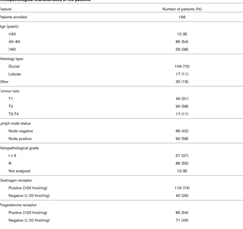
All patients underwent primary surgery before January 1996. Sixty-seven patients (43%) experienced disease recurrence within the median follow-up time of 57 months (range 27–88 months). Sixty-six patients (42%) were node negative, and 90 (58%) were node positive. Forty tumours (26%) were oestro-gen receptor (ER)-α negative. Ninety-five patients (61%) had ErbB-2-negative (<500 U/mg total protein) and 61 patients (39%) had ErbB-2 positive tumours [23]. None of the patients received neoadjuvant therapy. Patient and tumour characteris-tics are summarized in Table 1.
Cell lines and tissue culture
MCF-7 breast cancer cells were cultured in IMEM-ZO (improved minimal essential medium with zinc option) supple-mented with 5% foetal bovine serum, l-glutamine and antibiot-ics (penicillin/streptomycin) at 37°C in a 5% carbon dioxide incubator. For the phospho-standard preparation, subconflu-ent MCF-7 cells were serum starved for 48 hours in serum-free media, and were treated with NaF and Na3VO4 for 1 hour, and
then with 10% foetal bovine serum for 10 min. Cells were lysed for 5 min on ice in EB lysis buffer (20 mmol/l Tris-HCl [pH 7.4], 0.5 mol/l NaCl, 10 mmol/l EDTA, 1% Triton X100, 20 mmol/l NaF, 20 mmol/l glycerophosphate, 2 mmol/l Na3VO4, proteinase inhibitor cocktail [Roche, Indianapolis, IN,
USA]), centrifuged at 20,000 g for 5 min and supernatant was stored at -80°C.
Measurement of oestrogen receptor, progesterone receptor and ErbB-2 protein levels in tumour extracts by enzyme immunoassay
Immunoassay of phosphorylated Akt level
Neither antibody used in the CLISA discriminates between Akt isoforms. The catching antibody (anti-Akt/PKB, PH domain, clone SKB1; Upstate Biotechnology, Lake Placid, NY, USA) recognizes Akt1/PKBα, Akt2/PKBβ and Akt3/PKBγ (weak to none) based on immunoblot analysis using 100 ng recom-binant fusion protein for each isoform, as reported by the man-ufacturer. The detecting phospho-specific (S473) Akt monoclonal antibody (4E2) detects endogenous levels of Akt1 only when phosphorylated at serine-473. This antibody also recognizes Akt2 (S472) and Akt3 (S474) if they are phospho-rylated at the corresponding residues, according to the
infor-mation obtained from the manufacturer (Cell Signaling Technology, Inc., Beverly, MA, USA). However, 4E2 does not recognize other Akt phosphorylation sites.
[image:3.612.63.555.108.573.2]S473 phosphorylated Akt levels were measured using a novel two-site CLISA. Black 96-well microtitre plates (Nunc Black MaxiSorp Surface; Nalgen Nunc International, Rochester, NY, USA) were coated with coating antibody at a concentration of 3 mg/ml of coating buffer (phosphate-buffered saline with 0.6 mmol/l EDTA) in a volume of 100 µl/well and kept at 4°C over-night. To measure P-Akt, respective tumour extracts were pre-pared as described above in the presence of NaF and
Table 1
Clinicopathological characteristics of the patients
Feature Number of patients (%)
Patients enrolled 156
Age (years):
<40 12 (8)
40–60 85 (54)
>60 59 (38)
Histology type:
Ductal 109 (70)
Lobular 17 (11)
Other 30 (19)
Tumour size:
T1 49 (31)
T2 90 (58)
T3-T4 17 (11)
Lymph-node status
Node negative 66 (42)
Node positive 90 (58)
Histopathological grade
I + II 57 (37)
III 86 (55)
Not analyzed 13 (8)
Oestrogen receptor
Positive (>20 fmol/mg) 116 (74)
Negative (≤ 20 fmol/mg) 40 (26)
Progesterone receptor
Positive (>20 fmol/mg) 85 (54)
Na3VO4. Before sample applications, the coated microtitre
plates were washed five times with 200 µl/well washing buffer (25 mmol/l HEPES [pH 7.4], 300 mmol/l NaCl, 0.05% Tween-20) and then blocked for 2 hours at room temperature with 250 µl blocking buffer (25 mmol/l HEPES [pH 7.4], 300 mmol/l NaCl, 0.05% Tween-20, 3% TopBlock [Juro AG, Lucerne, Switzerland]). Blocked wells were washed five times with 200 µl washing buffer, and then 100 µl diluted tumour membrane extracts or reference material was added to the wells and incubated overnight at 4°C.
As a reference for each assay, an extract of MCF-7 cells, pre-pared as described above, was used. For use in the assay, MCF-7 cell extracts were sequentially diluted with sample dilu-tion buffer (blocking buffer, proteinase inhibitor cocktail, NaF and Na3VO4) at ratios of 1×, 0.75×, 0.5×, 0.25×, 0.125× and
0.025×, and then 100 µl aliquots were incubated on each microtitre plate, together with tumour tissue extracts and neg-ative controls (containing only dilution buffer). After incubation of the samples and reference material, wells were washed five times with 200 µl washing buffer at room temperature to elim-inate unbound particles. Biotinylated detection antibody was added, followed by incubation for 2 hours at room tempera-ture. Complexes were detected with horseradish peroxidase-conjugated streptavidin, diluted in conjugate diluents for 1 hour at room temperature. Horseradish peroxidase activity was detected using SuperSignal WestPico substrate (Pierce, Rockford, IL, USA) in a glow luminometer. The response data for diluted reference material was fitted, and the respective curve was used for the quantification of tumour extracts. The value of undiluted MCF-7 extracts was denominated as 1 U/ ml.
Quantitative real-time RT-PCR for the detection of proliferation markers
RNA was extracted using RNeasy kit (Qiagen, Hilden, Ger-many). Quality and quantity were checked using a Bioanalyzer 2100 (Agilent, Palo Alto, CA, USA). All genes were examined using SYBR Green I methods with Taqman 7000 (Applied-Biosystems, Foster City, CA, USA). Relative quantification (∆∆Ct) was obtained by normalization with ribosomal 18S and a standardization step with Human Universal Standard RNA (Stratagene, La Jolla, CA, USA). Quantitative real-time RT-PCR results were expressed in arbitrary units of reverse tran-scribed RNA (U/µg rt-RNA).
Statistical methods
The statistical significance of the association between P-Akt and other dichotomous variables (e.g. node status) was assessed using Mann–Whitney U-test. Spearman rank corre-lation (rs) was calculated to assess associations between
con-tinuous markers (e.g. ErbB-2 or tumour size and P-Akt protein expression levels). The continuous variable function of CLISA-determined P-Akt values was first tested for prognostic signif-icance by univariate Cox regression. A cutoff or prognostic
threshold value with respect to relapse-free survival was sought by means of classification and regression tree analysis [24,25]. Survival probabilities were calculated using the Kap-lan–Meier method and compared by means of log-rank analy-sis [26]. The Cox proportional hazards regression model was also applied over multivariate analyses, with the associated likelihood ratio test used to assess test-of-trend differences. The results of multivariate Cox regression analysis were sum-marized in a table and expressed as relative risk for relapse.
Results
Distribution of phosphorylated Akt levels and its correlation with tumour characteristics
CLISA quantified P-Akt levels have a left-tailed distribution ranging from 0 to 1.08 U/mg total protein, with a median of 0.17 U/mg (mean 0.19 U/mg; Fig. 1) and could be trans-formed to normality by means of the 10th root. There was no correlation between P-Akt and ErbB-2 protein expression lev-els. In this set of primary breast cancer samples, we did not find any significant difference in P-Akt levels with respect to nodal status, tumour size, ER status or grading, nor any corre-lation between P-Akt levels and the continuous variables tumour size and ER level.
Prognostic significance of phosphorylated Akt levels
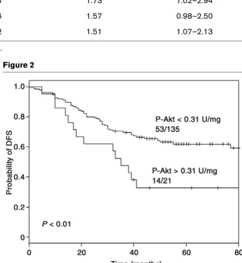
The prognostic value of P-Akt was investigated with respect to disease-free survival (DFS) in the patients overall (Fig. 2). Uni-variate Cox regression revealed a weak correlation between P-Akt levels and DFS (P < 0.05; likelihood ratio test). An optimal cutoff value for P-Akt (0.3 U/mg) was calculated using classi-fication and regression tree analysis, dividing the patients into two subgroups: 21 patients (14%) patients expressed high levels of P-Akt (>0.31 U/mg total protein) and 135 patients (86%) expressed low levels of P-Akt. Subsequently, Kaplan– Meier survival curves stratified according to low and high P-Akt levels were plotted (Fig. 2). Sixty-seven per cent of patients (14 out of the 21) with high P-Akt levels relapsed, whereas only 36% (49 out of 135) with low P-Akt developed a relapse of disease within the period of observation (P < 0.01; log-rank test). The 5-year DFS was 33% in the high P-Akt group versus 60% in the low P-P-Akt group. The 5-year DFS in node-positive patients was 50% versus 68% in node-nega-tive patients (P < 0.05; curves not shown).
Prognostic significance of phosphorylated Akt in ErbB2-overexpressing tumours
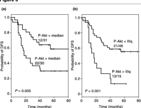
Although no correlation was found between P-Akt and ErbB-2 expression, the prognostic impact of P-Akt was greater in ErbB2-overexpressing tumours than in the samples overall. As shown in the Kaplan–Meier curves in Fig. 3a, patient progno-sis decreased significantly when tumours expressed P-Akt lev-els higher than the median value (P = 0.005). This effect was even more pronounced when P-Akt levels exceeded the third quartile value (P < 0.001), which, together with the multivari-ate Cox-analysis, indicmultivari-ates that P-Akt has independent and additive prognostic value in combination with ErbB-2 (Fig. 3b).
Correlation of phosphorylated Akt levels and mRNA expression of proliferation markers
Because involvement of P-Akt has been implicated in prolifer-ation and apoptosis, we compared the quantitative P-Akt
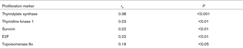
pro-tein levels with the quantitative mRNA expression levels of genes involved in these biological processes. Using Spear-man rank correlation, P-Akt levels were found to correlate with thymidylate synthase expression levels (rs = 0.38; P < 0.001)
and, to a lesser extent, with expression levels of thymidine kinase 1, survivin, topoisomerase IIα and the E2F transcription factor (Fig. 4, Table 3).
Discussion
Correlations between elevated P-Akt and higher risk for relapsehas already been demonstrated by other investigators in certain subsets of patients, specifically patients who received adjuvant endocrine therapy [17], patients treated with radiotherapy [18] and patients with a node-negative dis-ease [19]. Because ErbB-2 has been implicated in the activa-tion of Akt [27], we investigated the associaactiva-tion between P-Akt and ErbB-2 and its prognostic significance in tumours with known ErbB-2 expression levels. Our investigation
re-con-Table 2
Univariate and multivariate Cox analysis of relapse-free survival in patients with primary breast cancer
Factor Univariate P Multivariate P Relative risk for relapse 95% CI
P-Akt 0.01 0.02 2.09 1.14–3.85
Node status 0.0003 0.09 1.33 0.95–1.85
ER status 0.03 0.17 0.67 0.38–1.19
ErbB-2 status 0.002 0.04 1.73 1.02–2.94
Grading 0.03 0.06 1.57 0.98–2.50
Tumour size 0.00005 0.02 1.51 1.07–2.13
[image:5.612.313.554.189.451.2]CI, confidence interval; ER, oestrogen receptor; P-Akt, phosphorylated Akt.
Figure 1
Chemiluminescence-linked immunoassay (CLISA)-quantified phosphor-ylated Akt (P-Akt) levels
Chemiluminescence-linked immunoassay (CLISA)-quantified phosphor-ylated Akt (P-Akt) levels. (a) Histogram showing distribution of chemilu-minescence-linked immunoassay (CLISA)-determined phosphorylated Akt (Akt) expression levels in 156 primary breast cancer samples. P-Akt levels ranged from 0 to 1.08 U/mg, with a median of 0.17 U/mg. (b) Scatter plot of P-Akt versus ErbB-2 expression levels. No correlation was found between the levels of P-Akt and ErbB-2.
Figure 2
Kaplan–Meier survival curves for patients overall
[image:5.612.56.295.246.439.2]firmed the prognostic value of elevated P-Akt levels, and dem-onstrated that P-Akt expression levels are independent of other prognostic parameters, such as tumour size, grading, and node, ER and ErbB-2 status.
The lack of correlation between protein levels of ErbB-2 and P-Akt may be explained by the fact that Akt is also activated by various receptor tyrosine kinases [5-9], and by G-protein-cou-pled receptors [28]. Additionally, it was also observed that loss of PTEN activity is frequent in breast cancer and accompanied by increased activation of Akt [29], confirming that Akt can be activated by stimuli other than ErbB-2. The prognostic significance of P-Akt levels is increased if com-bined with ErbB-2 overexpression, suggesting that coactiva-tion of Akt and ErbB-2 may have a synergistic clinical impact.
Our study is the first report on P-Akt assessed by EIA using a phospho-specific antibody in breast cancer cytosols of cryopreserved tumour samples; the technique allowed us to obtain precise and quantitative results (for review [30]). In contrast to semiquantitative immunohistochemistry data, tumour marker profiles assessed by quantitative EIA are more sensitive and reproducible. EIA tests conducted with fresh fro-zen tissue extracts avoid the potential antigen damage due to formalin fixation, paraffin embedding and uncontrolled storage. Furthermore, the two-site (sandwich) CLISA assay used in this investigation ensures increased specificity as compared with single-antibody assays, such as immunohistochemistry and western blotting. In addition, chemiluminometric detection guarantees high sensitivity in the detection of antigen–anti-body complex.
We assayed for P-Akt in total breast tumour lysates, and not in tissue samples obtained from microdissection, both because we wished to correlate the protein expression levels of ErbB-2 and P-Akt levels directly in cells extracted from human tumour samples, and because it has been demonstrated that the activation status of Akt varies considerably in tumours of the same histotype, but not between different histotypes of the same tumour [31]. The CLISA assay used in the study was based on homogenized samples, which can include some stromal and normal tissue cells. The STB tissue samples con-tained at least 60% tumour cells, as observed by the pathologist. In addition, samples were previously analyzed for ErbB-2, ER and PgR using both EIA assays, as well as immunohistochemistry and/or fluorescence in situ hybridiza-tion. Importantly, good correlation between the assays was observed [23], suggesting that homogenization of samples does not play a crucial role in the final result. As in other assays that measure phosphorylation levels, the role played by phos-phatases should not be ignored. We used phosphatase inhib-itors in all steps of CLISA, as well as sample dilution. There could be some degradation before P-Akt testing, but all sam-ples were treated identically, and the study compared relative P-Akt levels among all tumours. Reference units (U) were used in order to establish a standard curve, but not to measure absolute P-Akt levels in separate samples.
Also of interest is the positive correlation between P-Akt and mRNA expression levels of tumour proliferation markers shown in the present study. Akt is known to promote cell cycle progression by modulating the expression [32] and stabiliza-tion of cyclin D1 [33], which in turn activates the E2F transcrip-tion factor. Our results also reveal a significant correlatranscrip-tion of P-Akt with E2F-1 transcription factor expression levels, as well
Kaplan–Meier survival curves for the subset of patients with ErbB-2 overexpressing tumours
Kaplan–Meier survival curves for the subset of patients with ErbB-2 overexpressing tumours. The curves stratified by (a) median and (b) last quartile values of phosphorylated Akt (P-Akt). Patients whose tumours express high levels of P-Akt exhibit a significantly worse out-come in terms of disease-free survival (DFS; P ≤ 0.005).
Scatter plot of phosphorylated Akt (P-Akt) versus thymidylate synthase (TS) mRNA expression
[image:6.612.57.297.89.274.2]as with genes regulated by E2F, such as thymidylate synthase, thymidine kinase 1, survivin and topoisomerase IIα.
Conclusion
Using a highly sensitive and specific CLISA assay, we demon-strated that elevated P-Akt is a marker of poor prognosis (decreased DFS). The prognostic value of Akt phosphorylation is independent of other characteristics, including tumour size and grade, and node, ErbB-2 and ER status. In a subset of patients with ErbB-2 overexpressing tumours, we demon-strated that P-Akt levels are of particular prognostic signifi-cance. In addition, Akt phosphorylation correlated with elevated mRNA expression levels of tumour proliferation fac-tors. Based on these findings, we suggest that P-Akt could play a predictive role with respect to Herceptin, topoisomer-ase IIα inhibitors and combination therapies using Akt inhibi-tors, which are currently in clinical trials and should primarily be assessed in patients with ErbB-2-overexpressing tumours.
Competing interests
The author(s) delcare that they have no competing interests.
Authors' contributions
JC carried out the development of CLISA assays, took meas-urements in breast cancer samples, participated in raw data analysis, participated in statistical analysis and drafted the manuscript. PU performed the statistical analysis. VV and ML carried out the RNA extraction and quantitative RT-PCR. WK participated in designing the study and participated in the raw data analysis. EW coordinated clinicians, providing tumour samples. MM helped in finalizing the manuscript. UE partici-pated in designing the study and coordination, and helped to draft the manuscript. SE participated in coordinating the study and in statistical analysis, helped to draft the manuscript and coordinated the selection of samples from the STB. All authors read and approved the final manuscript.
Acknowledgements
This work was supported by a grant Nr. 31-059819.99/1 (U. Eppen-berger) of the Swiss National Science Foundation and the Stiftung Tumorbank Basel (STB).
We thank Christine Wullschleger, Francoise David, Heidi Bodmer and Sabine Ehret for technical assistance, data management and tumour banking. We are indebted to A Almendral, M Anabitare, C Braschler, B von Castelberg, H Dieterich, D Fink, R Flury, R Gaudenz, K Lüscher, S Heinzl, M Mihatsch, H Moch, D Oertli, G Sauter, J Torhorst and M Zuber – clinicians and pathologists.
References
1. Vivanco I, Sawyers CL: The phosphatidylinositol 3-kinase AKT pathway in human cancer. Nat Rev Cancer 2002, 2:489-501. 2. Hanada M, Feng J, Hemmings BA: Structure, regulation and
function of PKB/AKT: a major therapeutic target. Biochim Bio-phys Acta 2004, 1697:3-16.
3. Staal SP: Molecular cloning of the akt oncogene and its human homologues AKT1 and AKT2: amplification of AKT1 in a pri-mary human gastric adenocarcinoma. Proc Natl Acad Sci USA 1987, 84:5034-5037.
4. Franke TF, Kaplan DR, Cantley LC: PI3K: downstream AKTion blocks apoptosis. Cell 1997, 88:435-437.
5. Bowers DC, Fan S, Walter KA, Abounader R, Williams JA, Rosen EM, Laterra J: Scatter factor/hepatocyte growth factor protects against cytotoxic death in human glioblastoma via phosphati-dylinositol 3-kinase- and AKT-dependent pathways. Cancer Res 2000, 60:4277-4283.
6. Hii CS, Moghadammi N, Dunbar A, Ferrante A: Activation of the phosphatidylinositol 3-kinase-Akt/protein kinase B signaling pathway in arachidonic acid-stimulated human myeloid and endothelial cells: involvement of the ErbB receptor family. J Biol Chem 2001, 276:27246-27255.
7. Chen Y, Li X, Eswarakumar VP, Seger R, Lonai P: Fibroblast growth factor (FGF) signaling through PI 3-kinase and Akt/ PKB is required for embryoid body differentiation.Oncogene 2000, 19:3750-3756.
8. Duan C, Liimatta MB, Bottum OL: Insulin-like growth factor (IGF)-I regulates IGF-binding protein-5 gene expression through the phosphatidylinositol 3-kinase, protein kinase B/ Akt, and p70 S6 kinase signaling pathway. J Biol Chem 1999, 274:37147-37153.
9. Franke TF, Yang SI, Chan TO, Datta K, Kazlauskas A, Morrison DK, Kaplan DR, Tsichlis PN: The protein kinase encoded by the Akt proto-oncogene is a target of the PDGF-activated phosphati-dylinositol 3-kinase. Cell 1995, 81:727-736.
10. Cantley LC, Neel BG: New insights into tumor suppression: PTEN suppresses tumor formation by restraining the phosph-oinositide 3-kinase/AKT pathway. Proc Natl Acad Sci USA 1999, 96:4240-4245.
11. Miwa W, Yasuda J, Murakami Y, Yashima K, Sugano K, Sekine T, Kono A, Egawa S, Yamaguchi K, Hayashizaki Y, Sekiya T: Isola-tion of DNA sequences amplified at chromosome 19q13.1-q13.2 including the AKT2 locus in human pancreatic cancer. Biochem Biophys Res Commun 1996, 225:968-974.
[image:7.612.56.554.114.215.2]12. Cheng JQ, Godwin AK, Bellacosa A, Taguchi T, Franke TF, Hamil-ton TC, Tsichlis PN, Testa JR: AKT2, a putative oncogene encod-ing a member of a subfamily of protein-serine/threonine kinases, is amplified in human ovarian carcinomas. Proc Natl Acad Sci USA 1992, 89:9267-9271.
Table 3
Spearman rank correlation of quantitative P-Akt levels and quantitative mRNA expression levels of proliferation markers
Proliferation marker rs P
Thymidylate synthase 0.38 <0.001
Thymidine kinase 1 0.23 <0.01
Survivin 0.22 <0.01
E2F 0.22 <0.01
Topoisomerase IIα 0.19 <0.05
DA, Wan M, Dubeau L, Scambia G, Masciullo V, et al.: Molecular alterations of the AKT2 oncogene in ovarian and breast carcinomas. Int J Cancer 1995, 64:280-285.
14. Roy HK, Olusola BF, Clemens DL, Karolski WJ, Ratashak A, Lynch HT, Smyrk TC: AKT proto-oncogene overexpression is an early event during sporadic colon carcinogenesis.Carcinogenesis 2002, 23:201-205.
15. Ruggeri BA, Huang L, Wood M, Cheng JQ, Testa JR: Amplifica-tion and overexpression of the AKT2 oncogene in a subset of human pancreatic ductal adenocarcinomas. Mol Carcinog 1998, 21:81-86.
16. Bacus SS, Altomare DA, Lyass L, Chin DM, Farrell MP, Gurova K, Gudkov A, Testa JR: AKT2 is frequently upregulated in HER-2/ neu-positive breast cancers and may contribute to tumor aggressiveness by enhancing cell survival. Oncogene 2002, 21:3532-3540.
17. Perez-Tenorio G, Stal O: Activation of AKT/PKB in breast can-cer predicts a worse outcome among endocrine treated patients. Br J Cancer 2002, 86:540-545.
18. Stal O, Perez-Tenorio G, Akerberg L, Olsson B, Nordenskjold B, Skoog L, Rutqvist LE: Akt kinases in breast cancer and the results of adjuvant therapy. Breast Cancer Res 2003, 5:R37-R44.
19. Schmitz KJ, Otterbach F, Callies R, Levkau B, Holscher M, Hoff-mann O, Grabellus F, Kimmig R, Schmid KW, Baba HA: Prognos-tic relevance of activated Akt kinase in node-negative breast cancer: a clinicopathological study of 99 cases.Mod Pathol 2004, 17:15-21.
20. Malik SN, Brattain M, Ghosh PM, Troyer DA, Prihoda T, Bedolla R, Kreisberg JI: Immunohistochemical demonstration of phos-pho-Akt in high Gleason grade prostate cancer.Clin Cancer Res 2002, 8:1168-1171.
21. Liao Y, Grobholz R, Abel U, Trojan L, Michel MS, Angel P, Mayer D: Increase of AKT/PKB expression correlates with gleason pattern in human prostate cancer. Int J Cancer 2003, 107:676-680.
22. Yamamoto S, Tomita Y, Hoshida Y, Morooka T, Nagano H, Dono K, Umeshita K, Sakon M, Ishikawa O, Ohigashi H, Nakamori S, Monden M, Aozasa K: Prognostic significance of activated Akt expression in pancreatic ductal adenocarcinoma. Clin Cancer Res 2004, 10:2846-2850.
23. Eppenberger-Castori S, Kueng W, Benz C, Caduff R, Varga Z, Bannwart F, Fink D, Dieterich H, Hohl M, Muller H, Paris K, Schou-macher F, Eppenberger U: Prognostic and predictive signifi-cance of ErbB-2 breast tumor levels measured by enzyme immunoassay. J Clin Oncol 2001, 19:645-656.
24. Chambers JM, Hastie TJ: Statistical Models in S London: Chap-man & Hall; 1971:414.
25. Breimann L, Friedman JH, Olsen RA, Stone CJ: Classification and Regression Trees Wadsworth: Belmont; 1984.
26. Mantel N: Evaluation of survival data and the two new rank order statistics arising its consideration. Cancer Chemother Rep 1966, 50:163-170.
27. Hellyer NJ, Kim MS, Koland JG: Heregulin-dependent activation of phosphoinositide 3-kinase and Akt via the ErbB2/ErbB3 co-receptor. J Biol Chem 2001, 276:42153-42161.
28. Murga C, Laguinge L, Wetzker R, Cuadrado A, Gutkind JS: Acti-vation of Akt/protein kinase B by G protein-coupled receptors. A role for alpha and beta gamma subunits of heterotrimeric G proteins acting through phosphatidylinositol-3-OH kinase gamma. J Biol Chem 1998, 273:19080-19085.
29. Mills GB, Lu Y, Fang X, Wang H, Eder A, Mao M, Swaby R, Cheng KW, Stokoe D, Siminovitch K, Jaffe R, Gray J: The role of genetic abnormalities of PTEN and the phosphatidylinositol 3-kinase pathway in breast and ovarian tumorigenesis, prognosis, and therapy. Semin Oncol 2001:125-141.
30. Ross JS, Fletcher JA: HER-2/neu (c-erb-B2) gene and protein in breast cancer. Am J Clin Pathol 1999:S53-S67.
31. Wulfkuhle JD, Aquino JA, Calvert VS, Fishman DA, Coukos G, Liotta LA, Petricoin EF 3rd: Signal pathway profiling of ovarian cancer from human tissue specimens using reverse-phase protein microarrays. Proteomics 2003, 3:2085-2090.
32. Muise-Helmericks RC, Grimes HL, Bellacosa A, Malstrom SE, Tsichlis PN, Rosen N: Cyclin D expression is controlled post-transcriptionally via a phosphatidylinositol 3-kinase/Akt-dependent pathway. J Biol Chem 1998, 273:29864-29872.