Open Access
Short Report
Two new splice variants in porcine
PPARGC1A
Tim Erkens
1, Karel Bilek
2, Alex Van Zeveren
1and Luc J Peelman*
1Address: 1Department of Nutrition, Genetics and Ethology, Faculty of Veterinary Medicine, Ghent University, Heidestraat 19, 9820 Merelbeke, Belgium and 2Department of Animal Morphology, Physiology and Genetics, Mendel University of Agriculture and Forestry, Zemedelska 1, 613 00 Brno, Czech Republic
Email: Tim Erkens - tim.erkens@ugent.be; Karel Bilek - kabyk@post.cz; Alex Van Zeveren - alex.vanzeveren@ugent.be; Luc J Peelman* - luc.peelman@ugent.be
* Corresponding author
Abstract
Background: Peroxisome proliferator-activated receptor γ coactivator 1α(PPARGC1A) is a coactivator with a vital and central role in fat and energy metabolism. It is considered to be a candidate gene for meat quality in pigs and is involved in the development of obesity and diabetes in humans. How its many functions are regulated, is however still largely unclear. Therefore a transcription profile of PPARGC1A in 32 tissues and 4 embryonic developmental stages in the pig was constructed by screening its cDNA for possible splice variants with exon-spanning primers.
Findings: This led to the discovery of 2 new splice variants in the pig, which were subsequently also detected in human tissues. In these variants, exon 8 was either completely or partly (the last 66 bp were conserved) spliced out, potentially coding for a much shorter protein of respectively 337 and 359 amino acids (aa), of which the first 291 aa would be the same compared to the complete protein (796 aa).
Conclusion: Considering the functional domains of the PPARGC1A protein, it is very likely these splice variants considerably affect the function of the protein and alternative splicing could be one of the mechanisms by which the diverse functions of PPARGC1A are regulated.
Background
Peroxisome proliferator-activated receptor γ coactivator 1α (PPARGC1A) is a transcriptional coactivator with many diverse functions and has a pivotal role in fat and energy metabolism. This cold- and exercise-inducible gene is cru-cial to adaptive thermogenesis and is an essential regula-tor of adipogenesis, adipocyte differentiation and mitochondrial biogenesis/respiration [1-4]. Recently, it has been shown that it is also involved in angiogenesis [5]. It exerts its function through a whole range of nuclear hormone receptors and other transcription factors, and is primarily expressed in tissues with high energy demands [6]. Besides having an important influence on the
regula-tion and composiregula-tion of the body weight, it also is an important factor in determining muscle fibre type compo-sition [7-9]. It has been shown that PPARGC1A increases the amount of oxidative muscle fibres, and that it also is expressed at a higher level in these muscle fibres.
For several reasons, porcine PPARGC1A is an interesting candidate gene for meat quality, an economically impor-tant and complex characteristic which is composed of many different traits. Associations have been found between mutations in the coding region of PPARGC1A and certain fat characteristics in the pig [10-12]. Other interesting findings are that PPARGC1A is the only
candi-Published: 29 December 2008
BMC Research Notes 2008, 1:138 doi:10.1186/1756-0500-1-138
Received: 4 August 2008 Accepted: 29 December 2008
This article is available from: http://www.biomedcentral.com/1756-0500/1/138
© 2008 Erkens et al; licensee BioMed Central Ltd.
date gene so far that was located in the QTL region for leaf fat weight and backfat on chromosome 8p21 [11,13], and that other candidate genes for meat quality, like GLUT4, are regulated by PPARGC1A [14].
As explained above, PPARGC1A has many functions which can strongly differ between tissues. It has been shown that there is a variation in mRNA expression in the pig, not only between tissues, but also between different
[image:2.612.54.554.215.732.2]locations within the longissimus dorsi muscle [15]. Only very little is known about this multifunctional gene in the pig and its possible use as a selection marker in the pig industry. In order to get a better understanding of the reg-ulation of the many functions of PPARGC1A, exon-span-ning primers were used to construct a detailed transcription profile of its presence in 32 different tissues and several embryonic developmental stages in the pig. The aim was to identify possible splice variants, because
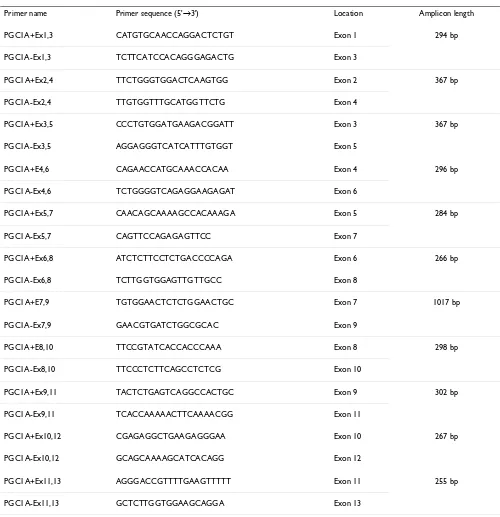
Table 1: Details on exon-spanning primers used for splice variant detection.
Primer name Primer sequence (5'→3') Location Amplicon length
PGC1A+Ex1,3 CATGTGCAACCAGGACTCTGT Exon 1 294 bp
PGC1A-Ex1,3 TCTTCATCCACAGGGAGACTG Exon 3
PGC1A+Ex2,4 TTCTGGGTGGACTCAAGTGG Exon 2 367 bp
PGC1A-Ex2,4 TTGTGGTTTGCATGGTTCTG Exon 4
PGC1A+Ex3,5 CCCTGTGGATGAAGACGGATT Exon 3 367 bp
PGC1A-Ex3,5 AGGAGGGTCATCATTTGTGGT Exon 5
PGC1A+E4,6 CAGAACCATGCAAACCACAA Exon 4 296 bp
PGC1A-Ex4,6 TCTGGGGTCAGAGGAAGAGAT Exon 6
PGC1A+Ex5,7 CAACAGCAAAAGCCACAAAGA Exon 5 284 bp
PGC1A-Ex5,7 CAGTTCCAGAGAGTTCC Exon 7
PGC1A+Ex6,8 ATCTCTTCCTCTGACCCCAGA Exon 6 266 bp
PGC1A-Ex6,8 TCTTGGTGGAGTTGTTGCC Exon 8
PGC1A+E7,9 TGTGGAACTCTCTGGAACTGC Exon 7 1017 bp
PGC1A-Ex7,9 GAACGTGATCTGGCGCAC Exon 9
PGC1A+E8,10 TTCCGTATCACCACCCAAA Exon 8 298 bp
PGC1A-Ex8,10 TTCCCTCTTCAGCCTCTCG Exon 10
PGC1A+Ex9,11 TACTCTGAGTCAGGCCACTGC Exon 9 302 bp
PGC1A-Ex9,11 TCACCAAAAACTTCAAAACGG Exon 11
PGC1A+Ex10,12 CGAGAGGCTGAAGAGGGAA Exon 10 267 bp
PGC1A-Ex10,12 GCAGCAAAAGCATCACAGG Exon 12
PGC1A+Ex11,13 AGGGACCGTTTTGAAGTTTTT Exon 11 255 bp
they could provide an explanation for the regulation of the tissue-dependent functions of PPARGC1A.
Methods
Tissue samples were collected from a freshly slaughtered female, commercial, hybrid pig and immediately sub-merged in RNA later (Sigma-Aldrich, Bornem, Belgium), according to the instructions manual. Testis was collected from a similar male pig. Total RNA was extracted with the Aurum Total RNA Fatty and Fibrous Tissue Kit (Bio-Rad, Nazareth, Belgium), according to the manufacturer's pro-tocol which included an on-column DNase treatment.
Ovaries were collected at a local slaughterhouse from pigs at slaughter age, and used for in vitro embryo production as described in Bijttebier et al. [16]. RNA extraction from embryonic samples (for the 2–4 cell, 8 cell, morula and blastocyst stage respectively 15, 12, 8 and 6 pooled embryos were used) was performed with the PicoPure RNA Isolation Kit (Arcturus, Mountain View, USA), according to the instructions manual, after which a DNase treatment was carried out with RQ1 RNase-free DNase (Promega, Leiden, The Netherlands). Both DNase treat-ments were verified by a minus reverse transcription (RT) control PCR and RNA integrity was checked, as described in Erkens et al. [15]. Also, RNA purity and concentration were measured with the ND-1000 Spectrophotometer (NanoDrop, Wilmington, USA). Next, the iScript cDNA Synthesis Kit (which contains both oligo dT and random primers; Bio-Rad, Nazareth, Belgium) was used to convert approximately 1 μg of RNA from each sample to cDNA, according to the manufacturer's protocol. This RT step was verified by a control PCR [15], in which a no-template control was included to check for DNA contamination. Ready-to-use human cDNA from kidney and liver was provided by Prof. Vandesompele (Department of Pediat-rics and Medical Genetics, Ghent University), to verify whether the detected splice variants also occur in human tissues.
Porcine sequences [GenBank:AH013726] and [Gen-Bank:AY346131], found in the NCBI database [17], were used for the design of exon-spanning primers with Primer3 [18] (Table 1). This way, possible splice variants for each of the 13 exons (except the outer ones) could be detected. During primer design, Mfold [19] and Blast [20] were used to check for possible secondary structures and primer specificity, respectively. PCR conditions for each primer were optimized with FastStart Taq DNA Polymer-ase (Roche, Vilvoorde, Belgium) and included a no-tem-plate control. Also, a genomic DNA control was included to check for possible amplification of pseudogenes. The annealing temperature used for all primer pairs was 60°C.
The amplicons from all primer pairs, except PGC1A+/-Ex7,9, were sequenced by direct sequencing. Because the use of PGC1A+/-Ex7,9 resulted in multiple amplicons, the GENECLEAN II Kit (Qbiogene, Brussels, Belgium) was used to first isolate and purify the separate amplicons from the agarose gel, before sequencing. Sequencing of the amplicons was conducted on an Applied Biosystems 3730xl DNA Analyser with the BigDye Terminator v3.1 Cycle Sequencing Kit (Applied Biosystems, Lennik, Bel-gium), according to the manufacturer's protocol.
Results and discussion
All exon-spanning primer pairs in all tissues resulted in one amplicon of the expected length (Table 1), except for
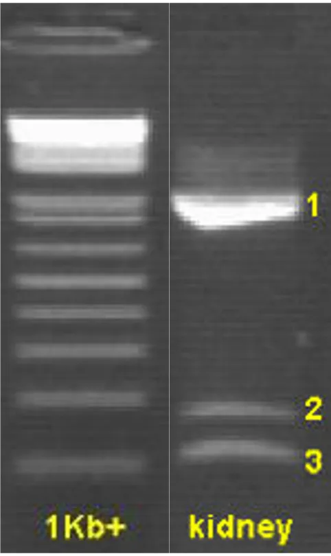
[image:3.612.54.295.85.490.2]Agarose gel showing the 3 different exon 8 amplicons from primer PGC1A+/-Ex7,9
Figure 1
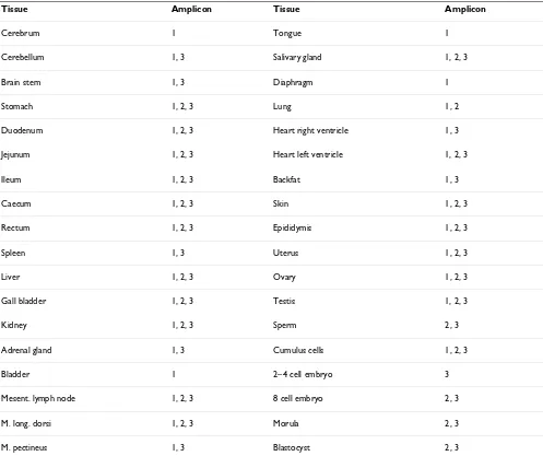
PGC1A+/-Ex7,9. With this last primer pair, 3 different amplicons were detected and the results were tissue-dependent (Table 2). The forward and reverse primer of PGC1A+/-Ex7,9 are located in exon 7 and exon 9, respec-tively, which means its amplicon completely contains exon 8. Besides the expected fragment of 1017 bp (which incorporates all 916 bp of exon 8), 2 new splice variants of PPARGC1A in the pig were detected. Sequencing revealed that in these splice variants exon 8 was either partly (the last 66 bp of exon 8 were conserved) or com-pletely spliced out, which resulted in amplicons of 167 and 101 bp, respectively (Figure 1, 2). Additional PCRs with longer amplicons (exon 4–9 and exon 4–12) con-firmed the existence of both exon 8 splice variants and indicate that the other exons were preserved in the rest of the transcript. Taken together with the fact that no pseu-dogenes for PPARGC1A are described in any species and the use of a genomic DNA control, this makes it unlikely that our observations are the result of pseudogenes.
As can be seen from Figure 2, both splice variant bounda-ries have GC-AG splice sites, instead of following the more common GT-AG rule. Table 2 shows that one or both splice variants were found in almost every tissue that was
tested. They could also be detected in the pre-implanta-tion embryonic stages, which implies PPARGC1A is involved in early development. An interesting finding however, is the fact that the complete amplicon (1017 bp) was not detected in any of the 4 embryonic stages. This could indicate certain functions of PPARGC1A are switched off or altered during early development, but without a functional analysis of the splice variants it is not possible to discuss the effects on its functionality or to give an explanation for these findings. In testis, a PCR artefact (221 bp) was detected with a 92% identity to human outer dense fibre of sperm tails 2 (ODF2) and was depos-ited in GenBank as an EST [GenBank:EY122774].
Only very little is known about the existence of splice var-iants of PPARGC1A in any species, and up until now noth-ing was known about it in the pig. There have been previous reports suggesting the existence of splice variants in which exon 8 was possibly spliced out, in rat skeletal muscle and brown adipose tissue, but their sequence was not determined [21,22]. However, this is the first study giving a detailed description of the actual presence and sequence of 2 splice variants in a whole range of porcine tissues.
[image:4.612.55.562.84.372.2]Nucleotide sequence of the 3 porcine PPARGC1A amplicons from primer PGC1A+/-Ex7,9
Figure 2
Nucleotide sequence of the 3 porcine PPARGC1A amplicons from primer PGC1A+/-Ex7,9. CAmpl: nucleotide sequence of the complete amplicon (1017 bp); SV2: nucleotide sequence of amplicon from splice variant in which last 66 bp of exon 8 are conserved (167 bp); SV3: nucleotide sequence of amplicon in which exon 8 is completely spliced out (101 bp).
1 exon7 exon8 130 CAmpl TGTGGAACTCTCTGGAACTGCAGGCCTAACTCCACCCACCACTCCTCCTCATAAAGCCAACCAAGATAACCCTTTTAGGGCTTCTCCAAAGCTGAAGCCCCCTTGCAAGACTGTGGTACCTCCGCCATCG SV2 TGTGGAACTCTCTGGAACTGCAG SV3 TGTGGAACTCTCTGGAACTGCAG 131 260 CAmpl AAGAAGACCCGGTACAGTGAGTCTTCGGGGACCCACGGCAACAACTCCACCAAGAAAGGGCCCGAGCAGTCCGAGCTGTACGCGCAGCTCAGCAAGACGTCCGCGCTCGGCGGCGGACACGAGGAACGGA SV2 SV3 261 390 CAmpl AGGCCAGGCGGCCCAGTCTGCGGCTATTTGGTGACCATGACTATTGTCAGTCGATTAATTCCAAAGCGGAAATCCTCATCAATATATCGCAGGAGCTCCACGACTCCAGACAACTAGACTCTAAAGATGC SV2 SV3 391 520 CAmpl CGCCTCTGACTGGCAGAGGCAGATGTGTTCTTCCACAGACTCAGACCAGAGCTACCTGACCGAGACGTCGGAGGCGAGCAGGCAGGTCTCTCCGGGCAGCGCCCGAAAACAGCTCCAAGACCAGGAAATC SV2 SV3 521 650 CAmpl CGAGCCGAGCTGAACAAGCACTTCGGTCATCCCAGTCAAGCTGTTTTTGACGACGAAGCAGACAAGACCAGTGAACTGAGGGACAGTGATTTCAGTAACGAACAATTCTCCAAACTACCTATGTTTATAA SV2 SV3 651 780 CAmpl ATTCAGGACTAGCCATGGATGGCCTGTTTGATGACAGCGAAGATGAAAGTGATAAACTGAACTCCCCTTGGGATGGCACGCAGTCCTATTCATTGTTCGATGTGTCGCCTTCTTGTTCTTCTTTTAACTC SV2 SV3 781 910 CAmpl TCCGTGTAGAGATTCCGTATCACCACCCAAATCCTTATTTTCTCAAAGACCCCAAAGGATGCGCTCTCGTTCAAGGTCCTTTTCTCAACACAGGTCGTGTTCTCGATCACCATATTCCAGGTCAAGATCA SV2 GTCGTGTTCTCGATCACCATATTCCAGGTCAAGATCA SV3 911 exon8 exon9 1017
The 2 newly identified splice variants possibly give rise to a much shorter protein of respectively 359 and 337 aa, depending on whether exon 8 is partly or completely spliced out (Figure 3). This is much shorter than the com-plete protein with 796 aa. The first 291 aa of both variants are identical to the complete protein, but the rest of the aa sequence is completely different. It can be expected that this will have an important influence on the function of the produced protein (Figure 3). In the study by Baar et al. [22] an increase of a smaller PPARGC1A protein was detected after exercise in rats. This was consistent with the increase of the smaller cDNA band they detected, although it was not established if that protein originated from the shorter mRNA and if this had any functional sig-nificance.
[image:5.612.57.554.100.517.2]The PPARGC1A protein can generally be divided into 3 regions (Figure 3a). The N-terminal region consists of a transcriptional activation domain which contains an essential LXXLL motif, and is involved in the activation of many transcription factors. A less distinct, central region contains both an inhibitory domain and several interac-tion domains (PPARγ, NRF-1, MEF2C). At the C-terminal end, an RNA processing domain is located, which com-prises 2 serine-arginine-rich domains (SR) and an RNA recognition motif (RRM) [1,23-25]. Figure 3 shows that in the putative protein from both splice variants the N-termi-nal activation domain is conserved. The central region however is only partly conserved. The aa in the first part of the inhibitory and NRF-1 domain are conserved, but the aa of the complete PPARγ interaction region are altered. Table 2: Porcine amplicons detected with primer pair PGC1A+/-Ex7,9.
Tissue Amplicon Tissue Amplicon
Cerebrum 1 Tongue 1
Cerebellum 1, 3 Salivary gland 1, 2, 3
Brain stem 1, 3 Diaphragm 1
Stomach 1, 2, 3 Lung 1, 2
Duodenum 1, 2, 3 Heart right ventricle 1, 3
Jejunum 1, 2, 3 Heart left ventricle 1, 2, 3
Ileum 1, 2, 3 Backfat 1, 3
Caecum 1, 2, 3 Skin 1, 2, 3
Rectum 1, 2, 3 Epididymis 1, 2, 3
Spleen 1, 3 Uterus 1, 2, 3
Liver 1, 2, 3 Ovary 1, 2, 3
Gall bladder 1, 2, 3 Testis 1, 2, 3
Kidney 1, 2, 3 Sperm 2, 3
Adrenal gland 1, 3 Cumulus cells 1, 2, 3
Bladder 1 2–4 cell embryo 3
Mesent. lymph node 1, 2, 3 8 cell embryo 2, 3
M. long. dorsi 1, 2, 3 Morula 2, 3
M. pectineus 1, 3 Blastocyst 2, 3
The second part of the inhibitory domain and NRF-1 interaction region is either altered or absent. The RNA processing domain at the C-terminal end of the complete protein is completely absent in the splice variants. These results suggest that the putative proteins from both splice variants show some remarkable alterations and this is likely to have a large impact on the function of PPARGC1A.
Currently, human medicine shows a great interest in PPARGC1A, because of the important role this gene plays in the worldwide problems concerning obesity, insulin resistance and correlated diseases, such as type 2 diabetes mellitus [26,27]. It has also been shown recently that a lower expression of PPARGC1A is involved in the onset of multiple neurodegenerative diseases, like Parkinson's, Alzheimer's and Huntington's disease [28]. Because of its
significance, human kidney and liver tissue were also tested for the presence of the newly detected splice vari-ants in the pig. This showed that both splice varivari-ants were also found in human liver and only the shortest splice iant in human kidney. In porcine kidney, both splice var-iants were detected, indicating the existence of possible species differences. The discovery of these new splice vari-ants could therefore be of importance for the human research regarding PPARGC1A as well.
Conclusion
The results from this study contribute to a better under-standing of this complex gene and are of possible use not only for research in the pig industry regarding meat qual-ity and carcass composition, but also for human research. Considering the functional domains of the PPARGC1A protein, it is very likely these splice variants considerably
[image:6.612.62.522.90.405.2]Porcine PPARGC1A protein and comparison with the putative aa sequence of both exon 8 splice variants
Figure 3
Porcine PPARGC1A protein and comparison with the putative aa sequence of both exon 8 splice variants. (a) The functional domains of the complete porcine PPARGC1A protein are shown, together with the part of its amino acid sequence (Cprot) that is altered in the splice variants. NRF-1, nuclear respiratory factor 1; PPARγ, peroxisome proliferator-activated receptor γ; MEF2C, myocyte enhancer factor 2C; SR, serine-arginine-rich domain; RRM, RNA recognition motif [1,22-24].(b) The putative protein and aa sequence of the exon 8 splice variant in which the last 66 bp of exon 8 are conserved (SV2). (c) The putative protein and aa sequence of the exon 8 splice variant in which exon 8 is completely spliced out (SV3). * indicates the stop codon of both splice variants.
291 360 Cprot GLTPPTTPPHKANQDNPFRASPKLKPPCKTVVPPPSKKTRYSESSGTHGNNSTKKGPEQSELYAQLSKTS
291 359
SV2 GRVLDHHIPGQDQGPQAVDPLQDLATTLSQATADTARTEILPCAPDHVQDLPTAGGPGMTATRNISTRG*
291 337 SV3 DLATTLSQATADTARTEILPCAPDHVQDLPTAGGPGMTATRNISTRG*
337 359
1 1
LXXLL NRF-33$5Ȗ15)-1 MEF2C SR SR RRM
1 activation domain inhibitory domain 796
(a)
(b)
affect the function of the protein and alternative splicing could be one of the mechanisms by which the diverse functions of PPARGC1A are regulated.
Competing interests
The authors declare that they have no competing interests.
Authors' contributions
TE participated in the study design, performed part of the experimental procedures and was the primary author of the manuscript. KB performed part of the experimental procedures and helped to draft the manuscript. AVZ and LJP participated in the design of the project, helped to draft the manuscript and supervised the study. All authors read and approved the final manuscript.
Acknowledgements
The authors would like to thank Jo Bijttebier and Isabel Lemahieu (Depart-ment of Obstetrics, Reproduction and Herd Health, Ghent University), Filip Barbé (Laboratory of Virology, Faculty of Veterinary Medicine, Ghent University), and Prof. Vandesompele and Nurten Yigit (Department of Pediatrics and Medical Genetics, Ghent University) for providing samples. This work was supported by Ghent University BOF-grant 01J02707.
References
1. Puigserver P, Wu Z, Park CW, Graves R, Wright M, Spiegelman BM:
A cold-inducible coactivator of nuclear receptors linked to adaptive thermogenesis. Cell 1998, 92:829-839.
2. Spiegelman BM, Puigserver P, Wu Z: Regulation of adipogenesis and energy balance by PPARγ and PGC-1. Int J Obes Relat Metab Disord 2000, 24 Suppl 4:S8-S10.
3. Yoon JC, Puigserver P, Chen GX, Donovan J, Wu ZD, Rhee J, Adel-mant G, Stafford J, Kahn CR, Granner DK, Newgard CB, Spiegelman BM: Control of hepatic gluconeogenesis through the tran-scriptional coactivator PGC-1. Nature 2001, 413:131-138. 4. Medina-Gomez G, Gray S, Vidal-Puig A: Adipogenesis and
lipotox-icity: role of peroxisome proliferator-activated receptor γ (PPARγ) and PPARγ coactivator-1 (PGC1). Public Health Nutr
2007, 10:1132-1137.
5. Arany Z, Foo SY, Ma Y, Ruas JL, Bommi-Reddy A, Girnun G, Cooper M, Laznik D, Chinsomboon J, Rangwala SM, Baek KH, Rosenzweig A, Spiegelman BM: HIF-independent regulation of VEGF and ang-iogenesis by the transcriptional coactivator PGC-1α. Nature
2008, 451:1008-1012.
6. Larrouy D, Vidal H, Andreelli F, Laville M, Langin D: Cloning and mRNA tissue distribution of human PPARγ coactivator-1. Int J Obesity 1999, 23:1327-1332.
7. Dulloo AG, Samec S: Uncoupling proteins: their roles in adap-tive thermogenesis and substrate metabolism reconsidered.
Br J Nutr 2001, 86:123-139.
8. Lin J, Wu H, Tarr PT, Zhang C-Y, Wu Z, Boss O, Michael LF, Puig-server P, Isotani E, Olson EN, Lowell BB, Bassel-Duby R, Spiegelman BM: Transcriptional co-activator PGC-1α drives the forma-tion of slow-twitch muscle fibres. Nature 2002, 418:797-801. 9. Mortensen OH, Frandsen L, Schjerling P, Nishimura E, Grunnet N:
PGC-1α and PGC-1β have both similar and distinct effects on myofiber switching toward an oxidative phenotype. Am J Physiol-Endocrinol Metab 2006, 291:E807-816.
10. Kunej T, Wu XL, Berlic TM, Michal JJ, Jiang Z, Dovc P: Frequency distribution of a Cys430Ser polymorphism in peroxisome proliferator-activated receptor-γ coactivator-1 (PPARGC1) gene sequence in Chinese and Western pig breeds. J Anim Breed Genet 2005, 122:7-11.
11. Jacobs K, Rohrer G, Van Poucke M, Piumi F, Yerle M, Barthenschlager H, Mattheeuws M, Van Zeveren A, Peelman LJ: Porcine PPARGC1A (peroxisome proliferative activated receptor gamma coacti-vator 1A): coding sequence, genomic organization, polymor-phisms and mapping. Cytogenet Genome Res 2006, 112:106-113.
12. Stachowiak M, Szydlowski M, Cieslak J, Switonski M: SNPs in the porcine PPARGC1a gene: interbreed differences and their phenotypic effects. Cell Mol Biol Lett 2007, 12:231-239.
13. Rohrer GA, Keele JW: Identification of quantitative trait loci affecting carcass composition in swine: I. Fat deposition traits. J Anim Sci 1998, 76:2247-2254.
14. Michael LF, Wu Z, Cheatham RB, Puigserver P, Adelmant G, Lehman JJ, Kelly DP, Spiegelman BM: Restoration of insulin-sensitive glu-cose transporter (GLUT4) gene expression in muscle cells by the transcriptional coactivator PGC-1. Proc Natl Acad Sci USA 2001, 98:3820-3825.
15. Erkens T, van Poucke M, Vandesompele J, Goossens K, Van Zeveren A, Peelman LJ: Development of a new set of reference genes for normalization of real-time RT-PCR data of porcine back-fat and longissimus dorsi muscle, and evaluation with PPARGC1A. BMC Biotechnol 2006, 6:41.
16. Bijttebier J, Van Soom A, Meyer E, Mateusen B, Maes D: Preovula-tory follicular fluid during in vitro maturation decreases polyspermic fertilization of cumulus-intact porcine oocytes in vitro maturation of porcine oocytes. Theriogenology 2008,
70:715-724.
17. National Center for Biotechnology Information [http:// www.ncbi.nlm.nih.gov]
18. Rozen S, Skaletsky HJ: Primer3 on the WWW for general users and for biologist programmers. Bioinformatics Methods and Proto-cols: Methods in Molecular Biology 2000:365-386 [http:// frodo.wi.mit.edu/cgi-bin/primer3/primer3_www.cgi]. Totowa: Humana Press
19. Zuker M: Mfold web server for nucleic acid folding and hybrid-ization prediction. Nucleic Acids Res 2003, 31:3406-3415 [http:// frontend.bioinfo.rpi.edu/applications/mfold/cgi-bin/dna-form1.cgi]. 20. Altschul SF, Gish W, Miller W, Myers EW, Lipman DJ: Basic local
alignment search tool. J Mol Biol 1990, 215:403-410 [http:// www.ncbi.nlm.nih.gov/blast].
21. Kakuma T, Wang Z-W, Pan W, Unger RH, Zhou Y-T: Role of leptin in peroxisome proliferator-activated receptor gamma coac-tivator-1 expression. Endocrinology 2000, 141:4576-4582. 22. Baar K, Wende AR, Jones TE, Marison M, Nolte LA, Chen M, Kelly
DP, Holloszy JO: Adaptations of skeletal muscle to exercise: rapid increase in the transcriptional coactivator PGC-1.
FASEB J 2002, 16:1879-1886.
23. Wu Z, Puigserver P, Andersson U, Zhang C, Adelmant G, Mootha V, Troy A, Cinti S, Lowell B, Scarpulla RC, Spiegelman BM: Mecha-nisms controlling mitochondrial biogenesis and respiration through the thermogenic coactivator PGC-1. Cell 1999,
98:115-124.
24. Puigserver P, Spiegelman BM: Peroxisome proliferator-activated receptor-γ coactivator 1α (PGC-1α): transcriptional coacti-vator and metabolic regulator. Endocr Rev 2003, 24:78-90. 25. Sano M, Tokudome S, Shimizu N, Yoshikawa N, Ogawa C, Shirakawa
K, Endo J, Katayama T, Yuasa S, Ieda M, Makino S, Hattori F, Tanaka H, Fukuda K: Intramolecular control of protein stability, sub-nuclear compartmentalization, and coactivator function of peroxisome proliferator-activated receptor γ coactivator 1α. J Biol Chem 2007, 282:25970-25980.
26. Ek J, Andersen G, Urhammer SA, Gæde PH, Drivshom T, Borch-Johnsen K, Hansen T, Pedersen O: Mutation analysis of peroxi-some proliferator activated receptor-γ coactivator-1 (PGC-1) and relationships of identified amino acid polymorphisms to type II diabetes mellitus. Diabetologia 2001, 44:2220-2226. 27. Hammarstedt A, Jansson P-A, Wesslau C, Yang X, Smith U: Reduced
expression of PGC-1 and insulin-signaling molecules in adi-pose tissue is associated with insulin resistance. Biochem Bio-phys Res Commun 2003, 301:578-582.
28. St-Pierre J, Drori S, Uldry M, Silvaggi JM, Rhee J, Jäger S, Handschin C, Zheng K, Lin J, Yang W, Simon DK, Bachoo R, Spiegelman BM: Sup-pression of reactive oxygen species and neurodegeneration by the PGC-1 transcriptional activators. Cell 2006,