Open Access
Vol 10 No 5Research article
ERalpha-status of disseminated tumour cells in bone marrow of
primary breast cancer patients
Tanja Fehm
1, Natalia Krawczyk
1, Erich-Franz Solomayer
1, Graziella Becker-Pergola
1, Silke
Dürr-Störzer
1, Hans Neubauer
1, Harald Seeger
1, Annette Staebler
2, Diethelm Wallwiener
1and
Sven Becker
11Department of Obstetrics and Gynecology, University of Tuebingen, Calwerstrasse 7, D-72076 Tuebingen, Germany 2Department of Pathology, University of Tuebingen, Liebermeisterstrasse 8, D-72076 Tuebingen, Germany
Corresponding author: Tanja Fehm, tanja.fehm@t-online.de
Received: 1 Jul 2008 Revisions requested: 14 Aug 2008 Revisions received: 11 Sep 2008 Accepted: 15 Sep 2008 Published: 15 Sep 2008
Breast Cancer Research 2008, 10:R76 (doi:10.1186/bcr2143)
This article is online at: http://breast-cancer-research.com/content/10/5/R76 © 2008 Fehm et al.; licensee BioMed Central Ltd.
This is an open access article distributed under the terms of the Creative Commons Attribution License (http://creativecommons.org/licenses/by/2.0), which permits unrestricted use, distribution, and reproduction in any medium, provided the original work is properly cited.
Abstract
Introduction Isolated disseminated tumour cells (DTC) are regarded as surrogate markers for minimal residual disease in breast cancer. Characterisation of these cells could help understand the known limitations of adjuvant therapy. Of particular interest is their oestrogen-receptor (ER) status because endocrine adjuvant therapy remains a cornerstone of breast cancer treatment.
Methods Bone marrow (BM) aspirates from 254 patients with primary breast cancer were included in this study. A double immunofluorescence staining procedure was established for the identification of cytokeratin (CK) positive/Erα-positive cells. ERα status of the primary tumour was assessed immunohistochemically using the same antibody against ERα.
Results In 107 of 254 (42%) breast cancer patients, CK-positive cells could be detected in the BM. More than one DTC
in the BM was observed in 38 of the 107 patients. The number of detected cells ranged between 1 and 55 cells per 2 × 10⁶
mononuclear cells. DTCs demonstrated ERα positivity in 12% of the patients. The ERα expression was heterogeneous in 10 of the 38 (26%) patients with more than one DTC. The concordance rate of ERα status between primary tumour and DTC was 28%. Only 12 of 88 patients with ERα-positive tumours also had ERα-positive DTCs.
Conclusions Primary tumours and DTCs displayed a concordant ERα status in only 28% of cases. Most of the DTCs were ERα negative despite the presence of an ERα-positive primary tumour. These findings further underline the distinct nature of DTCs and may explain the failure rates seen in conventional endocrine adjuvant therapy.
Introduction
Tumour cell dissemination is a common phenomenon in breast cancer where isolated disseminated cells can be detected in up to 40% of patients at the time of primary diagnosis [1-3]. Based on the pooled analysis of the bone marrow (BM) micrometastasis group, disseminated tumour cells (DTC) are a surrogate marker of minimal residual disease. Their presence is associated with a poor prognosis [4]. With their prognostic significance clearly demonstrated, efforts have been made to further characterise these cells using pheno- and genotyping techniques. Studies have shown that the persistence of DTCs
in the BM of patients with primary breast cancer after conven-tional adjuvant therapy is associated with a poor prognosis [5-7].
More detailed knowledge about their cellular and molecular characteristics could help define a targeted secondary adju-vant therapy in patients with primary breast cancer who have undergone conventional adjuvant therapy. It has already been shown that about 40% of DTCs express human epidermal growth factor receptor 2 (HER2) and that in some patients with recurrent breast cancer their HER2 status may differ from
that of the primary tumour [8]. Since the most widely used form of targeted therapy for breast cancer remains anti-oestrogen endocrine therapy, it is important to know if the ERα status of DTCs corresponds to the ERα status of the primary tumour, particularly in view of the 15 to 20% relapse rate in early stage ERα-positive tumours despite adjuvant endocrine therapy [9]. Furthermore, while ERα-negative tumours are not considered candidates for endocrine therapy, the ERα status of DTCs may differ from the primary tumour. The goal of this study was to determine the ERα status of DTCs in BM of breast cancer patients, and to compare the ERα status of DTCs and the cor-responding primary tumours.
Materials and methods
Collection and analysis of bone marrow
Prior to any therapy, between 10 and 20 ml of bone marrow were aspirated from the anterior iliac crest of 254 primary breast cancer patients undergoing surgical treatment from 2005 to 2007 at the Department of Gynecology and Obstet-rics, University Hospital Tuebingen, Germany.
The characteristics of the patients are shown in Table 1. All specimens were obtained after written informed consent was given and were collected using protocols approved by the institutional review board (114/2006A). Tumour cell isolation and detection was performed based on the recommendations for standardised tumour cell detection [10]. BM samples were separated by density centrifugation over Ficoll with a density of 1.077 g/ml (Biochrom, Germany). If necessary red blood cells were lysed with lysis buffer (155 mM NH4Cl, 10 mM
KHC03, 0.1 mM EDTA pH 7.2). Using a cytocentrifuge (Het-tich, Tuttlingen, Germany), 106 mononuclear cells were spun
onto a glass slide. The slides were air-dried overnight at room temperature. For detection and characterisation of DTCs, slides were fixed in a 0.5% neutral buffered formalin solution for 10 minutes. Control cytospins with ERα-positive MCF-7 cells were prepared, stored and fixed in the same way to ensure that ERα negativity of a patient's sample was not due to a handling error. Two slides per patient was analysed for the presence of DTCs (2 × 10⁶ cells per patient).
Optimising the ERα staining protocol
For establishing the ERα staining procedure, preparations of breast cancer cell lines MCF-7 and SKBR3 mixed with either BM or peripheral blood mononuclear cells (PBMCs) from a healthy volunteer were used (Figure 1). To optimise the stain-ing procedure, all relevant parameters of the protocol were evaluated as follows: types of primary ERα antibodies used were monoclonal mouse antibodies (NCL-L-ER-6F11, Novo-castra Laboratories, UK), polyclonal rabbit antibodies (H-184, Santa Cruz Biotechnology, Inc., CA) and monoclonal rabbit antibodies (SP1, Lab Vision, CA); antibody dilutions used were 1:200, 1:100, 1:50 and 1:25 made with DAKO Antibody Diluent (1% BSA in PBS, 0.1% Tween 20); incubation times for primary and secondary antibodies were 30, 45 and 60
[image:2.612.315.548.108.538.2]min-utes; selection of secondary antibodies was with Tex-Red labelled horse anti-mouse AB (Vector Laboratories, Inc., CA), Tex-red labelled goat anti-rabbit AB (CB 11, Biogenex, CA) and Alexa Fluor 594 labelled goat anti rabbit AB (Molecular Probes, Invitrogen, CA); cell fixation was 10 minutes of ace-tone at 4°C, 100% ethanol for 10 minutes or 0.5% neutral buffered formalin solution for 10 minutes, all three fixations at room temperature. The optimal ERα staining (low background, strong nuclear staining, no cytoplasmic staining) was deter-mined to be as indicated below.
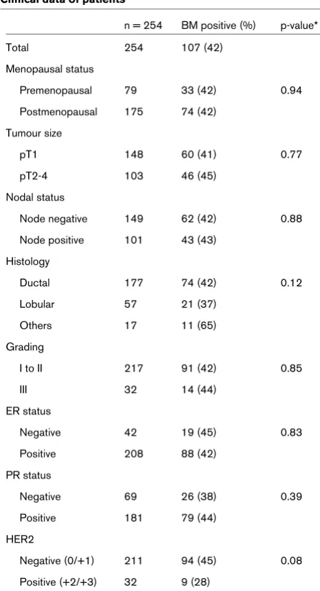
Table 1
Clinical data of patients
n = 254 BM positive (%) p-value*
Total 254 107 (42)
Menopausal status
Premenopausal 79 33 (42) 0.94
Postmenopausal 175 74 (42)
Tumour size
pT1 148 60 (41) 0.77
pT2-4 103 46 (45)
Nodal status
Node negative 149 62 (42) 0.88
Node positive 101 43 (43)
Histology
Ductal 177 74 (42) 0.12
Lobular 57 21 (37)
Others 17 11 (65)
Grading
I to II 217 91 (42) 0.85
III 32 14 (44)
ER status
Negative 42 19 (45) 0.83
Positive 208 88 (42)
PR status
Negative 69 26 (38) 0.39
Positive 181 79 (44)
HER2
Negative (0/+1) 211 94 (45) 0.08
Positive (+2/+3) 32 9 (28)
* by Chi-squared test. BM = bone marrow; ER = oestrogen receptor; HER2 = human epidermal growth factor receptor 2; PR =
Immunofluorescence staining of ERα-receptor
After an initial washing step with PBS (Sigma, Munich, Ger-many), cells were blocked for 30 minutes with normal goat serum (Dako, Glostrup, Denmark) at a 1:10 dilution. The auto-mated double immunofluorescence staining procedure was performed on the DAKO Autostainer using the monoclonal rabbit ERα-antibody SP1 (dilution 1:25, Lab Vision, Fremont, CA, USA) for 60 minutes and secondary detection with a goat anti-rabbit antibody, labelled with Alexa Fluor 594 (1:100, Inv-itrogen Molecular Probes, Carlsbad, CA, USA) for 30 minutes. Cytospins were then incubated with a pan-cytokeratin (CK) antibody (C11) directly conjugated to fluorescein isothiocy-anate (FITC) (1:100, Sigma, Munich, Germany) for 30 min-utes. This monoclonal antibody recognises human CKs 4, 5, 6, 8, 10, 13 and 18. Counterstaining was performed with 4'6-dia-midino-2-phenylindole (DAPI) in mounting media (Vector Lab-oratories, Burlingame, CA, USA). Preparations of the breast cancer cell line MCF-7 mixed with PBMCs from a healthy vol-unteer served as a positive control for CK and ERα staining. ERα negative control slides of SKBR-3/PBMC mixtures were also included with each batch of samples. Cytospins of PBMCs with no added tumour cells served as a negative con-trol for both.
Fluorescence microscopy
Slides were manually analysed for the presence of tumour cells using a computerised fluorescence microscope Axiophot (×40 oil immersion objectives, Carl Zeiss Micro Imaging GmbH, Göttingen, Germany). To screen for ERα-positive tumour cells, a single-pass filter for individual fluorochromes, FITC, Texas Red or DAPI, and a dual-pass filter for FITC/Texas Red were used. Criteria for evaluation of immunostained cells were based on the criteria of the International Society of Hematotherapy and Graft Engineering Working group for standardisation of tumour cell detection and the consensus statements [10,11]. Criteria for ERα positivity were either moderate or intense staining of the entire nucleus. Slides were evaluated by two, or in doubtful cases three, independent investigators (TF, NK and ES).
Immunohistochemical staining of the primary tumour
Immunohistochemical analysis was performed either on core biopsies or surgical resection specimens. The tissue was fixed in 4.5% buffered formalin (pH 7.0) and embedded in paraffin. Immunohistochemical staining was performed on 3 to 5 μm thick sections using a commercially available ABC kit (Vectastain, Vector Laboratories, Burlingame, CA, USA). The ERα antibody (clone SP1) was diluted 1:200 in Tris-HCl (pH 7.5) and applied according to the manufacturer's instruction (DCS, Hamburg, Germany). 3,3'diaminobenzidine (DAB) was used as a chromogen. Finally, the slides were counterstained with haematoxylin and mounted for examination. For assess-ment of the ERα status, the percentage of cells with nuclear reactivity (score 0: none, 1: > 10%, 2: 10 to 50%, 3: 51 to 80%, 4: > 80%) and the intensity of ER staining (score 0: none, 1: weak, 2: moderate, 3: strong) was determined. ERα expression was scored semi-quantitatively using the Rem-mele-score (score nuclear staining × score intensity of ER staining). Tumours with a score of 2 or more were considered ERα positive.
Statistical analysis
A chi-squared test or Fisher's exact test was used to evaluate the relation between ERα-positive DTCs and clinicopatholog-ical factors. Statistclinicopatholog-ical analysis was performed by SPSS, ver-sion 11.5 (SPSS Inc., Chicago, IL, USA). p < 0.05 was considered statistically significant.
Results
Patients' charateristics
[image:3.612.55.298.90.219.2]A total of 254 patients were included in the study. Clinical data are shown in detail in Table 1. Of patients, 82% had ERα -pos-itive primary tumours and DTCs were observed in 107 (42%) of them. Figure 2 shows the cytomorphology and immunophe-notype of a representative DTC of a patient with breast cancer. As can be seen, the nuclear to cytoplasmic ratio is high, the Figure 1
Oestogen receptor (ER) α staining of MCF-7 (positive control) and SKBR3 (negative control) breast cancer cells spiked in bone marrow Oestogen receptor (ER) α staining of MCF-7 (positive control) and SKBR3 (negative control) breast cancer cells spiked in bone marrow. A: MCF-7 cancer cells as positive control for ERα-staining. B: SKBR3 cancer cells as negative control ERα-staining.
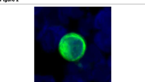
Figure 2
Typical cytomorphology (nuclear size clearly enlarged, high nuclear to cytoplasmic ratio) and immunophenotype (irregular cytoplasmic stain-ing for cytokeratin, cytokeratin filaments can be seen) of a representa-tive disseminated tumour cell from a breast cancer patient
[image:3.612.316.556.521.657.2]nucleus has irregularities and the CK stains the cytoplasm at the periphery of the cell causing a ring-like appearance. These are all accepted morphological criteria for malignant cells. The number of DTCs ranged from 1 to 55 cells/patient (2 × 10⁶
mononuclear cells). In 38 of the 107 (35%) BM-positive patients, more than one DTC could be detected. No correla-tion was observed between positive BM status and any of the established prognostic markers including the ERα status of the primary tumour (Table 1).
ERα expression in disseminated tumour cells
ERα status of DTCs was simultaneously evaluated using a double immunofluorescence staining procedure. The majority of patients (88%) had ERα-negative tumour cells in BM (Table 2). ERα-positive tumour cells could only be detected in 13 of 107 (12%) patients with BM involvement. ERα-positive but CK-negative cells were not observed. Figure 3 shows ERα -positive tumour cells from different patients. As can be seen, the nuclei are strongly stained with the ER antibody.
[image:4.612.57.558.115.205.2]Of the 107 patients, 38 had more than one DTC in the BM. Of Table 2
Correlation between ERα status of primary tumour and disseminated tumour cells
ERα status DTC Total (%)
ERα negative (%) ERα positive (%)
Tumour ERα negative (%) 18 (17) 1 (1) 19 (18)
ERα positive (%) 76 (71) 12 (11) 88 (82)
Total (%) 94 (88) 13 (12) 107 (100)*
*p = 0.8 (chi-squared-test). ER = oestrogen receptor; DTC = disseminated tumour cells.
Figure 3
Immunophenotyping of disseminated tumour cells from patients with primary breast cancer
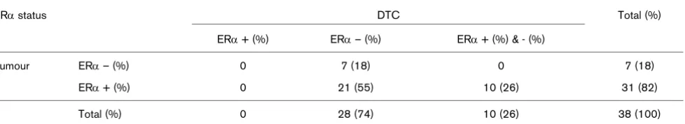
[image:4.612.56.552.377.679.2]these 38 patients, 28 had only ERα-negative tumour cells (Table 3). Heterogeneity of ERα expression could be detected in the remaining 10 (26%) patients (Figure 3i).
Comparison of ERα expression between primary tumour
and disseminated tumour cells
The ERα status of the primary tumour could be determined in all 107 patients with detectable DTCs in the BM. ERα positiv-ity of the primary tumour was demonstrated in 88 (82%) of these patients. The concordance rate between ERα status of DTCs and primary tumour was 28%. Only 12 of the 88 (14%) patients with ERα-positive primary tumour had ERα-positive DTCs in the BM. In contrast, 18 of 19 (95%) patients with ERα-negative primary tumours also had ERα-negative DTCs (Table 2). The extent of ERα expression (negative, low, moder-ate or strong) of the primary tumour was not correlmoder-ated to the ERα status of DTCs. The comparison of ERα expression between primary tumours and DTCs is summarised in Tables 2 and 3.
Discussion
Evaluation of ERα status of the primary tumour by immunohis-tochemistry has been part of routine clinical practice for many years and currently determines patient eligibility for adjuvant endocrine therapy. The assumption is that DTCs will share most characteristics with the primary tumour.
However, an increasing number of publications indicate a more complex relation between the primary tumour and DTCs, with considerable discrepancies noted at the genomic level [12,13]. Supporting this evidence at the phenotypic level are studies looking at HER2 status differences between primary tumours and isolated DTCs [8,14,15].
Similarly, the ERα status of DTCs could be completely differ-ent to that of the primary tumour which on the one hand (ERα -negative primary tumour, ERα-positive DTCs) could increase the number of patients eligible for endocrine therapy and on the other hand (ERα-positive primary tumour, ERα-negative DTCs) could explain why endocrine therapy fails in a subset of hormone receptor-positive patients.
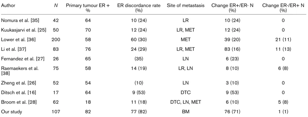
Looking at a large patient group, our data confirms findings of previous, smaller studies, indicating that the ERα status of the primary tumour does not necessarily reflect the ERα status of minimal residual disease (Table 4). In an observational study looking at 17 primary tumours and their corresponding DTCs, Ditsch et al. found that only two of 11 patients with ERα -pos-itive primary tumours (18%) had ERα-positive DTCs [16]. Reuben et al. investigated the ERα status of circulating tumour cells in metastatic breast cancer patients and their corre-sponding primary tumours: fourteen of 16 patients (88%) had ERα-positive primary tumours, but only three patients had ERα-positive circulating tumour cells [17]. Our results confirm the conclusions that DTCs do not reflect the ERα status of the corresponding primary tumour and a majority of DTCs tend to be ERα negative.
As mentioned above, these discrepancies between DTCs and the primary tumour are not confined to ERα-expression: Solo-mayer et al. compared the HER2 status of DTC and primary tumour in 137 cases [8] and found that DTCs were more likely to express HER2 than the primary tumour. Meng et al. reported HER2-positive circulating tumour cells in nine of 24 (38%) patients with recurrent breast cancer who had HER2-negative tumours [15]. It has been suggested that the high rate of HER2-positive DTCs reflects on their potentially more aggres-sive phenotype. Other studies looking at markers such as major histocompatibility complex (MHC) III and Ki-76 have reported similar discrepancies [18,19].
Different hypotheses need to be discussed with regard to our findings. One possible explanation is the clonal heterogeneity of the primary tumour: ERα-negative cells could be more likely to disseminate, corresponding to the worse prognosis of pre-dominantly ERα negative tumours and – inversely – to the demonstrated decreased invasiveness and metastatic poten-tial of ERα-expressing breast cancer cells [20,21]. MCF-7 cells, established from a pleural effusion, express ERα and are oestrogen-responsive breast cancer cells. MCF-7 cells do not form metastases in nude mice unless oestrogen supplementa-tion is provided [22-24]. MDA-MB-231 cells were also estab-lished from a pleural effusion; however, these cells are ERα -negative and highly invasive. Intravenous injection of MDA-Table 3
Correlation between ERα status of primary tumour and heterogeneity of ERα expression in patients with more than one disseminated tumour cell (DTC).
ERα status DTC Total (%)
ERα + (%) ERα – (%) ERα + (%) & - (%)
Tumour ERα – (%) 0 7 (18) 0 7 (18)
ERα + (%) 0 21 (55) 10 (26) 31 (82)
Total (%) 0 28 (74) 10 (26) 38 (100)
[image:5.612.65.554.125.214.2]MB-231 cells into the tail vein of nude mice produces tumours [24]. Furthermore, it is well known that about 20 to 30% of patients with ERα-positive primary tumours develop ERα -neg-ative metastatic diseases [25-28].
One interesting hypothesis currently under discussion is the theory that some or all DTCs, the presumed precursor cells of systemic metastatic disease, are in fact cancer stem cells. As recently published, this theory states that tumour growth and formation of secondary tumours can be traced to a small sub-population of tumour cells, so called cancer stem cells [29,30]. First, most DTCs do not respond to cytotoxic therapy because they are not proliferating and persist over many years in BM. This is also true for tumour stem cells. Secondly, it was also demonstrated that most DTCs in BM were CD44 positive and CD24 low/negative [31]. The CD44-/CD 24-/low pheno-type represents a minor population within primary tumours that is associated with self-renewal and tumourigenic potential. In addition, it has been shown that the CD44+/CD24- pheno-types correlated with a higher prevalence of metastases [32]. As breast cancer stem cells have been shown to be generally ERα negative, DTCs with an ERα-negative phenotype despite an ERα-positive primary tumour would agree with the cancer stem cell theory [33,34].
Conclusion
The phenotypic discrepancies between DTCs and their corre-sponding primary tumours have the potential to increase our understanding of why treatments are successful in some, but not in other patients, paving the way towards more individual-ised forms of treatment. The target of adjuvant therapy is the eradication of minimal residual disease. In order to optimise treatment strategies, the phenotypic properties of DTCs – the surrogate marker of minimal residual disease – should be
taken into account in addition to characterisation of the pri-mary tumour. Already, the available studies looking at pheno-typic properties of DTCs have often found them to be non-proliferative, ERα negative and HER2 positive [8,16,28]. For these patients, expanded treatment with HER2-specific thera-pies (e.g. trastuzumab and lapatinib) could prove especially beneficial. To further clarify these questions, the next step should be a more generalised and systematic characterisation of DTC-status before and after standard adjuvant therapy for all patients.
Competing interests
The authors declare that they have no competing interests.
Authors' contributions
TF, GPB, SD, NK, AS and ES made substantial contributions to the conception and design of the study, acquisition of data, and analysis and interpretation of data. TF, SB, HS and HN were involved in drafting the manuscript or revising it. All authors read and approved the final manuscript.
Acknowledgements
We would like to thank Dr Jonathan Uhr and Nancy Lane (UT South-western Medical School, Dallas, USA) for reviewing the manuscript. This work was supported by the IZKF-grant (1686-0-0) of the University of Tuebingen.
References
1. Diel IJ, Kaufmann M, Costa SD, Holle R, von Minckwitz G, Solo-mayer EF, Kaul S, Bastert G: Micrometastatic breast cancer cells in bone marrow at primary surgery: prognostic value in comparison with nodal status. J Natl Cancer Inst 1996, 88:1652-1658.
[image:6.612.58.555.118.308.2]2. Solomayer EF, Diel IJ, Salanti G, Hahn M, Gollan C, Schütz F, Bastert G: Time independence of the prognostic impact of tumor cell detection in the bone marrow of primary breast can-cer patients. Clin Cancer Res 2001, 7:4102-4108.
Table 4
Comparison of ERα status of the primary tumour and metastatic lesion§
Author N Primary tumour ER +
%
ER discordance rate (%)
Site of metastasis Change ER+/ER- N (%)
Change ER-/ER+ N (%)
Nomura et al. [35] 42 64 10 (24) LR 10 (24) 0
Kuukasjarvi et al. [25] 50 70 12 (24) LR, MET 12 (24) 0
Lower et al. [36] 200 58 60 (30) MET 39 (20) 21 (11)
Li et al. [37] 83 76 24 (29) LR, MET 83 (16) 11 (13)
Fernandez et al. [27] 26 65 (35) LN 6 (23) 0
Raemaekers et al. [38]
75 58 14 (19) LR, LN 8 (10) 6 (8)
Zheng et al. [26] 52 54 (10) LN 3 (10) 0
Ditsch et al. [16] 17 64 9 (53) DTC 9 (53) 0
Broom et al. [28] 62 18 11 (18) DTC, LN, MET 6 (10) 5 (8)
Our study 107 82 77 (82) BM 76 (71) 1 (1)
§distant metastasis, local recurrence, lymph nodes
3. Braun S, Pantel K, Müller P, Janni W, Hepp F, Kentenich CR, Gas-troph S, Wischnik A, Dimpfl T, Kindermann G, Riethmüller G, Schlimok G: Cytokeratin-positive cells in the bone marrow and survival of patients with stage I, II, or III breast cancer. N Engl J Med 2000, 342:525-533.
4. Braun S, Vogl FD, Naume B, Janni W, Osborne MP, Coombes RC, Schlimok G, Diel IJ, Gerber B, Gebauer G, Pierga JY, Marth C, Oruzio D, Wiedswang G, Solomayer EF, Kundt G, Strobl B, Fehm T, Wong GY, Bliss J, Vincent-Salomon A, Pantel K: A pooled anal-ysis of bone marrow micrometastasis in breast cancer. N Engl J Med 2005, 353:793-802.
5. Wiedswang G, Borgen E, Kåresen R, Qvist H, Janbu J, Kvalheim G, Nesland JM, Naume B: Isolated tumor cells in bone marrow three years after diagnosis in disease-free breast cancer patients predict unfavorable clinical outcome. Clin Cancer Res 2004, 10:5342-5348.
6. Braun S, Kentenich C, Janni W, Hepp F, de Waal J, Willgeroth F, Sommer H, Pantel K: Lack of effect of adjuvant chemotherapy on the elimination of single dormant tumor cells in bone mar-row of high-risk breast cancer patients. J Clin Oncol 2000, 18:80-86.
7. Becker S, Becker-Pergola G, Wallwiener D, Solomayer EF, Fehm T: Detection of cytokeratin-positive cells in the bone marrow of breast cancer patients undergoing adjuvant therapy. Breast Cancer Res Treat 2006, 97:91-96.
8. Solomayer EF, Becker S, Pergola-Becker G, Bachmann R, Krämer B, Vogel U, Neubauer H, Wallwiener D, Huober J, Fehm TN: Com-parison of HER2 status between primary tumor and dissemi-nated tumor cells in primary breast cancer patients. Breast Cancer Res Treat 2006, 98:179-184.
9. Breast International Group (BIG) 1–98 Collaborative Group, Thürl-imann B, Keshaviah A, Coates AS, Mouridsen H, Mauriac L, Forbes JF, Paridaens R, Castiglione-Gertsch M, Gelber RD, Rabaglio M, Smith I, Wardley A, Price KN, Goldhirsch A: A comparison of letrozole and tamoxifen in postmenopausal women with early breast cancer. N Engl J Med 2005, 353:2747-2757.
10. Fehm T, Braun S, Muller V, Janni W, Gebauer G, Marth C, Schindl-beck C, Wallwiener D, Borgen E, Naume B, Pantel K, Solomayer E: A concept for the standardized detection of disseminated tumor cells in bone marrow from patients with primary breast cancer and its clinical implementation. Cancer 2006, 107:885-892.
11. Borgen E, Naume B, Nesland JM, Kvalheim G, Beiske K, Fodstad O, Diel I, Solomayer EF, Theocharous P, Coombes RC, Smith BM, Wunder E, Marolleau JP, Garcia J, Pantel K: Standardization of the immunocytochemical detection of cancer cells in BM and blood. I. Establishment of objective criteria for the evaluation of immunostained cells. Cytotherapy 1999, 5:377-388. 12. Schmidt-Kittler O, Ragg T, Daskalakis A, Granzow M, Ahr A,
Blank-enstein TJ, Kaufmann M, Diebold J, Arnholdt H, Muller P, Bischoff J, Harich D, Schlimok G, Riethmuller G, Eils R, Klein CA: From latent disseminated cells to overt metastasis: genetic analysis of systemic breast cancer progression. Proc Natl Acad Sci USA 2003, 100:7737-7742.
13. Klein CA, Blankenstein TJ, Schmidt-Kittler O, Petronio M, Polzer B, Stoecklein NH, Riethmüller G: Genetic heterogeneity of single disseminated tumour cells in minimal residual cancer. Lancet 2002, 360:683-639.
14. Becker S, Becker-Pergola G, Fehm T, Wallwiener D, Solomayer EF: Her2 expression on disseminated tumor cells from bone marrow of breast cancer patients. Anticancer Res 2005, 25:2171-2175.
15. Klein CA, Blankenstein TJ, Schmidt-Kittler O, Petronio M, Polzer B, Stoecklein NH, Riethmüller G: uPAR and HER-2 gene status in individual breast cancer cells from blood and tissues. Proc Natl Acad Sci USA 2006, 103:17361-17365.
16. Ditsch N, Mayer B, Rolle M, Untch M, Schildberg FW, Funke I: Estrogen receptor expression profile of disseminated epithe-lial tumor cells in bone marrow of breast cancer patients. Recent Results Cancer Res 2003, 162:141-147.
17. Reuben JMLB, Li C, Broglio KR, Valero V, Jackson S, Ueno NT, Krishnamurthy S, Hortobagyi GN, Cristofanilli M: Genomic of cir-culating tumor cells in metastatic breast cancer. J Clin Oncol (Meeting Abstracts) 2007, 25:1002.
18. Pantel K, Schlimok G, Braun S, Kutter D, Lindemann F, Schaller G, Funke I, Izbicki JR, Riethmüller G: Differential expression of
pro-liferation-associated molecules in individual micrometastatic carcinoma cells. J Natl Cancer Inst 1993, 85:1419-1424. 19. Pantel K, Schlimok G, Kutter D, Schaller G, Genz T, Wiebecke B,
Backmann R, Funke I, Riethmüller G: Frequent down-regulation of major histocompatibility class I antigen expression on indi-vidual micrometastatic carcinoma cells. Cancer Res 1991, 51:4712-4715.
20. Thompson EW, Paik S, Brünner N, Sommers CL, Zugmaier G, Clarke R, Shima TB, Torri J, Donahue S, Lippman ME, Martin GR, Dickson RB: Association of increased basement membrane invasiveness with absence of estrogen receptor and expres-sion of vimentin in human breast cancer cell lines. J Cell Physiol 1992, 150:534-544.
21. Platet N, Prevostel C, Derocq D, Joubert D, Rochefort H, Garcia M: Breast cancer cell invasiveness: correlation with protein kinase C activity and differential regulation by phorbol ester in estrogen receptor-positive and -negative cells. Int J Cancer 1998, 75:750-756.
22. Price JE, Polyzos A, Zhang RD, Daniels LM: Tumorigenicity and metastasis of human breast carcinoma cell lines in nude mice. Cancer Res 1990, 50:717-721.
23. Mukhopadhyay R, Theriault RL, Price JE: Increased levels of alpha6 integrins are associated with the metastatic phenotype of human breast cancer cells. Clin Exp Metastasis 1999, 17:325-332.
24. Shafie SM, Liotta LA: Formation of metastasis by human breast carcinoma cells (MCF-7) in nude mice. Cancer Lett 1980, 11:81-87.
25. Kuukasjarvi T, Kononen J, Helin H, Holli K, Isola J: Loss of estro-gen receptor in recurrent breast cancer is associated with poor response to endocrine therapy. J Clin Oncol 1996, 14:2584-2589.
26. Zheng WQ, Lu J, Zheng JM, Hu FX, Ni CR: Variation of ER status between primary and metastatic breast cancer and relation-ship to p53 expression*. Steroids 2001, 66:905-910.
27. Fernandez D, Sauven P, Alaghband-Zadeh J, Burn JI: Variability of oestrogen and progesterone receptor status between primary breast cancer and nodal metastases: preliminary communication. J R Soc Med 1982, 75:719-722.
28. Broom RJ, Tang P, Simmons C, Bordeleau L, O'Malley FP, Miller N, Andrulis IL, Brenner DM, Clemons M: Changes in estrogen receptor (ER), progesterone receptor (PR) and HER2/neu sta-tus with time: Discordance rates between primary and meta-static breast pathology samples. Clin Oncol (Meeting Abstracts) 2007, 25:1024.
29. Reya T, Morrison SJ, Clarke MF, Weissman IL: Stem cells, cancer, and cancer stem cells. Nature 2001, 414:105-111.
30. Al-Hajj M, Wicha MS, Benito-Hernandez A, Morrison SJ, Clarke MF: Prospective identification of tumorigenic breast cancer cells. Proc Natl Acad Sci USA 2003, 100:3983-3988.
31. Balic M, Lin H, Young L, Hawes D, Giuliano A, McNamara G, Datar RH, Cote RJ: Most early disseminated cancer cells detected in bone marrow of breast cancer patients have a putative breast cancer stem cell phenotype. Clin Cancer Res 2006, 12:5615-5621.
32. Abraham BK, Fritz P, McClellan M, Hauptvogel P, Athelogou M, Brauch H: Prevalence of CD44+/CD24-/low cells in breast cancer may not be associated with clinical outcome but may favor distant metastasis. Clin Cancer Res 2005, 11:1154-1159.
33. Dontu G, El-Ashry D, Wicha MS: Breast cancer, stem/progeni-tor cells and the estrogen recepstem/progeni-tor. Trends Endocrinol Metab 2004, 15:193-197.
34. Asselin-Labat ML, Shackleton M, Stingl J, Vaillant F, Forrest NC, Eaves CJ, Visvader JE, Lindeman GJ: Steroid hormone receptor status of mouse mammary stem cells. J Natl Cancer Inst 2006, 98:1011-1014.
35. Nomura Y, Tashiro H, Shinozuka K: Changes of steroid hormone receptor content by chemotherapy and/or endocrine therapy in advanced breast cancer. Cancer 1985, 55:546-551. 36. Lower EE, Glass EL, Bradley DA, Blau R, Heffelfinger S: Impact of
metastatic estrogen receptor and progesterone receptor sta-tus on survival. Breast Cancer Res Treat 2005, 90:65-70. 37. Li BD, Byskosh A, Molteni A, Duda RB: Estrogen and