Available onlinehttp://breast-cancer-research.com/content/9/4/211
Abstract
This review addresses genes differentially expressed in the mammary gland transcriptome during the progression of mammary carcinogenesis in BALB/c mice that are transgenic for the rat neu
(ERBB2, or HER-2/neu) oncogene (BALB-neuT664V-E mice). The
Ingenuity knowledge database was used to characterize four functional association networks whose hub genes are directly linked to inflammation (specifically, the genes encoding IL-1β, tumour necrosis factor, interferon-γ, and monocyte chemoattractant protein-1/CC chemokine ligand-2) and are increasingly expressed during such progression. In silicometa-analysis in a human breast cancer dataset suggests that proinflammatory activation in the mammary glands of these mice reflects a general pattern of human breast cancer.
Introduction
Inflammation, the archetypal response to invasion, has evolved as a local protective response to life-threatening invasion; it is required to be rapid and devastating, sometimes regardless of the cost to the host. When viewed in the light of evolutionary pressure, the frequently disastrous long-term consequences of inflammation are a small price to pay for the protection against the immediate danger avoided.
It is now clear, however, that various distinct reactions are encompassed within the term ‘inflammation’. Development of the immune system has led to sophisticated decoding of danger signals elicited by incursion of foreign bodies, and retaliatory responses are adjusted accordingly. The type of cells recruited, their state of activation, the reactive substances that they release, their guidance by cytokines and soluble factors, and the time frame over which the reaction occurs will all depend on both the type of signals and the ability of the invader to resist responses. Therefore, it is hardly surprising that some of the molecular mechanisms that
sustain flares of inflammation may have opposing effects on the progression of early neoplastic lesions [1].
Acute inflammation cures lethal tumours
Acute inflammation can certainly destroy both initial and established neoplastic lesions. Indeed, much of our current understanding of tumour immunology can be traced back to Cooley’s observations of the ability of the acute inflammation elicited by bacterial infections to cure human carcinomas [2]. Similar observations have also been reported in countless studies of experimental tumours. However, it has also been shown that tumours can be destroyed by a more focused assault triggered by double-strand polynucleotides [3] and unmethylated-CpG oligodesoxynucleotides [4]. Moreover, because cytokines guide inflammatory reactions, most can initiate and guide inflammatory patterns that have a distinct ability to inhibit tumour growth. The powerful anti-tumour activity of tumour necrosis factor (TNF)-αdriven inflammation, for instance, is accompanied by dramatic adverse effects that confine its therapeutic use to isolated tissue perfusion [5]. IL-2 also recruits natural immunity mechanisms that exhibit marked anti-tumour activity in patients with advanced cancer [6]. Administration of minute amounts of IL-2 at the tumour site induces neutrophil infiltration, which produces dramatic, albeit temporary, regression [7].Several lines of experimental data have shown that tumour cells engineered to release a cytokine trigger a strong inflammatory reaction, which results in acute tumour destruc-tion and inducdestruc-tion of tumour-specific immune memory [8]. Systematic investigation of the reaction elicited by a mouse mammary adenocarcinoma cell line (TSA) transfected with several cytokine genes has shown that the cytokines produced induce distinct local inflammatory reactions. Neutrophils are
Review
Inflammation and breast cancer
Inflammatory component of mammary carcinogenesis in ErbB2
transgenic mice
Raffaele Adolfo Calogero, Francesca Cordero, Guido Forni and Federica Cavallo
Molecular Biotechnology Center, Department of Clinical and Biological Sciences, Via Nizza 52, 10126 Turin, Italy
Corresponding author: Guido Forni, guido.forni@unito.it
Published: 10 August 2007 Breast Cancer Research2007, 9:211 (doi:10.1186/bcr1745) This article is online at http://breast-cancer-research.com/content/9/4/211
© 2007 BioMed Central Ltd
the dominant infiltrating cells recruited by TSA-IL-2 cells (TSA cells transfected with the gene encoding IL-2) [9], eosinophils by TSA-IL-4 and TSA-IL-5 cells [10,11], natural killer cells by IL-12 cells [12], and macrophages by TSA-IFN-γ cells [10]. Other cytokines such as IL-7 [13], TNF-α [13] and IFN-α[14], or chemokines such as CC chemokine ligand (CCL)16 (liver expressed chemokine [LEC]) [15] activate acute inflammatory reactions and recruit mixed leucocyte populations. Although reactions elicited by cytokines are markedly different histologically, those elicited by TSA tumour cells engineered to release IL-2, IL-4, IL-7, IL-12, TNF-α, IFN-γand LEC/CCL16 rapidly neutralize a TSA challenge that would otherwise be lethal. Other cytokines only delay tumour growth (IFN-γ) or have no inhibitory effect (IL-5 and IL-6) [10,11,13]. Similarly, protective inflammatory reactions are triggered through repeated local injection of recombinant cytokines [8,16,17].
Inflammation modulates neu (ERBB2, or
HER-2/neu) driven mammary carcinogenesis
The time frame obviously differentiates acute inflammation from the chronic reactions that set the stage for initiation of carcinogenesis and its progression. Because chronic inflam-mation is a long-lasting response to an enduring invasion, it can persist for a significant period of time in the life of the individual. Prolonged release of a mixture of highly reactive oxidants secreted by infiltrating leucocytes may damage the genomes of nearby cells and increase their rate of mutation [18]. In addition to this direct oncogenic effect, a chronically inflamed microenvironment possesses many mechanisms with which to promote progression of a preneoplastic lesion, as described elsewhere in this series of review articles.Cancer-prone transgenic mice provide models of autoch-thonous tumour formation. We conducted a microarray gene expression analysis to determine how proinflammatory genes are switched on during the stepwise progression of
neu oncogene driven mammary carcinogenesis in BALB/c mice.
BALB/c mice transgenic for the transforming rat neu664V-E oncogene under the transcriptional control of the mouse mammary tumour virus promoter (BALB-neuT664V-Emice) are genetically predestined to develop one of the most aggres-sive forms of mammary carcinogenesis, with penetrance of all of their mammary glands. Starting from the week 3 or 4 of age, the protein product of the neu oncogene (p185neu) is diffusely over-expressed in the terminal buds of mammary glands of virgin mice. The over-expressing cells form foci of atypical hyperplasia. At around 8 weeks of age, these enlarge to in situ carcinomas that progress to invasive cancer between weeks 17 and 22. At around weeks 17 to 18 one or more tumours are palpable in the mammary pad of all mice, and at around week 33 a tumour is palpable in each of the 10 mammary glands [19]. The p185neu over-expression in the thymus and mammary gland has a marked effect on the T-cell repertoire of these mice, and CD8+T-cell clones reacting with dominant p185neuepitopes are deleted [20]. Moreover, during progression of the mammary lesions, both suppressor CD4+ CD25+ FOXP3+ GITR+ T
[image:2.612.58.532.94.324.2]reg cells [21] and CD11b+Gr1+immature myeloid cells [22] expand. This mammary carcinogenesis appears to be driven in a straightforward manner by the transgenic neu oncogene, which provides abnormal growth signals and inhibits cell death pathways [23].
Figure 1
Because the genetic alteration is the driving force of tumor development in the BALB-neuT664V-Emodel, and because the mammary gland is not commonly susceptible to chronic inflammation, interactions between the incipient tumour and its inflammatory microenvironment presumably do not play a major role. In contrast, progression of this devastating form of carcinogenesis is markedly modulated by the inflammatory
[image:3.612.60.552.116.623.2]microenvironment. The inflammation-related reaction elicited by repeated injections of recombinant IL-12 during the early stages of carcinogenesis results in restricted and delayed tumour formation [24], mostly resulting from the ability of IL-12 to trigger innate immunity reaction mechanisms and induce downstream factors that inhibit the angiogenic switch, which promotes progression from in situto invasive cancer Available onlinehttp://breast-cancer-research.com/content/9/4/211
Table 1
Functional network generated by Ingenuity knowledge database analysis
Hub gene Score GATMs Top functions
TNF↑ 34 35 Lipid metabolism, small molecule biochemistry, cell death
IFNG↑ 34 35 Lipid metabolism, molecular transport, small molecule biochemistry
IL1B↑ 34 35 Immune response, cellular movement, haematological system development and function
MYC↑ 34 35 Cell cycle, cellular movement, cancer
CCL2↑, KDR↓, VEGF↓ 34 35 Cellular movement, cancer, tumour morphology
SRC↑ 34 35 Haematological system development and function, immune and lymphatic system development and function, tissue morphology
BCL2↑ 34 35 Cancer, cell death, cell morphology
AGT↓ 34 35 Cell-to-cell signalling and interaction, haematological system development and function, immune and lymphatic system development and function
TIMP1↑, EDN1↓ 34 35 Cancer, cellular movement, cellular development
LEP↓ 34 35 Lipid metabolism, molecular transport, small molecule biochemistry
AR↓ 34 35 Cellular growth and proliferation, gene expression, cellular development
IGF2↓, ERBB3↑ 34 35 DNA replication, recombination, and repair, cancer, cell cycle
HOXA9↓, CDH1↑ 34 35 Cellular growth and proliferation, cell death, cancer
ARNT2↑, ZAP70↑ 34 35 Cellular growth and proliferation, haematological system development and function, immune response
PPARA↓, IL6, SERBF1 15 24 Lipid metabolism, small molecule biochemistry, molecular transport
CDC42, RAC1 13 22 Cell signalling, free radical scavenging, nucleic acid metabolism
ERBB2↑ 12 17 Cancer, tumour morphology, cell cycle
FN1 12 21 Cell-to-cell signalling and interaction, cellular movement, reproductive system development and function
FOS 12 21 Gene expression, cell cycle, haematological system development and function
IGF1, CCND1, BMP2 12 21 Cellular growth and proliferation, connective tissue development and function, skeletal and muscular system development and function
IL4 12 21 Cellular assembly and organization, cell cycle, DNA replication, recombination, and repair
NFYB 11 16 Cancer, cell cycle, cellular assembly and organization
IL13, MAPK8, NFKB1 11 20 Haematological system development and function, immune and lymphatic system development and function, tissue morphology
STAT3, INSR, PTPN 11 20 Cellular growth and proliferation, haematological system development and function, immune response
NFKBIA, CD40LG, HGF 11 20 Cell death, cellular movement, haematological system development and function
KRAS, CDKN1A 10 19 Cellular growth and proliferation, cell signalling, cancer
TNF↑ 10 19 Carbohydrate metabolism, molecular transport, small molecule biochemistry
[25]. Conversely, a marked modulation of the progression of
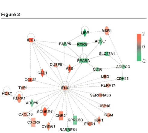
[image:4.612.58.298.92.276.2]neu carcinogenesis is evident in mice in which the gene encoding IFN-γ [26] or that encoding monocyte chemo-attractant protein (MCP)-1/CCL2 [27] has been knocked out. Figure 3
Network of functional association between IFN-γgene and other GATMs generated by Ingenuity database analysis. ADIPOQ, CCL22,
IFNG, IL1B, PPARA, RXRG, MSR1, GAD1and TAP1are genes associated with proliferation, whereas USP18and CDH13are genes linked to growth. ASS, DUSP5, ADCY5and UBDare genes associated with apoptosis/survival. CXCR6, CXCL16and CNR2are genes associated with chemotaxis/trafficking; KLRK1and HCSTare associated with cytolysis/cytotoxicity; and RARRES1and CD36are linked to migration. All of the other genes have unknown cell functions. Genes are shown by their symbols. GATM, gene associated with mammary tumour microenvironment; IFN, interferon.
Figure 2
Network of functional association between TNF gene and other GATMs generated by Ingenuity database analysis. RAET1B,KLRD1
andKLRK1are genes associated with cytotoxicity; TNFRSF21and
AATK are genes involved in apoptosis. LITAF, AK2and KLRC2are genes associated with proliferation regulation. PLA2G7is a gene associated with inflammatory response. All of the other genes have unknown cell functions. Genes are shown by their symbols [44]. The nodes represent the genes and the edges reflect direct links or connections between them. GATM, gene associated with mammary tumour microenvironment; TNF, tumour necrosis factor.
Figure 5
Network of functional association between CCL2 gene and other GATMs generated by Ingenuity database analysis. F3, F2R, F12, F10
and KNG1are part of the complement and coagulation cascades.
MMP12encodes a matrix metalloproteinase involved in tissue remodelling. HBEGFand PGFencode growth factors. Associations with VEGFand KDRhave been omitted for the sake of legibility. Genes are shown by their symbols. CCL, CC chemokine ligand; GATM, gene associated with mammary tumour microenvironment. Figure 4
Network of functional relationships between IL-1βgene and other GATMs generated by Ingenuity database analysis. AIF1, CCL4,
[image:4.612.55.300.392.612.2]Inflammatory gene expression during neu
carcinogenesis
We have used DNA microarray technology to compare patterns of transcription in 2-week pregnant wild-type BALB/c mice with those in the mammary glands of BALB-neuT664V-E mice during progression of neu carcinogenesis [28,29]. We also characterized the transcription profile in the gland when carcinogenesis is halted by immune mechanisms triggered by cell [30] and DNA-based anti-neu vaccines [28,31]. Meta-analysis of these transcription profiles [32] has provided suggestions regarding possible new onco-antigens for use in anti-tumour vaccines.
A recent transcription profiling study using full, genome-wide mouse arrays was run to identify changes in transcription in BALB-neuT664V-Emammary glands progressing from atypical hyperplasia and in situcarcinomas (10- to 15-week-old mice) to invasive cancer (19- to 22-week-old mice) (unpublished data; the microarray dataset has been released, GSE7395, on the GEO database, www.ncbi.nlm.nih.gov/geo). The total RNAs extracted from mammary glands were analyzed using Mouse Genome Survey Microarrays (Applied Biosystems, Foster City, CA, USA). Identification of differentially expres-sed genes associated with the transition from the pre-neoplastic state to neoplasia was done by means of a linear modelling approach [33]. An empirical Bayesian method [34] was used to assess differential expression, along with false
discovery rate correction of the Pvalue [35] to moderate the standard errors of the estimated log-fold changes. We identified 2,758 differentially expressed probes (2,651 transcripts) by applying a false discovery rate of 0.05, associated with an absolute log2(fold change) threshold of 1. These differentially expressed genes represent the element of the mammary gland transcriptome whose expression changes as the tumour microenvironment evolves and the tumour mass increases. Therefore, these transcripts are defined as genes associated with mammary tumour microenvironment (GATMs).
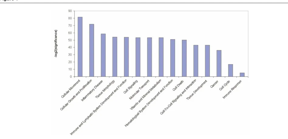
[image:5.612.116.552.101.354.2]The Ingenuity Pathways Knowledge Base [36] is currently the world’s largest database of knowledge on biological networks, with annotation curated by experts. We exploited this database to define the presence of functional associations within the GATMs and to identify differences between the ontological gene classes [37] that were enriched among the upregulated and downregulated genes (Figure 1). This ontological gene classification provides the controlled vocabulary to describe gene and gene product attributes. The principal Ingenuity ontological classes found to be enriched in BALB-neuT664V-E GATM sets are those related to cellular movement and cellular growth and proliferation. However, among the other significantly enriched groups were genes belonging to the classes inflammatory diseases, haematological system development and function, and immune and lymphatic system development and function. Available onlinehttp://breast-cancer-research.com/content/9/4/211
Figure 6
This highlights the collaboration between transformed cells and body defence systems during initial tumour development.
Furthermore, we generated significant functional association networks [37] encompassing GATMs (Table 1). These are graphical descriptions of literature associations identified by the Ingenuity knowledge database, in which gene products are linked if some kind of experimentally measured associa-tion has been reported. The 14 most significant gene networks, each composed of 35 GATMs (nodes), are characterized by the presence of one hub gene (the highest connected node), which is the main player in the biological events that connect the GATMs. Remarkably, four of these 14 networks are highly associated with the inflammatory response: TNF (Figure 2), IFN-γ (Figure 3), IL-1β (Figure 4) and MCP-1/CCL2 (Figure 5).
During neucarcinogenesis, these four functional association networks, centred on genes that encode proinflammatory molecules, can be defined. To ensure that these networks were not an artefact of the experimental model, we assessed their expression in breast cancer specimens in the light of recently published data exploring associations between recurrent copy number abnormalities, gene expression, and clinical outcomes in a set of aggressively treated early-stage breast tumours [38].
The presence of GATMs associated with TNF, IL-1β, IFN-γ, and MCP-1/CCL2 hub genes, and characterized by human orthologs (a total of 77 genes), was sought in the dataset reported by Chin and coworkers [38]. This is one of the largest collections of breast cancer samples (n= 118) analyzed by microarray and for which the clinical outcome is known. The study showed that the accuracy with which early-stage breast cancer patients can be stratified according to outcome can be improved by combining analyses of gene expression and genome copy number.
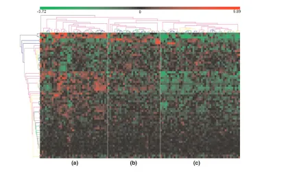
We identified 65 out of 77 GATMs. Hierarchical clustering of their expression profile (a method for dividing a dataset into subsets whose individual data ideally share some common traits [37]) was used to group the sample reported by Chin and coworkers into three subsets (Figure 6). This clustering was based on a proinflammatory gene signature, and it revealed different types or degrees of leucocyte infiltration. This suggests that proinflammatory activation in the mammary glands of BALB-neuT664V-E mice reflects a general pattern of human breast cancer. Only a limited overlap between the A group and a subset of samples characterized by negative oestrogen receptor status was observed (data not shown). This is not surprising, because our study addressed BALB-neuT664V-Ecarcinogenesis sequen-tially, whereas only full-blown cancers were considered in the report by Chin and coworkers [38]. Moreover, sampling bias in the human setting may mask any correlation with clinical outcome.
A complementary dataset that could corroborate our obser-vations is that report by Neve and coworkers [39]. Those investigators found that the recurrent genomic and trans-criptional characteristics of a large set of breast cancer cell lines mirrored those of primary breast tumours, although some significant differences were documented.
The work reported by Chin [38] and Neve [39] and their colleagues offers a unique opportunity to discriminate between transcripts that belong to the tumour micro-environment and those associated with tumour cells. Taking advantage of these transcriptional profiles [38,39] and specific data mining techniques [40], one can determine whether a set of genes shares little expression similarity between tumour specimens and cell lines. This would suggest that their expression is linked to cell infiltrates and does not belong to tumour cells. Such a scenario was actually observed for the set of proinflammatory genes that we identified in BALB-neuT664V-E mice because of the absence of correlation [40] between gene expression observed in tumour specimens [38] and in tumour cell lines [39] (Figure 7).
Figure 7
It should nonetheless be noted that the gene encoding MCP-1/ CCL2 is the only one of the four hub genes (those encoding IFN-γ, TNF, IL-1βand MCP-1/CCL2) that constantly exhibit relatively high levels of expression (Figure 8).
Conclusion
The neu oncogene is the central driving force of the mammary cancer that inexorably kills all female BALB-neuT664V-E mice. Even so, the timeframe within which neu carcinomas appear and progress is modulated by the pro-inflammatory components of the reactive stroma that surrounds the cancer lesions.
Our transcription profiling search for modulation of GATMs during the progression of neu mammary lesions in BALB-neuT664V-Emice generated a large body of data. The Ingenuity knowledge database was used to identify four functional association networks whose hub genes are directly linked to inflammation (namely, the genes that encode IFN-γ, TNF, IL-1β and MCP-1/CCL2). Data from such analyses clearly show that the progression of neu-driven autochthonous carcinogenesis is directly associated with increased expression of these four hub GATMs. These data were from a representative but artificial experimental model. However, an
in silico meta-analysis in a human breast cancer dataset suggests that proinflammatory activation in the mammary
glands of BALB-neuT664V-Emice reflects the general pattern of human breast cancer.
However, the way in which the proinflammatory cytokines encoded by these genes affect the progression of neu
carcinogenesis is neither simple nor unequivocal. Different amounts of the same cytokine in the tumour microenviron-ment may have different influence, whereas the significance of this influence can fluctuate during the distinct stages of cancer progression. For instance, increased expression of IFN-γ in the tumour microenvironment during neu tumour progression does not appear to favour tumour expansion but plays a major inhibitory role. In BALB-neuT664V-E IFN-γ knockout mice, absence of IFN-γ leads to accelerated progression of neu carcinogenesis [26]. Work in progress suggests that this more rapid progression is associated with a major increase in tumoural neoangiogenesis (Iezzi M, unpublished data). Indeed, it is well known that IFN-γ, as well as the downstream factors that it induces, have marked anti-angiogenic activity that naturally restricts tumour neoangio-genesis and hampers tumour growth [41].
[image:7.612.59.555.96.363.2]Moreover, our work on administration of recombinant mouse IL-12 shows that IL-12 mediated inhibition of neu carcino-genesis relies on production of IFN-γ in the tumour micro-environment [41]. In BALB-neuT664V-E IFN-γ knockout mice, Available onlinehttp://breast-cancer-research.com/content/9/4/211
Figure 8
IL-12 no longer impairs tumour growth [25]. We have also found that the ability of IL-12 induced IFN-γ to inhibit the progression of neucarcinogenesis is related to tumour stage. It is very marked during the tumour-induced angiogenic switch that accompanies the passage from an in situ lesion to invasive cancer. The newly formed capillary sprouts that characterize the early events in the angiogenic switch are sensitive to the inhibitory activity of IFN-γ, whereas this activity is nearly absent in the stages that precede and follow the switch [24].
Our data show that the expression of the gene that encodes the MCP-1/CCL2 chemokine increases as neu carcino-genesis progresses. The presence of this chemokine in the reactive stroma around the tumour appears to be directly associated with enhanced progression, because in BALB-neuT664V-E MCP-1/CCL2 knockout mice progression is slower and mice survive longer [27].
The influence of different levels of TNF and IL-1β has not been assessed in BALB-neuT664V-Emice. However, data from other experimental systems suggest that local increases in these factors may favour cancer progression. Skin carcinogenesis is dramatically impaired in TNF-α knockout mice [42]. In various experimental models, IL-1βappears to increase tumour invasiveness and metastasis [43].
In conclusion, our Ingenuity analysis identifies four genes that encode inflammatory cytokines whose increased expression in the tumour microenvironment is naturally associated with mammary cancer progression. Much greater concentrations of the very same cytokines, artificially reached in the tumour microenvironment either through their local injection or from cells engineered to release them, lead to both tumour rejection and induction of long-lasting tumour-specific immune memory [10].
Competing interests
The authors declare that they have no competing interests.
Acknowledgements
This work was supported by grants from Italian Association for Cancer Research; the Italian Ministero dell’Università e della Ricerca; the Uni-versity of Torino; the Compagnia di San Paolo, Torino; the Fondazione Denegri, Torino; and the Regione Piemonte: bando regionale sulla ricerca scientifica applicata 2004.
The Authors wish to thank Ms Irene F Merighi for her skilled technical assistance.
References
1. Karin M, Greten FR: NF-kappaB: linking inflammation and immunity to cancer development and progression. Nat Rev Immunol 2005, 5:749-759.
2. Nauts HC, McLaren JR: Coley toxins: the first century.Adv Exp Med Biol 1990, 267:483-500.
3. Weinstein AJ, Gazdar AF, Sims HL, Levy HB: Lack of correlation between interferon induction and antitumour effect of poly I-poly C.Nat New Biol 1971, 231:53-54.
4. Krieg AM: CpG motifs: the active ingredient in bacterial extracts?Nat Med 2003, 9:831-835.
5. Lejeune FJ, Lienard D, Matter M, Ruegg C: Efficiency of recom-binant human TNF in human cancer therapy. Cancer Immun
2006, 6:6.
6. Atkins MB, Lotze MT, Dutcher JP, Fisher RI, Weiss G, Margolin K, Abrams J, Sznol M, Parkinson D, Hawkins M, et al.: High-dose recombinant interleukin 2 therapy for patients with metastatic melanoma: analysis of 270 patients treated between 1985 and 1993.J Clin Oncol 1999, 17:2105-2116.
7. Cortesina G, De Stefani A, Galeazzi E, Cavallo GP, Badellino F, Margarino G, Jemma C, Forni G: Temporary regression of recurrent squamous cell carcinoma of the head and neck is achieved with a low but not with a high dose of recombinant interleukin 2 injected perilymphatically.Br J Cancer 1994, 69: 572-576.
8. Forni G, Santoni A, Frati L: Cytokine-Induced Tumor Immuno-genicity. London, UK: Academic Press; 1994.
9. Cavallo F, Giovarelli M, Gulino A, Vacca A, Stoppacciaro A, Modesti A, Forni G: Role of neutrophils and CD4+T
lympho-cytes in the primary and memory response to nonimmuno-genic murine mammary adenocarcinoma made immunononimmuno-genic by IL-2 gene.J Immunol 1992, 149:3627-3635.
10. Musiani P, Modesti A, Giovarelli M, Cavallo F, Colombo MP, Lollini PL, Forni G: Cytokines, tumour-cell death and immunogenicity: a question of choice.Immunol Today 1997, 18:32-36.
11. Di Carlo E, Modesti A, Coletti A, Colombo MP, Giovarelli M, Forni G, Diodoro MG, Musiani P: Interaction between endothelial cells and the secreted cytokine drives the fate of an IL4- or an IL5-transduced tumour.J Pathol 1998, 186:390-397.
12. Cavallo F, Signorelli P, Giovarelli M, Musiani P, Modesti A, Brunda MJ, Colombo MP, Forni G: Antitumor efficacy of adenocarci-noma cells engineered to produce interleukin 12 (IL-12) or other cytokines compared with exogenous IL-12. J Nat l Cancer Inst 1997, 89:1049-1058.
13. Allione A, Consalvo M, Nanni P, Lollini PL, Cavallo F, Giovarelli M, Forni M, Gulino A, Colombo MP, Dellabona P: Immunizing and curative potential of replicating and nonreplicating murine mammary adenocarcinoma cells engineered with interleukin (IL)-2, IL-4, IL-6, IL-7, IL-10, tumor necrosis factor alpha, gran-ulocyte-macrophage colony-stimulating factor, and gamma-interferon gene or admixed with conventional adjuvants. Cancer Res 1994, 54:6022-6026.
14. Ferrantini M, Giovarelli M, Modesti A, Musiani P, Modica A, Ven-ditti M, Peretti E, Lollini PL, Nanni P, Forni G: IFN-alpha 1 gene expression into a metastatic murine adenocarcinoma (TS/A) results in CD8+T cell-mediated tumor rejection and
develop-ment of antitumor immunity. Comparative studies with IFN-gamma-producing TS/A cells. J Immunol 1994, 153: 4604-4615.
15. Giovarelli M, Cappello P, Forni G, Salcedo T, Moore PA, LeFleur DW, Nardelli B, Carlo ED, Lollini PL, Ruben S, et al.: Tumor rejection and immune memory elicited by locally released LEC chemokine are associated with an impressive recruit-ment of APCs, lymphocytes, and granulocytes. J Immunol
2000, 164:3200-3206.
16. Forni G, Giovarelli M, Santoni A: Lymphokine-activated tumor inhibition in vivo. I. The local administration of interleukin 2 triggers nonreactive lymphocytes from tumor-bearing mice to inhibit tumor growth.J Immunol 1985, 134:1305-1311. 17. Bosco M, Giovarelli M, Forni M, Modesti A, Scarpa S, Masuelli L,
Forni G: Low doses of IL-4 injected perilymphatically in tumor-bearing mice inhibit the growth of poorly and apparently non-immunogenic tumors and induce a tumor-specific immune memory.J Immunol 1990, 145:3136-3143.
18. Weinberg RA: The Biology of Cancer. Oxford, UK: Garland Science; 2006.
This article is part of a review series on
Inflammation and breast cancer, edited by Mina J Bissell and Jeffrey W Pollard.
Other articles in the series can be found online at http://breast-cancer-research.com/articles/
19. Pannellini T, Forni G, Musiani P: Immunobiology of her-2/neu transgenic mice.Breast Disease 2004, 20:33-42.
20. Rolla S, Nicolo C, Malinarich S, Orsini M, Forni G, Cavallo F, Ria F: Distinct and non-overlapping T cell receptor repertoires expanded by DNA vaccination in wild-type and HER-2 trans-genic BALB/c mice.J Immunol 2006, 177:7626-7633. 21. Ambrosino E, Spadaro M, Iezzi M, Curcio C, Forni G, Musiani P,
Wei WZ, Cavallo F: Immunosurveillance of Erbb2 carcinogen-esis in transgenic mice is concealed by a dominant regulatory T-cell self-tolerance.Cancer Res 2006, 66:7734-7740. 22. Melani C, Chiodoni C, Forni G, Colombo MP: Myeloid cell
expansion elicited by the progression of spontaneous mammary carcinomas in c-erbB-2 transgenic BALB/c mice suppresses immune reactivity.Blood 2003, 102:2138-2145. 23. Yarden Y, Sliwkowski MX: Untangling the ErbB signalling
network.Nat Rev Mol Cell Biol 2001, 2:127-137.
24. Boggio K, Nicoletti G, Di Carlo E, Cavallo F, Landuzzi L, Melani C, Giovarelli M, Rossi I, Nanni P, De Giovanni C,et al.: Interleukin 12-mediated prevention of spontaneous mammary adenocar-cinomas in two lines of Her-2/neu transgenic mice.J Exp Med
1998, 188:589-596.
25. Cifaldi L, Quaglino E, Di Carlo E, Musiani P, Spadaro M, Lollini PL, Wolf S, Boggio K, Forni G, Cavallo F: A light, nontoxic inter-leukin 12 protocol inhibits HER-2/neu mammary carcinogen-esis in BALB/c transgenic mice with established hyperplasia. Cancer Res 2001, 61:2809-2812.
26. Spadaro M, Ambrosino E, Iezzi M, Di Carlo E, Sacchetti P, Curcio C, Amici A, Wei WZ, Musiani P, Lollini PL, et al.: Cure of mammary carcinomas in Her-2 transgenic mice through sequential stimulation of innate (neoadjuvant interleukin-12) and adaptive (DNA vaccine electroporation) immunity. Clin Cancer Res 2005, 11:1941-1952.
27. Conti I, Dube C, Rollins BJ: Chemokine-based pathogenetic mechanisms in cancer.Novartis Found Symp 2004, 256:29-41; discussion 41-52.
28. Quaglino E, Rolla S, Iezzi M, Spadaro M, Musiani P, De Giovanni C, Lollini PL, Lanzardo S, Forni G, Sanges R,et al.: Concordant morphologic and gene expression data show that a vaccine halts HER-2/neu preneoplastic lesions. J Clin Invest 2004, 113:709-717.
29. Astolfi A, Landuzzi L, Nicoletti G, De Giovanni C, Croci S, Palla-dini A, Ferrini S, Iezzi M, Musiani P, Cavallo F, et al.: Gene expression analysis of immune-mediated arrest of tumorigen-esis in a transgenic mouse model of HER-2/neu-positive basal-like mammary carcinoma.Am J Pathol 2005, 166: 1205-1216.
30. Astolfi A, Rolla S, Nanni P, Quaglino E, De Giovanni C, Iezzi M, Musiani P, Forni G, Lollini PL, Cavallo F,et al.: Immune preven-tion of mammary carcinogenesis in HER-2/neu transgenic mice: a microarray scenario. Cancer Immunol Immunother
2005, 54:599-610.
31. Quaglino E, Iezzi M, Mastini C, Amici A, Pericle F, Di Carlo E, Pupa SM, De Giovanni C, Spadaro M, Curcio C,et al.: Electropo-rated DNA vaccine clears away multifocal mammary carcino-mas in her-2/neu transgenic mice. Cancer Res 2004, 64:2858-2864.
32. Cavallo F, Astolfi A, Iezzi M, Cordero F, Lollini PL, Forni G, Calogero R: An integrated approach of immunogenomics and bioinformatics to identify new tumor associated antigens (TAA) for mammary cancer immunological prevention.BMC Bioinformatics 2005, Suppl 4:S7.
33. Wettenhall JM, Smyth GK: limmaGUI: a graphical user inter-face for linear modeling of microarray data. Bioinformatics
2004, 20:3705-3706.
34. Smyth: Linear models and empirical Bayes methods for assessing differential expression in microarray experiments. Stat Appl Genet Mol Biol 2004, 3:Article 1.
35. Westfall PH, Krishen A, Young SS: Using prior information to allocate significance levels for multiple endpoints. Stat Med
1998, 17:2107-2119.
36. Ingenuity Systems[www.ingenuity.com]
37. Hu P, Bader G, Wigle DA, Emili A: Computational prediction of cancer-gene function.Nat Rev Cancer 2007, 7:23-34.
38. Chin K, DeVries S, Fridlyand J, Spellman PT, Roydasgupta R, Kuo WL, Lapuk A, Neve RM, Qian Z, Ryder T,et al.: Genomic and transcriptional aberrations linked to breast cancer pathophys-iologies.Cancer Cell 2006, 10:529-541.
39. Neve RM, Chin K, Fridlyand J, Yeh J, Baehner FL, Fevr T, Clark L, Bayani N, Coppe JP, Tong F,et al.: A collection of breast cancer cell lines for the study of functionally distinct cancer subtypes. Cancer Cell 2006, 10:515-527.
40. Parmigiani G, Garrett-Mayer ES, Anbazhagan R, Gabrielson E: A cross-study comparison of gene expression studies for the molecular classification of lung cancer.Clin Cancer Res 2004, 10:2922-2927.
41. Cavallo F, Quaglino E, Cifaldi L, Di Carlo E, Andre A, Bernabei P, Musiani P, Forni G, Calogero RA: Interleukin 12-activated lym-phocytes influence tumor genetic programs. Cancer Res
2001, 61:3518-3523.
42. Moore RJ, Owens DM, Stamp G, Arnott C, Burke F, East N, Holdsworth H, Turner L, Rollins B, Pasparakis M,et al.: Mice defi-cient in tumor necrosis factor-alpha are resistant to skin car-cinogenesis.Nat Med 1999, 5:828-831.
43. Voronov E, Shouval DS, Krelin Y, Cagnano E, Benharroch D, Iwakura Y, Dinarello CA, Apte RN: IL-1 is required for tumor invasiveness and angiogenesis.Proc Natl Acad Sci USA 2003, 100:2645-2650.
44. Maglott D, Ostell J, Pruitt KD, Tatusova T: Entrez Gene: gene-centered information at NCBI. Nucleic Acids Res 2007, 35 (Database issue):D26-D31.