&
Structural Biology
Structural and Kinetic Profiling of Allosteric Modulation of Duplex
DNA Induced by DNA-Binding Polyamide Analogues
Khalid Aman
+,
[a]Giacomo Padroni
+,
[a]John A. Parkinson,
[a]Thomas Welte,*
[b]and
Glenn A. Burley*
[a]Abstract:A combined structural and quantitative biophysi-cal profile of the DNA binding affinity, kinetics and se-quence-selectivity of hairpin polyamide analogues is de-scribed. DNA duplexes containing either target polyamide binding sites or mismatch sequences are immobilized on a microelectrode surface. Quantitation of the DNA binding profile of polyamides containing N-terminal 1-alkylimidazole (Im) units exhibit picomolar binding affinities for their target sequences, whereas 5-alkylthiazole (Nt) units are an order of
magnitude lower (low nanomolar). Comparative NMR struc-tural analyses of the polyamide series shows that the steric bulk distal to the DNA-binding face of the hairpin iPr-Nt polyamide plays an influential role in the allosteric modula-tion of the overall DNA duplex structure. This combined ki-netic and structural study provides a foundation to develop next-generation hairpin designs where the DNA-binding pro-file of polyamides is reconciled with their physicochemical properties.
Introduction
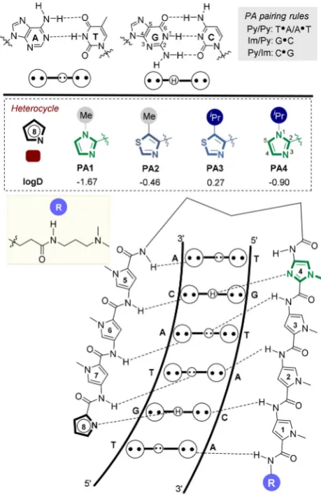
DNA-binding polyamides (PAs) are cell-permeable transcrip-tional modulators which function by inhibiting RNA poly-merase-mediated elongation and/or transcription factor bind-ing to its target double-stranded DNA (dsDNA) consensus se-quence.[1] Of the various designs reported,[2] hairpin PAs are
the most widely used[1b,c,f, 3]where the primary sequence ofN
-methyl pyrrole (Py) and N-methyl imidazole (Im) heterocyclic amino acids defines the selectivity of dsDNA binding ranging from 7 up to 24 base-pairs in length (e.g.,PA1, Figure 1).[1b, 4]
At present, an unmet challenge in their further development as a general tool to modulate gene-selective transcription is an in-depth understanding of the interplay between the dsDNA binding profile of PAs determined in vitro, with their overall physicochemical properties which impact cell uptake, and ulti-mately target engagement in vivo.[5]
We have recently expanded the heterocyclic repertoire of current Py-Im hairpin PA designs to include N-terminal
thia-Figure 1.General binding mode of hairpin PAs used in this study. [a]K. Aman,+Dr. G. Padroni,+Dr. J. A. Parkinson, Prof. G. A. Burley
Department of Pure and Applied Chemistry University of Strathclyde, Thomas Graham Building 295 Cathedral Street, Glasgow, G1 1XL (UK) E-mail: [email protected]
[b]T. Welte
Dynamic Biosensors GmbH, 82152 Planegg (Germany) E-mail: [email protected]
[+] These authors contributed equally to this work.
[image:1.595.311.541.374.728.2]zole-4-carboxylic acid units (Nt). Nt-building blocks (e.g.,
PA2–3) direct a hydrogen-bond-acceptor (N3) atom towards the floor of the minor groove and forms a hydrogen bond with the exocyclic hydrogen bond donor amine (N2) of G. A key structural difference with the incorporation of an Nt-unit in the N-terminal position of a hairpin PA is the endocyclic sulfur atom which changes both the geometry and hydrophobicity (logD) of this heterocycle (Figure 1).[7] Furthermore, when a
bulky isopropyl substituent is installed in the 5-position (i.e.,
iPr-Nt, PA3), a more pronounced compression of the major groove is observed relative to the archetypical hairpin
PA1·dsDNA complex.[6]These results imply that allosteric
mod-ulation of the DNA duplex imparted by PAs is influenced by both the nature of the N-terminal heterocycle pairing with the N2 of G, and the steric bulk of substituents not directly in-volved in selective minor groove recognition.[8]What is unclear
at present is how these changes to the N-terminus influence the kinetics of target versus mismatch binding to dsDNA se-quences.
In this manuscript, we report a label-free biophysical assay to profile the affinity, sequence-selectivity and binding kinetics of PA·dsDNA interactions where the N-terminal heterocycle is systematically altered (PA1–4). PAs containing N-terminal Im units (i.e.,PA1 andPA4) exhibit enhanced selectivity for their target sequences relative to cognate Nt units (i.e., PA2–3). Whilst increasing the steric bulk of the iPr-Im unit (PA4) does not impact DNA binding affinity for its target sequence, NMR structural analysis reveals the larger iPr-Im unit does induce more pronounced structural perturbation of the target dsDNA duplex relative to PA1, which contains an N-terminal Me-Im unit.
Results
Design and synthesis of hairpin polyamides (PA1–4)
The heterocyclic core of a known hairpin PA sequence (PA1) was chosen as our exemplar scaffold to explore the dsDNA binding profile as a function of four different N-terminal heter-ocycles.[4f, 5b, 8a]PA1has an established high affinity binding
pro-file for the general sequence 5’-WWGWWCW (where W=A/T), for which we used 5’-ATGTACTas the target sequence in an immobilized DNA duplex (ODN1).[6, 8a, 9] Compounds PA1–4
were prepared using Boc-based solid phase synthesis on ab -Ala PAM resin via amide coupling of the corresponding hetero-cyclic carboxylic acid (Scheme S1).[6, 10]
Polyamides incorporating N-terminal imidazole units exhibit picomolar binding affinity for their target dsDNA sequence
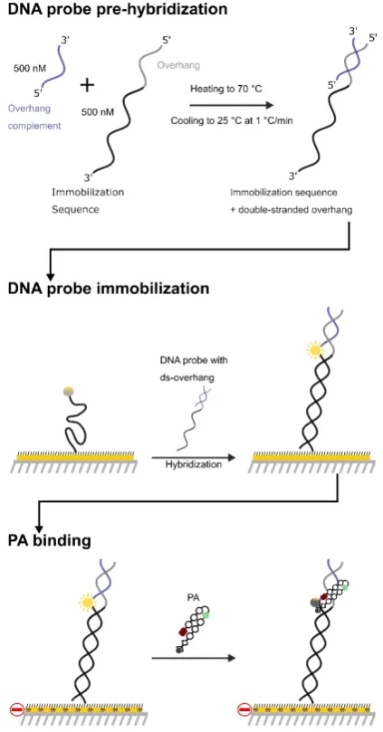
A schematic of the experimental setup is shown in Figure 2. DNA duplexes (ODN1–3, Table 1) were immobilized on a gold surface and contained a fluorophore reporter positioned in close proximity to the proposed PA binding site. PA binding to an immobilized DNA duplex containing the target binding se-quence (ODN1)[11] results in fluorophore quenching, which is
then restored upon dissociation. This provides an isothermal
reporter of the binding kinetics (i.e.,konandkoff) and the
equi-librium dissociation constant (KD).[12]The same fluorescence
re-porter setup was also used to determine the duplex stabiliza-tion profile (i.e.,DTm) of PA·ODN complexes as a function of a
temperature gradient.
Kinetic analyses of the binding profile of PA1–4 to ODN1
show all four PAs exhibit high-affinity binding (Table 1). Whilst the Im-containing PAs (PA1and PA4) exhibit KDvalues in the
picomolar range, the Nt-containing PAs (PA2–3) exhibited a binding affinity that is approximately an order of magnitude lower (i.e., in the low nanomolar range). Rate maps of PA1–4
targetingODN1provided deeper insight into the origin of the differences in the KDvalues of our PA set (Figure 3). Although
the dissociation rate (koff) for each PA was similar, the rate of
association (kon) ofPA2–3was approximately an order of
[image:2.595.320.532.65.470.2]mag-nitude slower relative toPA1andPA4.
G-selective dsDNA binding observed for all four polyamides
The sequence selectivity profile of PA1–4 was explored using duplexes where the target binding sequence inODN1was re-placed with mismatched sequences (ODN2–3). Analyses of the binding kinetics show that Im-containing PAs (PA1 and PA4) are more G-selective relative to Nt-containing PAs (PA2–3, Figure 4). Whilst the rates of association (kon) ofPA4for all
se-quencesODN1–3were similar, the dissociation rates (koff) were
significantly faster for mismatched sequences (ODN2–3). A less pronounced kon/koff trend was observed for PA1 binding to
ODN2, while no interaction was measured withODN3. Consistent with our previous DNA-foot-printing data,[6] the
most promiscuous dsDNA binding profile observed was PA2
(Figure 4 b) where theKDwas virtually the same for the target
(ODN1) and the mismatch (ODN2) sequence. Out of the PA series, PA3 displayed the most unique binding profile (Fig-ure 4 c). In this case, a decrease in both koff and kon was
ob-served for the binding profile ofPA3 forODN2, while no inter-action was observed forODN3.
This experimental setup was also used to determine duplex stabilization of PA·dsDNA complexes compared to the free
DNA duplex melts. A global Boltzmann fit over three inde-pendent runs was used to determine the mid-points of the melting transitions (Tm) for free ODN1–3 and in complex with 20 nm PA1–4. The UV/Vis melting profiles of the PA·dsDNA
complexes confirm a similar trend in dsDNA sequence selectivi-ty (i.e., higherDTm) observed in the fluorescence experiments
(Figure S3). Of particular note was the melting stabilization of
PA4, which displayed excellent G-selectivity relative toPA1–3. Consistent with our kinetics profiling (Figure 4) and previous DNA-foot-printing work,[6] PA2exhibited limited sequence
se-lectivity as highlighted by duplex stabilization observed for all three ODNs. Taken collectively, the kinetic and melting analyses show that the sequence selectivity of Im-containing PAs (i.e.,
PA1andPA4) is superior to Nt-containing analogues (i.e.PA2–
3). Furthermore, whilst enhancing steric bulk on the 5-position of the Nt-series enhanced G-selectivity, this had little effect on the Im-series (i.e.,PA1/PA4).
NMR structural analysis of the PA4·dsDNA complex
In order to gain insight into the influence of theiPr-Im unit in-corporated inPA4when in complex with its target dsDNA se-quence, NMR studies were undertaken using the self-comple-mentary dodecamer sequence d(CGATGTACATCG)2(ODN4).
Ti-tration experiments ofPA4into a solution ofODN4confirmed the formation of a 1:1PA4·ODN4complex. 2D NOESY studies at 4 different mixing times identified a suite of strong NOE cross-correlations from H4 of theiPr-Im building block to G5H1 and the G5N2 exocyclic amine, which implies that the iPr-Im N3 is directed towards the floor of the minor groove (Figure 5; Figure S9). NOE cross-peaks from H4 and H5 of the iPr-Im building block to Py1 and theb-alanine tail in thePA4·ODN4
complex is indicative of the PA binding to its target sequence in the hairpin conformation.
Comparative NMR structural analyses of polyamide·dsDNA complexes
Previous NMR structural work highlighted an increased pro-pensity of PA3 to compress the major groove when in com-plex with its target DNA sequence (PA3·ODN4) relative to
PA1·ODN4. A similar trend in enhanced major groove
com-Table 1.Equilibrium dissociation constant (KD[pm]) data forPA1–4binding to the target sequence (ODN1) versus mismatched sequences (ODN2–3).
PA1 PA2 PA3 PA4
2548 117070 1970240 1885
ODN1
132070 1250110 2880440 96735
ODN2
ND 15 4007700 ND 1100100
[image:3.595.51.549.85.219.2]ODN3
[image:3.595.60.276.210.432.2]pression was also observed with PA4·ODN4 relative to
PA1·ODN4 (Figure 6). However, the extent of major groove
compression was not as pronounced as that observed for the
PA3·ODN4complex.
The origins of these differences become apparent when comparing the extent of minor groove penetration of the three complexes (Figure 7). NMR-restrained molecular dynam-ics of the PA1·ODN4 complex reveal PA1 penetrating deep within the minor groove, exemplified by a hydrogen bond dis-tance of 2.01 between Me-Im N3 and the exocyclic amine G5N2 (Figure 7 a).[13]In contrast, thePA3·ODN4complex shows
a reduced level of minor groove penetration with an average distance of 2.36 between the iPr-Nt N3 and the exocyclic amine G5N2 (Figure 7 b).[13] The PA4·ODN4 complex on the
other hand shows a significant level of minor groove penetra-tion (2.10 ) relative toPA3·ODN4but it is not as extensive as that observed for thePA1·ODN4complex (2.01 ).[6]We
[image:4.595.99.239.55.669.2]there-fore conclude that both the nature of the N-terminal heterocy-cle and the steric bulk distal to the DNA-binding face of a PA scaffold influences the allosteric modulation of a target dsDNA sequence.
[image:4.595.357.495.62.434.2]Figure 4.Comparative analyses of the dsDNA sequence selectivity ofPA1–4 binding toODN1–3.
Figure 5.Strip plot analysis of 2D [1
H,1
Discussion
This combined kinetic and structural study has shown that the type of N-terminal heterocycle and its substituents influences the dsDNA binding profile and the overall structure of the duplex. We discuss here several conclusions that emerged from our results.
N-terminal heterocycle of a hairpin polyamide influences rate of association to target dsDNA sequence
Firstly, all four PAs exhibit high affinity (low nanomolar-picomo-lar) for its target dsDNA sequence. However, the two N-termi-nal Im-containing PAs (PA1/PA4) showed a higher binding af-finity relative to the Nt-containingPA2–3via an increase in the rate of association. Although there has not been a study dedi-cated to evaluating the influence of the hairpin PA N-terminus, a previous SPR-based study by Sugiyama et al. has shown that the number of Me-Im and their positioning in a hairpin PA scaffold can have a disproportionate impact on thekaandKD
relative to only small changes in thekd.[14]In contrast, replacing
internal Py/Im heterocycles with more flexible b-alanine units influences both ka and kd parameters.[15] Extensive work by
[image:5.595.318.530.65.681.2]Dervan et al. has investigated heterocyclic changes to the in-ternal positions of hairpin PA structures.[16]However, our results
[image:5.595.86.267.65.393.2]Figure 6.(a) Major groove width ofODN4(grey),PA1·ODN4(green), PA3·ODN4(blue), andPA4·ODN4(red). NMR-derived molecular model of (b) thePA4·ODN4complex.
highlight the N-terminal position can be used as a convenient site to tune parameters of dsDNA binding and overall physico-chemical properties.
The N-terminal heterocycle position of hairpin polyamides influence DNA structural perturbations
Our structural and binding analyses show that whilst an in-crease in the steric bulk of the iPr-Im unit does not impact dsDNA binding affinity to its target binding site (i.e.,
PA4·ODN4 complex), an improvement in G-selectivity relative
to the iPr-Nt unit (i.e., PA3·ODN4complex) is likely due to a greater level of minor groove penetration (see Figure 7), and in turn improved recognition of the N2 amine of G. However, the extent of major groove compression of the PA4·ODN4 com-plex (Figure 6a) is less than inPA3·ODN4(Figure 7). This sug-gests a fine interplay between minor groove penetration versus major groove compression, with enhanced major groove compression occurring if the hydrogen-bond between the N-terminal building block and the N2 of G is weaker as in
PA3·ODN4, thereby reducing penetration of the minor groove.
Conclusions
These experiments were designed to probe how an increase in the steric bulk of heterocyclic building blocks of PA impacts the binding kinetics and the allosteric distortion of dsDNA con-taining the target binding sequence. Although what superfi-cially appears to be a subtle increase in steric bulk at locations within a PA scaffold not directly involved in dsDNA base-read-out, these data suggest that strategic changes in the Im and Nt substitution pattern can be used to fine tune the sequence-selectivity of dsDNA binding as well as the overall physico-chemical properties of PA scaffolds.[17] We envisage that the
strategic incorporation of modified heterocyclic building blocks within a PA scaffold could be applied more broadly as a strategy to enhance cell uptake and potency of transcriptional modulation in cellulo.
Acknowledgements
G.P. thanks the University of Strathclyde for a University Stu-dentship. G.A.B. thanks the Biotechnology and Biological Scien-ces Research Council (BBSRC; BB/N016378/1) and the Science and Technology Facilities Council (STFC; ST/M000125/1) for funding this work. We thank the EPSRC U.K. National Mass Spectrometry Facility at Swansea University for HRMS analyses of compounds.
Conflict of interest
The authors declare no conflict of interest.
Keywords: allosterism · binding kinetics · minor groove binder·NMR characterisation·pyrrole-imidazole polyamide
[1] a) G. S. Erwin, M. P. Grieshop, A. Ali, J. Qi, M. Lawlor, D. Kumar, I. Ahmad, A. McNally, N. Teider, K. Worringer, R. Sivasankaran, D. N. Syed, A. Eguchi, M. Ashraf, J. Jeffery, M. Xu, P. M. C. Park, H. Mukhtar, A. K. Srivas-tava, M. Faruq, J. E. Bradner, A. Z. Ansari,Science2017,358, 1617 – 1622; b) A. A. Kurmis, F. Yang, T. R. Welch, N. G. Nickols, P. B. Dervan,Cancer Res.2017,77, 2207 – 2212 ; c) F. Yang, N. G. Nickols, B. C. Li, G. K. Marinov, J. W. Said, P. B. Dervan,Proc. Natl. Acad. Sci. USA2013,110, 1863 – 1868; d) J. A. Raskatov, J. L. Meier, J. W. Puckett, F. Yang, P. Ramakrishnan, P. B. Dervan,Proc. Natl. Acad. Sci. USA2012,109, 1023 – 1028 ; e) T. Hidaka, G. N. Pandian, J. Taniguchi, T. Nobeyama, K. Hashiya, T. Bando, H. Su-giyama,J. Am. Chem. Soc.2017,139, 8444 – 8447; f) K. Hiraoka, T. Inoue, R. D. Taylor, T. Watanabe, N. Koshikawa, H. Yoda, K. Shinohara, A. Taka-tori, H. Sugimoto, Y. Maru, T. Denda, K. Fujiwara, A. Balmain, T. Ozaki, T. Bando, H. Sugiyama, H. Nagase,Nat. Commun.2015,6, 6706; g) G. N. Pandian, S. Sato, C. Anandhakumar, J. Taniguchi, K. Takashima, J. Syed, L. Han, A. Saha, T. Bando, H. Nagase, H. Sugiyama, ACS Chem. Biol.
2014,9, 2729 – 2736.
[2] a) P. B. Dervan, R. M. Doss, M. A. Marques,Curr. Med. Chem. Anticancer Agents2005,5, 373 – 387; b) Y.-W. Han, H. Sugiyama, Y. Harada,Biomater. Sci. 2016, 4, 391 – 399; c) J. M. Withers, G. Padroni, S. M. Pauff, A. W. Clark, S. P. Mackay, G. A. Burley inReference Module in Chemistry, Molecu-lar Sciences and Chemical Engineering, Elsevier, Amsterdam, 2017, pp. 149 – 178; d) L. Pett, J. A. Hartley, K. Kiakos,Curr. Top. Med. Chem.
2015,15, 1293 – 1322 ; e) W. D. Wilson, F. A. Tanious, A. Mathis, D. Tevis, J. E. Hall, D. W. Boykin,Biochimie2008,90, 999 – 1014 ; f) Y. Kawamoto, T. Bando, H. Sugiyama,Bioorg. Med. Chem.2018,26, 1393 – 1411. [3] a) N. G. Nickols, J. O. Szablowski, A. E. Hargrove, B. C. Li, J. A. Raskatov,
P. B. Dervan,Mol. Cancer Ther.2013,12, 675 – 684; b) J. Syed, G. N. Pan-dian, S. Sato, J. Taniguchi, A. Chandran, K. Hashiya, T. Bando, H. Sugiya-ma,Chem. Biol.2014,21, 1370 – 1380 ; c) A. Yasuda, K. Noguchi, M. Min-oshima, G. Kashiwazaki, T. Kanda, K. Katayama, J. Mitsuhashi, T. Bando, H. Sugiyama, Y. Sugimoto,Cancer Sci.2011,102, 2221 – 2230; d) K. Haya-tigolkhatmi, G. Padroni, W. Su, L. Fang, E. Gomez-Castaneda, Y. C. Hsieh, L. Jackson, T. L. Holyoake, F. Pellicano, G. A. Burley, H. G. Jorgensen,
Blood Cells Mol. Dis.2018,69, 119 – 122.
[4] a) P. B. Dervan, B. S. Edelson,Curr. Opin. Struct. Biol.2003,13, 284 – 299; b) R. S. Edayathumangalam, P. Weyermann, J. M. Gottesfeld, P. B. Dervan, K. Luger,Proc. Natl. Acad. Sci. USA2004,101, 6864 – 6869; c) A. Hirata, K. Nokihara, Y. Kawamoto, T. Bando, A. Sasaki, S. Ide, K. Maeshi-ma, T. KasaMaeshi-ma, H. SugiyaMaeshi-ma,J. Am. Chem. Soc.2014,136, 11546 – 11554; d) G. S. Erwin, M. P. Grieshop, D. Bhimsaria, T. J. Do, J. A. Rodriguez-Mar-tinez, C. Mehta, K. Khanna, S. A. Swanson, R. Stewart, J. A. Thomson, P. Ramanathan, A. Z. Ansari,Proc. Natl. Acad. Sci. USA2016,113, E7418 – E7427; e) G. S. Erwin, D. Bhimsaria, A. Eguchi, A. Z. Ansari,Angew. Chem. Int. Ed. 2014, 53, 10124 – 10128 ; Angew. Chem. 2014, 126, 10288 – 10292 ; f) A. E. Hargrove, T. F. Martinez, A. A. Hare, A. A. Kurmis, J. W. Phil-lips, S. Sud, K. J. Pienta, P. B. Dervan,PloS One2015,10, e014316; g) X. Wang, H. Nagase, T. Watanabe, H. Nobusue, T. Suzuki, Y. Asami, Y. Shino-jima, H. Kawashima, K. Takagi, R. Mishra, J. Igarashi, M. Kimura, T. Takaya-ma, N. Fukuda, H. SugiyaTakaya-ma,Cancer Sci.2010,101, 759 – 766; h) T. G. Ed-wards, T. J. Vidmar, K. Koeller, J. K. Bashkin, C. Fisher,PLoS One2013,8, e75406 ; i) Y. Kawamoto, A. Sasaki, A. Chandran, K. Hashiya, S. Ide, T. Bando, K. Maeshima, H. Sugiyama,J. Am. Chem. Soc.2016,138, 14100 – 14107; j) Y. Kawamoto, A. Sasaki, K. Hashiya, S. Ide, T. Bando, K. Maeshi-ma, H. SugiyaMaeshi-ma,Chem. Sci.2015,6, 2307 – 2312.
[5] a) A. E. Hargrove, J. A. Raskatov, J. L. Meier, D. C. Montgomery, P. B. Dervan, J. Med. Chem. 2012, 55, 5425 – 5432; b) C. S. Jacobs, P. B. Dervan, J. Med. Chem. 2009, 52, 7380 – 7388; c) N. G. Nickols, C. S. Jacobs, M. E. Farkas, P. B. Dervan,Nucleic Acids Res.2007,35, 363 – 370. [6] G. Padroni, J. A. Parkinson, K. R. Fox, G. A. Burley, Nucleic Acids Res.
2018,46, 42 – 53.
[7] a) C. C. O’Hare, D. Mack, M. Tandon, S. K. Sharma, J. W. Lown, M. L. Kopka, R. E. Dickerson, J. A. Hartley,Proc. Natl. Acad. Sci. USA2002,99, 72 – 77; b) N. G. Anthony, B. F. Johnston, A. I. Khalaf, S. P. MacKay, J. A. Parkinson, C. J. Suckling, R. D. Waigh, J. Am. Chem. Soc. 2004, 126, 11338 – 11349.
[8] a) D. M. Chenoweth, P. B. Dervan,J. Am. Chem. Soc.2010,132, 14521 – 14529 ; b) D. M. Chenoweth, P. B. Dervan,Proc. Natl. Acad. Sci. USA2009,
106, 13175 – 13179.
[10] a) E. E. Baird, P. B. Dervan, J. Am. Chem. Soc. 1996, 118, 6141 – 6146; b) W. Su, S. J. Gray, R. Dondi, G. A. Burley,Org. Lett. 2009, 11, 3910 – 3913; c) A. J. Fallows, I. Singh, R. Dondi, P. M. Cullis, G. A. Burley,Org. Lett.2014,16, 4654 – 4657; d) L. Fang, Z. Pan, P. M. Cullis, G. A. Burley, W. Su,Curr. Protoc. Nucleic Acid Chem. 2015, 63, 8.11.1 – 8.11.14 ; e) S. M. Pauff, A. J. Fallows, S. P. Mackay, W. Su, P. M. Cullis, G. A. Burley,Curr. Protoc. Nucleic Acid Chem.2015,63, 8.10.1 – 8.10.41.
[11] J. Knezevic, A. Langer, P. A. Hampel, W. Kaiser, R. Strasser, U. Rant,J. Am. Chem. Soc.2012,134, 15225 – 15228.
[12] a) A. Clry, T. J. M. Sohier, T. Welte, A. Langer, F. H. T. Allain, Methods
2017,118 – 119, 137 – 145; b) M. Krepl, M. Blatter, A. Clry, F. F. Damberg-er, F. H. T. Allain, J. SponDamberg-er,Nucleic Acids Res.2017,45, 8046 – 8063 ; c) D. Ploschik, F. Rçnicke, H. Beike, R. Strasser, H.-A. Wagenknecht, ChemBio-Chem2018,19, 1949 – 1953.
[13] H. Y. Alniss, M. V. Salvia, M. Sadikov, I. Golovchenko, N. G. Anthony, A. I. Khalaf, S. P. MacKay, C. J. Suckling, J. A. Parkinson,ChemBioChem2014,
15, 1978 – 1990.
[14] Y.-W. Han, T. Matsumoto, H. Yokota, G. Kashiwazaki, H. Morinaga, K. Ha-shiya, T. Bando, Y. Harada, H. Sugiyama,Nucleic Acids Res. 2012, 40, 11510 – 11517.
[15] a) Y. W. Han, G. Kashiwazaki, H. Morinaga, T. Matsumoto, K. Hashiya, T. Bando, Y. Harada, H. Sugiyama,Bioorg. Med. Chem. 2013, 21, 5436 –
5441; b) B. B. Liu, S. Wang, K. Aston, K. J. Koeller, S. F. H. Kermani, C. H. Castaneda, M. J. Scuderi, R. S. Luo, J. K. Bashkin, W. D. Wilson, Org. Biomol. Chem.2017,15, 9880 – 9888; c) S. Wang, R. Nanjunda, K. Aston, J. K. Bashkin, W. D. Wilson,Biochemistry2012,51, 9796 – 9806.
[16] a) D. M. Chenoweth, A. Viger, P. B. Dervan,J. Am. Chem. Soc.2007,129, 2216 – 2217; b) D. M. Chenoweth, J. A. Poposki, M. A. Marques, P. B. Dervan,Bioorg. Med. Chem.2007,15, 759 – 770; c) M. A. Marques, R. M. Doss, S. Foister, P. B. Dervan, J. Am. Chem. Soc. 2004, 126, 10339 – 10349 ; d) D. Renneberg, P. B. Dervan, J. Am. Chem. Soc. 2003, 125, 5707 – 5716; e) S. Foister, M. A. Marques, R. M. Doss, P. B. Dervan,Bioorg. Med. Chem. 2003, 11, 4333 – 4340; f) M. A. Marques, R. M. Doss, A. R. Urbach, P. B. Dervan,Helv. Chim. Acta2002,85, 4485 – 4517.
[17] a) B. Liu, T. Kodadek,J. Med. Chem.2009,52, 4604 – 4612; b) S. Nishijima, K. Shinohara, T. Bando, M. Minoshima, G. Kashiwazaki, H. Sugiyama,
Bioorg. Med. Chem.2010,18, 978 – 983.
Manuscript received : October 24, 2018
Accepted manuscript online: November 8, 2018
FULL PAPER
&
Structural BiologyK. Aman, G. Padroni, J. A. Parkinson, T. Welte,* G. A. Burley*
&&–&&
Structural and Kinetic Profiling of Allosteric Modulation of Duplex DNA Induced by DNA-Binding Polyamide Analogues

![Figure 5. Strip plot analysis of 2D [1H, 1H] NOESY NMR data of PA4·ODN4.](https://thumb-us.123doks.com/thumbv2/123dok_us/1329925.86868/4.595.357.495.62.434/figure-strip-plot-analysis-noesy-nmr-data-odn.webp)
