0022-538X/89/114498-09$02.00/0
Copyright © 1989, American Society for Microbiology
Persistent Infection
of
Rabbits with Bovine Leukemia Virus
Associated with Development of
Immune
Dysfunction
CAROL R. WYATT,' DENISEWINGETT,2 JACQUELYNN S.
WHITE,'
CHARLES D. BUCK,'t DONALD KNOWLES,3 RAYMOND REEVES,2 ANDNANCYS.MAGNUSON'*
Departments of Microbiology,1
BiochemistrylBiophysics72
and VeterinaryMicrobiology andPathology,3Washington State University, Pullman, Washington 991644340
Received 13February1989/Accepted 5 July 1989
Bovine leukemia virus (BLV) infection of rabbits provides a safe and relatively inexpensive in vivo mammaliansystemfor the study of the mechanismscontrolling expression ofauniquegroupoflymphotropic retroviruses. This group of viruses, which includes C-type humanT-lymphotropic virus types I and II and lentiviruslike humanimmunodeficiency virustype 1, possesses genescoding for "trans-activating" products. Rabbits experimentally inoculatedwithBLV became persistently infected, as demonstrated by anumber of tests. All BLV-inoculated rabbits developed persistent serum antibody to BLV. Furthermore, all BLV-inoculated rabbits hadperipheral blood mononuclear cells which, whenstimulated, expressed the virus, as demonstrated by viral inductionofsyncytium formation inaBLV-susceptible fibroblast line. Thepresenceof BLV incirculatingcellswasconfirmed by using peripheral blood mononuclear cells fromrandomly selected BLV-inoculatedrabbits, which showedthe presenceofviral reversetranscriptaseactivity, BLV transcriptional activity,orBLVproviral DNA. Additionaltestsshowed thatinfectedlymphocytes maintained in culture with recombinant human interleukin-2 formed multinucleated giant cells and produced virus when incubated in cytokine-containing medium. BLV-infected rabbits also showed alterations in several parameters associated with immunity, beginning 6 months after inoculation. Thirty-eight percent of infected rabbits developed abnormallylowT-cellresponses, asmeasuredbyphytolectin stimulation, and T-cellresponsescycled between normal and abnormallylow over aperiod of 20to24months.Forty-fourpercentof rabbitsinfected for longer than 12 monthssuffered from recurrent
conjunctivitis
and rhinitis. By 24 months postinoculation, 28% of infected rabbitswere deador werekilled becauseofpoorclinical condition.Bovine leukemia virus (BLV) is a type C lymphotropic retrovirus which is associated with a disease complex termed enzootic bovine leukosis, often characterized by
persistent lymphocytosisanddevelopment of B-cell lympho-masafter irregular, long incubation periods (3, 5, 15). The
proviralgenome structureof BLVis similartothatof human T-lymphotropic virus type I (HTLV-I), thecausal agentof adult T-cellleukemia/lymphoma, and human
T-lymphotro-pic virus type II (HTLV-II), the causal agent ofa T-cell variant of hairy cell leukemia (41). Althoughmore distantly related to the lymphotropic lentivirus human
immunodefi-ciency virus type 1 (HIV-1), the causal agent ofacquired immunodeficiency disease, the BLV genome hasan impor-tantfeature incommonwith the HTLVs and HIV. Thatis, BLV, HTLV-I, HTLV-II, and HIV-1 all have a common structural organization in that they each possess a gene sequence that codes for a "trans-regulatory" protein that
activates viralgeneexpression (8, 23, 31).
Previously,BLVhad been reportedtoinfectanumber of small animal species, including cats, dogs, and rabbits. However, infection in these animals had only been defined by the continualpresenceofanti-BLV antibody(2, 32, 36). Thus, until this report, convincing evidence of persistent BLVinfectionintermsofthepresence of the virus or viral genes in rabbits had not been demonstrated. We used a variety of techniques todemonstrate the persistence of the BLV genome in rabbits infected for up to 18 months.
Additionally, wedemonstratedthatwithin6months of BLV
*
Corresponding
author.tPresentaddress: Department of Virology, American Type
Cul-tureCollection, 12301 ParklawnDrive, Rockville, MD 20852.
inoculation, about one-third of the rabbits had decreased
T-cell responsiveness to phytolectin stimulation, and we
observed that two-thirds ofBLV-infected rabbits exhibited
indications of clinical disease, including
conjunctivitis,
rhi-nitis, andsevereweight loss, ordiedunexpectedly.MATERIALSANDMETHODS
Animals. NewZealandWhite rabbits(male andfemale),2 to4monthsold,werepurchased fromaU.S.Department of
Agriculture-approved rabbitry and maintained at
Washing-tonStateUniversity, whichisfully accredited by the Amer-icanAssociationfor theAccreditation ofLaboratoryAnimal Care.
Cell separation. Peripheral blood mononuclear cells (PBMC) from BLV-inoculated and control rabbits were
separated from heparinizedwholebloodbyFicoll-Hypaque sedimentation(density, 1.09
g/ml)
asdescribed before(40).Inoculation with BLV. Overaperiod ofayear,groupsof
two to sixyoungadult rabbitswereinoculated with BLVor otherantigens. Twelve rabbits receivedtwoinoculations of
approximately 5 x 10⁶phytolectin-stimulatedPBMC froma
BLV-seropositive cow withpersistentlymphocytosis. Four rabbits received one inoculation ofapproximately 5 x 10⁶ cells fromaBLV-producingfetal lambkidney fibroblast line (BLV-FLK), and tested negative for serum antibody to bovine viral diarrhea virus (apotentialcontaminant of BLV-FLKcultures)2to4weeks later. Two rabbitsreceivedthree separateinoculations ofcell-freeBLV(400 mlofsupernatant from BLV-FLKcultures concentrated 100-fold; 0.5 ml given per
injection)
overan8-weekperiod.Electronmicroscopy ofboth BLV-FLK cells and virus purified from those cells confirmed the presence of type C virus particles. Two 4498
on November 10, 2019 by guest
http://jvi.asm.org/
control rabbits received 5 x 10⁶ phytolectin-stimulated PBMC from a BLV-seronegative cow, and 11 additional
control rabbits were given antigens other than BLV
(tRNAAla
linked to bovine serum albumin [one rabbit],gluteraldehyde-treated STa exotoxin from Escherichia coli [two rabbits], barley yellow dwarf virus inclusion bodies [two rabbits], mouse P815 cells [one rabbit], RPMI 1640 medium [onerabbit], ornothing [four rabbits]).
ELISA. Anenzyme-linked immunosorbentassay(ELISA) wasperformedonpurifiedBLVasdescribed previously(4).
The colorreaction between peroxidase and 5-aminosalicy-late was allowed to proceed for 2 h, after which color intensity was evaluated at 570 nm with an ELISA plate
reader (Cambridge Technology, Cambridge, Mass.). Anti-body titerwasdefinedasthereciprocal of theserumdilution
whichgave color intensity twice that of eithera
preinocula-tion serum sample from the same rabbit or serum from a
control rabbitatthesame dilution.
Syncytium formationassay. PBMC from BLV-inoculated
rabbitsweresuspendedat4 x 106 cellsperml ofRPMI 1640
containing 10 mM HEPES
(N-2-hydroxyethylpiperazine-N'-2-ethanesulfonic acid) and antibiotics (RPMI) with 5% normal rabbit serum and incubated with 2.5 mg of
phyto-hemagglutinin-L (PHA-L; Sigma Chemical Co., St. Louis, Mo.) per ml for48 h at 37°C in 5% CO2. The PBMCwere
thenwashed andresuspendedinRPMI. Thesuspensionwas
divided intotwoequal portions. Each portionwasmade10% serumwitheither anti-BLV bovine serum ornormal bovine serum and placed on a monolayer of bat lung fibroblasts
(TblLu; Wistar Institute), a cell line that is free of bovine
syncytial virus and parainfluenza virus type 3 and is not susceptible to infection by bovine herpesvirus type 1 or
bovine viraldiarrhea virus(AmericanTypeCulture Collec-tion). After 18 h at 37°C and 5% C02, the PBMC were
removed, and the monolayers were washed in Dulbecco modified Eagle medium containing 10 mM HEPES and antibiotics (DMEM) and incubated overnight in DMEM containing 10%fetalbovineserum(FBS). Monolayers were
maintainedingrowth phase for4to5days, andthen5 x 105 cells were placed in a 25-cm2 flask and incubated for an
additional 1 to 2 days. The relative numbers of syncytia (defined as cells containing five or more nuclei [9]) were
determined bycounting thesyncytiapresentin20
consecu-tivefields withaninverted microscopeat amagnificationof 10Ox. Backgrounds were subtracted, and results are
ex-pressed asnumber ofsyncytiaper square centimeter. Con-trolsconsisted of identicallytreated PBMC from 10 different control rabbits.
RTassay. Clarified supernatantsfrom 1 x 10⁷to2 x 107 PBMC stimulated with PHA-L for 48 to 72h were centri-fuged at90,000 x gfor 1 hat4°C,and the resulting pellets
were suspended in 160 ml of 0.1 M Tris, pH 7.8. Reverse
transcriptase(RT) activitywasdeterminedintriplicate50-,ul
samplesin thepresenceofmagnesiumbyanassaymodified for BLV (16, 19). Activity is expressed as picomoles per
hourper108cells. Negativecontrolsconsisted ofidentically obtained supernatants from 1 x 10⁷to2 x 107control rabbit PBMC. Positive controls were clarified, ultracentrifuged
virus pellets fromsupernatants of BLV-FLK cells.
Northern(RNA)blotanalysis.Bloodwasdrawn from three
BLV-inoculated rabbits, and PBMC were isolated as de-scribed before (40). Becauseonly alimited amountof blood could be drawn from each infected rabbitatagiven time,the PBMC were pooled to obtain sufficient RNA to analyze. RNA was isolated from PBMC of BLV-inoculated and control rabbits and a BLV-infected cow by CsCl gradient
centrifugation
asdescribed before(7).
Denaturing
formalde-hyde
gels
were run on atotalRNA from PBMC which hadbeenstimulated for24to48 h withconcanavalinA
(ConA;
5mg/ml).
Theprobe
(agift
ofA.Burny,
Brussels,
Belgium)
consisted of an 8.3-kilobase(kb)
clone of BLVproviral
DNA.PCR.
Samples
of DNA isolated as described before(7)
from PBMC of BLV-inoculated and control rabbits were
amplified by
thepolymerase
chain reaction(PCR).
Briefly,
thereactionmixturecontained 1 ,ugof PBMCDNAor30 ng
of control
plasmid
DNA, 200 mMeach of four deoxyribo-nucleosidetriphosphates,
10 mMTrischloride(pH 8.31),50 mMKCl,
1.5mMMgCl2,
2.5U ofthermoresistantTaqDNApolymerase,
and100pmol
each ofoligonucleotide
primers.
Theprimers
arecomplementary
to aregion
oftheenvgene and have thefollowing
sequences:First
primer:
5'-TAA AAGCAG GGC CCATGA TCTGAC AG-3'
Second
primer:
5'-TGG ATGACA GCA TAT AAC CAAGAG GC-3'
Astandard
amplification procedure
wasfollowedby
adding
primers
inlarge
excessto cellularDNA,
heatdenaturing
at95°C,
and thencooling
to37°C.
Primerextensionwascarriedout at
72°C
withheat-stableTaq
polymerase
(33).
Thiscycle
was carried out 35
times,
and Southern blotanalysis
wasperformed
on 1/10 of the reaction mixture with an 8.3-kb BLVcDNAprobe (27).
IL-2-dependent
lymphocyte
cultures. PBMC fromBLV-inoculatedorcontrol rabbitswere
suspended
at3 x 106/ml ofRPMI
containing
10%FBS andstimulatedwith 2.5 mgofthephytolectin
ConA perml. The cultures were maintainedby
adjusting
thecellcount toapproximately
3 x 10⁶cellsperml with fresh RPMIcontaining
10%
FBS and 50 U ofhumanrecombinant interleukin-2
(IL-2;
ChironCorp.,
Emeryville,
Calif.)
per ml every 3to 4days.
Induction ofBLV
production
fromIL-2-dependent
virus-infectedlymphocytes.
After 3 months or more inculture,
BLVproduction
wasinducedfromIL-2-dependent
lympho-cytesfromBLV-infected rabbits
by
suspending
the lympho-cytes in a mixture ofRPMIcontaining
50 UofIL-2 per mland 5% normal rabbit serum with 33%
supernatant
fromphytolectin-stimulated
PBMC from uninfected rabbits or33% RPMI
containing
5% normal rabbit serum. Viruspro-duction was monitored
by
incubating
theIL-2-dependent
lymphocytes
on TblLumonolayers
in the presence orabsenceof bovineanti-BLVserumand
counting
therelativenumbersof
syncytia. IL-2-dependent lymphocytes
obtainedfrom a control rabbit and treated in the same manner were
usedas
negative
controls.Response to
phytolectin
stimulation. Two hundredmillili-ters of 1:10 dilutions of blood
leukocytes
in RPMI wereadded to U-bottomed microtiter
plates.
Triplicate
cultures were stimulated with PHA-P(5
and 10mg/ml)
or ConA(5
and 10mg/ml).
After 48 hof incubationat37°C,
0.5 mCiof[methyl-3H]thymidine
(6.7
Ci/mmol;
Du Pont NewEngland
Nuclear Research
Products, Boston,
Mass.)
in 20,ul
wasadded to each well. After a further 22
h,
cultures were terminated with amicroharvester,
andradioactivity
was counted in aliquid
scintillationspectrometer
(Beckman
Instruments,
Inc.,
Fullerton,
Calif.).
Responses
wereevalu-ated
prior
to inoculation with BLVandatintervalsthereaf-ter. Controls consisted of identical cell
preparations
taken fromcontrolrabbits. Resultsareexpressed
as apercentage
of the response of
leukocytes
from anage-matched
controlanimalrun atthe sametime.
on November 10, 2019 by guest
http://jvi.asm.org/
TABLE 1. Summary of results
No.of rabbitspositive/no.tested No.of
source Inoculum rabbits Anti- PBMC RT Virus Multinu-
Clinical
inoculated BLV inducing activity genome cleatedgiant diseasebantibody syncytia presenta cells in culture
BLV-infectedlymphocytes 1 x 10⁷cells 12 12/12 12/12 1/5 1/5 8/8 6/12
BLV-FLK 5 x 10⁶ cells 4 4/4 4/4 2/3 1/2 2/2 4/4
Cell-freeBLV 100 x concentrated virus(2 ml) 2 2/2 2/2 1/2 1/1 N.T.c 2/2
a Analysis byNortherm blot orPCR.
bIncludes rabbits that died. cN.T., Not tested.
Pathology. Food andwaterconsumption and clinical
con-ditionwere visually monitored for all the BLV-inoculated
and control rabbits. Rabbits were weighed monthly, and
rabbits (three female, two male) which exhibited severe
decline in clinical condition or died unexpectedly were
necropsied. The followingstructuresweregrossly examined
at necropsy: eyes, integument, teeth, oral cavity, tongue, larynx, heart, lungs, esophagus, trachea, diaphragm, liver, gallbladder, spleen, pancreas, stomach, small intestine,
cecum, colon, adrenal glands, kidneys, ureters, urinary bladder, ovaries, uterus, cervices, vagina, vulva, brain, pituitary gland, bonemarrow, coxofemoraljoints, and stifle
joints. Tissues were placed in 10% buffered Formalin,
par-affin embedded, sectioned at 4 to 6 ,um, and stained with hematoxylin and eosin for histologic examination.
RESULTS
Evaluation of infection. Two major criteria were used to demonstratethat allBLV-inoculated rabbits become persis-tently infected with the virus. First, sera from all
BLV-inoculated rabbits wereperiodically tested for thepresence
and persistence of antibody to BLV. The development of persistentantibodytoBLV iscommonly usedtodetermine BLVinfection in cattle (31). Second, PBMC from all BLV-inoculated rabbitswereassayedfor virus expression by the
development of syncytia onBLV-sensitive fibroblasts.
Co-cultivation of BLV-susceptible fibroblastswith lymphocytes from BLV-infected cattle has been usedtodetect the
pro-duction of BLVby lymphoid cells (12). The syncytiaassay
had two functions. It confirmed that the rabbits were
in-fectedwithBLV, because TblLu issusceptibletoBLVbut isnotsusceptibletoseveral othercommonbovine pathogens
(see description in Materials and Methods). Additionally, it served as a means to demonstrate recovery of BLV from
circulating cells of inoculated animals, because BLV-in-duced cellfusion is the cytopathic effect seenin TblLu (9,
19). Additional confirmation of the presence of BLV in inoculated rabbit cells was achieved through evaluation of
PBMC from randomly selected animals for viralRTactivity,
presence of viral mRNA by Northern blot analysis, or
presence of provirus as detected by PCR (33). All
BLV-inoculated rabbits weremonitoredfor clinical signs
sugges-tive of suppressed immune function. A summary of the results of thetests is shown in Table 1.
AntibodyresponsetoBLV. To confirm and extend earlier reports of persistent anti-BLV antibodies in rabbits (6), 18 NewZealand White rabbitswereinoculated with BLV. Sera werecollected from these rabbitsover aperiod of 18 months
andanalyzed byELISA. For animals which survivedlonger
than 18 months, samples were collected up to 24 months postinoculation. Table 2 shows four separate examples of antibody titer patternsobserved in BLV-inoculated rabbits tested repeatedly over 24 months postinoculation. Titers
varied considerably with time, in some cases falling to
undetectable levels and thenbecoming detectable again later in the infection.Althoughnoconsistentpatternofrising and falling anti-BLV titerswasobserved, each rabbit produced
antibody over an extended period of time. In Table 3, the
ranges of anti-BLV antibody titers are shown for all
BLV-inoculated rabbits forearly (2to 3 months), middle (6to8 months), and late (12to18months) time points after inocu-lation. All of the BLV-inoculated animals produced anti-BLV antibodies for prolonged periods postinoculation. None of the rabbits which received inocula other thanBLV developed detectable levels of anti-BLV antibody.
Syncytium formation in BLV-susceptible indicator fibro-blasts and inhibition with anti-BLV antibody. In order to further demonstrate that BLV-inoculated rabbits had
be-comepersistently infected andtorecoverBLV from
circu-latingPBMC, weevaluated theabilityof
phytolectin-stimu-lated PBMC from BLV-inoculated rabbits to produce syncytiaupon cocultivationwithsusceptible bat lung fibro-blasts(TblLu). Table 4 shows syncytium formation induced by phytolectin-stimulated PBMC from 12 of the BLV-inoc-ulated rabbits randomly tested for virus expression begin-ning 2 months andcontinuingatleast 12 months postinocu-lation. In these experiments, two observations were made
consistently. First, syncytia always formed (8to26 syncytia
TABLE 2. ELISA titersovertime forfourBLV-inoculated rabbits
ELISAtiter' atmopostinoculation: Inoculum
2 4 6 8 10 12 14 16 18 20 22 24
Cell-free BLV 10,000 10,000 10,000 1,200 1,200 1,200 NT 1,200 1,200 NT NT 1,200
BLV-infected 1,000 1,200 1,000 250 250 NT 1,000 250 Dead
lymphocytes
BLV-FLK 250 1,000 100 100 BD BD NT NT BD NT BD 250
BLV-FLK 1,000 250 1,000 NT 1,000 50 NT 250 250 BD 50 50
aNT,Nottested; BD, below detectable levels.
on November 10, 2019 by guest
http://jvi.asm.org/
TABLE 3. Anti-BLV antibody titersa
Inoculum Titer range (no. of rabbits with indicated titer)
(no.ofrabbits) 2-3mo 6-8mo 12-18mo
BLV-infected lymphocytes (12) 100(4)-10,000(1) 100(7)-10,000(1) 100 (5)-6,000(1)b
BLV-FLK (4) 100 (1-1,000 (3) 100(2)-1,000(2) BDC (2)-250(1)
Cell-free BLV(2) 10,000 (2) 10,000 (2) 10,000 (2)
Uninfected lymphocytes (2) BD (2) BD (2) BD(2)
aTitersweredetermined by ELISA;serafrom rabbitsreceiving no BLV gave no detectabletiters.
bAnimal which had titer of 10,000was dead at 12 months.
cBD,Below detection limit.
per cm2) when PBMC from BLV-inoculated rabbits were incubated in normal bovine serum. Second, syncytium for-mation was alwaysdecreased (2.7- to >25-fold) when PBMC from BLV-inoculated rabbits were incubated with anti-BLV serum. Noneof 10 age-matched control rabbits tested at the sametime had PBMC which induced syncytium formation above background. The formation of syncytia in the pres-ence of BLV-inoculated rabbit cells and the inhibition of syncytium formation in the presence of anti-BLV antiserum provided further evidence that cells from BLV-inoculated
rabbits wereindeed persistently infected with the virus. RTactivity. ThepresenceofRTactivityinvirus-infected
cells has often been used as an indicator of retrovirus
infection.To assesswhether RT activity could be detected in
BLV-inoculated rabbits, PBMC from a number of animals were tested. Culture supernatants from phytolectin-acti-vated PBMCfrom control and BLV-inoculated rabbits were assayedfor RT activity inan assay mix containing magne-sium andmodifiedtomeasureBLV RT (9, 16). Whereas no RTactivity above background was detected in samples from seven uninfected rabbits, samplesfrom 4 of 10
BLV-inocu-lated rabbits sampled between 6 and 20 months
postinocu-lation exhibitedappreciable RT activity (16.4 + 8.0pmol/h
per 108 cells) (data not shown). Detectable RT activity in BLV-inoculated animals strongly suggests that circulating PBMC from such animalsareinfected with thevirus.
Northern blot analysis. Like HTLV-I, BLV is not
pro-ducedatdetectable levels infreshly isolated lymphocytes of the infected host but is oftenexpressed when those cellsare
[image:4.612.59.561.89.163.2]culturedinvitro (11, 14, 23, 24, 28). Todetermine whether BLVtranscripts couldbedetected in infected rabbit PBMC,
TABLE 4. Syncytium formation induced by PBMC from twelveBLV-inoculated rabbits
Timepost- No. ofsyncytia/cm2a
BLV source inoculation Normal Anti-BLV (mo) serum serum
BLV-FLK 2 19 1
2 24 9
5 25 0
8 19 4
Cell-freeBLV 10 10 1
11 18 0
BLV-infectedlymphocytes 5 26 8
6 26 4
7 8 0
7 10 0
7 15 0
12 9 0
aBackground level ofsyncytiumformation(fiveor morenucleipercell)for TblLuis less than1 per1,000 cells. PBMC from control rabbitsdidnotform syncytiaabovebackgroundlevels in either normalserum oranti-BLVserum.
Northern blot analyses with a BLV cDNA probe were
performed on total RNA obtained from phytolectin-stimu-lated PBMC from several of the BLV-inoculated rabbits.
Figure 1 shows anexperiment in which RNA was isolated
and pooled from a limited amount of blood from three
BLV-inoculated rabbits and compared with RNAobtained
from PBMC from an uninfected rabbit and from a
BLV-infectedcow. BLV transcripts were detected in RNA from the BLV-inoculated rabbits and the BLV-infected cow but notinthecontrol rabbit RNA.DifferenthybridizingmRNA
specieswereobserved intheinfected-rabbit mRNA popula-tionand mightbe the resultof alternative splicingevents.
PCR. PCR analysis was also performed to determine whether proviral DNA could be detected in cells from BLV-infected rabbits. In HTLV-I- and HIV-1-infected hu-mans, only a small percentage of lymphocytes can be
demonstratedtocontainproviral DNA (34). We suspected a similar situation might exist in cells from BLV-infected
rabbits because ofthedifficulty we encountered in
demon-strating RTactivityand BLVtranscripts. Inorder to detect such rareevents, an extremely sensitive method such as the PCR assay (33) is required. As shown in Fig. 2, BLV sequencescould be detected in one of two rabbits inoculated with BLV 12 months previously but could not be foundin
the DNA from the control rabbit. The rabbit with the
positive PCRwas not one of the three rabbits from which
pooledRNA was taken for Northern blotanalysis (Fig. 1). These dataindicatethat BLVproviruscanbe demonstrated in circulating PBMC fromatleast someBLV-infected rab-bits.
Multinucleated-cell formation in long-term rabbit PBMC cultures. To determine whether lymphocytes from
BLV-A B C D E
qenomic mRNAV t
28S
eRNARN J
18S
Dx niRNA jj
FIG. 1. Expression of BLV mRNA in BLV-infected rabbit
PBMC.Northernblotanalysiswasperformedontotal RNAisolated from ConA-stimulated bovine and rabbit PBMC. Lanes A and E containRNAisolatedfrom 24-h-stimulatedbovinePBMC;lane B, RNApooled from20-to 48-h-stimulatedPBMC fromthree BLV-infectedrabbits; laneC,RNAisolatedfrom96-h-stimulated BLV-infected rabbitPBMC;lane D,RNAisolated from48-h-stimulated uninfectedrabbit PBMC.px,Regioncontainingthetrans-activating
gene.
on November 10, 2019 by guest
http://jvi.asm.org/
[image:4.612.390.483.538.647.2] [image:4.612.59.301.555.704.2]A B C D E 6
51
4
475
bp-in
0
[image:5.568.303.525.66.259.2]0
FIG. 2. Amplification analysis of PBMC DNA from BLV-in-fected rabbits. PCR was performed on DNAisolated from PBMC fromBLV-infectedandcontrol rabbits. Theautoradiogram yielded thediagnostic 475-base-pair (bp) BLV-specific fragment. Lanes A and B, PCR-amplified DNA froma BLV-infected rabbit; lane C, PCR-amplified DNA from a different BLV-infected rabbit; lane D, amplified DNA from an uninfected rabbit; lane E, PCR-amplified DNA froma pBR322 plasmid containing the full-length BLVgenome.
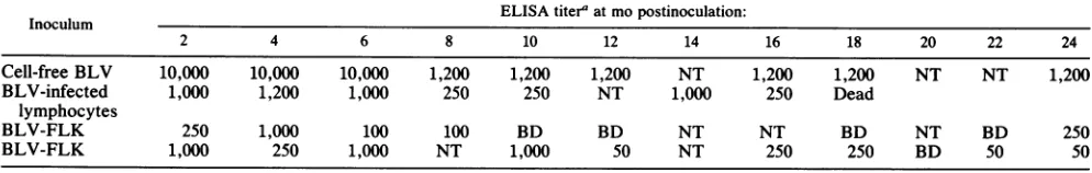
infected rabbits had altered growth characteristics in long-termculture, PBMCfrom BLV-infected and control rabbits were stimulated with phytolectinandmaintained in culture
withhumanrecombinantIL-2. During thefirst20 to 30 days
ofculture, majordifferenceswereobservedin thegrowth of
infected-rabbit PBMC compared with PBMC from normal
animals. Specifically, beginning at 4 to 5 days of culture,
giant multinucleated cells formed in cultures initiated from BLV-infected rabbits (Fig. 3). In some of the giant cells, as many as 25 to 30 nuclei were observed. Figure 4 shows
growthpatterns during the first 28 days in culture of PBMC
fromanormal rabbit andPBMC from a BLV-infected rabbit:
During that period, while giant cells formed in the
infected-31
I~~~
II
I
*
I~
II I I
2
F
IF
| I I I I
4
8
12
16
20
24
28
Age
of
culture
(days)
FIG. 4. Typical growth in continuous culture of PBMC from BLV-infected and control rabbits. The data represent cell growth from one control (solid circles) and one infected (open circles) rabbit. Similar growth patterns were obtained for four additional control and sevenadditional infected rabbits. The vertical dashed lines indicatetimes and cell densitiesatwhich cultureswereeither dividedorconcentrated.
PBMC cultures, total cell numbers did not increase.
IL-2-dependent cell proliferation could be detected in
BLV-infected cultures only after giant-cell formation ceased
(by
30days).
BLV production incultures oflong-term cells. The use of
PBMC to show that BLV-inoculated rabbits were infected withthevirus indicatedthat viruscouldbe demonstratedin
circulating leukocytes. However,PBMCcontainmonocytes as well as
lymphocytes. Therefore,
to determine whetherA
4w
1
..ko .. g ...q
'% 4 II
ftf
FIG. 3. Multinucleated giant-cell formation in BLV-infected rabbit PBMC cultured with recombinant IL-2. BLV-infected and control rabbit PBMC were stimulated with ConA and cultured with human recombinant IL-2.Toevaluatecellmorphology,1 x 105to2 x 105cells werecentrifuged onto glass slides(Cytospin, ShandonSouthernInstruments, Sewickley, Pa.) and stained withWright-Giemsa stain. (A) Cells obtained froma3-week-old culture of control rabbit PBMC. (B) Cells obtained froma3-week-old culture of BLV-infected rabbit PBMC. The large arrowheads pointtothecytoplasmic margin ofasingle cell. Each small arrowhead pointsto anucleus within thatcell. Magnification, x200.
..Wlllllc
on November 10, 2019 by guest
http://jvi.asm.org/
[image:5.568.108.201.68.201.2] [image:5.568.57.505.462.666.2]TABLE 5. Induction by cytokine-containing medium of BLV expression in infected lymphocytes in long-term culture
No. ofsyncytia/cm2 a
Expt BLV
no. inoculated'b m NnC CM +
anti-no.inoculateCM Non-CM BLV serum
1 Y 24 2 0
2 Y 24 0 NTC
Y 25 2 NT
3 Y 25 8 NT
N 0 2 NT
a CM, Cytokine-containing medium.
bYand N,BLV-infected(Y)andcontroluninfected(N)lymphocytes.
cNT,Nottested.
BLV could be produced by lymphocytes from infected rabbits, IL-2-dependent lymphocyte cultures were derived
from PBMC of infected rabbits. BLV expression was
in-duced fromlymphocytes maintained in long-term culture for 3months orlonger by adding cytokine-containing superna-tants from phytolectin-stimulated PBMC from uninfected
rabbits tothelong-term cultures of IL-2-dependent lympho-cytes derived from BLV-infected rabbits. Virus expression was monitored by induction of syncytium formation on
susceptible fibroblasts (TblLu) (Table 5). In three separate
experiments, lymphocytes from three different
BLV-infected rabbits induced increased numbers of syncytia in the presence of supernatant compared with the numbers seeneitherin the absenceofsupernatant,in the presence of
anti-BLV antiserum, or in comparison with cells from an
uninfected rabbit.
Response of peripheral blood leukocytes to phytolectin stimulation. To assess whether BLVinfection was affecting
rabbit T-lymphocyte function, a method which measures
general responsiveness ofTlymphocyteswas used. Circu-lating leukocytes from the first 11 rabbits toreceive
BLV-infected bovine lymphocytes and the first two rabbits to
receiveBLV-FLK cells were evaluated at regular intervals
forresponse tostimulation bytheT-lymphocyte-stimulatory phytolectinsPHA andConA. The stimulation obtained with BLV-inoculated rabbit leukocytes was compared with the
stimulation obtainedwithage-matchedcontrolrabbit leuko-cytes run at the same time. The BLV-inoculated rabbits
were evaluated for 18 to 24 months or until they died. The
data(Fig. 5) revealed thatleukocyteresponses to phytolec-tin stimulation fluctuated in an abnormal and apparently random mannerwhichwasdifferent foreach BLV-infected rabbit.
Specifically, analysis ofthe data after 6 months showed that responses of five animalshad become abnormally low,
five had remainednormal, and three had become elevated.
Figure 5 shows the responses to PHA of the five animals
which had abnormally low responses (panel A) and those with responses withinthe normal range(panel B)inthefirst
6monthspostinoculation. The shaded areas show therange
of responses of age-matched control animals. Ofthe five
rabbits withabnormally low responsesby 6months
postin-oculation (panel A), one animal was killed at 12 months
postinoculation
(seePathology,below).
Theresponsesofthe otherfourreturned tonormal and have fluctuated betweennormal and
abnormally
low. Three had abnormally low responsesby20 months(1, 8, and44% of normal), while the fourth animal appeared normal. Of the five animals with PHAresponsiveness within the normal rangeforthe first6 months postinoculation (panel B), two developedabnor-mallyhighresponses over thenext6 to8months,andathird
0
c
0
0
E
0
z ...I. I
'b
bD D
Time post infection(months)
I I *
4 8 12 16 20
Time post infection(months) FIG. 5. Response to phytolectin stimulation of peripheral blood leukocytes from BLV-infected rabbits. Heparinized blood was diluted and cultured in the presence of PHA-Pfor 3 days. Cell proliferation was determined by uptake of [methyl-3H]thymidine. Panels A and B show the phytolectin responsiveness of BLV-infected rabbitsover18to 24monthspostinoculation. The shaded areas represent the range of responses of leukocytes from age-matched normal rabbitsrun atthesametimeastheBLV-infected rabbits. (A) Responsiveness of five infected rabbits which had abnormally low responses by 6 months postinoculation. (B) Respon-siveness offive infected rabbits which had normal responses by 6 monthspostinoculation.Adashed line followed bya Dindicatesthe death of the rabbit being evaluated. Results are expressed as a percentage of the responsiveness of leukocytes from an age-matched control rabbitrunsideby side with those from the infected animal.Uptake of[methyl-3H]thymidine into PHA-stimulated con-trol rabbitleukocytes ranged between 20,000 and 45,000 cpm for control rabbits less than 18 months old and between 6,000 and 15,000 cpm for control rabbits over18 months old. Unstimulated leukocytes incorporated between100and700cpmregardless ofthe age oftheanimal.
animal was killed(see Pathology). At 20 months
postinocu-lation,the tworabbitswhichhaddevelopedabnormallyhigh
responseswithin6 to 14monthspostinoculationwerefound
dead (see Pathology). The remaining two rabbits have re-sponses within the normal range at this time. The three animals with responses above normal within the first 6 months still haveabnormally high responses after 19to 20 months(148, 225,and286%ofnormal). Together,these data show that most BLV-infected rabbits develop unique and
abnormalpatternsofT-lymphocyteresponsiveness.
Pathology. Within 12 to 18 months ofinoculation, three
rabbits displayedmarked
weight
loss(one oftheserabbits,
no.5,was euthanized). Insixadditional
rabbits,
therewereclinicalindications ofupper
respiratory
disease.The clinical indications of upper respiratory disease most often found wereconjunctival and nasal exudates. Theextentofweight
loss in one of the BLV-infected rabbits is showninFig. 6. The additional two rabbits with marked weight loss had similar appearance.
Currently,
of the nine infected rabbitsexhibiting either
weight
loss or clinicaldisease,
five have died(tworabbits,
nos.2and5,
werekilledduetodecline in their clinicalcondition,
and three were founddead).
This represents amortality
rate of 28%(5
of18)
for rabbitsB
.1..
on November 10, 2019 by guest
http://jvi.asm.org/
FIG. 6. Severeweight lossinaBLV-infected rabbit.Appearance of a BLV-infected rabbit (left) compared with that of an
age-matched control rabbit (right). The BLV-infected rabbit had
re-ceived BLV-infected cells 23 months earlier. Both animals were
maintained under thesameenvironmentalconditions.
inoculated with BLV. During the same time period, no
illnesses or deaths were observed in our colony of 25
uninfected rabbits held under the same environmental
con-ditions.
Gross and histological examination of these five rabbits revealedinflammatoryprocesses of the colonin tworabbits (nos. 1 and 2). Rabbit 1 exhibited a moderate plasmacytic and lymphocytic colitis, and rabbit 2 had a focal chronic transmural colitis. The colonic lesion ofrabbit 2was
asso-ciated with afocal perforation. Although we hypothesized
that a foreign body might have caused the perforation, a
foreign body was not found. Inflammatory changes of the epicardium, pulmonary pleura, kidney,and liverwerefound in rabbits 3 and 4. Rabbit 3 exhibited a fibrinous and suppurative pleuritis and pericarditis, and rabbit 4 had moderate interstitial lymphocytic nephritis and periportal
lymphocytic hepatitis. Pasteurella multocida was isolated
from thepericardial and pleural lesions; however,thecause
ofthe hepatic and renal inflammation was not determined. Rabbit 5, the last one necropsied, had only a moderate
increasedcellularity of peribronchial lymphoid tissue. Thegrossandhistological examinations of rabbits 2 and 3
revealedprocesses ofadequate severityto explain the clin-icalcoursein theseBLV-infected rabbits. Furthermore, the
pleural and pericardial lesions caused by P. multocida in rabbit 4 certainly contributed to the death of this rabbit; however, the reason for the proliferation of this common
rabbit pathogen in this rabbitwasnotdetermined. Although the gross and histological examinations of the five rabbits
necropsied did not reveal neoplastic, degenerative, or
in-flammatory processes thatcould be directly attributable to BLV infection, an indirect contribution of BLV to the clinicaldeterioration and death seenin these rabbitscannot be ruled out.
DISCUSSION
Inthisstudy,weinoculatedhomogeneous preparations of
the type C lymphotropic retrovirus BLV or circulating
lymphocytes fromacow infected with BLV into 18 young
adult rabbits to determine whether rabbits could become
persistently
infectedwith the virus. We found that all rabbitsreceiving
BLVpreparations
produced
persistent
antibody
to BLV and had PBMC which inducedsyncytia
inBLV-susceptible
TblLu fibroblasts when tested aslong
as 16months after inoculation. Additional tests on PBMC from
randomly
selected rabbits revealed the presence of RTactivity,
BLVmRNA,
and BLVproviral
DNA.Long-term
IL-2-dependent
lymphocyte
cultures initiated fromBLV-infected rabbits
developed
multinucleatedgiant
cells withinthefirst30
days
ofculture,
during
which timenoincrease incell numbers was observed. After
IL-2-dependent
cellpro-liferation
commenced,
lymphocyte
cultures were shown tocontain cells which could induce
syncytia
inTblLu
fibro-blasts uponstimulation with a
cytokine-containing
superna-tant from PBMC of control rabbits.
Abnormally
lowre-sponsesto
phytolectin
stimulation ofcirculating
leukocytes
developed
within 6monthsin 38% ofthe rabbits tested andsubsequently
fluctuated between normal andabnormally
low.
Forty-four
percent of BLV-infected rabbits sufferedrecurrent
conjunctivitis
andrhinitis,
and28% oftheanimalswere found dead or werekilled due to poor clinical
condi-tion. Noneofthecontrol rabbits has hadclinical
signs,
andnone havedied.
The
major
conclusion tobe drawnfromthesecumulative observations is that allrabbits inoculated withBLVprepa-rations become
persistently
infected with the virus. Thepersistent
presence ofanti-BLVantibody
and titers ofanti-BLV
antibody
which rise andfall in the absence of admin-istration of additional BLVantigen
in and of themselves suggestthatthevirus which initiatesantibody
production
isbeing
made in theinoculated rabbits.Inaddition,
thecapac-ity
of PBMCtoinducesyncytia
in TblLumonolayers
when tested upto1 yearafterinoculation withBLVindicates thatBLV-infected cellsare
present
amongthecirculating
PBMC.The
finding
ofpersistent
BLVinfectionin rabbitsreceiving
BLV-infected cells is consistent with several studies which
have demonstrated the presenceofinfectious BLV in
lym-phocytes
from infectedcattleand ininfectedcell
lines(1, 9,
35, 38).
There are two
important
findings
in these studies.First,
two-thirds ofthe BLV-infectedrabbits showedone or more ofthe
following changes:
low response tophytolectin
stim-ulationby
T-cellmitogens,
clinicalindications ofrespiratory
disease,
severeweight
loss,
ordeath. Noneofthesechanges
has beenseenamong thecontrolrabbits,
strongly
suggesting
that BLV infection is involved in the
development
oftheseabnormalities. The decreased in vitro T-cell responses to
phytolectin
stimulationthatwe saw aresimilartoreports
forother
trans-acting
lymphotropic
retrovirussystems,
suchasHTLV-I-induced chronic T-cell
lymphocytosis
(26)
and HIV-1-inducedacquired immunodeficiency
syndrome (10).
Furthermore,
ourobservations ofrecurrent clinical indica-tions ofrespiratory
disease aresuggestive
ofan increased incidence ofsecondary
infections in BLV-infected rabbits.Secondary
infectionshavebeen found both inpatients
withHIV-1-induced
acquired
immunodeficiency syndrome
and those with HTLV-I-induced leukemia(reviewed
inrefer-ences 17and
41).
Second, long-term
IL-2-dependent lymphocyte
culturesinitiated from BLV-infected rabbits
initially
showed abnor-malgrowth
characteristics consistentwith the presence ofatrans-acting
retrovirus. Multinucleatedgiant
cells similarto thoseseeninourrabbit PBMCcultures have beenobservedin
HTLV-I-, HTLV-II-,
and HIV-1-infected human cell cultures(30, 37,
41,
42).
The presenceof suchcellssuggests
on November 10, 2019 by guest
http://jvi.asm.org/
[image:7.612.67.294.80.270.2]that BLV expression induced by stimulating infected cells resulted in theirsubsequentfusion. Preliminary results indi-cate thepresence ofintensely stained nonspecific esterase-positivecells in thefusions,suggestingthat somemonocytes
might be infected with the virus. Studies are in progress to evaluate BLVexpressioninthese fused cells.
Furthermore, once fusion has ceased andIL-2-dependent growth has become established, a portion of the remaining lymphocytesmustcarry the virus. Our data show that BLV
expression can be induced from cultured long-term
IL-2-dependent lymphocyteswhenthose lymphocytes are
incu-bated with a cytokine-containing medium derived from PBMC of control rabbits. IL-2 does not appear to be
sufficienttoinduce virusexpression, because cultures con-taining IL-2 without cytokine-containing medium did not
inducesyncytium formation. Ourresults are similar to those reported forinductionof HIV-1 from a monocyte cell line by
cytokine-containingmedium (13) in thatlong-term-cultured, infected cells require cytokine stimulation in order to ex-press virus. These findings are important because they
indicate that both monocytes andlymphocytes can be
res-ervoirs ofBLV in infected rabbits.
While allBLV-inoculated rabbits showed persistentBLV
infection,theconfirmingtests for BLV (RT assay,Northern blot analysis, and PCR) done on PBMC did not show the presenceof virus in all animals tested. Itis interesting that RTactivitywasdetectedincells fromanimalsthat had been
infected for longer than 1 year and were showing signs of
disease. Twoanimals whichwere RT negative at 6 months
postinoculation wereRTpositive over 1 year later,
suggest-ing that too fewinfected cellswerepresentinthose rabbits early in the infection toproduce enough virus to detect in
this assay. Failure to consistently detect BLV message by
Northernblotanalysis andBLVproviral sequencesbyPCR could also be dueto the presenceofveryfew infected cells
in the preparations from which the nucleic acids were
extracted. The presenceofveryfewcirculating infected cells
hasbeenatypicalobservation byotherinvestigators
study-inglymphotropic retroviruses. Forexample, althoughsheep areverysensitive toinfection withBLV(22), BLV-infected sheephave beenestimatedto have between 1 in 2x 10³ and 1in 5 x 10⁵ infected circulating cells earlyininfection (25).
Similarly, HIV-1-infected people have fewer than 1 in 104
infectedcirculating lymphocytes (20, 34).Thesefindingsare
consistentwith ourobservations.
Recently, attentionhasbeen giventothe presence ofan
additional bovine retrovirus, which hasbeen classifiedasa
lentivirus. Tentatively named bovineimmunodeficiencylike virus, or BIV (18), it was isolated from some cattle with
persistent
lymphocytosis,
wasting,andcentral nervous sys-tem signs (39). While BIV infectiondoes not appear to bewidespread among cattle intheUnitedStates(M. A.Gonda,
10th Annu. W.Food Anim. Dis.Res.Conf., 1989, abstr.),it
is of concern that some ofour rabbits, notably those that
receivedlymphocytes fromaBLV-seropositive, persistently
lymphocytotic
cow, couldhavereceivedamixed BLV-BIVinfection. While we know that all of our rabbits became
infected with BLV, we do not believe that the changes observed in our rabbits werethe result ofa
superimposed
BIV lentivirus infection. BLV-infected rabbits developed clinical signs of disease whether the animals were given homogeneousBLV
preparations
(BLV-FLK cellsorpurified
BLV) orbovine lymphocytes. In fact, homogeneous BLVpreparations appeared to be more effective in inducing
diseasesignsinrabbits(six ofsix rabbits withclinical
signs)
than were bovine lymphocyte suspensions (6 of 12 rabbits
withclinical signs) (Table1). To date, we have noevidence
to support infection by BIV in rabbits. Furthermore, no
direct
relationship
between BIV infection and disease incattlehasbeendemonstrated(M. C.Thurmond,J. Stott,G.
Theilen, and J. P. Picanso, 10th Annu. W. FoodAnim. Dis. Res. Conf., abstr.;M. J. vanderMaaten, C. A.Whetstone,
J. A. Roth, and J. M. Miller, 10th Annu. W. Food Anim.
Dis. Res. Conf., abstr.). Based on the abundance of evi-dencethatBLV causesdisease incattle, wesuggest that the
changes in parameters associated with
immunity
thatoc-curred in our rabbits are more consistent with infection
by
BLV.
A
possible
alternative explanation for thedepression
inimmune functionseeninourBLV-infected rabbitsmight be that infection with BLV activated a latent, endogenous rabbit retrovirus, andthe changes seenin therabbits were duetotheactivated rabbit virus. Suchan
explanation
cannotbe
unequivocally
ruled out because of theprevalence
of endogenous retroviruses ina largenumber of other animalspecies
(Infectious
Diseases ofRodents, short course,Mas-sachusetts Institute of Technology, May 1986). However,
we believe that endogenous retrovirus activation does not
explain
our findingsbecause endogenous retroviruses have notyetbeenconfirmed in rabbits. Infact, rabbitshave beenrecommendedas
experimental
hosts tostudy
murine retro-viruses(MITshort course, 1986), suggestingthatactivation ofendogenous retroviruses inrabbits isanunlikely
event.We have demonstratedthat BLV infects
rabbits,
and we suggest that this viral infection can result in immune dys-function which maypredispose
infected animals tosecond-aryinfections and death. Because BLV is oneofa
unique
groupof
trans-acting
lymphotropic
retroviruses, the use ofBLV infection in rabbits
provides
a safe andrelatively
inexpensive
in vivo system for studies ofexpression
ofviruseswhich contain
trans-acting
genes.ACKNOWLEDGMENTS
We thank R. Scofield, M. Trumble, and I. Smith for excellent technical assistance.
Thisworkwassupportedin partbyfundsprovidedtoWashington
StateUniversity throughthe NationalInstitutes of Health Biomed-ical Research Support Grant, American CancerSociety grant
IN-119H,
Public Health Service grant Al 26356 from the National Institutes ofHealth,andbythe ElsaU. PardeeFoundation.LITERATURE CITED
1. Baliga, V., and J. F. Ferrer. 1977. Expression of the bovine leukemia virus and its internalantigen in blood lymphocytes.
Proc.Soc.Exp. Biol. Med. 156:388-391.
2. Baumgartener,L.E.,and C. Olson. 1980.Host rangeof bovine leukosis virus: preliminaryreport,p. 338-347. In0. C. Straub (ed.), Fourth International Symposium on Bovine Leukosis,
Brussels-Luxembourg. MartinusNijhoff,TheHague.
3. Bouillant,A. M.P., andS. A. W. E. Becker. 1984. Ultrastruc-tural comparison ofOncovirinae (type C), Spumavirinae, and Lentivirinae: three subfamilies of Retroviridae found infarm animals. JNCI 72:1075-1084.
4. Buck,C.,A.McKeirnan,J. Evermann,N. S.Magnuson,and R. Reeves.1988. Arapidmethodfor thelarge scale
preparation
of bovine leukemiavirusantigen. Vet. Immunol. 17:107-116. 5. Burny,A.,C.Bruck,H.Chantrenne,Y.Cleuter,D.Dekegal,J.Ghysdael, R. Dettmann, M. Leclerq,J. Leunen, M. Mammer-ickx, and D.
Portelie.
1980. Bovine leukemia virus: molecular biologyandepidemiology,p. 231-289.InG. Klein(ed.), Viral oncology.RavenPress, NewYork.6. Burny,A., C. Bruck, Y. Cleuter,D. Couez, J. Deschamps,J. Ghysdael, D. Gregoire, R. Kettmann, M. Mammerickx, G. Marbaix,D. Portetelle,and L. Willems. 1985. Bovineleukemia
on November 10, 2019 by guest
http://jvi.asm.org/
virus, a versatile agent with various pathogenetic effects in various animalspecies. CancerRes. 45:4578-4582.
7. Davis, L. G., M. D. Dibner, and J.F. Battey. 1986. Basic methods in molecularbiology. Elsevier SciencePublishingCo., NewYork.
8. Derse,D.1988.trans-Acting regulationof bovine leukemia virus mRNAprocessing. J.Virol. 62:1115-1119.
9. Diglio, C.A.,andJ.F. Ferrer. 1976. Induction ofsyncytia by the bovine c-type leukemia virus. CancerRes.36:1056-1067. 10. Epstein, J. S.,W. R. Frederick, A. H. Rook,L.Jackson, J.E.
Manischewitz, R.E. Mayner, H. Masur, J.C. Enterline, J.Y. Djeu,andG. V. Quinnan, Jr. 1985. Selective defects in cyto-megalovirus-andmitogen-inducedlymphocyteproliferationand interferon release inpatients with acquired immunodeficiency syndrome. J.Infect. Dis. 152:727-732.
11. Feinberg,M.B.,R. F.Jarrett,A.Aldovini,R.C.Gallo,and F. Wong-Staal.1986.HTLV-IIIexpressionandproductioninvolve complex regulation atthe levels ofsplicingandtranslation of viral RNA. Cell46:807-817.
12. Ferrer, J. F.,andC.A.Diglio. 1976.Developmentofanin vitro infectivityassayfor the c-type bovine leukemia virus. Cancer Res. 36:1068-1073.
13. Folks,T. M., J. Justement,A.Kinter,G. A.Dinarello, and A. Fauci. 1987. Cytokine-inducedexpressionofHIV-1ina chron-icallyinfected promonocyte cell line. Science238:800-803. 14. Franchini, G., F. Wong-Staal, and R.C. Galo. 1984. Human
T-cellleukemia virus(HTLV-I) transcriptsinfresh and cultured cells ofpatientswith adult T-cell leukemia. Proc. Natl. Acad. Sci. USA 81:6207-6211.
15. Ghysdael, J., C. Bruck, R. Kettmann, and A. Burny. 1984. Bovine leukemia virus. Curr. Top. Microbiol. Immunol. 112: 1-19.
16. Gilden,R.V.,C. W.Long,M.Hanson,R.Toni,H. P.Charman, S. Oroszlan, J.M. Miller, and M.J. Van der Maaten. 1975. Characteristics of the majorinternalprotein and RNA-depen-dentDNApolymeraseof bovine leukaemia virus.J.Gen. Virol. 29:305-314.
17. Gluckman, J. C., D. Klatzmann, and L. Montagnier. 1986. Lymphadenopathy-associated-virus infection andacquired im-munodeficiency syndrome.Annu. Rev.Immunol.4:97-117. 18. Gonda, M. A., M. J. Braun, S. G. Carter, T. A. Kost, J.W.
Bess, Jr., L.0. Arthur, and M. J. Van der Maaten. 1987. Characterization and molecularcloning ofabovine lentivirus related to human immunodeficiency virus. Nature (London) 330:388-391.
19. Graves,D. C., C. A. Diglio, andJ.F. Ferrer. 1977. A reverse transcriptaseassayfor detection of the bovine leukemia virus. Am. J.Vet. Res. 38:1739-1744.
20. Harper,M.E.,L. M.Marselle,R. C.Gallo,and F.Wong-Staal. 1986. Detection oflymphocytes expressing human T-lympho-tropicvirus typeIIIinlymphnodes andperipheralbloodfrom infectedindividuals byinsituhybridization.Proc. Natl. Acad. Sci. USA 83:772-776.
21. Katoh, I., Y. Yoshinaka, N. Sagata,and Y. Ikawa. 1987. The bovine leukemia virusXregionincodesatrans-activator of its longterminal repeat.Jpn.J. CancerRes.(Gann) 78:93-98. 22. Kenyon, S. J., J.F. Ferrer,R. A. McFeely,andD.C. Graves.
1981.Inductionoflymphosarcomainsheep by bovineleukemia virus.JNCI 67:1157-1162.
23. Kettmann, R., G.Marbaix, Y.Cleuter, D.Portetelle,M. Mam-merickx, and A. Burny. 1980. Genomic integration ofbovine leukemiaprovirusand lack of viralRNAexpressioninthetarget cells of cattle with different responses to BLVinfection. Leu-kemiaRes. 4:509-519.
24. Kettmann,R.,J. Deschamps,Y.Cleuter, D. Couez,A.Burny, and G. Marbaix. 1982. Leukemogenesis by bovine leukemia virus:proviralDNAintegrationandlack ofDNAexpressionof viral long repeats and 3' proximate cellular sequences. Proc. Natl. Acad. Sci. USA 79:2465-2469.
25. Lagarias,D.M.,andK. Radke.1989.Transcriptionalactivation of bovine leukemia virus in blood cells from experimentally infected, asymptomatic sheep with latent infections. J. Virol. 63:2099-2107.
26. Levitt, L. J., G. R. Reyes, D. K. Moonka, K. Bensch, R. A. Miller, and E.G. Engleman. 1988. Human T cell leukemia virus-I-associatedT-suppressorcellinhibition of erythropoiesis in apatientwithpure red cellaplasiaand chronic Tg-lympho-proliferativedisease. J. Clin. Invest. 81:538-548.
27. Maniatis, T., E. F.Fritsch,and S.Sambrook. 1982. Molecular cloning:alaboratory manual.ColdSpringHarborLaboratory, ColdSpring Harbor, N.Y.
28. Miller,J. M.,L. D.Miller,C. Olson,andK.G. Gillette.1969. Virus-like particles in phytohemagglutinin-stimulated lympho-cytecultures with reference to bovinelymphosarcoma.J. Natl. CancerInst. 43:1297-1305.
29. Miller, J.,and M.J.Van der Maaten. 1976.Serologicdetection of bovine leukemiavirus infection. Vet. Microbiol. 1:195-202. 30. Montefiori,D.C.,and W. M.Mitchell.1987. Persistent
coinfec-tion of TlymphocyteswithHTLV-IIandHIV and the role of syncytium formation in HIV-induced cytopathic effect. Virol-ogy160:372-378.
31. Muesing,M.A.,D.H.Smith,and D.J. Capon.1987.Regulation of mRNA accumulation by ahuman immunodeficiency virus trans-activator protein. Cell48:691-701.
32. Olson, C., R. Kettmann, A. Burny, and R. Kaja. 1981. Goat lymphosarcoma from bovine leukemia virus. JNCI67:671-673. 33. Oste, C. 1988. Polymerase chain reaction. Biotechniques 6:
162-167.
34. Ou, C., S. Kwok, S. W. Mitchell, D. H. Mack, J. J. Sninsky, J. W. Krebs, P. Feorino, D. Warfield, and G. Schochetman. 1988. DNAamplification fordirect detection ofHIV-1inDNA ofperipheral blood mononuclear cells. Science 239:295-297. 35. Paul, P.S., K. A. Pomeroy, A. E. Castro, D. W. Johnson, C. C.
Muscoplat, and D. K. Sorensen. 1977. Detection of bovine leukemia virus in B-lymphocytes by the syncytia induction assay. J.Natl. CancerInst. 59:1269-1272.
36. Sentsui, H., Y. Kono, S. Itohara, and S. Ishino. 1988. Experi-mentalinfection ofbovine leukemia virus in small laboratory animals.Jpn. J. Vet. Sci. 50:1245-1251.
37. Sodroski, J., W. C. Goh, C. Rosen, K. Campbell, and W. A. Haseltine. 1986.Roleof the HTLV-III/LAVenvelopein syncy-tium formation and cytopathicity. Nature (London) 322:470-474.
38. Stock,N. D., and J. F. Ferrer.1972.Replicating C-type virusin phytohemagglutinin-treated buffy-coat cultures of bovine origin. J. Natl. CancerInst. 48:985-996.
39. Van der Maaten, M. J., A. D. Boothe, and C. L. Seger. 1972. Isolation ofavirusfrom cattle with persistent lymphocytosis.J. Natl. Cancer Inst. 49:1649-1657.
40. Walstra,K., A. Gratwohl, I. Riedener, and B. Speck. 1985. B/T cell ratioof rabbitperipheral lymphocytes. Influence of separa-tion techniqueonresults. J.Immunol. Methods79:143-150. 41. Wong-Staal, F., and R. C. Gallo. 1985. HumanT-lymphotropic
retroviruses. Nature(London) 317:395-403.
42. Yoffe, B., D. E. Lewis, B. L. Petrie, C. A. Noonan, J. L. Melnick, and F. B. Hollinger. 1987. Fusion as a mediator of cytolysisinmixtures of uninfected CD4+lymphocytesand cells infected byhuman immunodeficiencyvirus. Proc. Natl. Acad. Sci. USA84:1429-1433.