0022-538X/85/070096-05$02.00/0
Copyright C 1985,American SocietyforMicrobiology
Monkey
B-Lymphotropic Papovavirus
Mutant
Capable of
Replicating in
T-Lymphoblastoid
Cells
TADAHITO KANDAt* AND KENNETH K. TAKEMOTO
Laboratory of MolecularMicrobiology, NationalInstituteof Allergy and Infectious Diseases, Bethesda, Maryland 20205
Received13 November1984/Accepted 22 March1985
Monkey B-lymphotropic papovavirus (LPV) DNA present as free copies in LPV-transformed hamster embryo cells was molecularly cloned in Escherichia coli. Twenty-two of 24 cloned DNAs were 4.9 kilobases long andshorter than the wild-type LPV DNA (5.1 kilobases). The shorter DNA was nondefective and generated infectious virus(designated LPV-76) upon transfection of human B-lymphoblastoid BJA-B cells. LPV-76 DNA had asmall deletion in the early region and a deletion and an insertion in the control region for transcription. LPV-76 VP-1 was apparently larger than that of the wild-type LPV. LPV-76 could grow in human T-lymphoblastoid MOLT-4 cells, whereas the wild-type LPV replicated only in B-lymphoblastoid cells.
Characterization ofconstructed recombinant viruses between wild-type LPV and LPV-76 showed that the
mutationresponsible for the extended host range of LPV-76 was within the PstI Bfragment, which includes the VP-1coding region. These data strongly suggest that the mutation of VP-1 altered the host range of LPV.
Monkey B-lymphotropic papovavirus (LPV) was isolated
from a B-lymphoblastoid cell line derived from the lymph
node ofan African green monkey (19). Although LPV has
characteristics common to polyomaviruses (14, 19), it is
distinct from other polyomaviruses intwo aspects: thewide
distribution of antibody amongprimatesanditsuniquehost
range in vitro. Serological surveys have shown that
mon-keys,apes,andhumans haveanti-LPV antibody, indicating
that viruses closely related to LPV are widely prevalent
among primates (1, 14). Replication of LPV is confined
exclusivelytocertain B-lymphoblastoid cell lines (1, 2, 14).
The physical map of LPV (14) was correlated to its
functionalmapfromlocation ofthe DNAreplication origin
(7),hybridization ofLPVand simian virus 40 DNAs(7), and
determination ofthe DNA sequence aroundthe replication
origin
(4). Initiation of replication of circular 5.1-kilobase (kb) LPV DNA is at apoint withinBamHIIHpaII-B2.
The LPV DNA has partial homology overthe entiregenome ofsimian
virus40 orBK virus,as revealedby low-stringencyhybridization. DNA sequencinghas shown that, despite its
unique structure in the control region, the structure ofthe
DNAreplication originand thegenomic organization ofLPV
resemble those ofother polyomaviruses. Therefore, it was
possible to align the LPV DNA to the simian virus 40
genomewith thereplication originasthereference point.
LPV was recently demonstrated to be capable of
trans-forming hamster embryo cells (15). The transformed cells
containedabout 10 freecopies of LPV DNA per cell (15). In thepresent study we molecularly cloned the free LPV DNA
in the transformed cells. The cloned DNA was infectious
and, upon transfection of B-lymphoblastoid BJA-B cells,
yielded infectious virus, designated LPV-76. This mutant
was found to be capable of growing in both B- and
T-lymphoblastoid cells. Thispaper describescharacterization
of the new host range mutant of LPV rescued from
LPV-transformed hamstercells.
*Correspondingauthor.
tPresent address: Department ofEnteroviruses, National Insti-tuteofHealth,Shinagawa-ku, Tokyo 141, Japan.
MATERIALS AND METHODS
Virus.Thewild-typeLPVstock virus (LPV-02)was apool
of nondefectivevirusobtained from molecularly clonedLPV
DNA (14). The infectivity titer was approximately 106 50%
infective doses per ml.
Cells. Human BJA-B cells, a
B-lymphoblastoid
line (8),were obtained from C. Yee, National Cancer Institute,
Bethesda, Md. Human MOLT-4 cells, a T-lymphoblastoid
line (13), were kindly provided by Y. Hinuma, Kyoto
University, Kyoto,Japan. These lymphoblastoid cellswere
grown in RPMI 1640 medium supplemented with 5% fetal calf serum. LPV-transformed hamster embryo cells
[HE-LPV(E)] (15) were grown in Eagle minimum essential
me-dium supplemented with 5% calfserum.
FA procedure. The indirect fluorescent-antibody (FA)
procedure with anti-LPV serum made in hamsters and
fluorescein-conjugated goat anti-hamster immunoglobulin
was used. This anti-LPV serum contained both anti-T and
anti-V antibodies (15). Infected cells were centrifuged at
2,500 rpmfor5 min. Thepelleted cellswere suspended in a
small volume of
phosphate-buffered
saline (pH 7.2) andsmeared onto cover slips. The air-dried cover
slips
werefixed in ethanolfor3 min and stained afterdrying.
Enzymes. Restriction endonucleases BamHI, HindIII,
HhaI, HpaII,
andPstI and T4 ligase were purchased fromBethesda Research Laboratories Inc., Gaithersburg, Md.,
andreactionswerecarriedoutaccordingtotheinstructions
ofthe manufacturer.
Preparation of LPV DNA. Viral DNA was extractedfrom
LPV-infectedBJA-BcellsorLPV-transformedhamster
em-bryocells(15)bythemethodof Hirt(6).
Supercoiled
formI DNA waspurified
bytheethidiumbromidebuoyant
density
method (11).
Molecular cloning. The LPV DNA cleaved
with
BamHI wascloned in Escherichia coliplasmid pBR322 at aBamHI site and then used to transform E. coli K-12 strainHB101,
essentially asdescribedpreviously (14). RecombinantDNA
wasextractedfrom
chloramphenicol-amplified
bacterialcul-96
on November 10, 2019 by guest
http://jvi.asm.org/
=
-x _F
co :1
_ q
0 7
:02 76
kb
23.13
-
9.42-6.68
-5.10
-4.36
-_:Z=
CI :l
02 76
kb kb
3.99-
0.78--
4.90
02
76kb
kb2.36-
1.70-1.04
--0.75
02
76kb
kb
3.89--2.36
-1.54
-1.01
0.60\
0.59r
0.32-02
76
kb
kb
2.07-
1.07-
0.78-
0.62-
0.51--0.62
-0.43
2.32
--
-
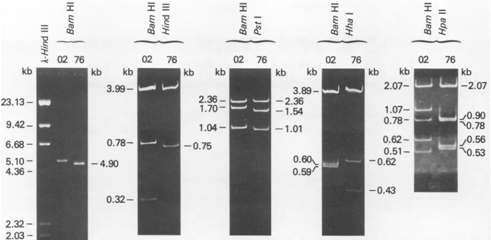
-FIG. 1. Comparison of LPV-02 andLPV-76 DNA. Purified formI DNAfrom LPV-02-orLPV-76-infected BJA-Bcellswasdigestedwith therestriction endonucleases indicated andelectrophoresed. Agarose gels,0.8%,wereusedforBamHI-digested samplesandpolyacrylamide gels, 3.5%,wereusedfor the others.DNAbandswere stainedwithethidium bromide.
tures (3)by amodification ofthe clearlysate procedure (5)
andpurified by the dye-buoyant density methodtil).
Infection with LPV DNA. Molecularly cloned LPV DNA was cleaved with BamHI, released from bacterial plasmid
pBR322,andtested forinfectivity with BJA-B cells. BJA-B
cellswere centrifuged and suspendedat aconcentration of
106cellspermlinserum-freeRPMI 1640medium containing
100
jig
ofDEAE-dextran per ml. A 2-,ug portion of vitalDNAwasaddedtothecells, andthe
preparation
wasshakenandallowedtoincubateat roomtemperaturefor30 min.The
cells were then centrifuged and
suspended
in RPMI 1640mediumcontaining 5% serum.
Gel electrophoresis. For determination of
cleavage
pat-terns, DNA samples digested with various restriction
endonucleaseswere adjustedto 10%glycerol and subjected
to electrophoresis in either agarose or polyacrylamide slab
gels, depending on the size ofDNA to be analyzed. The
running bufferwasTris-borate(pH 8.2;89 mMTris,89 mM
boric acid, 2.5 mM EDTA). DNA bands were stained with
ethidium bromide (0.5
jig/ml
for 30min) and photographedon aUVtransilluminator.
Analysis of viral proteins by immunoprecipitation. BJA-B cells (107 cells) infected with LPV for 3 to 5 days were labeled with [35S]methionine for3 h,and cell extractswere
immunoprecipitated with anti-LPV hamsterserum. The
im-munoprecipitates were electrophoresed in a 12.5% sodium
dodecyl sulfate-polyacrylamide gel and fluorographed
ac-cording topreviously describedprocedures (15).
Constructionof recombinant DNA. Thecloned LPV DNA
containing pBR322 was digested with BamHI and Pstl and
electrophoresed in a 1.2% agarose slab gel. The separated
fragments(Al,A2, andBfrom LPV-02 andAl',A2',and B' from LPV-76) were extracted by the method of McDonell et al. (10). Four mixtures of fragments (Al + A2, Al + A2', Al' + A2,andAl' + A2') were incubated with T4 ligase at
12°C for 14 h and cleaved with PstI to eliminate DNA
molecules ligated at the Pstl site, and then the B or B'
fragmentwasligated. AllsamplesweredigestedwithBamHI
and inserted intopBR322 atthe BamHI site. Orientationof
eachfragment in constructed recombinants was determined
from the HpaII digestion patterns, and recombinants
con-taining thecomplete viralgenomes were selected.
RESULTS
Rescue of virus from
LPV-transfokrmed
hamster cell line.LPV was rescued by transfection of BJA-B cells with
molecularly cloned LPV DNAfrom HE-LPV(E) cells
con-taining about10copiesof free LPVDNA percell (15). Form
I LPV DNAfrom
HE-LPV(E)
cellsatpassages 12to15was extracted andpurified. This DNAwasdigestedwith BamHI and inserted into bacterial plasmidpBR322
at the BamHIsite. Viral DNAsfrom2of24cloneswereabout 5.1kblong
and showed the same HpaII cleavage pattern as that of
LPV-02, which was initially used to transform hamster
embryo cells (15) (datanotshown). DNAsoftheremaining
22 cloneswere about4.9 kblong, and theirHpaII cleavage
patternswerethesameamongthembutweredifferent from
thatofLPV-02 DNA (datanotshown).
The shorter 4.9-kb DNA cloned from HE-LVP(E)
cells
was nondefective. One of the 22 clones (pL76) was
transfected to BJA-B cells by the DEAE-dextran method
aftercleavage with BamHI. Viralantigenswere detectedby
the FAprocedure 3 days after infection, and 14 days after
infection more than 60% of the cells were positive. The
culturefluid was collected and usedas avirus stock
(desig-nated LPV-76) for further characterization. The infectivity titerwas
approximately
106
50%infective doses per ml.DNA oftherescued virus(LPV-76). The LPV-76 DNAwas
analyzed by restriction endonuclease cleavage of the viral
DNA purified from LPV-76-infected BJA-B cells (Fig. 1).
The
HpaIIIBamHI
cleavage pattern(Fig. 1, right lane) was x-Z
mC. no
:c
-ct) m -Z
ad
_-t
on November 10, 2019 by guest
http://jvi.asm.org/
[image:2.612.62.560.71.315.2]Early region origin
-4----
A1Late region
e--* A2 -% B
;I* Al' * e-a A2'-- - B'
-LPV-76
I_
-160 * -T0
+25
*
~-~
Large TSmall
TSmall T
VP-3
VP-2
VP-i
[image:3.612.128.486.71.259.2]?Hpall tPstl
tHhal
tHindll |BamH IFIG. 2. Restrictionmapof LPV-76 DNA. Themaps wereconstructed from thedata in Fig. 1. Location of the replication origin, the early andlate regions of the LPVgenome,is from Kandaetal.(7) and Furunoetal. (4). The codingregionsarefor simianvirus 40 proteins and
aretaken from Tooze(16).
identicaltothatof clonedDNA (datanotshown). The sizes
of LPV-76DNAfragmentswere determined with reference
to LPV-02 DNA anditsfragments, the sizesof whichwere
based on previously reported data (14), and to XDNA
fragments obtained by HindlIl digestion. From the results
shown in Fig. 1, the LPV-76genome sizewas estimatedto
be 4.9 kb in length and its physical map was constructed
(Fig.2).These datarevealed three changesinLPV-76 DNA.
These consisted ofa 160- to 170-base-long deletion in the
early region, an insertion ofapproximately 20 bases,and a
50- to 60-base-long deletion in the control region for
tran-scription.
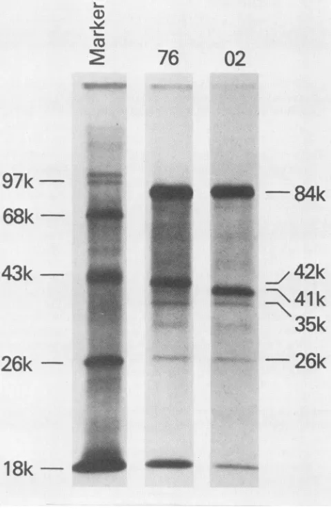
Viralproteins of the rescued virus (LPV-76). The sizes of
LPV-76 viralproteinswere compared withthoseof LPV-02
(Fig. 3). Cell extracts from [35S]methionine-labeled BJA-B
cells infected withLPV-02or LPV-76were
immunoprecipi-tated with anti-LPV serum, and precipitates were
electrophoresed in sodium dodecyl sulfate-polyacrylamide
slab gels. Although the sizes of large T antigen (84,000
daltons[84K]),VP-2(35K),and VP-3(26K) ofLPV-76were
identicalto those ofLPV-02 (12), themajor capsid protein
(VP-1) of LPV-76 was 42K and apparently larger than the
41K VP-1 ofLPV-02(12).
Host range of LPV-76. The growth ofLPV-76 in T- and
B-lymphoblastoid cell lineswas tested to examine whether
LPV-76hadanalteredhostrange.BJA-B
(106)
andMOLT-4(106)
cells were infected with 1 ml of LPV-02 or LPV-76stock
virus,
and virusreplication
wasmonitoredby
theFAtest at3, 6, and 18days
postinfection.
The results(Table
1)showed that in BJA-B cells both LPV-02and LPV-76 grew well. By6days,over60%of the cellswereFApositiveand
cytopathic effects were observed. LPV-02 did not
replicate
in MOLT-4 cells as previously reported (14); however,
FA-positive cells in the LPV-76-infected culture increased
gradually and reached 37% by day 18. When the LPV-76
virus harvested from MOLT-4 cells was grown in fresh BJA-Bcells,the resultant virus induceda
higher
percentage ofV-antigen-positive
cells(45%)
in MOLT-4 cells. These results indicate that LPV-76 is a new host range mutantcapableofreplicatingin both B- and T-cell lines.
Construction of recombinant viruses betweenLPV-02 and LPV-76. We constructed recombinant viruses between
co~
76
97k-68k
43k
-
26k-02
IIPP-84k
42k
.~ ~ 4
ki.
....42k
M::
---L
-
26k
1
8k
*5t
FIG. 3. Immunoprecipitation of LPV proteins in LPV-02- or
LPV-76-infectedBJA-B cells. The infected cellswerelabeled with [35S]methionine, and cellular extracts were immunoprecipitated
with anti-LPVserum.
LPV-02
?
I
?
*t
I?o
t
T?
0
on November 10, 2019 by guest
http://jvi.asm.org/
[image:3.612.314.554.313.682.2]TABLE 1. Growth of LPV-02 and LPV-76 in BJA-B and MOLT-4cells
Days FA-positivecells(%) Virus after
infection BJA-B MOLT-4
LPV-02 3 1 0
6 62 1
18 NDa 0
LPV-76 3 2 2
6 68 12
18 ND 37
aND,Notdone.
R-1 R-2 R-3 R-4 R-5 R-6 76 02
84K
42K,
41K-35K
-*
d!!!'a
m.40
4-
4-m26K- - -mmm
4-LPV-02 and LPV-76 to examine which part of the LPV-76 genome wasresponsiblefor itsbroadenedhost range. Diges-tion of LPV DNA with BamHI and PstI gave rise tothree fragments: Al, A2, and B from LPV-02DNA; Al',A2', and B' from LPV-76 DNA (Fig. 1 and 2). Each segment ofone virus was replaced with the corresponding fragment of the other, and the recombinants were molecularly cloned into pBR322 at the BamHI site. Figure 4 shows the cleavage
patternsof recombinantswith BamHI andPstI. Orientation of the insertedfragments was examined byHpaII digestion
(data not shown), and recombinants containing complete
viral genomes were used fortransfection of BJA-B cells after BamHI cleavage. The recombinant DNAs of all possible
combinations were infectious in BJA-B cells, and
culturefluids were collected for virus stocks when
ap-proximately 60% ofthe cells were FApositive.
One partoftransfected cells with each recombinant DNA
- N ' C CD N
OL
a: zcL
cr: a: r- coc:
cr
Q1
-3.23kb (pBR)
[image:4.612.315.563.72.285.2]-1.13kb
(pBR)
FIG. 4. Cleavage ofrecombinant DNAs between LPV-02 and LPV-76 with BamHI and PstI. Fragments B, Al, and A2 were
derived from LPV-02 DNA, and fragments B', Al', and A2' were
derived from LPV-76 DNA. Recombinants R-1, R-2, and R-3
contained the B' fragment, and R-4, R-5, and R-6 had the B
fragment. All samples including pBR322 DNA were cleaved with BamHI andPstI andelectrophoresed ina 1.2% agarosegel. DNA
[image:4.612.59.298.89.190.2]bandswere stained with ethidiumbromide.
FIG. 5. Immunoprecipitation of LPV proteins in LPV-02, LPV-76, and the recombinant DNAs between 02- and LPV-76-transfected BJA-Bcells.
was labeled with [35SJmethionine, and the sizeof VP-1 was analyzed by immunoprecipitation (Fig. 5). Three re-combinants (R-4, R-5, and R-6) that contained the PstI B fragment derived from LPV-02 synthesized 41K VP-1, and three recombinants (R-1, R-2, and R-3) that had thePstI B' fragment derivedfrom LPV-76 synthesized 42K VP-1.These results indicate that the PstI B' fragment carries the muta-tion responsible for the altered VP-1 of LPV-76.
Hostrange ofrecombinant viruses. Growth of recombinant viruses in MOLT-4 cells was tested (Table 2). Each virus stock(1ml)wasmixed with 106MOLT-4orBJA-B cells and the presence ofFA-positive cells was examined. In BJA-B cells all viruses grew well, and FA-positive cells reached about 50to60% at5days afterinfection. In MOLT-4 cells, although the infection of LPV-02 R-4, R-5, and R-6 was abortive, FA-positive cells infected with LPV-76 R-1, R-2, and R-3 increased gradually to 30 to 40% by 18 days after infection. Thus, only recombinants that contained the PstI
B'fragmentderived fromLPV-76replicatedinMOLT-4 cells.
TABLE 2. Growth of recombinant viruses in BJA-B and MOLT-4 cells
FA-positive cells(%) Size of
Virus compositionof BPM
fragments" BJA-B51 MOLT-4(' (x
10')
LPV-02 B + A1 + A2 55 0 41
LPV-76 B' + A1' + A2' 60 37 42
R-1 B' + A1 + A2 53 34 42
R-2 B' + A1 + A2' 48 31 42
R-3 B' + A1' + A2 65 39 42
R-4 B + A1' + A2' 55 0 41
R-5 B + A1 + A2' 52 0 41
R-6 B + A1' + A2 50 0 41
aFragments A,A2.and Bwerederived from LPV-02DNA,andfragments Al', A2',and B' were derived from LPV-76.
b5daysafter infection. ' 18daysafterinfection.
Al-
Al'-A2 _
A2' '
on November 10, 2019 by guest
http://jvi.asm.org/
[image:4.612.56.300.417.648.2]DISCUSSION
LPV-76, derived from the LPV genome present in the HE-LPV(E) cell line (15), was found to be a viabledeletion mutant with an extended host range. Whereas wild-type LPVisa strictly B-lymphotropic virus forreplication(1, 2, 14), LPV-76 has acquired the ability to replicate in the T-lymphoblastoid cell lineMOLT-4. To examine which part
ofthe genome was reponsible for the altered host range of
LPV-76, we constructedrecombinants between LPV-02 and LPV-76 and found that only those containing the PstI B'
fragment of LPV-76 could replicate in MOLT-4 cells. The
data indicate that, from the correlation of physical and
functional maps of LPV (4, 7), the rearrangements of
LPV-76 DNA detected in theearly region and controlregion
(in the PstI A' segment in Fig. 2) are not related to the
capacityto growinT-lymphoblastoid cells andthatthePstI
B' segment contains the mutation which determined the
altered host range of LPV-76.
From the physical andfunctional maps (4, 7), the PstI B segment of LPV is believed to contain the entire coding
regions forVP-1 and VP-3, the greater partofthe VP-2 gene,
andapart(thecarboxyl-terminal end) ofthelargeT-antigen
gene. Thus, each ofthese genes may contain a mutation.
Sincethe data on viralproteins (Fig.3) clearly show that the
mobility of VP-1 inelectrophoresis has changed in LPV-76,
themutation inthe VP-1 geneisprobably responsible forthe
broadened host range ofLPV-76. The precise location and
type of mutation will be determined in future studies by
sequencingPstI B and PstI B' segments.
Apparently, LPV-76originatedfrom DNAconstitutingthe
major portion ofthe free viral DNA population in
trans-formed hamster cells. The initial characterization of viral
DNA in HE-LPV(E) showed that the free LPV DNA was indistinguishable in size and cleavage pattern from the wild-type LPV DNA from the inputtransformingvirus (15). However, themajorpopulation ofthefree DNAisolated in thisstudy was different from the DNA of the input virus. The DNAweanalyzedpreviouslywas extractedfrom
early-pas-sage HE-LPV(E) (passage 3), but the DNA used in the present studywasextractedfromthecells at passages 12 to 15. LPV-76 DNA, therefore, seems to have become
domi-nantinhamstercellsduringrepeated cell passages,although
itisnotknown whether LPV-76 emergedfromthewild-type
LPVbeforeoraftertheestablishment of cell transformation
by LPV. Possibly, LPV-76 DNA may have some selective
advantageoverthewild-type LPV DNA in hamster cells.
The significance ofrearrangements in LPV-76 DNA(Fig.
2) is not clear at present. A deletion of 160 to 170 bases occurs in theintervening sequenceoflargeT mRNA or the
carboxyl endofthe probable smallT-antigen coding region
(4). If LPV has a small T antigen, the small T antigen of
LPV-76 mustbeaffected by this deletion.The DNAchanges in the controlregionof LPV-76 may affecttranscriptionand have different biological significance, since the DNA re-arrangement in thisregion affects variousbiological
proper-ties ofpolyomaviruses, including the host range of mouse
polyomavirus (9) and JC virus (18) and the transforming
ability of BK virus(17). During the courseof serialpassage
of LPV-76 in MOLT-4 cells, a variant emerged which
replicated in MOLT-4 cells much more efficiently. This
variant has an additional change in the control region
(un-published data). Comparative studies of the controlregions
ofLPV-76 and its new variant are under way.
ACKNOWLEDGMENT
We thankKunito Yoshiike, National Institute ofHealth, Tokyo, Japan,for critical review ofthemanuscript.
LITERATURECITED
1. Brade, L., N. MuIIer-Lantzsch, and H. zur Hausen. 1980. B-lymphotropic papovavirus and possibility of infections in hu-mans.J. Med.Virol. 6:301-308.
2. Brade, L., W. Vogl, L. Gissman, and H. zur Hausen. 1981. Propagation of B-lymphotropic papovavirus (LPV) in human B-lymphoma cells and characterization of its DNA. Virology 114:228-235.
3. Clewell, D. B. 1972. Nature of ColEl plasmid replication in Escherichiacoli in the presenceof chloramphenicol. J. Bacte-riol. 110:667-676.
4. Furuno, A., T. Miyamura, and K. Yoshiike. 1984. Monkey B-lymphotropic papovavirus DNA: nucleotidesequence ofthe region around theorigin of replication. J. Virol. 50:451-456. 5. Guerry, P., D. J. LeBlanc, and S. Falkow.1973.Generalmethod
for the isolation ofplasmid deoxyribonucleicacid. J. Bacteriol. 116:1064-1066.
6. Hirt, B. 1967. Selective extraction of polyoma DNA from infectedmousecell cultures.J. Mol. Biol. 26:365-369. 7. Kanda, T., K. Yoshiike, and K. K. Takemoto.1983. Alignment
ofthe genome of monkey B-lymphotropic papovavirus to the genomesof simian virus40andBKvirus.J.Virol.46:333-336. 8. Klein, G., T. Lindahl, M. Jondal, W. Leibold, J. Menezes, K. Nilsson, andC.Sundstrom.1974.Continuouslymphoid cell lines with characteristicsof B cells(bone-marrow-derived), lacking the Epstein-Barr virusgenome and derived from three human lymphomas. Proc.Natl. Acad. Sci. U.S.A. 71:3283-3286. 9. Levine, A. J. 1982. The natureof the host rangerestriction of
SV40 andpolyoma viruses in embryonal carcinoma cells. Curr. Top. Microbiol.Immunol. 101:1-30.
10. McDonell, M. W., M. N. Simon, and F. W. Studier. 1977. Analysisof restriction fragments ofT7 DNAand determination of molecularweights byelectrophoresisinneutralandalkaline gels.J. Mol. Biol. 110:119-146.
11. Radloff, R., W. Bauer, and J. Vinograd. 1967. Adye buoyant-densitymethodforthedetection andisolation of closed circular duplex DNA: the closed circular DNA in HeLa cells. Proc. Natl. Acad. Sci. U.S.A. 57:1514-1521.
12. Segawa, K., and K. K. Takemoto. 1983. Identification of B-lymphotropic papovavirus-codedproteins.J.Virol.45:872-875. 13. Srivastava, B. I. S., and J. Minowada. 1973. Terminal deoxynucleotidyl transferase activity in a cell line (MOLT-4) derived from the peripheral blood of a patient with acute lymphoblastic leukemia. Biochem. Biophys. Res. Commun. 51:529-535.
14. Takemoto, K. K., A. Furuno, K.Kato,and K. Yoshiike. 1982. Biological and biochemical studies of African green monkey lymphotropicpapovavirus. J. Virol.42:502-509.
15. Takemoto, K. K., and T. Kanda. 1984. Lymphotropic papovavirus transformation ofhamsterembryo cells. J. Virol. 50:100-105.
16. Tooze, J. (ed.).1981.Molecularbiologyoftumorviruses,part2, 2nd ed., revised. DNA tumor viruses. Cold Spring Harbor Laboratory, ColdSpring Harbor, N.Y.
17. Watanabe,S.,andK.Yoshiike. 1982.ChangeofDNAnearthe origin of replication enhances the transforming capacity of human papovavirusBK.J. Virol. 42:978-985.
18. Yoshiike, K., T.Miyamura,H. W.Chan,and K. K. Takemoto. 1982. TwodefectiveDNAsofhumanpolyomavirusJCadapted
to growth in human embryonic kidney cells. J. Virol. 42:395-401.
19. zur Hausen, H., and L. Gissmann. 1979. Lymphotropic papovaviruses isolated from Africangreenmonkeyandhuman cells. Med. Microbiol. Immunol. 167:137-153.