Vol.65, No. 7 JOURNALOFVIROLOGY, July1991,p. 3746-3758
0022-538X/91/073746-13$02.00/0
Copyright C) 1991, AmericanSociety for Microbiology
Overexpression in Bacteria and Identification in Infected Cells of the
Pseudorabies Virus Protein Homologous
to
Herpes
Simplex
Virus Type
1ICP18.5
NELS E. PEDERSONt AND LYNN W. ENQUIST*
ViralDiseasesResearch, Experimental Station E3281B31, DuPont Merck PharmaceuticalCompany,
P.O. Box80328, Wilmington, Delaware 19880-0328 Received 24 January 1991/Accepted 15 April 1991
TheICP18.5gene(UL28) of herpes simplexvirustype1isamember ofawell-conservedgenefamilyamong
herpesvirusesandisthought toplayarole in localizationof viral glycoproteins. Wehavecloned, sequenced, and expressed the entire pseudorabies virus (PRV) ICP18.5 open reading frame in Escherichia coli as a
Cro-ICP18.5 fusion protein. Rabbit antiserum against Cro-ICP18.5 immunoprecipitated a 79-kDa protein
from PRV-infected cells as well as a 79-kDa protein from in vitro translation of a T7 RNA polymerase
transcript oftheICP18.5 gene. ICP18.5could be detected ininfectedcells by2 hpostinfection. Analysis by
indirect immunofluorescence demonstrated that ICP18.5 became associated with the nucleus. Subcellular fractionation confirmed that ICP18.5 synthesized during a pulse-chase experiment appeared in the nuclear
fractionwithtimeandwasstableforatleast 2.5 hafter synthesis. Pulse-chaseanalysisrevealed that ICP18.5 wassynthesizedas a monomerduringa2-min pulse labeling but formed fastersedimenting complexeswhich weresensitivetosodiumdodecylsulfate(SDS)treatment.ThemajorityofICP18.5appearedincomplexeswith anantigenicallyunrelated 70-kDaprotein.Immunoblotanalysisof total infected-cell extractsusing polyvalent
anti-ICP18.5 serum demonstrated that a 74-kDa cellular protein in addition to the 79-kDa ICP18.5 was detected. This cellularproteinwaspresent atsimilar levels in uninfected cells and in PRV-infected cellsatleast 12 h intotheinfectious cycle.
Herpes simplex virus type 1 (HSV-1) mutants that are
defectiveinlocation of viralglycoproteinstothe cellsurface
have been identified (24). Cells infected with one of these temperature-sensitive (ts) mutants, HSV-1(KOS)icr ts78,
were notlysed by a polyvalent antiserum to viral
glycopro-teins in the presence of complement at the nonpermissive
temperature. HSV-1(KOS)icr ts78 expressed normal levels
ofglycoproteinsgB andgCintracellularlyatboth permissive
andnonpermissive temperatures (24). Atthenonpermissive temperature, the infected cell expressed greatly reduced amounts of these glycoproteins on the cell surface (24). Markerrescueanalysis localized thetsmutation between the ICP8 and gB genes (24). An open reading frame was deter-mined from DNA sequence analysis, and the predicted ICP18.5 protein sequence was used to generate an
anti-peptide serum and to identify a 95-kDa protein in
HSV-1-infected cells (26). Despite the finding of homologs to
ICP18.5invirtuallyeveryherpesvirusgenomethat hasbeen
sequenced (1, 3, 4, 7, 9, 13-15, 20, 23, 25, 30, 35, 41, 43),
little is known about the nature or function of the ICP18.5
protein.
Previously we reported the identification and DNA se-quence of the pseudorabies virus (PRV) ICP18.5 homolog.
Inthis report, we identify the PRV ICP18.5 gene product as
a79-kDa protein. This was accomplished by expressing the
open reading frame in Escherichia coli and producing a
polyvalent, monospecific antiserum in rabbits. This
antise-*Correspondingauthor.
tPresent address:DepartmentofMicrobiologyandImmunology, School of Medicine, East Carolina University, Greenville, NC 27858-4354.
rum immunoprecipitated the 79-kDa protein translated in
vitrofrom ICP18.5-specific RNA as well asa79-kDa protein
fromPRV-infected cell extracts.
(A preliminary report of these findings was presented at
the 1990 International Herpesvirus Workshop, Georgetown University, Washington, D.C.)
MATERIALSANDMETHODS
Cellsand virus. The PK15 cells and Becker strain of PRV have been described previously (33).
Antibody reagents. The antisera usedincluded goat poly-valent 282 antiserum raised against a denatured E. coli-produced Cro-gll fusion protein (reactive with native and
denatured glll) (36), goat polyvalent 284 antiserum raised
against immunoaffinity-purified gll protein (reactive with
native and denatured gIl) (31), and rabbit polyvalent D71
antiserum raised against E. coli-produced Cro-ICP18.5 (de-scribedbelow).
Bacterial strains. ThefollowingE. coli strainswereused: KK2186 [A(lac-pro) thi strA endA sbcB15 hsdR4 supE (F' traD36 proAB lacIfZVM15)], LE392 (37), MBM7060 (37), MC1000 (37), NF1829 (39), XL1-Blue (Stratagene), and NFPU2 (describedbelow).
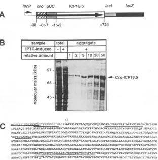
Construction ofpN50. Theexpression plasmid pN50 (Fig.
1A) is capable of expressing two distinct fusion proteins
containing the ICP18.5 gene. The first 23 codons of the
lambdacrogene arefusedto ICP18.5 sequences startingat
the secondcodon. The natural terminator codon of ICP18.5 is followed by an in-frame lacI-lacZ gene fusion. In a
suppressor-negative host, onlythe82-kDa Cro-ICP18.5
pro-tein should beexpressed. Inasuppressor-positive host, the 3746
on November 10, 2019 by guest
http://jvi.asm.org/
IacP cro pUC ICP1 8.5 lac/ lacZ
A
B
C
-30 -8:-7 -1:+2 +724
-2
MTv-- - -A^v"s7jjQTKAK --%SttA; RSAERRYAVL%V2YVQLEMKCDPAVVEAPRVKNALMCRYARR :
LPLEAQTTPLTCAIRLALAYAPAEGDRVI.GAIAAGDDAEAYFERTMGGACRFHARVAIDTYGGRVETELQFLHDAMNLL KQLNYCRLITPlAVDLSAVDFLARTIGGGLVVPPELYDPAQPCAVCFEELCTANQGE-ATHRRLLGCVCDELTRQLAVR VDPEDVAKNLPEVEGLDEARRGRALAALAAVDAAZAREAZ.AASTAA.GAC.AGDAGETARRADALLDA)VFPASRRLY
AVSELQFWLASTNQAVRALDI3THNLDDLERR.RRAEVP.AVELALFGRRPEFDRADAAR.LDIIDGLVGGCAASPD ERLF-ALIRACYDBERMSTPMI.RMPDPRANRDALERLLDEGDDADADGG&AGGADAGDGGGVGDEDGPGAPPPA.DAVAWADL
PAAALRDAEIRRRILYAD)RLSRRSAASLAQCVREQRREL.KTLRVNVYGDALLHTYVAVAAGFRARRAFCEAAARAGTVVD
ERETGCFDARSFMKATVQRfPVDAALLPALTH FELVNGPLFADrTBAFAQPPNTALYFAVENVGLLPHLKEELAMV ARDWCVSEFRGFYRFQTAGVTATQRQAWRYIRELVLAVAVERSVFHCGD,VEVIRADRFAGRDGLYLTYEASCPLVAVFGA
GPGGIGPGTTAVLASDVFGLLETTLQLRGAPSR* -24
FIG. 1. pN50andCro-ICP18.5 expression. (A) The protein expressed frompN50 includes23amino acids of Cro (-30 to -8), 7 amino acids from the pUC18 polylinker (-7 to -1), and 723 amino acids of ICP18.5 (+2 to +724). Amino acids are numbered relative to the translation initiationcodon of nativeICP18.5. Translation of the753-amino-acidproteinterminates at the natural amber codon of ICP18.5, which isin frame with downstreamlacI-lacZcodons.(B)NFPU2 containingpN50wasincubatedin Luriabroth media and induced with 1 mMIPTGfor4h, and aggregate protein was purified as described in Materials and Methods. Whole-cell lysates are from uninducedand induced cultures. Aggregate protein was titrated as indicated. Protein samples were separated bySDS-PAGE anddetected by Coomassie staining.(C) The entire aminoacidsequenceof theCro-ICP18.5fusion protein is shown. Amino acids in bold type mark ICP18.5 sequences between +2 and +724 ofnativeICP18.5. Underlined amino acids were verified by direct amino acid analysis of either the N terminusor internalcyanogenbromide-derived fragments.
ICP18.5 terminator should not be translated, and an
-200-kDa
Cro-ICP18.5-LacI-LacZ
fusion protein should result.Expression is under control of the Lac repressor and is
inducedbyisopropyl-thiogalactoside (IPTG).
ThepN50plasmidwasconstructedin three steps by using
coordinates relative to the A of the translation initiation
codon as follows. The ends ofaninsert spanningfrom +3
(Bal31 derived)to +1120(XhoI)werefilled in with Klenow
polymerase (New England BioLabs) and ligated into the
HincII site ofpUC18(44)tocreate pN8A2'5. The 3' end of
ICP18.5 was added by replacing the StuI (+272) to XbaI
(pUC18) fragment from
pN8A2'5
withaStuI(+272)toNheI(+2175) fragment to generate pN40. This ICP18.5 cassette
(+3 to +2175) wastransferred by flankingBamHI(pUC18)
andHindIII(pUC18)sites into the BamHI andHindlllsites
ofthepHK412expressionvector(31)togeneratepN50(Fig.
1A).
Isolation ofaspontaneousE.coli mutationenabling expres-sion of Cro-ICP18.5. Expression ofeither Cro-ICP18.5 or
Cro-ICP18.5-LacZ fusions inE. coliwas lethalto the cell.
Wemodified atechnique described by Silhavyetal. (37)to
selectexpression of nonlethal fusion
proteins.
In the pN50construction (Fig. 1A), the termination codon of ICP18.5
was placed in frame between ICP18.5 and lacZ. When this
plasmid is introduced intoE. coli lacking a nonsense
sup-pressor mutation, ,B-galactosidase should not be produced.
Certain spontaneous deletions that fuse Cro-ICP18.5 to
LacZin frame andremove segmentsofICP18.5 that leadto
lethalityshould be seenas
Lac'
papillaeonlactoseindicatormedia.
NF1829 (39) harboring pN50 was streaked onto lactose
MacConkey agar (37) with ampicillin and incubated
over-night at 37°C to allow many single colonies to occur. The
petridishesweresealed and incubatedat roomtemperature
anadditional10days.Severaldozenred,
Lac'
papillae
werethen picked and
purified
onMacConkey
platescontaining
ampicillin. Analysisof theplasmidDNAfrom each of these
Lac'
isolates revealed thatessentially all the PRV ICP18.5DNA hadbeendeleted,confirmingourinitial
hypothesis
thatexpression of this open
reading
frame was lethal inE. coli.In addition to red,
Lac'
papillae, white, Lac-papillae
alsoarosewithtime. Several dozenwere
purified,
andtheirplasmidDNAwasscreenedasdescribed above. Whilemost
strains contained
plasmids
with substantialdeletions,
plas-mids fromafew ofthese strains
appeared
intactby
restric-tion enzyme analysis. One isolate was chosen for further
rool-I
on November 10, 2019 by guest
http://jvi.asm.org/
[image:2.612.146.458.73.389.2]3748 PEDERSON AND ENQUIST
analysisandwasfoundtoproduceanovelproteinof 82 kDa
upon IPTG induction. This was the predicted size of the
Cro-ICP18.5 fusionprotein. Plasmid DNAfrom this isolate
waspurified and assayed for transformation into severalE.
coli strains along with pN50. Significantly, these plasmids
would nottransform strains lackingthe lacIq repressor,just as the parent pN50 plasmid would not transform strains
lackingthe lacIq repressor. Thissuggestedthat themutation
conferring
escapefrom fusionprotein lethality
wasnotin theplasmid but rather in the bacterial chromosome. This was
confirmedby curingtheplasmidfrom thenewE. coliisolate
by repeated streakingfromsinglecolonies in the absence of
ampicillin. The cured strain (NFPU2) was readily
trans-formedwithparental pN50. Uponinduction with
IPTG,
theretransformed NFPU2expressed anovelprotein of82kDa
(Fig. 1B). Weconcluded that NFPU2 contained a
chromo-somalmutation permitting overexpressionandaccumulation
of Cro-ICP18.5 fusion
protein.
Fusionprotein purification. Thefusionprotein producedin
E. coli (NFPU2) formed insoluble aggregates. Aggregated
protein
from induced cultures was isolatedessentially
aspreviously
described (40). Anovernight
culture ofNFPU2containing
pN50wasdilutedintofreshmedia,
shakenfor2 hat 37°C, and induced by addition of IPTG to 1 mM. The
induced culture was shaken for 4 h at 37°C, at which time
cells were harvested by
centrifugation.
The cellpellet
wasfrozenon dry ice, thawed, and resuspended in lysis buffer
(50 mMTris [pH 7.9], 200 mMNaCl, 2 mMEDTA, 2 mM
3-mercaptoethanol),
andlysozymewasaddedto 100p.g/ml.
After 20min, the cellextract wasbroughtto1% TritonX-100 andchilledonicefor10min,and
Zwittergent
Detergent3-14(Calbiochem)
was added to 0.5%. After10 minonice,
thesample was sonicated
briefly,
broughtto5 mMMgCl2,
andtreated with DNase and RNase at37°C for25 min. Protein
aggregateswerepelleted througha40%sucrosecushionand
resuspended in deionizedwater.
Production ofD71antiserum.Partially purified aggregated
proteinswereprepared forinjectioninto rabbits asfollows.
Approximately 100 ,ug ofpartially purified insolublefusion
protein was suspended in 200 ml of 50 mM NaOH and
solubilizedby heating at 65°C for 10min. Thesolution was
neutralizedbythe additionof20
RI
of500mMHCland 20RI
of 1 M Tris pH 7.4. This preparation was
injected
into arabbitby usingtheprotocoldescribedbyRobbinsetal.(33). In vitro transcription. Plasmids encoding sequences for ICP18.5andgIIIwerecloned downstreamfromthe T7 RNA
polymerasepromoterin pBluescript II SK(-) (Stratagene).
The plasmid containing the entire ICP18.5 open reading
framewasgenerated by usingcoordinates relativetothe A
of the translation initiation codonasfollows. The ends ofa
DNA fragment spanning -123 (Bal 31 derived) to +1120
(XhoI)
werefilledin with Klenow polymerase,insertedintotheHinclI siteofpUC18, and named
pN8A2'12.
This 5' endof ICP18.5 was transferred to PstI-cleaved pBluescript II
SK(-)
by restriction ofpN8A2'12
at a flankingPstI
site(pUC18)
and the internal PstI site (+361) to create pN56. The 3' end of ICP18.5 was added by replacing theMluI
(+108) to XbaI [pBluescript II SK(-)] fragment of pN56
with theMluI(+ 108) to
NheI
(+2175) fragment from PRV.This plasmid, pN59, encompasses the entire ICP18.5 gene
from-123to+2175. TheentiregIIIopenreadingframe was cloned in plasmid pN61 as follows. A 2.4-kb
NcoI-NcoI
fragment
of PRV was filled in with Sequenase (U.S.Bio-chemical)
andinsertedintotheuniqueBamHI
site ofpBlue-script
II SK(-) previously filled in with Sequenase. Allrestrictionrecognitionsequences wererestored. The 5'
NcoI
site encompasses the translation initiation codon of gIII.
Plasmidswerelinearized andprepared for in vitro
transcrip-tion and mRNA capping as described by Stratagene. RNA was analyzed by denaturing gel electrophoresis prior to in vitro translation.
Invitro translation. RNAwastranslated in the presence of 50 ,uCi of [35S]cysteine with rabbit reticulocytes (New En-gland Nuclear). Aliquots were diluted in
radioimmunopre-cipitation assay (RIPA) buffer for analysis by
immunopre-cipitation.
Radioimmunoprecipitation analysis. Preparation of infect-ed-cell extracts in RIPA buffer and precipitation of immune complexes were performed essentially as previously de-scribed(33). RIPA buffer is 20 mM Tris-Cl (pH 8.0), 100 mM NaCl, 1 mM EDTA, 1% deoxycholate, 1% Nonidet P-40 (NP-40), 0.1% sodium dodecyl sulfate (SDS). Immune com-plexes were adsorbed to Staphylococcus aureus (ICN) on ice for 15
min,
pelleted at 6,000 x g for 2min, and serially washed with buffer B (50 mM Tris [pH 8.0], 120 mMNaCl, 0.5% NP-40), buffer B containing 500 mM LiCl, and buffer B. The complex was resuspended in sample buffer (60 mM Tris [pH 6.8], 3% SDS, 5% ,-mercaptoethanol, 10% glyc-erol, 0.001% bromphenol blue) and heated to boiling. Bac-teria were pelleted, and the immune complexes in the supernatant were separated by denaturingelectrophoresis.PAGE. Immunoprecipitates were fractionated by poly-acrylamide gel electrophoresis (PAGE) on SDS-polyacryl-amide slabgelsof various concentrations (31). Fluorography was performed with 1 M sodium salicylate (6) and was
followed by autoradiography.
Quantitation of autoradiographs. Regions of dried gels were quantitated with an AmBis radioanalytical imaging system (AmBis Systems, San Diego, Calif.).
Amino acid sequence determination. Aggregate protein in deionized water was dissolved in 50% acetic acid. The amino-terminal sequence wasdetermined by Edman degra-dation chemistry on an Applied Biosystems, Inc. 470A gas-vapor sequencer interfaced withanApplied Biosystems 120A PTH analyzer. All fragments were runwitha
phenyl-thiohydantoin (PTH) standard and blank. Cyanogen
bro-mide-derived fragments weregenerated by overnight diges-tion (38). Fragments were resolved on a Hewlett-Packard 1090 reversed-phase high-performance liquid chromatogra-phy (HPLC) column. Selected fractions were sequenced as described above.
Precipitation oftotalprotein. Sampleswereincubated with 3 volumes of acetone at -20°C for at least 30 min. The
precipitatewaspelletedby centrifugationat14,000 x gfor 5
min. The supernatant wasaspirated and discarded, and the pellet wasdried, resuspended in sample buffer, and heatedto
boilingprior to separation by SDS-PAGE.
Affinity purification of serum. Serum was purified by binding to immobilized protein as described by Harlow and Lane (16). Cro-ICP18.5 was separated from minor compo-nents of the aggregate preparation by SDS-PAGE, trans-ferredtoImmobilon-P,andincubatedwith D71 antiserumor
preimmune serum. The filters were washed with phosphate-buffered saline (PBS) and PBS with 0.4% polyoxyethylene sorbitan monolaurate (Tween 20; Sigma). Antibodies were
eluted with two washes of 100 mM glycine, pH 2.5, and the eluate wasneutralized with 1 M Tris-Cl, pH 8.0, and dialysis against PBS. The purified serum was then used for
immuno-fluorescence.
Indirect immunofluorescence. The protocol described by Larjavaet al. (21) wasfollowed. PK15 cells were seeded on
coverslips and infected with the Becker strain of PRV at a
J.VIROL.
on November 10, 2019 by guest
http://jvi.asm.org/
multiplicity of infection (MOI) of 10. At various times postinfection the cells werewashed with PBS andfixed for
20min in 3.7% formaldehyde and4%sucroseinPBSatroom
temperature. The cells were washed with PBS and
perme-abilizedwith0.2% Triton X-100 in PBS for10 minatroom temperature.The cellswereincubated with primary antisera
inPBS with 2.5% bovine serumalbumin (BSA)andwashed
with PBS, andthesecondary antiserawereincubatedinPBS
with 2.5% BSA. Coverslips were washed with PBS and
mounted. Immunofluorescence and phase-contrast fields
were visualized with a Zeiss microscope and documented
with Tri-X Pan 400 film (Eastman Kodak Co., Rochester,
N.Y.).
Pulse-chase analysis. The pulse-chase analyses were
per-formed essentially as described previously (36). PK15 cells wereinfectedatanMOI of10,andat4.5hpostinfectionthe
cellswerestarved forcysteine. At 5 h postinfection the cells were labeled for 2 min with 100 ,uCi of[35S]cysteine, the
monolayerwasthenwashed, and the cellswereincubated in
fresh media containinganexcessof nonradioactive cysteine for varioustimes.
Subcellular fractionation. Cells were scraped in 4 ml of
ice-cold PBS, pelleted, and resuspended in bufferL(50mM
Tris [pH 7.4], 150 mM NaCl, 5 mM MgCl2) with 0.5% NP-40. Nuclei were pelleted at1,000 x g for2 min and set aside. The supernatant was saved, and an equal volume of 2x
RIPA bufferwasadded. Remaining debris wasremoved by
centrifugation at 100,000 x g for 45 min. This supernatant was defined as the cytoplasmic fraction. The nuclei
previ-ously setasidewereresuspended inbufferLandwashed by being sedimented through a sucrose cushion (buffer L, 5% sucrose, 0.5%NP-40) at1,000 x gfor 2 min. The superna-tant was discarded, and the pellet containing nuclei was
lysed by resuspension in RIPA buffer and brieflysonicated. Debriswasremoved by centrifugationat100,000 x gfor 45
min. This supernatantwasdefinedas the nuclearfraction.
Sucrosegradient sedimentation. Infected-cellextractswere
prepared, andsucrosegradient sedimentation was as
previ-ously described(42). Samples layered on 12-mlgradientsin BeckmanUltraclear tubes (14 by 95 mm)weresedimentedin an SW4OTi rotor at 196,000 x g for 18 h. Twenty 600-,u
fractions were collected and analyzed as described in the text.
Western immunoblot. Proteins were electroblotted onto Immobilon-P (Millipore) membranes with an Attoblot(Atto
Corp., Tokyo, Japan) semidry apparatus. Two sheets of filter paper saturated with 300 mM Tris, pH 10.4, were
placed on theanode, andtwo sheets saturated with 25 mM
Tris, pH 10.4, were layered next and were followed by hydrated Immobilon-P and the polyacrylamide gel. Three filterpapersheets saturated with cathode buffer(25mM Tris
[pH 10.4], 40 mM 6-hexanoic acid) were layered on top. Electrotransferwasconducted for 30minatacurrentof 1.5 mA/cm2. Filterswerethen incubated with diluted antisera in PBS (137 mM NaCl, 2.7 mM KCl, 1.6 mM Na2HPO4, 1.5 mM KH2PO4) with 5% BSA and washed as previously described (33). Antigen-antibody interactions were visual-ized by incubating the filters with 1 ,uCi of 251I-protein A (New England Nuclear)inPBS with 5%BSA, washingthem asbefore,and exposingthemto Kodak X-OmatAR film.
RESULTS
Expression of ICP18.5 in E. coli. Our objective was to identify the protein product of the PRV ICP18.5 open
[image:4.612.315.558.93.146.2]reading frame. Our first approach was to produce the
TABLE 1. Revision of the PRVICP18.5 DNAsequencea
Sequence
Reported Revised
A v T R A L T H
GCa £TC ACG C£C GCG aTC ACG CAC
aThe PRV ICP18.5 DNA sequencefrom2026 to 2037(lowersequence)and thepredicted protein sequence of ICP18.5from 560 to563(upper sequence) areshown. Underlinednucleotideshave been revised, and underlined amino acids have been affected.Coordinatesare from Pederson and Enquist(25).
ICP18.5 protein in E. coli by using the expression plasmid
pN50 described in Materials and Methods (Fig. 1A). After
severalexperiments, it was readily apparent that expression of both Cro-ICP18.5 and Cro-ICP18.5-LacI-LacZ fusion
proteins were lethal. Forexample, pN50 was readily
prop-agated in either nonsense suppressor-positive (LE392 with
F'/lacIq, KK2186, XL1-Blue) or nonsense
suppressor-nega-tive (NF1829) strainsof E. colias long asthey carried the lacIq mutation. pN50 would not transform either nonsense
suppressor-positive (LE392, MBM7060) or nonsense
sup-pressor-negative (MC1000) strains lacking the lacIq muta-tion. Furthermore, despite avariety of protocols, we could
never detect any fusion protein after IPTG induction of suppressor-positive or -negative strains carrying the lacIq mutation.Cross-streaking experiments with IPTG on plates suggested that cells with pN50 were killed even with low levels of IPTG. To overcome the apparent lethality of Cro-ICP18.5,asecond strategywasdesigned. Asaresult of
the approach described in Materials and Methods, we
iso-lated anovel E.coli strain (NFPU2) that allowed significant production of the 82-kDa Cro-ICP18.5 fusion protein from
pN50(Fig. 1B). Upon induction,the82-kDaproteinformed
insoluble aggregates inE. coli that werepartially purifiedas
described in Materials and Methods.
We confirmed the authenticityof the fusion protein pro-duced in this manner by direct protein sequencing. The
amino-terminalsequenceof the 82-kDafusionprotein agreed
with thatpredicted for the Cro leader(Fig. 1C). Cyanogen
bromidecleavage ofthe82-kDaproteinresultedinanumber
of fragments which were separated by HPLC and
se-quenced. Several peptides were sequenced and identified
within the predicted sequence of Cro-ICP18.5 (Fig. 1C).
These results confirmed that the 82-kDa fusion protein
isolatedfrom IPTG-induced NFPU2 cells containing pN50
was theexpected Cro-ICP18.5protein.
A secondary result of peptide sequencing revealed a
discrepancy between the determined protein sequence and
the predicted protein sequence. The DNA sequence from
thisregionof the ICP18.5 genecontained anumberof band
compressions thatwere
particularly
difficulttoresolve. TheDNA sequenceofthis
region originally
wasdetermined fiveindependenttimesfrom both strands. The number of
nucle-otideswas
correctly
interpreted,
but the sequencewasnot.Theoriginaland corrected DNA andprotein sequencesare
listed in Table 1.
Aggregates ofCro-ICP18.5were
partially
purified,
solubi-lized,andinjected intotworabbitsasdescribed in Materials
and Methods. D71 serum is from one of the rabbits and reacted strongly with the Cro-ICP18.5 fusion
protein
in a Western blot (data not shown). This serum was used in furtheranalysisof the ICP18.5protein,
asdescribed below.Theprimary protein productof the ICP18.5 open
reading
frame is a 79-kDa protein.
Expression
vectors for in vitroon November 10, 2019 by guest
http://jvi.asm.org/
3750 PEDERSON AND ENQUIST
lanes
12
i
4L5II
67
8I9 110
111protein relative amount
antisera antiseradilution
-en
V13
E
L.
3
0
z 92
-68
-46
-gill
lx 9x
1 2 200 200
ICP18.5 9x
pre
1
1
50 800
lx 9x
D71 J0|ol20040 800
FIG. 2. Invitroprotein synthesis.Invitro-transcribedRNAfrompN59 and pN61wastranslated invitro with rabbitreticulocytesin the presenceof[35S]cysteine.Relativeamountsofproteinextractsaregiven above each lane. Dilutions of antiserawereincubatedwithconstant volumesofextracts. Immunoprecipitatedproteinswere separated by SDS-PAGE.
transcription of ICP18.5 andacontrol PRV gene,
glycopro-tein gIII, were constructed as described in Materials and Methods. Sense strand RNAwassynthesizedandcappedby usinglinearized plasmid DNAasthetemplate. Analiquotof RNA from each in vitro reaction was added to a rabbit reticulocyte in vitro translation system in the presence of
[35S]cysteine, as described in Materials and Methods.
Ali-quots wereeither addedtoloadingbufferordiluted in RIPA buffer and immunoprecipitated. Proteins were then sepa-ratedby SDS-PAGE.
Translation of glll RNA produced one prominently
la-beledspeciesin theabsence ofimmunoprecipitation (Fig. 2,
lane 1). The invitro-translated gIII productwas 57kDa, as
previouslydescribed (34), andwasimmunoprecipitatedwith
282 antiserum essentially quantitatively, as determined by
radioanalytical imaging on an AmBis system (Fig. 2, lane 2). Translation ofICP18.5 RNA (Fig. 2, lane 6) produced a
prominently labeled protein of 79 kDa, as predicted from
DNA sequenceanalysis (25), and several smallerspecies of 65and 52 kDa. D71 quantitatively immunoprecipitated the 79- and 65-kDa proteins at dilutions ofup to 1:200 (Fig. 2,
lanes7 to 11) but did not react with in vitro-translatedglll
(Fig. 2, lane 3). Pooled rabbit preimmune sera did not
immunoprecipitate either the ICP18.5 or the gIII proteins
(Fig. 2, lanes 4 and 5). We conclude that the primary
translationproduct of the ICP18.5 open reading frame is a
79-kDaprotein. The smaller species may result from
prema-ture translation termination products or from strong pause
sites within the ICP18.5 message. Although D71 antiserum
efficiently immunoprecipitatedICP18.5 at dilutions of 1:200,
significant amounts of ICP18.5 were still
immunoprecipi-tatedatadilutionof 1:800,indicating that the antiserum had
ahigh titer.
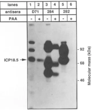
Identification of ICP18.5 in PRV-infected cell extracts.
PK15 cells were infected with PRV, and
[35S]cysteine
wasadded at4hpostinfection. At 10 h postinfection, cells were
harvested andlysed,and labeledproteinswereanalyzed by
immunoprecipitation (Fig. 3). The D71 serum
immunopre-cipitated amajor speciesof 79 kDa(Fig. 3, lane2)aswell as
twominor speciesat92 and120 kDa. Thepreimmuneserum
immunoprecipitated only the minor species at 92 and 120
kDa. Control 284 serum immunoprecipitated the predicted
gIlfamily(110, 100,68,and 55kDa)ofglycoproteins (Fig. 3,
lane 4), and control 282 serum immunoprecipitated the
lanes 1 2
1
3451
7
[image:5.612.156.480.82.311.2] [image:5.612.352.534.456.645.2]antisera pre D71 284 |282
|Cro-ICPI8.5
- +ICP18.5
-q2
d
co 0
-68 as Ca
E
L.)
-46
FIG. 3. Competition of infected-cellextracts with Cro-ICP18.5. PK15 cells were infected with PRV at an MOI of 10 and were continuously labeled from 4 to 10 h postinfection with 100 ,uCiof
[35S]cysteine.
The cells were lysedat 10 hpostinfection in RIPA buffer. Solubilized Cro-ICP18.5 was addedto halfof the samples priorto antisera addition. Polyvalent ICP18.5-specific D71 antise-rum, polyvalent gIl-specific 284 antiserum, or polyvalent gIII-specific 282 antiserum was then added. Immunoprecipitated pro-teinswereresolvedby SDS-PAGE and imaged byfluorography.*-ICP18.5
___
=_
J. VIROL..Ai- ,A&..
.8
on November 10, 2019 by guest
http://jvi.asm.org/
antisera D71
time (hours) O
1
2 3 4 |6 |8112
tn
0
co
E
.3
92
-68
-- ICP18.5
FIG. 4. Timecourse of ICP18.5 accumulation in PRV-infected
cells. PK15 cells were infected with PRV at an MOI of 10 and
harvested atvarious times by lysis in RIPA buffer. Samples were
incubated with D71 antiserum, and immunoprecipitated proteins
wereseparated by SDS-PAGEand transferredtoImmobilon-P. The
filterwasthenreacted with D71 antiserum inaWestern blotformat
and identified by 125I-protein A. The time postinfection is shown
above each lane.
expected precursor(74 kDa) and mature (92 kDa) forms of glll (Fig. 3, lane 6).
To demonstrate that the 79-kDa protein
immunoprecipi-tated by D71 serum corresponded to bona fide ICP18.5, immunoprecipitation of 35S-labeled PRV-infected extracts
was competed with unlabeled E. coli-derived Cro-ICP18.5
fusion protein. The Cro-ICP18.5aggregatewassolubilized in
9.5 Murea with 800 ,M P-mercaptoethanol and heated to
100°C for5min. Denaturantswereremoved by dialysis, and
thedialysatewas used in subsequent experiments.
The extracts in Fig. 3 indicated with a plus (+) were
incubated with denatured Cro-ICP18.5 prior to addition of the antisera. The Cro-ICP18.5 fusion protein did not
com-pete in the control immunoprecipitations with 284 and 282 sera. However, specific competition was observed for the 79-kDa species precipitated by D71 serum. The 92- and 120-kDaspecies presentinboth preimmune and D71 immu-noprecipitations remained unchanged in intensity, confirm-ing that similar quantities were loaded and that these are
unrelatedtoICP18.5. Moreover, ICP18.5 from infected cells
has the same mobility as the in vitro-synthesized ICP18.5,
suggesting that no posttranslational modifications that may affect electrophoretic mobilityhad occurred.
Time course of expression of ICP18.5. PK15 cells were infected with PRV and harvested at various times after
infection. Extracts wereimmunoprecipitated with D71 anti-serum, separated by SDS-PAGE, transferred to
Immo-bilon-P, and reacted with D71 antiserum in a Western blot format(Fig. 4). Noprotein reactingwith D71antiserumwas detected in uninfected cells or at 1 h after infection. A
specific band at 79 kDa predicted to be ICP18.5 that in-creased inintensity as the infectionproceeded was detect-ableby2 hpostinfection.
Whentheexperimentshown inFig.4wasperformedwith totaluninfected- and infected-cell proteinon the membrane ratherthanimmunoprecipitated protein,anadditional cellu-lar protein of approximately 74 kDa reacted with D71 antiserum and was observed at all times. Both uninfected andinfectedcellscontained thisprotein (datanotshown,but see Fig. 8, lane 1). The cross-reactivity ofD71 antiserum withthis 74-kDacellular proteinis discussed later.
Viral DNAreplicationisnotrequiredforICP18.5synthesis.
* -92 X
ICP18.5 -68 E
_ ~~~~~~~~~a)
[image:6.612.360.521.77.272.2]-46 0
FIG. 5. Protein expression in the presenceofa DNAsynthesis
inhibitor.PK15 cellswereinfected with PRVatanMOI of 10 with
either 0or300 pLgof PAA perml present atthestart of infection. [35S]cysteine (100 ,uCi) wasaddedtothemedia between 4 and 10 h postinfection. The cells were lysed in RIPA buffer, and aliquots
were incubated with ICP18.5-specific polyvalent D71 antiserum, gll-specific polyvalent 284 antiserum,orglll-specific polyvalent 282
antiserum. Immunoprecipitated proteins were resolvedon a 10%
SDSgel and visualized by autoradiography.
In experiments not shown, we determined that 300 p.g of
phosphonoacetic acid (PAA; Aldrich) per ml added at the onset ofinfectionwassufficienttoblockviralDNA
replica-tion. To study the effectofviral DNA synthesis on
expres-sion of ICP18.5, cells were infected in the presence or absence ofPAA, incubated with 100 ,uCi of [35S]cysteine
between 4 and 10 h postinfection, and harvested in RIPA buffer. Proteinswereimmunoprecipitated from the extracts and separated by SDS-PAGE (Fig. 5). Expression of the
viralcontrol protein, glycoproteinglll, wasnot detected in the presence of PAA (Fig. 5, lanes 5 and 6), which is
consistentwith thepremisethatglllisalategenesimilarto
HSV-1 gC (32; unpublished data). Accumulation of the second control viral protein, glycoprotein gII, was slightly diminished in the presence of PAA (Fig. 5, lanes 3 and 4),
consistent with its assignment as an early gene similar to HSV-1 gB (31). ICP18.5accumulation wasalso diminished butnotabolished in thepresenceof PAA(Fig. 5,lanes1 and 2). Theseresultsare consistent with ICP18.5beingan early genewhose synthesisdoesnotrequireDNA synthesis. The twobands above ICP18.5 seenpreviously with both
preim-muneand D71serainFig.3appearsensitivetoPAA but not relevantto ICP18.5.
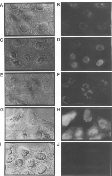
Indirect immunofluorescence. Wedetermined the intracel-lular localization of ICP18.5 in infected cells by indirect immunofluorescence withimmunoaffinity-purifiedantiserum to reduce background. The purified antibodies were
pre-paredbyelutingbound antibodies from Cro-ICP18.5 fusion
protein attached to Immobilon-P (16). PRV-infected PK15 cells were fixed and permeabilized at 1-h intervals after infection and incubated with affinity-purified antisera as indicated inFig. 3.Boundrabbitimmunoglobulin G antibod-ies were identified by incubation with rhodamine
tetrachlo-ride-conjugatedgoatanti-rabbitimmunoglobulinG(Jackson
Immunoresearch Laboratories, West Grove, Pa.).
,*.*'
11_
_
_..
,&
A
* ;4 i:A
% .Son November 10, 2019 by guest
http://jvi.asm.org/
[image:6.612.80.287.78.208.2]A
C
E
G
D
F
H
I v>
-.=,*H
I~~~~~~~~~~~~~I
FIG. 6. Intracellular localizationofICP18.5 in PRV-infected cells.PK15cellswereinfected withPRVat anMOI of10and thenfixed and permeabilizedat1 h(AandB),2 h(CandD),3h(EandF),4h(GandH),or6h(IandJ)postinfection.Theprimaryantiserumusedwas eitherD71 antiserum(1:250)immunopurifiedtoCro-ICP18.5(B, D,F,andH)or
preimmnune
serum(1:250)immunopurified toCro-ICP18.5(J). The secondary antiserumwasrhodaminetetrachloride-conjugatedgoatanti-rabbitimmunoglobulinG(1:50).Eachfieldwasvisualizedby phasecontrast(A, C, E,G,andI)orfluorescence(B, D,F, H, andJ).
3752
on November 10, 2019 by guest
http://jvi.asm.org/
[image:7.612.110.502.64.677.2]Visible immunofluorescence from D71 antiserum could be detected by 1 h postinfection in PRV-infected cells as a punctate pattern of fluorescence in the nucleus of most cells (Fig. 6B). By 2 h postinfection, the nucleus was fairly well delineated by diffuse fluorescence in addition to the punctate pattern(Fig. 6D). At 3 h postinfection the punctate regions had grown to sizeable segments within the nucleus (Fig. 6F). From 4 h onward all portions of the nucleus except the nucleolus contained D71-reactive material. The cytoplasm of infected cells at 1 to 3 h postinfection exhibited slightly increased fluorescence. The cytoplasmic pattern wasdiffuse, and the fluorescence was not as striking as that in the nucleus. Preimmune serum produced background levels of fluorescence when incubated with infected cells (Fig. 6J), as did D71 and preimmune serum on uninfected cells (data not shown).
Kinetic analysis of ICP18.5 synthesis and subcellular frac-tionation. PRV-infected PK15 cells were pulse-labeled with
[35S]cysteine
and chased for various times as indicated in Fig. 7. Cells were lysed in the presence of NP-40, and the cytoplasmic fraction was separated from intact nuclei. Nu-clei were washed and lysed, and each fraction was incubated with antiserum. Immune complexes were analyzed by SDS-PAGE (Fig. 7A). ICP18.5 protein immunoprecipitated by D71 antiserum was made in the 2-min pulse and found only in the cytoplasmic fraction. Moreover, the protein was stable for at least 2.5 h after synthesis. ICP18.5 made in the 2-min pulse was undetectable in the nuclear fraction; how-ever, after 30min of chase, ICP18.5 could easily be detected in the nuclear fractions.As controls for this fractionation, two PRV glycoproteins were examined by using the same infected-cell lysates. The precursor forms of these glycoproteins are synthesized in the rough endoplasmic reticulum which is contiguous with the outer nuclear membrane and should be found in the nuclear fractions. As the glycoproteins are exported from the endo-plasmic reticulum to the Golgiapparatus for final processing, we would expect the precursors to disappear from the nuclear fractions. These experiments with gIl and gIll are shown in Fig. 7B and C, respectively. ThegIl precursor is 100 kDa and thegIll precursor is 74 kDa. Immediately after the 2-min pulse, the gIl and gIII precursor forms and incompletely synthesized forms are found in the nuclear fraction. Full-length precursor products appear in the cyto-plasmic fraction. The precursor forms in the cytoplasm chase to themature forms with kinetics previously described (36, 42). Mature gIl is 110 kDa, and mature gIll is 92kDa. No mature glycoproteins were found in the nuclear frac-tions, and all precursor forms disappear from the nuclear fraction between 30 and 45 min after synthesis. This time coincides with the predicted time of export of the 74-kDa
gIII and 100-kDagIl precursors to the Golgi apparatus (36, 42). These control experiments suggest that ICP18.5 is synthesized in the cytoplasm and then becomes associated with the nucleus.
Subcellular fractionation of unlabeled ICP18.5. Infected cells were harvested and separated into nuclear and cyto-plasmic fractions as described in Materials and Methods. Aliquots of each fraction were either immunoprecipitated with D71antiserum (Fig. 8, right 2 lanes) or precipitated with acetone (Fig. 8, left 2 lanes). Proteins were separated by SDS-PAGE and transferred to Immobilon-P. The filterwas
incubated with D71 antiserum, and reactive proteins were
identified with 125I-protein A. ICP18.5 is found in both the cytoplasm and the nuclear fractions in all four lanes. Recall that a 74-kDa cellular protein is also recognized by D71
antiserum (Fig. 8, lane 1). This
protein
remained in thecytoplasmand was not
immunoprecipitated
by
D71. Theseresults are consistent with the
pulse-chase
experiments
which showed that ICP18.5 issynthesized in the
cytoplasm
andbecomesassociatedwith the nucleus.
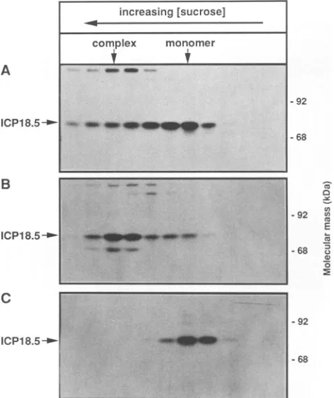
Sedimentation analysis of ICP18.5. PRV-infected PK15
cells were pulse-labeled for 2 minat 6 h
postinfection
with[35S]cysteine. Extractsweremadeaftera2-or90-minchase
period. Cellextractswere solubilizedwith1%Triton X-100
and loadedonto a 5 to 15% sucrose
gradient.
ICP18.5 wasimmunoprecipitated from each fraction and
separated
by
SDS-PAGE. The
primary
ICP18.5translationproduct
made in the 2-minpulse sedimentedinitially
to aposition
consis-tentwitha monomerof79kDa(Fig.
9A).However,
after 90minof chase, ICP18.5 sedimented more
rapidly,
suggesting
thatICP18.5formeda
complex
aftersynthesis
(Fig.
9B).
Anadditionalprotein of
approximately
70kDawasalso presentincomplex fractionsand may be
responsible
for this shift insedimentation. When the 90-min chase
sample
was treatedwith SDS prior to
sedimenting
the extract on the sucrosegradient, the ICP18.5
complex
wasdisrupted
(Fig.
9C).ICP18.5 was detected
exclusively
in fractions consistentwith ICP18.5 monomers. The smaller protein was not de-tected after SDS treatment,
indicating
that it does not reactdirectly with D71 antiserum. We suggest that the 70-kDa
protein complexes with ICP18.5 in a noncovalent fashion
soonafter
synthesis.
Steady-state analysis of monomer and
complex
forms ofICP18.5.The steady-state distribution ofICP18.5 in
mono-merandcomplexformswasexaminedasfollows. Unlabeled
PRV-infectedextracts(5h
postinfection)
wereprepared
andsedimented
through
sucrosegradients.
Totalprotein
fromeachfraction was
separated
by
SDS-PAGE,
transferred toImmobilon-P, and
probed
with D71 antiserum(Fig. 10).
The70-kDa
protein
in thecomplex
fraction was notrecognized
by the D71 antiserum. Moreover, ICP18.5 was detected
exclusively in the
complex
form;
no monomericprotein
could be detected. This is consistent with the idea that
virtually all ICP18.5 formsa
complex.
As notedabove,
D71antiserum reacted with a 74-kDa cellular
protein
thatsedi-mentedas
predicted
for its molecular mass andwas auseful internal standard.DISCUSSION
The HSV-1 ICP18.5 gene is
highly
conserved amongherpesviruses forwhich DNA sequencesare
currently
avail-able. Comparison of the PRV ICP18.5
predicted
protein
sequence withother
sequenced
herpesviruses
revealssignif-icant conservation in
regions
throughout
theprotein
(Fig.
11). Complete and
partial
sequence informationwasaligned
with the assistanceofthe
University
ofWisconsin GeneticsComputer
Group
programs(10).
BESTFIT and LINEUPwere used
reiteratively
toproduce
theprotein
alignments
shown in
Fig.
11.Every
aminoacid identical among all thealpha-herpesvirus
sequences was marked. Those residueswhichwerealsoidentical in the beta-or
gamma-herpesvirus
sequences were markedas well. Themost
highly
conservedregionsareshown withthis
stringent
scheme.Comparison
ofthese sequences indicates that the ICP18.5
proteins
fromneurotropic
herpesviruses
(bovine
herpesvirus, equine
her-pesvirus,
herpesvirus
saimiri,
infectiouslaryngotracheitis
virus, PRV, and varicella-zoster
virus)
are moreclosely
related to each other than any of them are to the ICP18.5
proteins
oflymphotropic
herpesviruses
(Epstein-Barr
virus,
human
cytomegalovirus,
andherpesvirus
saimiri)
with theon November 10, 2019 by guest
http://jvi.asm.org/
3754 PEDERSON AND ENQUIST
fraction cytoplasm nucleus
chasetime 15 30 45 60 90 150 0 15 30 45 60 90 150
A
ICP18.5 -'
B
gil
_--C
gill _-0
...a,...
'
*6
at..
aot
-92
68
0 *92 wc6 E 68
c2
0
0
92
68
FIG. 7. Kineticsof ICP18.5synthesisandsubcellular fractionation. PK15 cellswere infectedwithPRVatanMOIof10. Infected cells werepulse-labeledat5 hpostinfection for2min with[35S]cysteineandchased for thetimesindicated.Cellswere
processed
into nuclearand cytoplasmic fractions. Each fractionwasimmunoprecipitatedwith D71, 282,or284 antiserum.Immunoprecipitated
proteins
wereresolved onanSDS-7%polyacrylamide geland visualizedbyautoradiography.sample total P.
fraction
I
NI
CI
NNICP18.5
-_-Ca
0
-92o() Cm E
68 .s a)
CD
[image:9.612.121.481.70.437.2].5
FIG. 8. Subcellular fractionation of ICP18.5. PK15 cells were
infected with PRV at an MOI of10 and subjected to subcellular fractionationasdescribedaboveat6 hpostinfection. Fractionswere
precipitated with acetone or immunoprecipitated with D71
antise-rum, separated by SDS-7% PAGE, transferred to Immobilon-P,
reactedwithD71 antiserum, and identified with 125I-protein A.
exception of Marek's disease virus. The clusters of amino
acids which are conserved in all
herpesviruses
may markdomains that provide clues to the function of ICP18.5. It seemslikelythatmostherpesvirusICP18.5
proteins
functionin much the same way,
considering
theirhigh degree
ofsimilarity. A consideration of ICP18.5 homology concerns
the upstream major DNA binding protein, ICP8
(HSV-1
UL29), and downstream gB (HSV-1 UL27) genes. Both of
these genesareessential. While there isnoevidence that the upstream HSV-1ICP8 gene affects ICP18.5, transcriptional
mapping placesthe promoter and mRNAstartsite of HSV-1
gB within the domain ofthe ICP18.5
coding
region (5, 18;
unpublished data). Insome
herpesviruses
(bovine
herpesvi-rus type 1, Epstein-Barr virus, equine herpesvirustypes 1
and 4, infectious laryngotracheitis
virus,
and PRV) thecoding regions ofgB and ICP18.5 overlap,
suggesting
thatthese regions of conservation among ICP18.5 genes are likelytobe the resultof featuresrequiredforexpressionof
gB. However, it is clear from Fig. 11 that ICP18.5 is
conserved in many differentregionsnotbelievedtoinfluence
gBexpression. Comparisonsof the DNA sequences suggest
that only one region of protein homology is due to
gB
.|. S.
*
ai
J. VIROL.
on November 10, 2019 by guest
http://jvi.asm.org/
[image:9.612.99.264.552.665.2]PRV ICP18.5 PROTEIN IDENTIFICATION 3755
-92
-68
-92
-68 0
Ca
E
.3
0 FIG.
10. Sedimentation analysis of ICP18.5. PRV-infected cells
werelysedat6hpostinfection in1%TritonX-100and layeredon5to 15%sucrosegradients.Fractionswerecollected,acetoneprecipitated, resuspendedinsamplebuffer, and heatedtoboiling. Treated samples
were separated by SDS-7% PAGE, transferred to Immobilon-P, reacted withD71antiserum, and identified with "25I-proteinA.
-92
FIG. 9. Kineticsof ICP18.5 synthesis and sedimentation analy-sis. PK15 cells were infected with PRV at an MOI of 10 and pulse-labeled for 2 min at 5 h postinfection. (A) The cells were
chased for 2 min, solubilized in 1% Triton X-100, sedimented througha5 to15%sucrosegradient, fractionated,
immunoprecipi-tated with D71, and separated by SDS-7% PAGE. (B) The cells
werechased for 90minand treated exactlyasdescribedabove. (C)
Thecellswerechased for90minand harvestedasdescribed above.
Theextractwasbroughtto1% SDSandincubatedat4°Cfor 30 min priortosucrosegradient sedimentation. Fractionswerethen treated exactlyasdescribed above.
promoter elements. This is the sequence of PRV
(641-YIREL-645) immediately tothe right of the bovine herpes-virus type2 carboxy terminus.
Despite the level of conservation, the product of this
conserved gene has not been studied in any detail in any
herpesvirus. HSV-1 ICP18.5 is essential, as demonstrated with ts mutants (24), and HSV-2 ts mutants have been assignedto twocomplementationgroupswithin theICP18.5 locus (12). ICP18.5 thus appears to be essential in at least two human herpesviruses, although the nature of these mutations is unknown.Pellettetal.(27)usedananti-peptide serumto identifythe 95-kDa ICP18.5 protein in HSV-1(F), whichwas10 kDa larger than predicted fromDNAsequence
analysis. In thisreportweexpressed the PRVICP18.5open
reading frame as a fusion protein in E. coli and made a
polyclonal serumspecificfor ICP18.5.
Expressionofthe fusionproteininbacteriawas an impor-tant part ofour analysis. However, our initial efforts
sug-gestedthateven smallquantities of the Cro-ICP18.5fusion protein were lethal to E. coli. We subsequently isolated a spontaneous E. coli mutant that enabled expression of significantamountsof Cro-ICP18.5. We have taken care to demonstrate that the fusionproteinproduced bythis mutant strain is thatpredictedfrom the DNAsequence. The nature
of thespontaneousmutation(s) which enabled expression of
Cro-ICP18.5 is intriguing but beyond the scope of this
report. We do know that the strain may be specific for
alleviating the lethal phenotype of PRV ICP18.5, since it does not appear tobe of general use in the expression of
foreign proteins inE. coli(unpublished data). Wenote that
H.-J. Rziha(Federal Research Center for Virus Diseases of Animals, Tubingen, Germany) (36a) has expressed portions
of PRV ICP18.5 inadifferent E.coliexpressionsystemwith
no problems of lethality. Further work is necessary to characterize ourstrain and the lethality phenotype of Cro-ICP18.5.
We have alsotakencaretodemonstrate that the reactivity
of the antiserum directed against the Cro-ICP18.5 fusion
protein is specific for PRV ICP18.5. We transcribed ICP18.5
RNA invitro andtranslatedthe RNA invitrotodemonstrate that theprimary product of the PRV ICP18.5 open reading
frame migrated in SDS-PAGE as a 79-kDa protein, as
expectedfrom the DNA sequence. Weused the polyclonal
serum D71 and the E. coli-produced Cro-ICP18.5 fusion
proteintodemonstrate that themajor productof the ICP18.5 gene in PRV-infected cells is also a79-kDa protein. Thus, PRV ICP18.5 does not appear to be posttranslationally
modified inanywaythat affects itselectrophoretic mobility.
We detected PRVICP18.5proteinininfected cells within 2 h after infection even in the presence of the viral DNA synthesis inhibitor PAA. This is consistent withthe finding that HSV-1 ICP18.5mRNAwasdetectedatearlytimes(17).
ICP18.5 accumulated throughout infection and was stable foratleast2.5 haftersynthesis. Thekinetics ofappearance of ICP18.5wereconfirmedbyindirect immunofluorescence. Some ICP18.5wasdetectedinmost cells at 1 hpostinfection
andingreateramountsinvirtually allcellsby2 h
postinfec-tion. ICP18.5 waslocalized to the nucleus, as measuredby
indirect immunofluorescence and by subcellular fractiona-tion. TheappearanceofICP18.5 at discrete locationswithin the nucleuswas striking. As the infectionprogressed,these localized regions diffusedfrom the initial sites ofreactivity until the entire nucleus contained ICP18.5. It isnoteworthy
that the nucleolus was devoid of ICP18.5
immunofluores-cenceduringthe entire infection. increasing [sucrose]
complex monomer
- _ a ...
_ __ w
A
ICP18.5-1
B
ICP18.5-I-C
ICP18.5
--increasing [sucrose]
complex monomer
to*q
-m
- m+
+~~~~~~~~~~~~~~~~~~
I~~~~~~~~~~~~~~~~~~~~~~~~~~.._.._ .. ...
..4
....
~ ~ ....- J .. ..=_6 -_
I
92
68
CI
0
U,cn
E
Cu
n
0
0
VOL.65, 1991
ICP'8.5 _*,
on November 10, 2019 by guest
http://jvi.asm.org/
[image:10.612.326.564.72.239.2] [image:10.612.66.305.73.358.2]3756 PEDERSON AND ENQUIST
.U
3iEi
*:
0 0;>
*;
*;
(a H@
R1>0><t>.
>9
> >-- > >9 CG:>~- > >g>4
-- rq>-- > >Q>
|
a:.
>x
> >12.1 rs:m
v g.Hs
n-
K5
Es||sms
m ::m 3: m m
i s. Ha
:
i
in
ttE
H n
iii3
E
R
4
I
J. VIROL.
.
1-. -. C.0>.
-. C.D
.6
H
H01H
40v -Ip---4m>--4N> >
. ..l.-.- K .-.v m9
CQ,4m MT
on November 10, 2019 by guest
http://jvi.asm.org/
The subcellularfractionationprocedure demonstrated that
ICP18.5 was synthesizedin the cytoplasm before it
associ-ated with the nucleus. While ICP18.5 was detected immedi-ately in cytoplasmic fractions, it was not found in nuclear
fractions until at least 30min later. The ICP18.5
fractiona-tion results were distinctly different from those of the glycoprotein fractionation. Precursor glycoproteins gll and glll were detected in nuclear fractions immediately after
synthesis. Precursor forms were then detected in the
cyto-plasmic fractions as they left the nuclear fractions. The
cytoplasmic precursor forms chased into mature
glycopro-teinswithpreviously observed kinetics (36, 42). The results
obtained by using the subcellular fractionation procedure
describedabovecomparedwell to those observed by others.
Analysis of HSV-1 glycoprotein synthesis by electron mi-croscopy showed that precursor glycoproteins appeared
rapidlyin the nuclearenvelope before they were transported
to theGolgi complex (28). Transiently expressed HSV-1 gB has also been identified in the nuclear envelope by indirect
immunofluorescence (2).
Although we have demonstrated the association of
ICP18.5 with the nucleus, the amino acid sequence of
ICP18.5 does not contain a consensus nuclear localization
signal (8, 11, 19, 22, 29). The most promising sequence of
PRVICP18.5(461-RRRR-464)is not absolutelyconserved in
the ICP18.5 homologs among herpesviruses (Fig. 11). One
possibility is thatICP18.5 is-directed to the nucleus by the
70-kDaprotein observed ina complex with ICP18.5 that is
not antigenically related to ICP18.5 by Western blot
analy-sis. Most ICP18.5 associates with this 70-kDa protein in
infected cells, yetthis interactionisnoncovalent, as
demon-strated by the sensitivity of the complex to SDS. The
identity of this protein as a virally or cellularly encoded
proteinis not known. Sincethe70-kDaproteinwasdetected
by pulse-labeling 5 hpostinfection, it issynthesized at times when the
majority
of host protein synthesis is reduced, which suggests that it may be virally encoded. Thesedimen-tationrateofthecomplex form is consistentwith one70-kDa
monomerinteractingwith one ICP18.5 monomer.
Ourfindingsdo notreadily explainhow ICP18.5 functions
in the model proposed by Pancake et al. (24). That model
suggests that ICP18.5 may bind viral
glycoproteins
andeither assistin theirtransport or mark them fortransportto
the cell surface and virions. We did notfind ICP18.5
com-plexed to glycoproteins, but we saw it complexed to a
70-kDa protein. Another
possibility
might be that ICP18.5would function in virusparticles to coordinateglycoprotein
distribution. We did not see ICP18.5 in virus particles
(unpublished data); insteadwefound ICP18.5 in the nucleus
indiscrete,punctateregions.Significantprogresstowardour
understanding can be expected once viruses deleted for
ICP18.5are established.
ACKNOWLEDGMENTS
AlanRobbins andMary Whealyprovidedreagents, advice, and encouragement duringthe courseof these studies. We thank Kim Solomon and DanTenneyfor critical commentsand support. We thankBarbaraKriegerandJimMarshallfortheproteinsequencing work. We thank Hanns-Joachim Rziha for sharing unpublished
preliminary information onPRVICP18.5. WeareindebtedtoDan Chelsky fortheuseof his microscope.
ADDENDUMINPROOF
Addison et al. (C.
Addison,
F. J.Rixon,
and V. G. Preston, J. Gen. Virol. 71:2377-2384,1990)
havesuggested
thatICP18.5wasinvolvedin
capsid
maturation. Our resultsareconsistent withthis idea.
REFERENCES
1. Albrecht,J.-C., and B.Fleckenstein. 1990. Structural organiza-tion of the conserved geneblock ofherpesvirussaimiricoding forDNApolymerase, glycoproteinB, andmajorDNAbinding
protein. Virology 174:533-542.
2. Ali, M., M. Butcher, and H. P. Ghosh. 1987. Expression and nuclearenvelope localization ofbiologically active fusion
gly-coprotein gB ofherpessimplexvirus inmammalian cellsusing clonedDNA. Proc. Natl. Acad.Sci. USA 84:5675-5679. 3. Baer, R., A. T. Bankier, M. D.Biggin, P. L. Deininger, P. J.
Farrell,T.J.Gibson,G.Hatfull,G. S.Hudson,S. C.Satchwell, C. Seguin, P. S. Tuffnell, and B. G. Barrell. 1984. DNA sequence and expression of the B95-8 Epstein-Barr virus
ge-nome.Nature(London)310:207-211.
4. Bzik, D. J., C. DebRoy, B. A. Fox, N. E. Pederson, and S. Person. 1986. The nucleotidesequence of thegB glycoprotein
geneof HSV-2 andcomparisonwiththecorrespondinggeneof HSV-1. Virology155:322-333.
5. Bzik, D. J., B. A. Fox, N. A. DeLuca, and S. Person. 1984. Nucleotide sequence specifyingthe glycoprotein gene,gB, of
herpes simplexvirustype 1. Virology 133:301-314.
6. Chamberlain,J.P. 1979.Fluorographicdetection of
radioactiv-ity inpolyacrylamide gelswiththe watersoluble fluor sodium
salicylate.Anal. Biochem.98:132-135.
7. Chee,M.S.,A.T.Bankier,S.Beck,R.Bohni,C. M.Brown,R. Cerny, T. Horsnell, C. A. Hutchison III, T. Kouzarides, J. A. Martignetti, E.Preddie, S. C. Satchwell, P. Tomlinson, K. M. Weston,and B.G. Barrell. 1990.Analysisof the
protein-coding
content ofthe sequence ofthehuman cytomegalovirus strain
AD169, p. 125-169. In J. K. McDougal (ed.),Currenttopicsin microbiologyandimmunology. Springer-Verlag KG, Berlin.
8. Chelsky,D., R.Ralph, andG.Jonak. 1989. Sequence
require-ments for syntheticpeptide-mediated translocation to the nu-cleus. Mol.Cell. Biol. 9:2487-2492.
9. Davison, A. J., and J. E. Scott. 1986. The complete DNA sequenceof Varicella-Zoster virus.J.Gen.Virol.67:1759-1816. 10. Devereux,J.,P.Haeberli,and0.Smithies.1984.A comprehen-sive set ofsequence analysis programsfor the VAX. Nucleic Acids Res. 12:387-395.
11. Dingwall,C.,J.Robbins,S. M.Dilworth,B.Roberts,and W. D. Richardson.1988. Thenucleoplasminnuclearlocationsequence islargerandmorecomplexthan thatofSV40largeT
antigen.
J. Cell. Biol. 107:841-849.12. Dixon, R. A. F., D. J. Sabourin, and P. A. Schaffer. 1983. Geneticanalysisof
temperature-sensitive
mutantswhich define the genes for the major herpes simplex virus type 2DNA-bindingproteinanda newlate function. J. Virol. 45:343-353.
13. Gompels, U. A., M. A. Craxton, and R. W. Honess. 1988. Conservation ofgene
organization
in thelymphotropic
herpes-viruses herpesvirus saimiri and Epstein-Barr virus. J. Virol. 62:757-767.
14. Griffin,A. M.1991. Thenucleotidesequenceof the
glycoprotein
gB gene of infectious
laryngotracheitis
virus:analysis
andevolutionary relationship to the
homologous
gene from otherherpesviruses.J.Gen. Virol. 72:393-398.
15. Hammerschmidt, W., F. Conraths, J. Mankertz, G. Pauli, H. Ludwig, and H.-J.Buhk. 1988. Conservation ofagenecluster
includingglycoproteinBinbovine
herpesvirus
type2(BHV-2)andherpessimplexvirustype1(HSV-1).
Virology
165:388-405.16. Harlow, E.,and D. Lane. 1988. Antibodies: alaboratory
man-ual.ColdSpringHarborLaboratory,Cold
Spring
Harbor,N.Y.17. Holland, L. E., R. E. Sandri-Goldin, A. L. Goldin, J. C. Glorioso, and M. Levine. 1984.
Transcriptional
andgenetic
analysesof theherpes
simplex
virustype1 genome:coordinates0.29to0.45. J. Virol. 49:947-959.
18. Homa,F. L.,T. M.Otal,J.C.Glorioso,andM.Levine. 1986.
Transcriptionalcontrol
signals
ofaherpes
simplex
virustype1late (Y2) gene lie within bases -34 to +124relative to the 5' terminus ofthemRNA. Mol. Cell. Biol.6:3652-3666.
19. Kalderon,D.,B.L.Roberts,W.D.Richardson,andA. E.Smith.
on November 10, 2019 by guest
http://jvi.asm.org/
3758 PEDERSON AND ENQUIST
1984. A short amino acid sequence able to specify nuclear location. Cell 39:499-509.
20. Knopf, C. W. 1987. The nucleotide sequence of the herpes simplex virus type 1 late gene ICP18.5 of strain Angelotti. Nucleic AcidsRes. 15:8109-8110.
21. Larjava, H., J. Peltonen, S. K. Akiyama, S. S. Yamada, H. R. Gralnick, J. Uitto, and K. M. Yamada. 1990.Novelfunctionfor P1 integrinsin keratinocyte cell-cell interactions. J. Cell Biol. 110:803-815.
22. Lyons, R. H., B. Q. Ferguson, and M. Rosenberg. 1987. Pen-tapeptide nuclear localization signal in adenovirus Ela. Mol. Cell. Biol. 7:2451-2456.
23. McGeoch, D. J., M. A. Dalrymple, A. J. Davison, A. Dolan, M. C. Frame, D. McNab, L. J. Perry, J. E. Scott, and P. Taylor. 1988. The completeDNA sequenceofthelong unique region in the genome of herpes simplex virus type 1. J. Gen. Virol. 69:1531-1574.
24. Pancake, B. A., D. P. Aschman, and P. A. Schaffer. 1983. Geneticandphenotypic analysis of herpes simplex virustype 1 mutants conditionally resistant toimmune cytolysis. J. Virol. 47:568-585.
25. Pederson, N. E., and L. W. Enquist. 1989. The nucleotide sequence of apseudorabies virus gene similar to ICP18.5 of herpes simplexvirus type 1. Nucleic Acids Res. 17:3597. 26. Pellett, P. E., M. D. Biggin, B. Barrell, and B. Roizman. 1985.
Epstein-Barr virus genome may encode a protein showing significant amino acid and predicted secondarystructure homol-ogy with glycoprotein B ofherpes simplex virus 1. J. Virol. 56:807-813.
27. Pellett, P. E., F. J. Jenkins, M. Ackermann,M.Sarmiento, and B.Roizman. 1986.Transcription initiation sites andnucleotide sequence of a herpes simplex virus 1 gene conserved in the Epstein-Barrvirus genomeandreportedtoaffectthe transport of viralglycoproteins. J. Virol.60:1134-1140.
28. Poliquin, L., G. Levine, and G. C. Shore. 1985. Involvementof theGolgiapparatusandarestructurednuclear envelopeduring biogenesis andtransportof herpes simplex virus glycoproteins. J.Histochem. Cytochem. 33:875-883.
29. Richardson, W. D., B. L. Roberts, and A. E. Smith. 1986. Nuclearlocationsignals in polyoma virus large-T. Cell 44:77-85.
30. Riggio, M. P., A. A. Cullinane, and D. E. Onions. 1989. Identification and nucleotide sequenceofthe glycoprotein gB geneofequine herpesvirus4.J. Virol.63:1123-1133.
31. Robbins, A. K., D. J. Dorney, M. W. Wathen, M. E. Whealy, C. Gold,R.J. Watson, L. E. Holland, S. D. Weed, M. Levine, J. C. Glorioso,and L. W.Enquist. 1987. Thepseudorabies virus gll gene isclosely related to the gB glycoprotein gene ofherpes simplex virus.J. Virol. 61:2691-2701.
32. Robbins, A. K., R. J. Watson, M. E.Whealy, W. W. Hays, and L. W. Enquist. 1986. Characterization of apseudorabies virus glycoprotein gene with homology toherpes simplex virus type 1 and type 2glycoprotein C. J. Virol. 58:339-347.
33. Robbins, A. K., J. H. Weis, L. W.Enquist, and R. J. Watson. 1984. Construction of E. coli expression plasmid libraries: localizationof a pseudorabies virusglycoproteingene.J. Mol. Appl. Genet. 2:485-496.
34. Robbins, A. K., M. E. Whealy, R. J.Watson, and L. W. Enquist. 1986. Pseudorabies virus geneencoding glycoproteinglllis not essential for growth in tissue culture. J. Virol. 59:635-645. 35. Ross, L. J. N., M. Sanderson, S. D.Scott, M. M. Binns, T. Doel,
and B. Milne. 1989. Nucleotide sequence and characterization ofthe Marek's disease virus homologue of glycoprotein B of herpes simplex virus. J. Gen. Virol. 70:1789-1804.
36. Ryan, J. P., M. E. Whealy, A. K.Robbins, and L. W. Enquist. 1987.Analysis of pseudorabies virusglycoprotein glIl localiza-tion and modificalocaliza-tion by using novel infectious viral mutants carrying unique EcoRI sites. J. Virol.61:2962-2972.
36a.Rziha, H.-J.Personalcommunication.
37. Silhavy, T. J., M. J. Berman, and L. W.Enquist. 1984. Exper-imentswith genefusions. Cold SpringHarborLaboratory, Cold Spring Harbor, N.Y.
38. Simpson, R. J., and E. C. Nice. 1984. Insitucyanogenbromide cleavage of N-terminally blocked proteins in agas-phase
se-quencer.Biochem. Int. 8:787-791.
39. Watson, R. J., J. H.Weis, J. S. Salstrom,and L. W.Enquist. 1982. Herpes simplexvirus type-1glycoproteinDgene: nucle-otide sequence and expression in Escherichia coli. Science 218:381-384.
40. Watson, R. J., J. H.Weis, J. S. Salstrom,andL. W.Enquist. 1984. Bacterialsynthesisofherpes simplexvirus types 1 and 2 glycoproteinDantigens. J. Invest. Dermatol. 83:102s-111s. 41. Whalley, J. M., G. R. Robertson, N. A.Scott, G. C. Hudson,
C. W. Bell, and L. M. Woodworth. 1989. Identification and nucleotidesequenceofa genein equineherpesvirus1analogous totheherpessimplex virus geneencoding the major envelope glycoproteingB. J.Gen.Virol.70:383-394.
42. Whealy, M. E., A. K. Robbins, and L. W. Enquist. 1990. The export pathway of the pseudorabies virus gB homolog gll involvesoligomer formationin the endoplasmic reticulum and protease processingin theGolgi apparatus. J. Virol. 64:1946-1955.
43. Whitbeck, J. C., L. J. Bello, and W. C. Lawrence. 1988. Comparisonof the bovineherpesvirus1gI geneandtheherpes simplexvirus type 1gB gene. J. Virol.62:3319-3327.
44. Yanisch-Perron, C., J. Vieira, and J. Messing. 1985. Improved M13 phage cloning vectors and host strains: nucleotide se-quencesoftheM13mpl8andpUC19 vectors.Gene33:103-119. J. VIROL.

![FIG. 2.presencevolumes In vitro protein synthesis. In vitro-transcribed RNA from pN59 and pN61 was translated in vitro with rabbit reticulocytes in the of [35S]cysteine](https://thumb-us.123doks.com/thumbv2/123dok_us/1314613.84892/5.612.352.534.456.645/presencevolumes-protein-synthesis-transcribed-translated-rabbit-reticulocytes-cysteine.webp)