0022-538X/84/020621-05$02.00/0
Copyright © 1984,AmericanSociety forMicrobiology
Characterization
of
a
Metastable, Partially Replicated Dimeric
Intermediate of Minute
Virus of
Mice
EMANUEL A. FAUST* AND GREGGLOOR
Cancer Research Laboratory, University of Western Onittairio, Lonidoni, Ontar-io, Cantiada N6A5B7 Received 5 July 1983/Accepted 10October1983
Intracellular, replicative-form DNAofminutevirusof micewascharacterizedbyagarosegel electropho-resis, velocity sedimentation, electron microscopy, restriction endonuclease digestion, and sensitivity to the single-stranded nuclease S1. This analysis demonstrated the presence in murine cells infected with minute virus ofmiceofa 10.0-kilobase pairdimer replicativeform, a5-kilobase pair monomerreplicative form, as well as a 5-kilobase viral single-stranded DNA species. Two additional viral DNA species that migratedin0.5%agarosegelswithapparent sizes of 8.0 and 5.5 kilobasepairswerealso observed. Further investigation indicated that the 8.0-kilobase pair DNA represents a novel class of metastable, partially replicated, dimeric intermediates. This findinghasimportant implications for the mechanismofparvovirus
DNA replication.
Minute virus of mice (MVM), a member of the helper-independent parvovirus group (12),contains a linear single-stranded DNA molecule of 5,081 nucleotides (3) that
repli-cates in the S-phase nucleus of susceptible rodent cells grownintissue culture (10, 12, 15, 17). TheterminiofMVM
single-stranded(SS) DNAarefoldedin abase-paired hairpin configuration (1, 2, 4, 5) and play an essential role in viral DNAreplication. Deletionmutantsthat retainboth terminal self-complementary sequences can be propagated when complementedintransbywild-type virus, despitetheloss in these mutants ofup to 90% of the internal viral sequences (7).
Infection of rodent cells intissueculture withMVMleads
to the accumulation of linear duplex replicative
intermedi-atesthat have been characterized as monomeric (5 kilobase pairs [kbp])ordimeric (10kbp) replicative forms (RFs) (16). Both monomeric and dimeric RF DNA molecules are capa-ble of instantaneous reannealing. This indicates that viral andcomplementary strandsare covalently linked, an obser-vation which suggests that terminal palindromic sequences
serve as primers during the initiation of viral DNA replica-tion.
Dimersconsistingof monomericequivalents orientedin a
head-to-head fashion have been observed such that the 3' and 5' ends of viral and complementary strands, respective-ly, are juxtaposed at the junction of the two monomeric
units.Discontinuities, locatedat or nearthejoint region,are
present in dimer RF DNA extracted from infected cells by the method of Hirt (8) and are thought to reflect the in vivo action of an as-yet-unidentified site-specific nuclease in-volved in processing replicative intermediates during viral DNAreplication (14, 16). Such aprotein may remain bound
to the DNA afterendonucleolytic scission since the mono-merRFDNAofH-1,aparvovirus relatedtoMVM,existsin the cellinacomplexwithaproteinof unknownoriginwhich
is covalently bound to the 5' terminus ofthe DNA(9).
We undertook adetailed investigation ofthe intracellular forms ofMVM DNAinanefforttoclarify furthertheroleof these DNA molecules in viral DNA replication. In the course of these experiments, we discovered a novel
"8.0-*Correspondingauthor.
kbp" intracellular viral DNA species. The results of a
structural analysis of this DNAare presented in this report. Asshown in Fig. 1, when murine cells infected with MVM were extracted by the Hirt procedure (8) to selectively
recoverlow-molecular-weight viral DNA molecules, a
vari-I ety ofintracellular forms of viral DNA were released into the
I Hirt supernatant. Five discrete DNA species with apparent
I molecular sizes of 10.0, 8.0, 5.5, 5.0, and 3.5 kbp were
I detected upon electrophoretic analysis of Hirt supernatant I fractions resulting from infections in various host-virus
combinations. The 10.0-, 5.0-, and 3.5-kbp viral DNA spe-cies were invariably found to be present upon infection of fourdifferent susceptible mouse cell lines including Ehrlich ascites (EA) cells, Ltk aprt cells, As cells, and 3T6 cells. Based ontheirsize, as determined both bysedimentation in neutral sucrose gradients and by their mobility during ag-arose gel electrophoresis, as well as their sensitivity to S1 nuclease and restriction enzyme digestion pattern (see be-low), it is clear that the 10.0-, 5.0-, and 3.5-kbp viral DNA species represent dimer RF, monomer RF, and viral SS DNA, respectively.
These findings confirm the interpretation of a previous characterization of intracellular forms of MVM DNA by Ward and Dadachanji (16). In addition to these viral DNA species, infections of EA and 3T6 cells yielded a novel 8.0-kbp viral DNA species which had not been characterized previously. An Ltk- aprt- cell line yielded a 5.5-kbp viral DNAspecies,which has also been observed on a number of separate occasions upon MVM infection ofEA cells; in such DNA preparations the 8.0-kbp species was invariably
ab-sent.Thus, the 5.5- and8.0-kbpviral DNAspecies appear to be mutually exclusive.
To investigate the structure of the 8.0-kbp DNA species further, total intracellular viral DNA labeled in vivo with [3H]thymidine was extracted from infected EA cells and sedimented in a neutral sucrose gradient (Fig. 2). The location of each MVM DNA species in the gradient was then determined by agarose gel electrophoresis of portions taken from each gradient fraction. As expected, viral SS DNA sedimented relatively rapidly under neutral, high-ionic-strength conditions withapeakat20S(4). ThemonomerRF (1SS) and the dimer RF DNA species also sedimented as
expected according to their relative sizes. However, the apparent 8.0-kbp DNA sedimented at approximately 20S,
621
on November 10, 2019 by guest
http://jvi.asm.org/
622 NOTES
<1-i. '.<
Kbp
FIG. 1. Analysis of intracellular forms of MVM DNAbyagarose gel electrophoresis. Murine cell lines were infected with wild-type MVMvirionswhich had been purified byCsCIequilibrium density gradient centrifugation as described previously (7). Cells were
grown in monolayer in 125-cm2 plastic tissue culture flasks (Cor-ning)in Dulbecco minimal essential mediumsupplementedwith 10% fetal bovine serum. Whencells reached 50% confluencetheywere
infectedwith 100hemagglutination units of virus per125-cm2flask. One hemagglutination unit was taken to be equivalent to 5 x 10⁵ PFU,assumingaparticle-to-PFUratioof 200. To prepare intracellu-lar viral DNA, infected cellsweretrypsinizedat21 h postinfection and lysed in 50 mMTris-hydrochloride buffer. pH 8.0. containing 0.15 M NaCl, 1 mM EDTA, and 0.6% sodium dodecyl sulfate.
more rapidly than its electrophoretic mobility alone would predict if this DNA species were simply a linear 8.0-kbp
double-stranded DNA molecule. Sinceelectron microscopic
analyses ofelectrophoretically purified 8.0-kbp DNA didnot
revealanycircular molecules (unpublished data), this
anom-alous sedimentation behavior couldmostlikely beexplained if the8.0-kbp DNA possessedextensive regionsof SS DNA as part of a partially replicated dimeric intermediate. This interpretation is supported by the results of the restriction endonuclease and S1 nuclease digestionpatterns presented below.
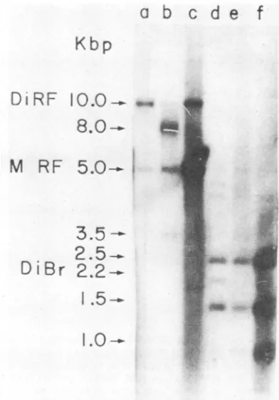
The restriction endonuclease EcoRI cleaves 5.0-kbp MVM monomer RF DNAtwice to yield three unique DNA fragments of2.4, 1.5, and 1.0kbp; the lattertwofragments
map tothe 5' and3'termini oftheviralgenome,respectively (3, 5, 16). TheEcoRIdigestionpatternsof electrophoretical-ly purified 10.0-kbp dimer RF DNA and 8.0-kbp DNA, as
well as sucrose gradient-purified 5.0-kbp monomer RF DNA, werecompared inthe Southern blotanalysisdepicted in Fig. 3.
Upon EcoRI digestion, the 10.0-kbp dimer RF DNA species yielded fragments of 2.5, 2.2, 1.5, and 1.0 kbp(Fig. 3, lane d), whereas 5.0-kbp monomer RF DNA yielded fragments of 2.5, 1.5, and 1.0kbp, asexpected (Fig. 3, lane f). The extra 2.2-kbp fragment derived from the 10.0 kbp dimer RF DNA species is believed to represent a dimer bridge fragment (16) and is consistent with the arrangement
oftwomonomericunitsin ahead-to-head configuration(i.e., with juxtaposed 3' termini). Cleavage of the 8.0-kbp DNA
species yielded fragments of 2.5, 1.5, and 1.0 kbp, which
were indistinguishable in their mobility from the EcoRI
fragments derived from monomer RF DNA. In addition,
Subsequentsteps werecarried out as described by Hirt(8). Approx-imately5% ofthe total DNApreparation wasanalyzedby electro-phoresis in a 0.5% horizontal agarose slab gel asdescribed previous-ly(6). The gel was stained in a 1 ,ug/ml solution of ethidium bromide, and the DNA was visualized by illuminating the gel with short-wavelength UV light.
,. ).
FIG. 2. Velocity sedimentation analysis of intracellular viral DNA. Virus wasgrown in EA mouse cells in the presence of 20 ,uCiof
[3H]thymidine
per ml(30Ci/mmol; New EnglandNuclear). Radiolabeled intracellular viral DNAwasappliedtoa5to20% neutralsucrose gradient madeup in50 mMTris-hydrochloridebuffer, pH 8.0,containing 1.0MNaCI and1mM EDTA.Thegradientwascentrifugedat4°C for20 hat28,000rpm inaBeckmanSW40rotor. Portions(50,ul) of each250-,Jl gradientfractionwereanalyzedbyelectrophoresisina0.5% horizontal agarose slabgel. To visualize the radiolabeled viralDNA. thegelwassoakedfor1h inagarosegelrunningbuffercontaining1.0 M sodium salicylate. Aftera second1.0 M sodiumsalicylatesoak in freshbuffer, thegelwasdried andexposedto X-rayfilmat -70°C inthe presenceof a Dupont LightningPlus intensifyingscreen.J.VIROL.
A&I Awl
0.,* ..
lw w
r
t
on November 10, 2019 by guest
http://jvi.asm.org/
[image:2.612.85.272.69.338.2] [image:2.612.134.461.493.652.2]there was some viral DNAin these digests which migratedas a relatively broad band between 4.0 and 4.5 kbp. The
possible significance of this material is discussed below.
Virtually none of the 8.0-kbp DNA remained in the
EcoRI-treated DNA samples, indicating quantitative cleavage of the 8.0-kbp DNA species. Thus, the 8.0-kbp DNA structure contains the entire sequence of MVM DNA in a double-stranded form which can be cleaved by EcoRI.
Although the intensity of the various bands in Fig. 3 was not accurately quantified, it appears that in general the variousEcoRI fragments werepresentincloseto the
expect-edstoichiometricquantities. For example,the2.2-kbp dimer
bridge fragmentisless intense than either the 5' terminal (1.5 kbp) or internal (2.5 kbp) fragments in lane d of Fig. 3, consistent with the expected 1:2:2 stoichiometry of these respective fragments in 10.0-kbp dimer RF DNA. Similarly,
in the EcoRI digests of 8.0-kbp
DNA,
an approximate 1:1stoichiometry for 5' terminal (1.5kbp) and internal (2.5 kbp)
fragments was also observed (seebelow).
In an attempt tofurther clarify its
structure,
electrophoret-ically purified 8.0-kbp DNA was treated with S1 nuclease.
After SI nuclease treatment, the DNA was again analyzed
by agarose gel electrophoresis to determine whether the
mobility of the DNA hadbeen affected. Undigested 8.0-kbp
DNA was analyzed in parallel. Dimer RF and monomer RF
DNA were included in the experiment as controls and for
purposes ofcomparison. The results are depicted in Fig. 4.
Although monomer and dimer RF DNA samples remained
largely unaffected by SI nuclease digestion, the 8.0-kbp
DNA species was no longer present in the Si
nuclease-treated samples, revealing the presence ofone or more S1 nuclease-sensitive regions of SS DNA in the 8.0-kbp DNA structure. Moreover, quantification of the 5.0-kbp monomer
RF band by densitometric analysis of autoradiograms
re-vealed a 2.5-fold increase in the intensity of this band after treatment of 8.0-kbp DNA with SI nuclease. Therefore, the
8.0-kbp DNA species can be converted by SI nuclease to a kbp DNA species which comigrates with bona fide
5.0-kbp monomer RF DNA.
It is evident upon examination of the data in Fig. 3 and 4 that electrophoretically purified 10.0-kbp dimer RF and 8.0-kbp DNA preparations contain some 5.0-kbp monomer RF DNA. The 8.0-kbp DNA preparation also contains some unit-length viral SS DNA. (This species is no longer present in SI nuclease-treated samples; compare lanes b and e in Fig. 4.) A possible interpretation of these observations is thatboth the 10.0-kbp dimer RF DNA and 8.0-kbp DNA are metastable and break down to yield the viral DNA species observed when electrophoretically purified DNA prepara-tions are examined by agarose gel electrophoresis.
The experimental evidence presented in this paper sug-gests theexistence in MVM-infected murine cells of a novel class ofpartially replicated dimeric intermediates consisting of an unreplicated portion as well as a fully replicated portion. This partially duplex structure (Fig. 5) appears to reconcile the mobility of the 8.0-kbp DNA in agarose gels with its sedimentation behavior, although it is difficult to predict precisely how such a molecule would behave in either case based only on acomparison with either duplex or SS molecules as standards. Other structures which do not behave like linear duplexes when their sedimentation prop-erties are compared with their mobility in agarose gels include open circular or supercoiled structures or rolling circle intermediates. However, only linear molecules were observed when electrophoretically purified preparations of 8.0-kbp DNA were examined in the electron microscope,
a
b
c
d
e
Kbp
Di RF
10.0-.
*
8.0--
ee
M RF
5.0-.
.I3.5
-2.5-_
DiBr
2.2-.
1.5-.
I.0-..
f
FIG. 3. EcoRl digestion ofelectrophoretically purified 8.0-kbp DNA. Dimer RF DNA and8.0-kbpDNAwere electroelutedfroma preparative0.7%agarose slabgelaccordingtothe method ofYang
et al. (18). The purified DNA wasextracted twicewith phenoland once with diethyl ether and then precipitated in 70%ethanolin the presence of 10 to 20 p.g of tRNA. The ethanol precipitate was redissolved in 10 mMTris-hydrochloride buffer. pH 7.4,containing 0.1 mM EDTA and treated with EcoRI (2 U/,ugofDNA) for 1 h at
37°C in the presence of0.1 M
Tris-hydrochloride.
pH 7.4, 5 mM MgCl, and 6 mMdithiothreitol. Theenzymedigestswere analyzed byelectrophoresis in ahorizontal 0.5% agaroseslabgel. Untreated samples were analyzed in parallel. Afterelectrophoresis, the DNA in the gel was transferred to a nitrocellulose filter which was then incubatedwith anick-translatedMVM DNAprobebythe methodof Southern (11). The32P-labeledDNAhybridizedtothenitrocellulose filter was visualized byautoradiography. Nick-translated viral DNA was prepared byincubating 2 p.gofmonomerRF DNA with 10 U of Escherichia coli DNA polymerase I and 10 p.Ci of[Qx-32P]dATP
in the presence of 50 mM Tris-hydrochloride, pH 7.4, 10 mMP-mercaptoethanol, 5 mM MgCl,. 50pLgofbovine serum albumin per
ml. 1.5 ,uM each ofdATP, dCTP, dGTP. and TTP, and 0.1 ng of DNase I per ml. The mixture was incubated for 1 h at 15°C. Nick-translated DNAwasextractedoncewith phenolandpurified bygel
filtration on Sephadex G-100. Untreated samples: lane a, 10.0-kbp dimer RE: lane b, 8.0-kbp DNA, lane c, 5.0-kbp monomer RF, sucrose gradient purified. EcoRl-treated samples: lane d. 10.0-kbp
dimer RF lane e. 8.0-kbp DNA; lane f. 5.0-kbp monomer RF, sucrose gradient purified.
and therefore the data remain consistent with the proposed
structure.
This structure is also consistent with the results of EcoRI digestion. The fully replicated duplex portion ofthe
8.0-kbp
DNA structure could yield a 2.5-kbp EcoRI internal
frag-ment as well as a 1.5-kbp 5' terminal fragment
(i.e.,
with respect to the V strand) and these were indeed observed inapproximately stoichiometric amounts as expected
(Fig.
3,lane e). The 1.0-kbp 3' terminal
ELoRI
fragment present inthese digests is presumably derived from the
5.0-kbp
break-.,j-,,I
.c
I 4*9W,
AAW
on November 10, 2019 by guest
http://jvi.asm.org/
[image:3.612.342.538.72.353.2]624 NOTES
001
I. ' t
FIG. 4. Sensitivity of 8.0-kbp DNA to S1 nuclease. Electropho-retically purified samples of 10.0-kbp dimer RF, 8.0-kbp DNA,and 5.0-kbpmonomer RF were treated with S1 nuclease(500 U)in the presence of 250 mM NaCI, 4 mM ZnSO4, and 50 mM sodium acetate, pH 5.0.Incubations were performed at22°Cfor 15 min. The digests were thenanalyzed by electrophoresis in a0.5%agaroseslab gel followed by Southern blot hybridization and autoradiography (see thelegend to Fig. 3). Untreated DNAsampleswereanalyzedin
parallel. Untreated samples: lane a, 10.0-kbp dimerRF;laneb, 8.0-kbp DNA; lanes c and g,5.0-kbp monomer RF. S1nuclease-treated samples: lane d, 10.0-kbp dimer RF; lane e, 8.0-kbp DNA: lane f,
5.0-kbp monomer RF.
down products of the 8.0-kbp DNA species. A discrete 3' terminal EcoRI fragment attached to a 5-kilobase SS DNA
molecule isnot readily apparent in E(oRI digestsof the 8.0-kbp DNAspecies, indicatingthat termination sites in partial-ly replicated dimers may be somewhat heterogeneous. This notion was supported by the presence of a heterogeneous DNApopulation of 4.0 to 4.5 kbp inEcoRIdigestsof8.0-kbp DNAthat was notfound in undigested DNAsamples(Fig. 3, compare lanes b and e).
Treatment of 8.0-kbp DNA with the SS nuclease SI clearly demonstrated the presence of regions of SS DNA within this structure. Moreover, removal of these SS DNA
regions resulted in conversionof the 8.0-kbp DNA to a 5.0-kbp DNA species. This interpretation is consistent with the results of the EcoRI digestion, which also indicated that a
unit-length monomer RF DNA constituted a portion of the
a /Z pol
bIL
N Vp Abao
aBA Vpr
FIG. 5. Structure and proposed origin of partially replicated dimeric intermediates ofMVM parvovirus. The structure for
8.0-kbpDNAas deduced from theresults of this studyis diagrammed onthe right and is depictedas aproduct ofhairpin-primed
replica-tion of 5.0-kbp monomer RF (see text). N denotes a hypothetical site-specific telomere nuclease.Complementary sequencesare
des-ignatedby upper- and lower-case letters. Abbreviations: pol, DNA
polymerase; Vp, parental viral strand; Vpr, progeny viral strand.
8.0-kbp DNA. The metastable properties of 8.0-kbp DNA have suggested in addition that unit-length viral SS DNA forms part of the 8.0-kbp DNAstructure since such mole-cules can be derived together with 5.0-kbp monomer RF DNA by spontaneous breakdown of the 8.0-kbpDNA
spe-cies. Moreover the results also indicate the presence of a
nick (or a small gap) at or nearthe axis ofsymmetry in the dimer-length strand asindicated in Fig. 5. Although electro-phoretically purified 10.0-kbp dimer RF DNA was also observedtobemetastable, the breakdownproducts included only the 5.0-kbp monomer RF DNA species-as expected from the fully duplex nature ofthe parent DNA molecule.
The existence of partially replicated dimers among the intracellular forms of parvovirus DNA has a number of important implications for the mechanism of parvovirus
DNA replication. The partially replicated dimerstructure is
alikely replicative intermediate since itcan be envisionedto
arise simply by hairpin-primed synthesis of monomer RF
from viral SS DNA followed by conversion to the partially replicated dimer by displacement ofthe parental viral strand (Fig. 5). According to this scheme, which has been consid-ered in detail by Tattersall and Ward (14), partiallyreplicated dimers would arise ifDNAsynthesis terminated prematurely
at or nearthe parental 3' terminal V strand hairpin sequence (abA) covalently positioned at the axis of symmetry in the dimer-length strand. Theexistenceof suchstrong-stop
inter-mediates may be a consequence of the recognition of the
telomeric viral sequenceabA in the dimer-lengthstrand bya
site-specific telomere nuclease. Interactions between this
protein(s)and the DNA may arrestDNA synthesisat or near thesite ofnuclease action, possiblyas partof the mechanism
for segregating progeny viral SS DNA.
We thank D. Denhardt for critically reading the manuscript and for many helpful suggestions inregard to these experiments. We are indebted to George Chaconas, Janet Miller, and Aileen Hogan for their advice and assistance in spreading DNAsamples for electron microscopy.The valuable technicalassistance of Mary Clement and Randy Nagy is gratefully acknowledged. We thank DaleMarsh and Linda Bonis for typing the manuscript.
This work was supported by grants from the Medical Research Council ofCanada and the National Cancer Institute of Canada.
LITERATURE CITED
1. Astell, C. R., M. Smith, M. B. Chow, and D. C. Ward. 1979.
Structure of the 3' hairpin termini offour rodent parvovirus genomes: nucleotide sequence homology at origins of DNA replication. Cell 17:691-703.
2. Astell, C. R., M. Smith, M. B. Chow, and D. C. Ward. 1979. Sequence of the 3' terminus ofthe genome from Kilham rat virus, anondefective parvovirus. Virology 96:669-674. 3. Astell, C. R.,M. Thompson, M. Merchlinsky,and D. C. Ward.
1983. Thecomplete DNA sequence of minute virus ofmice, an autonomous parvovirus. Nucleic Acids Res. 11:999-1018.
4. Bourguignon, G. J., P. Tattersall, and D. C. Ward. 1976. DNA of minute virus of mice: self-priming, nonpermuted single-stranded genome with a 5'-terminal hairpin duplex. J. Virol. 20:290-306.
5. Chow, M. B.,and D. C. Ward. 1978.Comparison of the terminal nucleotide structures in the DNA ofnondefective parvoviruses, p. 205-217. InD. C. Ward and P.Tattersall(ed.), Replication of mammalian parvoviruses. Cold Spring Harbor Laboratory, Cold SpringHarbor, N.Y.
6. Faust, E. A., and C. D. Rankin. 1982. In vitro conversion of MVM parvovirus single-stranded DNA to the replicative form by DNA polymerase ot from Ehrlich ascites tumour cells.
Nucleic Acids Res. 10:4181-4201.
7. Faust, E. A.,andD.C.Ward. 1979. incompletegenomes of the J. VIROL.
on November 10, 2019 by guest
http://jvi.asm.org/
[image:4.612.61.292.73.296.2] [image:4.612.62.298.615.649.2]parvovirus minute virus of mice: selective conservation of
genome termini, including the origin of DNA replication. J.
Virol.32:276-292.
8. Hirt, B. 1967. Selective extraction of polyoma DNA from
infectedmouse cell cultures. J.Mol. Biol. 26:365-369.
9. Revie, D.,B. Y. Tseng,R. H.Grafstrom,and M. Goulian. 1979.
Covalent association ofprotein with replicative formDNA of
parvovirus H-1. Proc. Natl. Acad. Sci. U.S.A. 76:5539-5543.
10. Rhode,S.L.,III.1973.Replicationprocessof theparvovirus H-1. 1. Kinetics in a parasynchronous cell system. J. Virol.
11:856-861.
11. Southern, E. M. 1975. Detection ofspecific sequences among DNA fragments by gel electrophoresis. J. Mol. Biol.
38:503-517.
12. Tattersall, P, 1972. Replication of the parvovirus MVM. I. Dependence ofvirusmultiplicationandplaqueformationoncell
growth. J. Virol. 10:586-590.
13. Tattersall, P., and D. C. Ward. 1978. The parvoviruses: an
introduction, p. 3-12. In D. C. Ward and P. Tattersall (ed.), Replication of mammalian parvoviruses. Cold Spring Harbor Laboratory, Cold SpringHarbor, N.Y.
14. Tattersall, P., and D. C. Ward. 1976. The rolling hairpin: a modelforthereplication of parvovirus and linear chromosomal DNA. Nature(London) 263:106-109.
15. Tennant, R. W., K. R.Layman,and R. E. Hand, Jr. 1969. Effect of cell physiological state on infection by rat virus. J. Virol. 4:872-878.
16. Ward, D. C., andD.K.Dadachanji. 1978. Replication of minute virusof mice DNA, p. 297-313. In D. C. Ward and P. Tattersall (ed.), Replication of mammalian parvoviruses. Cold Spring Harbor Laboratory, Cold Spring Harbor, N.Y.
17. Wolter, S., R. Richards, and R. W. Armentrout. 1980. Cell cycle-dependent replication of the DNA of minute virus of mice, aparvovirus. Biochim. Biophys. Acta 607:420-431.
18. Yang, C., J. Lis, and R. Wu. 1979. Elution of DNA from agarose gels afterelectrophoresis. Methods Enzymol. 68:176-182.
![FIG.2.gradientfor[3H]thymidinehorizontalpresencesodium Velocity sedimentation analysis of intracellular viral DNA](https://thumb-us.123doks.com/thumbv2/123dok_us/1437721.96205/2.612.85.272.69.338/fig-gradientfor-thymidinehorizontalpresencesodium-velocity-sedimentation-analysis-intracellular-viral.webp)