0022-538X/90/052280-10$02.00/0
CopyrightC 1990, AmericanSociety for Microbiology
Orthopoxvirus
Gene
Expression
in
Xenopus
laevis
Oocytes:
a
Component of
the
Virion
Is
Needed
for
Late
Gene
Expression
ROBERT F. MASSUNG ANDRICHARD W. MOYER*
Department of Immunology and Medical Microbiology, College of Medicine, P.O. BoxJ-266,J. Hillis Miller Health
Center, University of Florida, Gainesville, Florida 32610
Received 1 December1989/Accepted 27 January 1990
We haveexaminedthe feasibility ofusing Xenopus laevisoocytesmicroinjected withrabbitpoxvirusas a system to study poxvirus gene expression. The injection of either intact virus or subviral cores resulted in
accurate synthesis of viralproteins. This expressionwasdependentonthemultiplicity of
injected
virus, withthe optimal
injected
dose being equivalent to approximately 300 PFU per oocyte. Extensive viral gene expression including late viral protein synthesiswasobserved whenintact virionsweremicroinjectedintotheoocyte. However, the injectionof subviralcoresresulted in only early protein synthesis. When oocyteswere
injected withamixtureof subviralcoresandthenonionicdetergent-soluble fractionwasremoved from virus
duringthepreparation ofcores,bothearly and late viralproteinsweresynthesized. Therefore, the
detergent-solublefractionappears tocontainafactor(s) required for the transition fromearlytolategeneexpression. Rabbit poxvirus (RPV) isa member of theorthopoxvirus
family and is characterized by its ability to replicate and develop within the cytoplasm of the infected cell (9). Gene expression is temporally regulated. Early or prereplicative gene expression begins rapidly after infection, is indepen-dent of denovoproteinsynthesis,and leadstothe inhibition of host transcription and translation. Lategene expression depends on prior earlygene expression andwasclassically thoughtto beginwith theonsetof viral DNAreplication.
The cytoplasmic nature of this class of complex DNA viruses has sparked interest in the delineation of the
en-zymes and factors involved in the regulation of poxvirus geneexpression. Enzymes whichareknowntobeinvolved in this process include a virion-encoded, DNA-dependent RNApolymerase (14, 32), capping and methylatingenzymes (11, 19-21, 24, 25, 35, 39, 41, 42), andapoly(A) polymerase
(27-29). The development of in vitro systems to more precisely study viral transcription atthe biochemical level has made possible the search for regulatory factors of transcription (5, 13, 36-38). One of the most surprising results of these studies has been the discovery that the capping enzyme, in addition to providing the 5'-guanylyl residue of thecap for mRNAs, also functionsas a
termina-tion factor in the transcriptermina-tion of earlygenes (3).
While it is acknowledged that the poxviruses develop in the cytoplasm, recent evidence has suggested that certain specificcomponentsof the host cell nucleusaremobilizedto the cytoplasm. These nuclearcomponentsinclude subunits of RNApolymerase II (Pol II) (23, 30, 43, 44). In addition,a
cellularlaminlike protein normally found in the perinuclear
area redistributes in infected cells so that much more is
found inthecytoplasm (1). Some of the published studies in which the mobilization of Pol II was examined suggested
that the movement of the enzyme to the cytoplasm takes place in the absence of viral protein synthesis. This implies
thatacomponentof input virus is inpartresponsible for the
process (23). An elucidation ofwhich component(s) of the virion is involved in mobilization is technically difficult
because of the problems inherent in selectively introducing
*Corresponding author.
specific components, normally packagedas partsofmature virions, into cells.
Theuse offrogoocytesoffersasystemwhichpotentially obviates these problems, provided the virus is capable of functioning normallyintheoocyteenvironment. For
exam-ple, microinjection of the oocyte would allow the introduc-tion of intact virions, subviral cores, protein fractions, or
evenindividual purified viralcomponents. The oocyte
sys-tem also allows for the unambiguous separation of the nuclear and cytoplasmiccompartments,aproblem which is
nottrivial in typical mammalian cells. The abilityto repro-ducibly obtainpure cytoplasmic and nuclearfractions from oocytes (12) facilitates experiments designed to determine partitioning of either viralorhostcellcomponents between thesetwocellularcompartments. Finally, theoocytesystem
hasbeen characterized extensively and shownto faithfully translateinjected heterologous mRNAs (6, 18, 40).
In this paper, we describe experiments to examine the
feasibility of using frogoocytesas asystemfor thestudy of orthopoxvirus gene expression. We show that following injection of either intact virusorsubviralcores,both exten-sive translation and presumably accurate viral gene tran-scriptionoccur. In thecaseof intactvirus, uncoating ofthe particlewas efficient, rapid, and productive. We also show
that while injection of whole virus leads to expression of both early and late viral genes, injected cores synthesize
only early proteins. Finally, we presentdatademonstrating that a factor(s) within the material removed by nonionic
detergent treatmentofvirus during the preparationof
sub-viralcoresiscapable of restoring the ability of subviralcores
to synthesize late viral proteins.
MATERIALSANDMETHODS
Virus and cells. Wild-type RPV Utrecht strain was
ob-tained from the American Type Culture Collection. Viral stocks were prepared and purified as described by Moyer
andRothe(31). Rabbit kidney (RK-77) cells were obtained
fromJ. DeMarchi, and human lung carcinoma (A549) cells
were obtained from C. Tibbets of Vanderbilt University,
Nashville, Tenn. The cell lines werepropagated as
mono-layers in Eagle minimal essential medium (F-li; GIBCO Laboratories) supplemented with 10% fetal bovine serum,2
2280
on November 10, 2019 by guest
http://jvi.asm.org/
POXVIRUS-INFECTED OOCYTES 2281
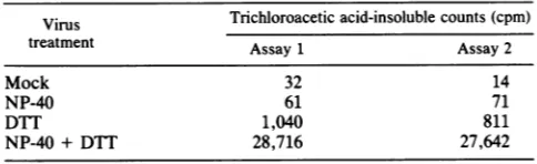
TABLE 1. RPV RNA polymerasetranscription assay'
Virus Trichloroaceticacid-insoluble counts (cpm)
treatment Assay1 Assay2
Mock 32 14
NP-40 61 71
DTT 1,040 811
NP-40 + DTT 28,716 27,642
a Treatments of virus and assays for transcriptions were performed in duplicate as described in Materials and Methods.
mMglutamine, 100 Uofpenicillin, 100 ,ug ofstreptomycin,
and 0.1 mgof pyruvate per ml.
Preparation and labeling of infected tissue culture extracts.
Confluent monolayersofRK-77 or A549 cells were infected
with RPV at a multiplicity of 5 to 10 PFU per cell. The
inhibitor cytosine arabinoside (40 ,ug/ml), rifampin (100
,ug/ml), orhydroxyurea (50 mM), as indicated below, was present throughout the infection. Radiolabeled samples of
early viral proteins were prepared from infected tissue culture cells labeled from 3 to 5 h postinfection in the presence ofcytosine arabinoside orhydroxyurea.
Radiola-beled late viral proteins were prepared from infected cells
labeled 13 to 15 h postinfection either in the presence of rifampin or with no inhibitors present. Specifically, a 150-mm dish of infected cells was labeled with 250 ,uCi of
[3H]leucine
(ICN Pharmaceuticals) for 2 h in 10 ml ofleucine-free medium. Immediatelythereafter, the cells were
scraped intothemedium,chilled, pelleted bycentrifugation
at800x gfor5min at4°C, and suspendedin 0.5 to 1.0 mlof
RIPA buffer (0.15 mM NaCl, 1% sodium dodecyl sulfate
[SDS],1% TritonX-100,0.1% sodium deoxycholate, 10 mM
Tris hydrochloride [pH7.4], 100,000 U ofaprotinin per ml) plus 1 mM phenylmethylsulfonyl fluoride. The cells were
incubated at 4°C for 30 min and then sonicated briefly. Cellular debriswaspelleted bycentrifugationat 12,800 x g
for 10min at4°C. Thesupernatant wasremoved and stored
at-70°C priortoimmunoprecipitation.
Preparation of subviral cores and soluble proteins from
purifiedvirus. Thefractionation ofvirusinto subviralcores andsolubleproteinswasperformedessentially aspreviously described(34). Purified RPV (3 x
105
PFU) was suspendedin solubilization solution (0.05% Nonidet P-40 [NP-40], 10 mM dithiothreitol [DTT], 50 mM Tris hydrochloride [pH
8.5], 10 mM MgSO4) and incubatedat4°C for60min. The
subviralcores were thenpelleted by centrifugationat12,800
x g for 1minat 4°C. Thesupernatantwas removed and used as the soluble fraction. Complete removal of the viral
membranefrom the corepellet was assured byresuspending
the core preparation in 10 times the original volume of
solubilization solution followed by incubationand centrifu-gation as previously described. This twice-extracted core
pelletwas suspendedin TE(50mMTris hydrochloride [pH 7.5], 1 mMEDTA) andusedfor oocyteinjections.
Viral RNA polymerase assay. Viral RNA polymerase
ac-tivity was assayed essentially as described previously (14, 32, 34). Eachreaction sample contained 4 mM ATP, CTP,
andGTP;0.8 mMUTP;3
p.Ci
of[4,5-3H]UTP (ICN),50mMTris hydrochloride(pH 8.5), 10mM MgSO4, and 1.7 x
106
PFUofpurifiedRPVin a finalvolume of 250p.1.
Inaddition,somesamplesalsocontained 0.5%NP-40, 10mMDTT,or a
mixture of both (Table 1). The samples were incubated at
37°C for30min andstopped bythe addition of 2 ml of cold
20% trichloroacetic acid containing 50 mM sodium phos-phateand 50 mM sodiumpyrophosphate.
Precipitation
wasenhanced
by
the addition of 50 ,ul of 1% bovine serumalbuminto each
sample.
Following
a 15-minincubation
at4°C,
theacid-insoluble nucleic acidwas collectedon aglass
fiber filter, dried, and counted in a
liquid
scintillationcounter.
Oocyte
injection
andmethodology. Xenopuslaevis oocyteswere collected from mature female
frogs
anesthetizedby
hypothermia
andwereinjected essentially
as describedby
Colman (6). Individual oocytes were
manually
defollicu-lated,
andunblemishedstage 5and 6 oocyteswere selected forinjection.
Theinjections
were directed intothevegetal
pole
ofthe oocyte. The intact virus or subviral fractionswere
suspended
in TEimmediately prior
toinjection.
The total volumeinjected
wasapproximately
40 nl per oocyte. Theinjections andsubsequent
incubationswere carriedout in eitheramphibian Ringer
or modified Barth solutioncon-taining
100Uofpenicillin
and100 ,ugofstreptomycin
perml. Theincubationtemperature forall oocyteswasbetween 25 and27°C.
Radiolabeling andcollection of oocytes.
Groups
of3 to 10 oocyteswere labeledwith 250,uCi
of[4,5-3H]leucine (ICN)
inatotal volumeof100to250 ,ulof incubation medium. Cells
were labeled for 2 h at the normal incubation temperature and collected
immediately
thereafter. Labeledoocyteswerewashed two times with incubation
medium, collected,
and storedindividually
in 100,u1
of RIPA bufferplus
1 mMphenylmethylsulfonyl
fluoride.Storage
was at-70°C
prior
to use.Immunoprecipitations.
All steps were at4°C.
Individualoocytes were thawed and
briefly
Douncehomogenized
in 100p.l
of RIPA storage buffer in order to rupture andhomogenize
the cell. NET-NP-40(0.5%
NP-40,
150 mMNaCl,
5 mMEDTA,
50 mM Trishydrochloride
[pH
7.4])
(100
p.l)
was then added to eachsample
followedby
theaddition of25 to50
,u1
ofa10%suspension
ofStaphylococ-cus aureus
(Cowen
strain)
whichhad been heatkilled, fixed,
and stored in NET-NP-40
(17).
Thesample
wasmixed,
incubated for 15 min, mixed
again,
and incubated for an additional 15 min.Samples
were thencentrifuged
for3 min at10,000
x g to remove cellular debris as well as anyproteins
which adherednonspecifically
to thebacterialsur-face. The
supernatant
wasremoved and used forsubsequent
immunoprecipitations.
Undilutedpolyclonal
rabbitanti-RPVserum
(1
to3p.1)
or amousemonoclonalantibody
(MAb
94)
(50
,ul)
culturesupernatant
was then added to eachsample.
The
samples
were mixed and incubated for 12 to 15 h. For theexperiments
inwhichthemonoclonalantibody
wasused,
2
p.l
of a secondantibody,
anti-mouseimmunoglobulin
G(Sigma
ChemicalCo.),
wasthen added(undiluted)
and thesample
was incubated an additional 2 h. Allsamples
werethenmixed with 25to50
p.l
of the10%S.aureussuspension,
incubated for 15 min,
remixed,
and incubatedan additional15 min. Thebacterial
pellets
werecollectedby
centrifugation
at
10,000
x g for 1 min and 20 sec. The supernatant wasdiscarded,
and thepellet
wassuspended
in 800 p.1 ofNET-NP-40. The
suspension
waspelleted
twoadditional timesin ordertominimizenonspecific
adherenceofprotein.
Thefinalpellet
wassuspended
in 50pl
ofimmunoprecipitation
lysis
buffer(2%
SDS,
30mM Trishydrochloride [pH 6.8],
1.5% DTT, 20%glycerol,
and0.05%bromophenol blue)
by
water bath sonication. Thesamples
werethen heatedto100°C
for2minand cooledtoroomtemperature. Bacterial debris was
pelleted by
centrifugation
at10,000
x gfor 3 min,and theresulting sample
supernatants
wereanalyzed by
polyacryl-amide
gel
electrophoresis.
Ascontrols,
comparable
amounts of radiolabeled extracts of uninfected and infected tissueVOL.64, 1990
on November 10, 2019 by guest
http://jvi.asm.org/
2282 MASSUNG AND MOYER
culture cells were
immunoprecipitated
in parallel with the oocyte samples by using the same procedures as those described forthe oocytes.Resolution of immunoprecipitated proteins.
Samples
ofradiolabeled, immunoprecipitated proteins were separated
byelectrophoresison10%
SDS-polyacrylamide
(30:1 acryl-amide/bisratio) gels.Samples (50 ,ul)containingthe materialcollected from a single oocyte or comparable amounts of material derived fromtissue culturesampleswere subjected
to electrophoresis at70 Vfor 16 h at room temperature in buffercontaining6.1g of Trisbase,28.8gof
glycine,
and 1.0 g of SDS per liter. Gels were processed for fluorography priortoautoradiography
asdescribedby BonnerandLaskey(2).
Monoclonal and polyclonal antibodies. The monoclonal
antibody
MAb 94, whichrecognizes
boththe 94-kilodalton(kDa) structuralprotein precursor of theRPV virion andits
maturecleavage product
(p62),
wasgenerated
aspreviously describedby Morrisonetal.(22).Polyclonal antibodieswereprepared from rabbits immunized with eitheracetone-fixed
tissue culture cells infected with RPV or acetone-fixed
purified
RPV. Both immunization regimens involved theinjection
oftheantigens dilutedat a 1:1ratio withcomplete Freundadjuvant. Eachrabbit receivedaseries offour suchimmunizations witha time interval of7days between each
injection.
At 2 weeks afterinjection
4, the animals weregiven
a booster injection of antigen minus the adjuvant. Another boosterinjection
was repeated 2 weeksfollowing
thefirst.The animalswerebled7days after boost 2,and the
antiserawere storedat
-70°C prior
to use.Electron microscopy. To prepare
samples
for electronmicroscopy,
oocyteswereinjected
withorwithout virusintothedistal
region
of thevegetalpole. Colloidal gold particles (15 nm) were included in the injection mix and used as amarker for the site of
injection.
Immediately
or at 2 hpostinjection,
the oocyteswere fixedovernight
at4°C with3%
glutaraldehyde,
2%paraformaldehyde,
2.5% dimethylsulfoxide,
1% acrolein,and0.14%CaCl2
in 0.1 Mcacodylate buffer. The oocytes were then further treated at roomtemperature for 2 h in 2%
OS04.
The oocytes were flatembedded in Spurr
embedding
medium. The sections werepoststained
withuranyl
acetateand lead citrate andexam-inedat 60 kV withaJEOL 100-CXmicroscope.
RESULTS
Response of theoocyte to
injection
ofRPV subviralcores.Amphibians
are notnatural hosts for RPV.Therefore,
wefirst had to determine whether the X.
laevis
oocyte wouldrespond to
injected
virus. Our firstexperiments, however,employed
theinjection
not ofintact virus but ofsubviralcores. Thechoice of subviralcoresfortheseinitial
microin-jection
experiments wasfor the followingreason. During anatural
infection,
after attachmentof the virus to the cell,uncoating
of the virion occursvia fusionof the viralmem-brane with the cell membrane or endocytic vesicle mem-brane
during
orimmediately
afterviralpenetration (4, 7,8).The
microinjection
oftheoocyteeffectively
eliminatescon-tactof the virus with these membranes and therefore might
preventuncoatingof the viralparticleto yield active subviral
cores.
Any potential
difficulties with the uncoatingprocessarecircumvented by instead injecting subviralcoresdirectly intothecytoplasm. However,in orderforsubviral cores to beeffectivein theoocyte, core-associatedtranscriptionand
subsequent
translationstill must takeplace. Furthermore, iftheoocyteenvironmentis to beconsideredanalogousto that
A B
2 3 4 5 1 2 3 4
- 80 K - 16&K -84K
-58 K
,,
0 *448.W. KJill.g
~~~
4im~~4~
~ -
36.5
K
>..m*.'al
a r d S S _ -26. 6 K
FIG. 1. Response of X. Iaevis oocytes to injection of RPV subviralcores. Singleoocytes maintained in eitherRingeror mod-ified Barthmediumwereinjectedwith40nl of TE medium
contain-ing 300-PFU equivalents ofRPV cores prepared as described in Materials and Methods. At the times after injection indicated,
proteins were radiolabeledasdescribed in Materials and Methods. Soluble radiolabeledproteinswithin theoocyteaswellassamplesof radiolabeled early and late viral proteins derived from infected rabbitkidneycellswereimmunoprecipitated asdescribed in Mate-rials and Methods. All samples were then analyzed by
SDS-polyacrylamide gel electrophoresis followed by fluorography and
autoradiography of thedried gel. (A) Lanes: 1, 2, and 3,
samples
derived from uninfected andearlyandlateinfected cellspermissive
for RPV; 4, immunoprecipitable proteins derived from
mock-in-jected oocytes; 5, sample derived fromoocytesinjectedwith sub-viralcores, collected 24 h afterinjection. (B)Lanes: 1, 2,3,and4,
samples derived from oocytes injected with subviral cores and collected 12,24, 36,and48hpostinjection,respectively. Molecular masses areindicated at the right. K, Kilodaltons.
of the mammalian cell,then the pattern ofproteins
initially
expressed should be similar to the early pattern of viral
protein synthesisobservedduringnormalinfections of
mam-malian cells.
The results observed when oocytes were injected with subviralcores are shown inFig. 1. Also shown arecontrols for immunoprecipitated mock-injected oocytes (Fig.
1A,
lane4)anduninfectedearlyorlateviral
immunoprecipitable
proteins derived from infected rabbit kidney (RK-77) cells
(Fig. 1A, lanes 1, 2, and 3, respectively). Comparison with infected cell cultures shows that within 24 h after the
injectionofcores,anearly viral pattern of protein
synthesis
had been established. The early pattern of viral
protein
synthesis in oocyteswasalready apparent by 12 hfollowing injection and persisted forperiods up to 48 h (Fig. 1B). In
J. VIROL.
on November 10, 2019 by guest
http://jvi.asm.org/
[image:3.612.322.560.71.383.2]POXVIRUS-INFECTED OOCYTES 2283
A
B
C
D
- 40.
__mo
4_
XE
F
A
Ba
5....w
- 94 K
- 65 K
C
I -I
FIG. 2. Response of X.laevisoocytes toinjection of intactRPV.
Injections, preparation, and analysis of sampleswere asdescribed in thelegend toFig. 1 exceptthat injections were withintact virus. Lanes A, B, and C contain radiolabeled immunoprecipitable viral proteins synthesized 24, 36, and 48 h postinjection, respectively. Also shown, as controls, are samples of immunoprecipitable late
andearly viral proteins derived from infected permissivecells(lanes
Dand E, respectively) and fromoocytesinjected with subviralcores
(lane F). The molecularmassesof viral structuralprecursorproteins p4A(94 K)andp4B(65 K)areindicated. K,Kilodaltons.
experiments not shown, we have observed that this early
pattern of viralgene expression persisted forup to96 h or until oocyte death. The results ofthis experiment suggest that subviral cores can direct viraltranscription within the oocyteenvironment and that thosetranscriptsareaccurately translated into early proteins. However, unlike what was observed in a normal productive infection, there was no progressiontotheexpression oflategenes.
Response of the oocyte to injected intact RPV virions.
Injection of subviral cores into oocytes resulted in the establishmentofapersistentpattern ofexpression ofearly
viral proteins (Fig. 1). However, unlike what occurs in a naturalinfection,therewas notransitiontolategene
expres-sion. We thenexamined the response of the oocyte tothe
injection of intact RPV virions. The results of this experi-ment(Fig. 2) surprisinglyrevealedamorecomplete pattern of viral gene expression than we observed following the
injectionof subviral cores. Unlike whatwas observed with subviralcores, by24 ha patternof viralproteins was seen thatwas both moreextensive than that ofcores and more similartoalate pattern ofproteins. Additional viralproteins
continued to appearat evenlater times, andeach ofthese proteins corresponded to proteins seen at late times in a natural infection. Solelyonthe basis of molecularmasses,it
wouldappearthattwoof the proteinbandscorrespondtothe
FIG. 3. Effects ofhydroxyurea onoocytes injected with intact RPV virions. Virions were injected into duplicate oocytes in the absence (lanes A and B) or presence (lanes C and D) of 50 mM
hydroxyurea and radiolabeledasdescribed in Materials and Meth-ods. Samples were collected 42h afterinjection, immunoprecipi-tated, and analyzedonpolyacrylamide gels. Asacontrol,asample
derived from tissue culture(A549) cells infected with virusin the
absence of inhibitors(lane E) is included.The molecularmassesof
p4A(94 K) andp4B (65 K)areindicated. K, Kilodaltons.
precursor proteinsof the majorlate corepolypeptidesP4A and P4B(94and 65kDa, respectively). By48h,thepattern of viralproteinswasquitesimilarthoughnotidenticaltothat of lateproteinsobserved for cellsproductivelyinfected with RPV. Unlike subviral cores, which expressed only early proteinswithinoocytes, itwould appear that intact virions
can synthesize both early and at least some late viral proteins. Intact virions,therefore, candirectmorecomplete expressionof the viralgenome thancansubviralcores.
Analysis of oocytes injected with intact virus for the
pres-enceof late viral proteins. The results inFig. 2 suggestthat late viralgeneproductsaresynthesizedinoocytesfollowing injection with complete virus. The synthesis of late viral proteins shouldhave been blockedby drugssuchas
hydrox-yurea,which inhibit late viralprotein synthesis by
prevent-ingviral DNAsynthesis.The effect of thedrugwastoinhibit
expression ofmany of the viral proteins, and the resulting patternofexpressed proteinswhichweredrugresistantwas similar to that of an early viral protein pattern (Fig. 3; compare lanes A and B withC and D). It should be noted that hydroxyurea was generally suppressive in terms of
poxvirus gene expression. However, various exposures of the gels, whilenot shown, still support the notion that the patternofproteinsobservedin thepresenceof thedrugwas
early.
Further evidence of late viralprotein synthesisinresponse
D
E
m - 94 K
- 65 K
%W
w
VOL.64, 1990
-Aft ..
on November 10, 2019 by guest
http://jvi.asm.org/
[image:4.612.360.511.68.370.2] [image:4.612.82.278.69.366.2]A B C
D
F
.-G
Hw_"c
* o= -so -mo - 94 K
[image:5.612.63.297.71.374.2]- 62 K
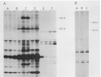
FIG. 4. Immunoprecipitation of viral proteins synthesized in
oocyteswithmonoclonal antibody directed againstalatestructural
protein. Proteins from infected A549 cell cultures orfrom oocytes
injected with virus wereimmunoprecipitated (48 h after injection)
with either polyclonal anti-RPV sera (lanes A to D) or with a
monoclonal antibody (MAb 94) whichrecognized both theprecursor
(94-kDa) and processed form (62-kDa) of the viral structural protein p4A (lanes E to I). K, Kilodaltons. Cells were infected with
wild-typevirus in the absence ofanyinhibitor (lanesBandE),in the
presence of CAR (lanes A andI), orin the presence of rifampin
(lanes C and F). Infected cell samples were collected 5 h after
infection in thepresence of CARorat15 hpostinfection otherwise. Oocyteswereinjected in the absence of inhibitors with either intact virus(lanesDand G) orwith subviralcores(lane H).
toinjected intact viruswasprovided by the experiment (Fig.
4) in which we used a monoclonal antibody (MAb 94) we
prepared against p4A, one of the two major late structural
proteins of the virion. This protein is synthesized late in infection, following the onset of DNA replication, as a
94-kDaprecursorwhich isproteolytically cleaved duringthe
maturation process to a 62-kDa form of the protein found
within mature virions (15, 16, 26, 35). MAb94 recognizes both forms of the protein. As controls, we have included
immunoprecipitations ofcell cultures infected with RPV in
thepresenceand absence oftheinhibitors cytosine
arabino-side(CAR)andrifampin. CAR isaninhibitorwhichprevents
DNA replication and subsequent synthesis of late viral
proteins; rifampinhas noobviouseffecton gene expression
butinsteadblocks theproteolytic processing oflate
precur-sorproteins.
Wheninfected tissueculture cellswereexamined, itcould be seenthattheaddition ofCARblockedtheappearanceof
boththe94- and 62-kDa forms ofp4A(Fig. 4;comparelanes
AandB). Addition ofrifampintoinfectedcells(Fig.4,lane
C) drasticallyreduced levels of themature62-kDacleavage
products of p94 but had relatively little effecton levelsofthe
94-kDa precursor. Oocytes injected with intact virus
ap-peared to synthesizethe94-kDa structuralprecursorprotein (Fig. 4, lane D).
Similar experiments using MAb94 are shown in Fig. 4, lanes E through I. The monoclonal antibody precipitated both forms of p4Afrominfectedtissueculturecells(Fig. 4, lane E),only the94-kDa precursor from cells infectedin the
presence of rifampin (Fig. 4,laneF), and neither form when cellswere infected in the presenceofCAR(Fig. 4, lane I). Examination of immunoprecipitated oocytes in theabsence
of inhibitors revealedthepresence oftheprecursor94-kDa
protein but not of the processed form of the protein (Fig. 4, lane G). While we interpret these results to be a further
indication of late viralprotein synthesis, it is also interesting to note that we have never seen any indication that this
protein is processed in oocytes. As expected, none of the protein was detected in oocytesinjected with subviral cores (Fig. 4, lane H).
Analysis of the integrity ofinjected whole virions. Itisclear that the injection of intact virions results in extensive
tran-scription and translation of viral genes. Normally, intact
virions are transcriptionally inactive because the virions
mustfirst bepartially disruptedtoform cores eitherduring
the adsorption and penetration of the cell in vivo or by
disruptionof the viral membrane in vitroby the addition of detergent and a reducing agent before the transcriptional machinery becomes functional. Therefore, in order for the virions to function within the oocyte environment, it is
implicitthat either the virus isactivatedoruncoatedwithin the oocyteorthe virusmustbe somehowactivated priorto
injection. We have examined thetranscriptionalstateofthe viruspreparationsused intheseexperiments. The results, in
duplicate, are shown in Table 1. It is evident that without
treatment with bothdetergent andreducing agent, the
viri-ons are transcriptionally inactive. It is also clear from this
experiment that the virion preparation, when activated by thenormal detergent and reducing-agent treatment, has the expected transcriptional potential. Therefore, since the vir-ions are transcriptionally inactive prior to injections, they
must be activated within the oocyte in the absence of the usual passage of the virus through the cellular membrane.
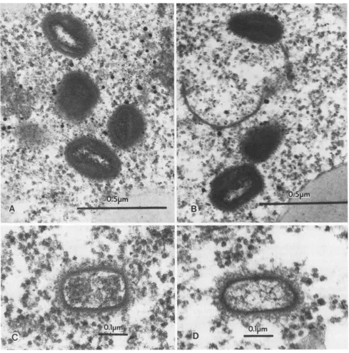
Electronmicroscopicanalysis of virions afterinjection into
oocytes. Since virions were activated when injected into
oocytes, we attempted to visualize the processing of the virionsfollowing their introduction into the oocyte. Colloidal
goldwascoinjected withthevirustoaidinlocatingthe virus within the oocytes. It is clear that there were marked
changes incurred followingthe injection ofthe virions and that these changes were induced rapidly (Fig. 5). Immedi-ately followinginjection,virions which appeared normal and unaltered were readily found within the cytoplasm of the oocyte(Fig. 5A andB). However, within 2h postinjection,
the appearanceof the virions had been markedly altered and visually appeared identical to cores as defined by Easter-brook(10)(Fig.SC and D). Theseresults would indicate that
changestothe virusbeginrapidly. Although we have exam-ined manyfields, there did not appear to be localization of theviralparticles with any particular subcellular structure,
membrane, orother site within the oocyte. After about 4 to 6h,itbecameincreasinglydifficult to find any discrete viral
structure.
Effectof inputmultiplicity of
injected
virus on geneexpres-sion. After repeated experiments ofinjecting oocytes with intact virus, it became apparent that viral expression was
highly multiplicity dependent. For example, theinjection of 49mok 4..,-*-. 1
.1111" .14,Kommklbl
on November 10, 2019 by guest
http://jvi.asm.org/
POXVIRUS-INFECTED OOCYTES 2285
FIG. 5. Electron micrographs ofoocytes injected with RPV. Oocytes were injected and prepared for electron microscopy as described in Materials and Methods. Samples were taken for analysis either immediately (A and B) or 2 h (C and D) after injectionwith RPV.
concentrated stocksofvirus
(>105
PFU per oocyte)rapidlykilled theoocytes. At verylowconcentrations ofvirus,we noted significant variations in both the amount of viral protein synthesis and the length of time needed for late viral proteinsynthesis to become apparent. Therefore, we sought
to quantitate the effect of virus input multiplicity on the response seen in the oocyte. Theresults of this experiment
(Fig. 6) demonstrate the pattern of viral protein synthesis observed 24 h after injection with various input levels of virus ranging from 3 to 12,000 PFU per oocyte. At input doses of 3 or 30 PFU per oocyte, little, if any, late viral
protein synthesis was observed at 24 h postinjection (Fig.
6A, lanes A and B). Input levels of virus of 300 PFU per oocyte appeared to be ideal (Fig. 6A, lane C). The input
optimum appeared fairly narrow, because expression was notnearlyas extensive whenhigher levels (1,500PFUper
oocyte)wereused(Fig. 6A,laneD). Largerdoses(6,000or
12,000 PFU peroocyte) actually appearedtobe detrimental to the oocyte (Fig. 6A, panels E and F) and resulted in a
strongdiminutionof viralexpression.Theinhibitionof viral
protein synthesisobservedathigherconcentrations ofvirus
appearedtobenonspecificasoverall oocyteprotein synthe-sis,asmeasuredby totalacid-precipitableradiolabeled
pro-teins,was also decreased. Weroutinely titrated eachbatch
VOL.64,1990
on November 10, 2019 by guest
http://jvi.asm.org/
[image:6.612.63.552.76.571.2]A
A B C D E F
- 94 K
- 94 K
_--_
FIG. 6. Viralgeneexpression inoocytesas afunctionofmultiplicityofinjection. Oocyteswereinjectedatvariousinput multiplicitiesof
virus or cores as describedinMaterials andMethods. Proteins were radiolabeled 22 h after injectionfor2 h, collected thereafter, and
immunoprecipitated and analyzedonpolyacrylamide gelsaspreviouslydescribed.(A)Responses ofoocytestointactvirus.Themultiplicities ofinjectionwere3 (laneA), 30(lane B),300(lane C), 1,500 (lane D),6,000 (lane E),and12,000 (lane F)PFUperoocyte.Duplicate samples representingtwoindividualoocytesarepresentedforeachmultiplicity. (B)Responses ofoocytestoinjectionofcoresatinputmultiplicities of300(lane A), 1,200 (lane B),and12,000 (lane C) PFUperoocyte.Themolecularmassesofp4A(94 K)andp4B (65K)areindicated. K, Kilodaltons.
of virus prior to use to maximize both the response and
reproducibility ofthe oocytes. The response ofoocytes to subviralcores was notasfastidious, as asimilarpattern of
protein expressionwasobservedoverquiteawiderangeof
inputcores(30to 12,000coresperoocyte) (Fig. 6B, lanesA toC).
Addition ofsolubilizedcomponents back to subviral cores allowedlateviral geneexpression. Injection of oocytes with subviralcoresresultedinonly early viralprotein synthesis,
whereasinjection ofintactvirionsallowed additional expres-sion of late viral gene products. We would predict, on the basis ofthese observations, that some material is removed duringthepreparation ofsubviralcoreswhichfacilitatesthe
expression of late viral genes. This prediction has been
testedbymeasuring theeffectonproteinsynthesiswhen the
solubilizedmaterial wasadded backto subviralcoresprior toinjection intooocytes(Fig. 7).The solublefraction itself,
when injected alone into oocytes, elicited only minimal
levels ofactivity (Fig. 7, lanes A and B). Injection of the purified cores suspended in TE or in solubilizing solution alone (see Materials and Methods) yielded the expected
limited early pattern of protein synthesis as previously shown (Fig. 1, 4, and 6B) (datanot shown). High levels of solubilizing solution (NP-40, DTT, and MgSO4)were
even-tually toxic and killed the oocytes. However, when the soluble fraction was added to the cores, we observed an extensive patternofproteinsynthesis (Fig. 7, lanesEandF) virtuallyidenticaltothat fromoocytesinjected withpurified
virus(Fig. 7, lanes C and D). It wouldappear,therefore, that
expressionof viralgenesbypurifiedcoresislimitedtoearly proteins because some component(s) essential forthe tran-sition to lategene expression isremoved when viruses are
extractedtoproducecores. Furthermore, the component is stable and canbe added back to cores to reconstitute late
geneexpression.
DISCUSSION
The initial reason for exploring the X. laevis oocyte system was to examine whether the oocyte was a suitable environment to study the communication that occurs be-tweenthe nucleus andcytoplasminRPV-infected cells. Our
work here focuses on the ability of the oocyte to allow
poxvirus-directedgeneexpression following microinjections
of either intact RPV or subviral cores. The expression of viral genes was assayed by gel electrophoresis of
radiola-beled, immunoprecipitated proteins. The
immunoprecipita-tions withapolyclonal anti-RPVantiserum servedto mini-mize thebackgroundof host oocyteprotein synthesis.From these experiments, we were able to make several conclu-sions. First, RPV genes werecapable ofbeingtranscribed andaccuratelytranslated in theoocyte. Second,therewas a remarkabledifference between thepatternsof viralproteins
observed which depended on whether subviral cores or intactvirionswere injected. The injection of subviralcores resulted in theexpression ofonly early genes, whereasthe injectionof intact virions resulted in the expressionofboth
early and late genes. Furthermore, the early and late viral
proteinsobserved inresponsetoinjectedintact virionswere
expressed in atime-dependent, sequential order, asis seen in anatural infection.
In a natural infection, the expression of late genes is generally believed to be dependent on prior viral DNA replication. We have attempted to measure viral DNA
B
A B C
- 65 K
- 65 K
on November 10, 2019 by guest
http://jvi.asm.org/
[image:7.612.141.471.74.326.2]POXVIRUS-INFECTED OOCYTES 2287
A B C
D
E
Fw - 94K
-~~
- 65 KFIG. 7. Effectsof virion soluble extract on protein expression in
oocytes by RPV cores. Purified virions were fractionated into subviralcoresand asolublefraction aftertreatmentwithdetergent
and DTT.Oocyteswereinjectedwith soluble fraction alone(lanesA and B), purified intact virions (lanes C and D), or subviral cores
whichweresupplementedwithanamountof solubleextract
equiv-alenttotheamountstrippedfrom theinjectedcores(lanesE andF).
Newly synthesized proteinswereradiolabeled for 2 hat46hafter
injection, oocytes were harvested, and proteinswere
immunopre-cipitated and analyzed on polyacrylamide gels as described in Materials and Methods. The molecularmassesofp4A(94 K) and
.B(65 K)areindicated. K, Kilodaltons.
synthesis within virus-injected oocytes by several
proce-dures (datanot shown). We have failed to detect anyviral
DNA synthesis in the oocyte under any conditions. After input virus was uncoated, we were unable to detect any
immature ormature viruswithin oocytes up to48 h
postin-fection (unpublished results), even though extensive late
protein synthesishadoccurred.
In naturalhost cells, all poxvirus late proteins by defini-tion are thought to depend on viral DNA synthesis for
expression. Based onthis definition, the protein precursors
ofthe major core polypeptides referred to as p4A andp4B have beenconsideredaslate proteins. Considering thelack of detectable DNA replication in the oocyte, we wanted
further proof that a protein of 94 kDa-synthesized in
re-sponseto injected wholevirus was indeed p4A. Therefore,
we used the monoclonal antibody MAb 94 to immunopre-cipitatesamplesof infected oocytesinjectedwithvirus. This
monoclonal antibody recognized both the p4A protein (94
kDa)andthe 62-kDacleavage productwhichwasgenerated
fromthe 94-kDaproteinduringmorphogenesisofthevirus.
Theseexperiments (Fig. 4) revealed not onlythatauthentic
p4Awasproduced in theoocytebutalso that the proteinwas not cleaved to the mature 62-kDa form. The failure to process p4A was seen during a natural infection in the
presence of rifampin, an inhibitor of viral morphogenesis.
This lack of the propercleavage ofone of the major core
polypeptides mightevenbeexpected in theoocytebecause there isnoviral DNAsynthesis.Under these conditions,no new DNA would be available for the initiation of viral morphogenesis.
Toprovide further evidencefor the synthesisof late viral proteins in oocytes, we used aninhibitor of DNA
replica-tion, hydroxyurea, that blocksthe expressionof lategenesin
a natural infection. Oocytes injected with virus and
incu-batedin the presence ofhydroxyurea showed adiminished
overall pattern of viral protein synthesis but nevertheless appeared to maintain an early viral protein pattern. Both observations are consistent with what is seen in a natural infection in thepresence ofhydroxyurea and further support the contention that many ofthe proteins seen without any inhibitors in the oocyte in response to intact virus are indeed late viral proteins, despite the fact we cannot demonstrate anyviral DNA synthesis.
It has been well documented that intact virions are
tran-scriptionally inactive and that gene expression commences only when the virion is uncoated. Since viral genes were
expressedinthe oocytefollowing injection of the virus, the virions must be uncoated within the oocyte, assuming the
viruswasintact and transcriptionally inactive prior to injec-tion. Our results demonstrate that the virus we injected into the oocyte was intact, i.e., inactive unless somehow proc-essed. We usedanin vitro assay of RNApolymerase activity
toprovethat the viruswasactiveonly after treatment which
permeabilized and removed the viral membrane. The un-treated virionslacked any abilitytotranscribe(Table 1).
Inanaturalinfection, theinitial stage of uncoatingof the virus is believed to occur viafusionof the viral membrane with either the cell membrane or an endocytic vesicle.
Assuming fusion of the viral membrane with a cellular
membraneto be aprerequisite for uncoating, weemployed
electronmicroscopytolookforpossible
compartmentaliza-tion of the virus or fusion of the virus with an oocyte cytoplasmic membrane early after injection of the oocyte
(Fig. 5). Our results did not reveal any particular site of
localization or cytoplasmic membrane involvement in the
uncoating
process. However, electron microscopydidindi-cate that within 2 h postinjection, nearly all of the virions
were altered. Therefore, we conclude that productive
un-coatingofthe virus within theoocyteisrelatively rapidand
quiteefficient.However,the actual mechanismbywhichthe
virus isuncoated remains unclear.
We havealso demonstrated that viral gene expressionin the oocyte is clearly dependent on the multiplicity ofthe
injectedvirus. We have shown that the optimal multiplicity for oocyte injections is 300 PFU per oocyte and that the
expressionof viral geneproductsdecreases if eithermoreor less virus isinjected. However,itshould be noted thateach
purified virus preparation can vary in its optimal oocyte
multiplicitybecause of variations in thepurityof thestock.
Perhaps this reflects differences in the particle/PFU ratio.
Therefore, each stockmustbe tested todetermine itsown
optimal multiplicity. Theinjection oflarge numbersof
viri-onsinto anoocyte appearstobe lethal. Oocytes injected at
high multiplicities (Fig. 6) reveal not only little or no viral
gene expression but also a shutoff of endogenous oocyte
expression
andmorphological changes
consistent with se-vereandrapidoocytedeterioration (unpublished results). VOL. 64,1990on November 10, 2019 by guest
http://jvi.asm.org/
[image:8.612.60.298.79.403.2]Perhaps our most interesting observation relates to the transitionfrom early tolategeneexpression. As previously
stated, the injection ofsubviral coresresulted inthe expres-sion of only early genes, whereas the injection of intact
virionsresulted in theexpressionof bothearly and lategene
products. During the preparation of subviral cores, the
fraction of the virus that was solubilized by a nonionic
detergent and areducing agent was removed. This fraction
has previously been shown to contain a number of viral
proteins (33). We have shown that the injection of this soluble fraction together with subviral cores is capable of
reconstitutingtheexpressionoflateviralgeneproducts. We
conclude thatthis solublefractioncontainsafactor(s) which
is requiredfor the transitionfrom earlytolategene
expres-sion.
These studies potentially reveal new aspects of poxvirus
generegulation. Inthis regard,the oocyte system mayoffer aratherunique experimentalsystemin whichtostudy these
viruses. On theother hand, theoocyte isnotanatural host
cell for the orthopoxviruses. Therefore, additional studies areneededtodetermine whetherourobservations aretruly
reflectionsof natural regulatory events.
ACKNOWLEDGMENTS
We express our appreciationto CarlFeldherrfor tireless assis-tanceontheuseof frogoocytes.Weacknowledge theUniversity of Florida InterdisciplinaryCenterforBiotechnology Research Elec-tronMicroscopy Core for the data shownin Fig. 5.
This workwassupported by Public HealthServicegrantAl 15722
from the National Institutesof Health. LITERATURE CITED
1. Bloom, D. C., R. Massung, L. Savage, D. K. Morrison, and
R.W. Moyer. 1989. Recruitmenttothe cytoplasmofacellular
lamin-likeprotein fromthenucleusduringapoxvirus infection.
Virology169:115-126.
2. Bonner, W. M., and R. A. Laskey. 1974. A film detection
methodfor tritium-labelled proteins and nucleic acids in
poly-acrylamidegels. Eur. J. Biochem. 46:83-88.
3. Broyles,S. S., L. Yuen, S. Shuman, and B. Moss. 1988.
Purifi-cation ofafactor required for transcription of vaccinia virus
earlygenes. J.Biol. Chem. 263:10754-10760.
4. Chang,A., andD. H. Metz. 1976. Furtherinvestigationsonthe
mode of entry of vaccinia virus into cells. J. Gen. Virol.
32:275-282.
5. Cochran, M. A., M. Mackett, and B. Moss. 1985. Eukaryotic
transientexpression systemdependentontranscription factors
and regulatory DNAsequences ofvaccinia virus. Proc. Natl.
Acad. Sci. USA82:19-23.
6. Colman,A. 1984. Translation of eukaryoticmessengerRNA in
Xenopusoocytes,p. 270-302. B. D. Hamesand S. J.Higgins,
(ed.), Transcription and translation: apractical approach.
Ox-ford University Press, Oxford.
7. Dales,S. 1973. Early events in cell-animal virus interactions.
Bacteriol. Rev. 37:103-135.
8. Dales, S.,and R. Kajioka. 1964. The cycle of multiplication of
vacciniavirus in Earle's strainLcells. I. Uptake and
penetra-tion.Virology24:278-294.
9. Dales, S., andB. G. Pogo. 1981. Biology of poxviruses. Virol.
Monogr. 18:1-109.
10. Easterbrook, K. B. 1966. Controlled degradation of vaccinia
virions invitro: anelectron-microscopic study. J. Ultrastruct.
Res.14:484-496.
11. Ensinger,M. J., S.A. Martin, E. Paoletti, and B. Moss. 1975.
Modificationofthe 5'-terminus ofmRNAbysoluble guanylyl
and methyltransferasesfrom vacciniavirus. Proc. Natl.Acad.
Sci. USA72:2525-2529.
12. Feldherr,C. M.,and P. A.Richmond.1978.Manualenucleation
of Xenopus oocytes, p. 75-79. InG. Stein,J. Stein, and L. J.
Kleinsmith (ed.), Methods in cell biology, vol. 17. Chromatin
and chromosomal protein research
II.
Academic Press, Inc., New York.13. Golini, F., and J. R. Kates. 1985. A soluble transcription system derived from purifiedvaccinia virions. J.Virol. 53:205-213. 14. Kates, J. R., and B. R. McAuslan. 1967. Poxvirus
DNA-dependent RNA polymerase. Proc.
Natl.
Acad. Sci. USA 58:134-141.15. Katz, E., and B. Moss. 1970. Vaccinia virus structural polypep-tide derived from ahigh-molecular-weight precursor: formation and integration into virus particles. J. Virol. 6:717-726. 16. Katz, E., and B. Moss. 1970. Formation of a vaccinia virus
structural polypeptide from a higher molecular weight precur-sor: inhibition by rifampicin. Proc.
Natl.
Acad. Sci. USA 66:677-684.17. Kessler, S. W. 1975. Rapid isolation of antigens from cells with a staphylococcal protein A-antibody absorbent: parameters of the interaction of antibody-antigen complexes with protein A. J. Immunol. 115:1617-1624.
18. Marbaix, G., and G. Huez. 1980. Expression of messenger RNAs injected into Xenopus laevis oocytes, p. 347-381. In J. E. Celis, A. Graessmann, and A. Loyter (ed.), Transfer of cell constituents into eukaryotic cells (NATO study series A). Plenum Publishing Corp., New York.
19. Martin, S. A., and B. Moss. 1975. Modification of RNA by mRNA guanylyltransferase and mRNA (guanine-7-)methyl-transferase from vaccinia virions. J. Biol. Chem.250:9330-9335. 20. Martin, S. A., and B. Moss. 1976. mRNA guanylyltransferase and mRNA (guanine-7-)methyltransferase from vaccinia
viri-ons. Donor and acceptor substrate specificities. J. Biol. Chem. 251:7313-7321.
21. Martin, S. A., E. Paoletti, and B. Moss. 1975. Purification of mRNA guanylyltransferase and mRNA (guanine-7-)methyl-transferase from vaccinia virions. J. Biol. Chem. 250:9322-9329. 22. Morrison, D. K., J. K. Carter, and R. W. Moyer. 1985. Isolation and characterization of monoclonal antibodies directed against two subunits of rabbit poxvirus-associated, DNA-directed RNA polymerase. J. Virol. 55:670-680.
23. Morrison, D. K., and R. W. Moyer. 1986. Detection of a subunit of cellular PolII within highly purified preparations of RNA polymerase isolated from rabbit poxvirus virions. Cell 44: 587-596.
24. Moss, B., M. J. Ensinger, S. A. Martin, and C. M. Wei. 1975. Modification of the 5'-terminus of mRNA by guanylyl and methyl transferases from vaccinia virus, p. 161-168. INSERM (Inst. Natl. Sante Rech. Med.) Colloq., Paris.
25. Moss, B., A. Gershowitz, C. M. Wei, and R. Boone. 1976. Formation of the guanylylated and methylated 5'-terminus of vaccinia virus mRNA. Virology 72:341-351.
26. Moss, B., and E. N. Rosenblum. 1973. Protein cleavage and poxvirus morphorenesis: tryptic peptide analysis of core pre-cursors accumulated by blocking assembly with rifampicin. J. Mol. Biol. 81:267-269.
27. Moss, B., and E. N. Rosenblum. 1974. Vaccinia virus polyri-boadenylate polymerase: covalent linkage of the product with polyribonucleotide and polydeoxyribonucleotide primers. J. Vi-rol. 14:86-98.
28. Moss, B., E. N. Rosenblum, and A. Gershowitz. 1975. Charac-terization of a polyriboadenylate polymerase from vaccinia virions. J. Biol. Chem. 250:4722-4729.
29. Moss, B., E. N. Rosenblum, and E. Paoletti. 1973. Polyadenylate polymerase from vaccinia virions. Nature New Biol. 245:59-63. 30. Moyer, R. W. 1987. The role of the host cell nucleus in vaccinia
virus morphogenesis. Virus Res. 8:173-191.
31. Moyer, R. W., and C. T. Rothe. 1980. The white pock mutants of rabbit poxvirus. I. Spontaneous host range mutants contain deletions. Virology 102:119-132.
32. Munyon, W., E. Paoletti, and J. T. Grace, Jr. 1967. RNA polymerase activity in purified infectious vaccinia virus. Proc. Natl. Acad. Sci. USA58:2280-2287.
33. Oie, M., and Y. Ichihashi. 1981. Characterization of vaccinia polypeptides. Virology 113:263-276.
34. Paoletti, E. 1977. In vitro synthesis of a high molecular weight virion-associated RNA by vaccinia. J. Biol. Chem.252:866-871.
on November 10, 2019 by guest
http://jvi.asm.org/
POXVIRUS-INFECTED OOCYTES 35. Pennington, T. H. 1973.Vaccinia virus morphogenesis:a
com-parison of virus-induced antigens and polypeptides. J. Gen. Virol. 19:65-79.
36. Puckett, C., and B. Moss. 1983. Selective transcription of vaccinia virusgenes in template dependent solubleextracts of infected cells. Cell35:441-448.
37. Rohrmann, G., and B. Moss. 1985. Transcription of vaccinia virus early genes by a template-dependent soluble extract of purified virions. J. Virol. 56:349-355.
38. Rohrmann, G., L. Yuen, and B. Moss. 1986. Transcription of vaccinia virus early genes by enzymes isolated from vaccinia virions terminates downstream ofaregulatory sequence. Cell
46:1029-1035.
39. Shuman, S., M. Surks, H. Furneaux, and J. Hurwitz. 1980. Purification and characterization of a GTP-pyrophosphate
exchange activity from vaccinia virions. Association of the GTP-pyrophosphate exchange activity with vaccinia mRNA
guanylyltransferase. RNA (guanine-7-)methyltransferase
com-plex(capping enzyme). J. Biol. Chem. 255:11588-11598. 40. Turner, P. C., P. A. C. Watkins, M. Zaitlin, and T. M. A.
Wilson. 1987. Tobacco mosaic virus particles uncoatand ex-press their RNA in Xenopus laevis oocytes: implications for early interactions between plant cells and viruses. Virology 160:515-517.
41. Wei, C. M., and B. Moss. 1974. Methylation of newly synthe-sized viral messengerRNA by an enzyme in vaccinia virus. Proc. Natl. Acad. Sci. USA 71:3014-3018.
42. Wei, C. M., and B. Moss. 1975. Methylated nucleotides block 5'-terminus of vaccinia virus messenger RNA. Proc. Natl. Acad. Sci. USA 72:318-322.
43. Wilton,S., andS.Dales. 1986. InfluenceofRNApolymeraseII
uponvaccinia virus-related translation examined by meansof alpha-amanitin. Virus Res. 5:323-341.
44. Wilton, S., and S. Dales. 1989. Relationship between RNA polymerase II and efficiency of vaccinia virus replication. J.
Virol.63:1540-1548.
VOL. 64, 1990 2289