0022-538X/99/$04.00⫹0
Copyright © 1999, American Society for Microbiology. All Rights Reserved.
Expression of Mouse Mammary Tumor Virus Superantigen
mRNA in the Thymus Correlates with Kinetics of Self-Reactive
T-Cell Loss
ANNA BARNETT,† FARAH MUSTAFA, THOMAS J. WRONA, MARY LOZANO, ANDJAQUELIN P. DUDLEY*
Department of Microbiology and Institute for Cellular and Molecular Biology, The University of Texas at Austin, Austin, Texas 78712
Received 12 October 1998/Accepted 7 May 1999
Mouse mammary tumor virus (MMTV) encodes a superantigen (Sag) that is expressed at the surface of antigen-presenting cells in conjunction with major histocompatibility complex (MHC) type II molecules. The Sag-MHC complex is recognized by entire subsets of T cells, leading to cytokine release and amplification of infected B and T cells that carry milk-borne MMTV to the mammary gland. Expression of Sag proteins from endogenous MMTV proviruses carried in the mouse germ line usually results in the deletion of self-reactive T cells during negative selection in the thymus and the elimination of T cells required for infection by specific milk-borne MMTVs. However, other endogenous MMTVs are unable to eliminate Sag-reactive T cells in newborn mice and cause partial loss of reactive T cells in adults. To investigate the kinetics of Sag-reactive T-cell deletion, backcross mice that contain single or multiple MMTVs were screened by a novel PCR assay designed to distinguish among highly related MMTV strains. Mice that containedMtv-17alone showed slow kinetics of reactive T-cell loss that involved the CD4ⴙ, but not the CD8ⴙ, subset. Deletion of CD4ⴙor CD8ⴙ
T cells reactive withMtv-17Sag was not detected in thymocytes. Slow kinetics of peripheral T-cell deletion by
Mtv-17 Sag also was accompanied by failure to detect Mtv-17 sag-specific mRNA in the thymus, despite
detectable expression in other tissues, such as spleen. Together, these data suggest that Mtv-17Sag causes peripheral, rather than intrathymic, deletion of T cells. Interestingly, the Mtv-8 provirus caused partial deletion of CD4ⴙV12ⴙ cells in the thymus, but other T-cell subsets appeared to be deleted only in the
periphery. Our data have important implications for the level of antigen expression required for elimination of self-reactive T cells. Moreover, these experiments suggest that mice expressing endogenous MMTVs that lead to slow kinetics of T-cell deletion will be susceptible to infection by milk-borne MMTVs with the same Sag specificity.
The mouse mammary tumor virus (MMTV) superantigen (Sag) protein is necessary for the efficient transmission of virus from infected mother’s milk to the mammary glands of off-spring (23, 54). MMTV transmission requires infection of both B and T lymphocytes to facilitate transport of virus from the gut of newborns to mammary cells, and lymphocytes appear to be a reservoir of virus in the postnatal period prior to mam-mary gland development (7, 23, 26). Viral infection of B cells in the gut-associated lymphoid tissue leads to the expression of Sag at the B-cell surface as a type II transmembrane glyco-protein (32, 33). The C-terminal portion of Sag in conjunction with major histocompatibility complex (MHC) class II protein is recognized by entire classes of T cells that have specific
chains as part of the T-cell receptor (TCR) (4, 54, 59). Exper-iments that switch the N-terminal and C-terminal regions of different Sags as well as site-directed Sag mutations suggest that the C-terminal 30 to 40 amino acids are critical for TCR interaction (38, 54, 59). Interaction of Sag with the TCR leads to a T-cell signaling pathway that results in the production of cytokines and/or T-cell proliferation (3, 45, 50). The
produc-tion of cytokines leads to further B- and T-cell proliferaproduc-tion and amplification of MMTV-infected cells (3, 29).
Another consequence of T-cell stimulation by MMTV Sag is the ultimate loss of Sag-reactive T cells from the immune repertoire of the infected mouse (36). For example, milk-borne infection with C3H MMTV leads to stimulation and, in sub-sequent months, deletion of V14⫹T cells reactive with C3H Sag (5, 10). Similarly, endogenous MMTVs express Sag pro-teins at the surface of antigen-presenting cells, and Sag expres-sion results in the deletion of specific T-cell subsets (52). Pre-vious data have shown that expression of the milk-borne C3H MMTV Sag from a transgene results in complete deletion of V14⫹T cells that are required for C3H virus transmission to the mammary gland (23). Suchsagtransgenic mice are resis-tant to infection by C3H MMTV through nursing on infected mothers. These experiments suggested that T-cell deletion in-duced by endogenous MMTV Sags would provide protection against exogenous MMTV infection with the same Sag speci-ficity for T cells (23, 28, 36).
Deletion of T-cell classes due to endogenous MMTV pro-viruses may be incomplete (43). For example, both theMtv-8 andMtv-9proviruses delete V5⫹, V11⫹, and V12⫹T cells. The Mtv-8provirus causes incomplete deletion of V5⫹and V11⫹cells, whereas theMtv-9provirus causes complete de-letion of these T-cell subsets (17, 43, 52), despite the fact that theMtv-8andMtv-9long terminal repeats (LTRs) are nearly identical (8). Because complete deletion of a T-cell subset by an endogenous MMTV Sag appears to require effective anti-* Corresponding author. Mailing address: Department of
Microbi-ology, ESB 226, The University of Texas at Austin, Austin, TX 78712-1095. Phone: (512) 471-8415. Fax: (512) 471-7088. E-mail: jdudley @uts.cc.utexas.edu.
† Present address: Howard Hughes Medical Institute, Brigham and Women’s Hospital and Harvard Medical School, Boston, MA 02115.
6634
on November 9, 2019 by guest
http://jvi.asm.org/
gen presentation in the thymus to eliminate self-reactive T cells during development of the immune repertoire (3, 44), incom-plete deletion of Sag-reactive cells may reflect a failure to express certain endogenous MMTVs in thymic antigen-pre-senting cells, perhaps dendritic cells (39). Incomplete deletion also may reflect the expression and presentation of these viral Sags on other cell types that result in the deletion of self-reactive cells in the periphery (31, 49). Alternatively, some endogenous MMTVs may be expressed in thymic antigen-presenting cells at a reduced level, resulting in an avidity that borders the threshold for T-cell signaling necessary to elimi-nate self-reactive cells by apoptosis (50). Because little is known about the level of endogenous MMTVsag-specific ex-pression (56), we investigated whether there was a correlation between thymic expression of spliced sagmRNA and the ki-netics of Sag-specific T-cell deletion. Using mice bred to con-tain single endogenous MMTV proviruses, we showed that partial deletion of Sag-reactive T cells was correlated with characteristics typical for the establishment of T-cell tolerance in the peripheral immune system. Slow kinetics of Sag-reactive T-cell deletion also correlated with poor expression of sag -specific mRNA in the thymus.
MATERIALS AND METHODS
Mice.C58/J, BALB/cJ, and PERA/Ei (Peru-Atteck) mice were obtained from the Jackson Laboratories (Bar Harbor, Maine); these animals, F1hybrids, and
backcross animals were bred in the Animal Resources Center at the University of Texas at Austin. Sentinel animals tested negative for the presence of mouse hepatitis virus and other common murine pathogens (except for those tested for the experiments reported in Table 1).
Antibodies and FACS analysis.Lymph nodes (in most cases, only inguinal nodes) were removed, and cells were released into fluorescence-activated cell sorter (FACS) wash buffer (phosphate-buffered saline containing 0.1% NaN3
and 2.5% bovine serum) by being crushed with the blunt end of a 3-ml syringe. Clumps were removed on ice by settling for 5 min, and single cells were washed several times in FACS buffer before staining. Peripheral blood lymphocytes were obtained and purified as described previously (53, 54). Cells (approximately 106)
were incubated with antibody specific for V3, -5, -6, -7, -8, -9, -11, -12, or -14 labeled with fluorescein and CD4- or CD8-specific antibody labeled with phy-coerythrin (PharMingen, San Diego, Calif.). Thymocytes were prepared similarly except that the staining was performed with fluorescein-labeled Vantibodies, phycoerythrin-labeled CD4, and Cy-chrome-labeled CD8 (also from PharMin-gen). Cells were incubated for 45 min on ice prior to being washed with FACS buffer and fixed in 1% paraformaldehyde in FACS buffer. Cells were analyzed by using the CELLQuest program and a FACSCalibur cytometer (Becton Dickin-son, San Jose, Calif.). Statistical analysis was performed with a two-tailed Student ttest.
RNA extractions and RNase protection assays.The guanidine isothiocyanate method was employed for RNA extractions as described previously (55). DNA and low-molecular-weight RNAs were removed by precipitation with sodium acetate (41). RNase protection assays were performed essentially as described by Yang and Dudley (58) except that hybridizations were performed at 56°C. The riboprobe was derived from theSau3A fragment (⫺455 to⫺116 relative to the ⫹1 start site for transcription at the U3/R junction of the LTR ofMtv-17(24). This probe should detect all known MMTV mRNAs.
DNA extractions and Southern blotting.High-molecular-weight DNA was obtained from tails or livers of backcross mice as described by Choi et al. (11) or Dudley and Risser (13). In later experiments, a simplified procedure was used for isolating tail DNA. A tail section (ca. 1 in.) was added to 1 ml of 20 mM Tris-HCl (pH 7.4)–25 mM EDTA–0.5% sodium dodecyl sulfate–75 mM NaCl–100g of proteinase K per ml and digested for at least 3 h at 53°C. The protease was inactivated by being boiled for 5 min, and the solution was cooled to 4°C prior to centrifugation at the same temperature for 10 min at 1,700⫻g. The supernatant (500l) was clarified further by centrifugation at 10,000⫻gfor 5 min at 4°C to remove sodium dodecyl sulfate prior to ethanol precipitation. Pellets were washed with 70% ethanol and resuspended in 500l of 10 mM Tris-HCl (pH 7.4)–0.1 mM EDTA. Southern blotting of high-molecular-weight DNA or PCR products was performed as described by Dudley and Risser (13), and blots were hybridized to a C3H LTR probe (13) followed by autoradiography.
PCR and reverse transcription-PCR (RT-PCR).In the majority of experi-ments, PCR was used for determining the number and identity of endogenous MMTVs in individual mice of the BALB/cJ⫻PERA/Ei and C58/J⫻PERA/Ei crosses. In BALB/c crosses, mice were typed forMtv-6,-8, and-9, whereas in C58/J crosses, mice were typed forMtv-3,-7, and-17. Although C58/J mice also appear to contain theMtv-30provirus, expression of this provirus has not been detected (43) and may represent a solo LTR; therefore,Mtv-30was not
consid-ered in this analysis. Since all endogenous MMTVs are highly related (8), primers were designed by using variable portions of the LTR and/or the DNA sequence flanking the provirus. Flanking sequences adjacent to the 3⬘ends of the Mtv-8,-9, and-17proviruses were obtained by sequencing of plasmid subclones of DNA extracted from the lambda phages AACl14, AACl7, and AACl6, re-spectively (14, 15).
PCR mixtures for provirus typing contained 1l of each primer (diluted to 50 ng/l), 3l (ca. 400 ng) of tail DNA, and 45l of PCR SuperMix (Gibco BRL, Gaithersburg, Md.). The following primer pairs were used to detect different MMTV proviruses:Mtv-3andMtv-6, LTR926⫹(5⬘AGGCATTGCCCTTAGC TTTC 3⬘) and LTR 1191⫺(5⬘GTGAATGTTAGGACTGTTGCA 3⬘) (product size, 265 bp); Mtv-7, LTR970⫹(5⬘ ATACAATCAGGTCTACTTGC 3⬘) and LTR 1191⫺(product size, 252 bp);Mtv-17, LTR958⫹(5⬘AACCTTTATGAGC CCAACCTTG 3⬘) and AC6A⫺(flanking DNA) (5⬘GTTCCCCATTCAAGAA AGCCCT 3⬘) (product size, 434 bp);Mtv-8, LTR 958⫹andMtv-F2⫺(flanking DNA) (5⬘GGAATAGAGGAGAATGAAGATTCC 3⬘) (product size, 488 bp); andMtv-9, LTR958⫹and pCl7F⫺(5⬘GTATAAGAGTCCCCCAAGAGGCT 3⬘) (product size, 537 bp). PCR mixtures forMtv-3, -7, and -17were incubated at 94°C for 5 min and for 44 cycles with denaturing at 94°C for 1 min, annealing at 50°C for 1 min, and polymerization at 72°C for 1 min followed by incubation at 72°C for 5 min. PCRs forMtv-6, -8, and -9were performed similarly except that the annealing temperature was 46°C.
RT-PCRs were performed essentially as described previously (55, 56) with the following modifications. After RNA extractions, each sample was digested for 20 min in a 500-l reaction mixture containing 50 mM KCl, 20 mM Tris-HCl (pH 7.4), 2 mM MgCl2, 50 U of RNase-free DNase I (Boehringer Mannheim,
Indi-anapolis, Ind.), and 20 U of RNasin (Promega, Madison, Wis.). Control RT-PCRs with glyceraldehyde-3-phosphate dehydrogenase (GAPDH) primers were performed as described previously (55). After addition of EDTA to 100 mM, reaction mixtures were extracted with phenol-chloroform-isoamyl alcohol (25: 24:1). RNA was precipitated with ethanol, and the concentration was determined by absorbance at 260 nm. For cDNA synthesis, 4g of total RNA was used in a 25-l reaction mixture containing 240 U of murine leukemia virus reverse tran-scriptase (Gibco BRL). One-tenth of each reaction mixture was added directly to PCR mixtures as described previously (56).
RESULTS
Partial deletion of cognate T cells byMtv-17.Previously, it has been reported that V5⫹, V11⫹, and V12⫹T cells are not deleted in C58/J mice (1, 2, 20, 46), although C58 mice harbor the endogenous provirusesMtv-3, -7, -17, and -30(43). Mtv-8, -9, -11(also calledMlsf[1, 20]), and -17Sags are highly
related and have been shown to cause complete or partial deletion of V11⫹and V12⫹T-cell subsets in inbred strains resulting from a CBA/CaJ⫻ C58/J cross (43). Furthermore, C58/J mice have theI-Ekhaplotype that should allow efficient
presentation of Sag on antigen-presenting cells (43, 46). To confirm this result, we tested lymph nodes from C58/J mice for the percentage of CD4⫹T cells bearing different TCRchains (Table 1). Compared to PERA/Ei mice that lack endogenous MMTV proviruses (58a), there was a 30% deletion of V12⫹ cells, but this deletion was variable (6.7%⫾ 2.3% in C58/J mice compared to PERA mice [9.5%⫾0.5%]), and no dele-tion of V11⫹cells was observed. Therefore, deletion of cog-nate T cells due to theMtv-17provirus appears to be minimal and somewhat variable in C58/J mice, as previously described (43, 46). In contrast, there was virtually complete deletion of V3⫹T cells reactive withMtv-3Sag and V6⫹and V9⫹T cells reactive withMtv-7Sag as noted previously (3, 44) (Table 1).
We also analyzed the deletion of specific classes of CD4⫹T cells from the lymph nodes of F1 hybrids between C58/J and
PERA/Ei mice (Table 1). In these animals, the deletion of CD4⫹V12⫹T cells was approximately 70% but variable, and some animals showed minimal deletion of V11⫹cells. Thus, as previously observed (46), F1animals that were haploid for
each MMTV provirus, includingMtv-17, showed increased de-letion ofMtv-17cognate T cells compared to animals that were diploid for the provirus. This may be due to competition for limiting MHC class II molecules (35).
To further analyze deletion of specific T cells by individual MMTVs, we tested mice from the (C58/J⫻PERA)⫻PERA
VOL. 73, 1999 MMTV sagmRNA EXPRESSION CORRELATES WITH T-CELL LOSS 6635
on November 9, 2019 by guest
http://jvi.asm.org/
backcross (Fig. 1). Although a relatively small number of ani-mals were tested, results from such backcross mice again indi-cated thatMtv-3 deleted CD4⫹V3⫹ T cells, Mtv-7 deleted CD4⫹V6⫹ and -9⫹ T cells, and Mtv-17 variably deleted CD4⫹V12⫹T cells (Table 1).
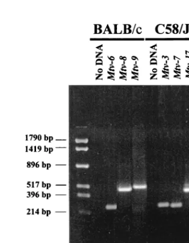
Analysis of (C58/JⴛPERA)ⴛPERA backcross mice. Be-cause we observed considerable variability of T-cell loss due to theMtv-17provirus, we analyzed a larger number of animals from the (C58/J⫻PERA)⫻PERA cross for the kinetics of T-cell deletion. The initial backcross mice were tested for the presence of specific MMTV proviruses by Southern blotting analysis since the pattern of proviral integration will distinguish highly related MMTV proviruses in different chromosomal positions (12). However, the testing of a larger number of animals was expedited by the development of PCR assays that could distinguish among highly related MMTV proviruses ex-pressed in C58/J mice (Fig. 2).
Analysis for the presence of theMtv-3andMtv-7proviruses was based on a 5⬘primer with sequence polymorphisms in the MMTV LTR hypervariable region that encodes the C-terminal portion of Sag (8) and a 3⬘conserved primer within the LTR. Testing for theMtv-17provirus used a 5⬘primer from the LTR polymorphic region combined with a 3⬘primer specific for the cellular DNA flanking this provirus. With appropriate condi-tions, fragments of 265, 252, and 434 bp were obtained with primer sets specific forMtv-3, -7, and -17, respectively (Fig. 2, lanes 7 to 9). These bands were not obtained in the absence of DNA template (lane 6) and could be used to distinguish among the three MMTV proviruses in DNAs from backcross mice previously tested by Southern blotting (data not shown). Backcross mice containing single MMTV proviruses were
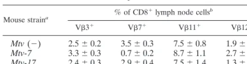
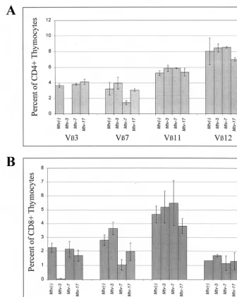
[image:3.612.53.553.84.219.2]tested over a period of 7 months for deletion of CD4⫹T cells expressing V3, V7, V11, and V12 (Fig. 3A to D). As expected, Mtv-3-only animals showed complete deletion of V3-specific T cells but not T cells expressing V7, -11, or -12, and this deletion was complete when the mice were 6 weeks of age. Animals containing onlyMtv-7had 68% deletion of V 7-specific T cells by 6 weeks after birth, whereas deletion of T cells expressing V3, -11, and -12 was unaffected. Interestingly, Mtv-17-only mice showed no deletion of the T-cell subsets when tested at 6 weeks of age; however, deletion of V11- and V12-specific T cells was apparent after 3 to 7 months. At 7 months, deletion of V11-specific cells was only 34% and de-letion of V12-specific cells was 65%. Scherer et al. reported TABLE 1. Deletion of specific T-cell classes in (C58⫻PERA)F1and (C58⫻PERA)⫻PERA N1backcross mice
Mice Provirus(es) na % of CD4
⫹lymph node cellsb
V3 V6 V8 V9 V11 V12 V14
C58 Mtv-3, -7, -17 3 0.4⫾0.6 0.2⫾0.4 10.2⫾1.2 0.1⫾0.2 10.0⫾0.3 6.7⫾2.3 7.3⫾0.8 PERA None 3 4.2⫾0.7 6.7⫾0.6 12.2⫾1.3 1.0⫾0.1 7.2⫾0.4 9.5⫾0.5 4.8⫾0.1 (C58⫻PERA)F1 Mtv-3, -7, -17 4 0.1⫾0.1 0.5⫾0.3 14.9⫾0.7 0.2⫾0.1 7.5⫾1.8 3.1⫾1.6 8.6⫾0.8
Backcross N1 None 2 3.2 6.6 11.6 0.9 6.8 7.4 4.4
Backcross N1 Mtv-3 4 0.2⫾0.2 8.6⫾1.8c 15.2⫾1.2 0.9⫾0.2 8.0⫾0.8 10.1⫾0.4 5.2⫾0.9
Backcross N1 Mtv-7 1 4.3 0.4 10.6 0.1 8.4 9.5 6.8
Backcross N1 Mtv-17 3 4.4⫾0.9 9.4⫾3.1c 15.4⫾0.6 1.0⫾0.2 6.9⫾3.9 1.7⫾1.3 5.7
Backcross N1 Mtv-3, -7 2 0.2 1.0 11.1 0.1 11.9 12.7 6.6
Backcross N1 Mtv-7, -17 2 0.3 9.6 16.6 1.2 6.8 3.5 6.2
Backcross N1 Mtv-7, -17 1 5.3 1.2 9.0 0.1 6.0 NDd 6.0
Backcross N1 Mtv-3, -7, -17 2 0.2 0.5 12.3 0.1 9.1 4.3 8.4
aNumber of mice analyzed. The age of mice tested wasⱖ5 months. bAverage⫾standard deviation.
cThe high standard deviation observed in these samples was due to the use of limiting antibody in early experiments and was not observed in later experiments. dND, not done.
[image:3.612.333.522.435.678.2]FIG. 1. Scheme for generation ofMtvsingle-positive mice. Each animal was analyzed in a series of three PCRs containing primers that were specific for the MMTV proviruses in C58/J or BALB/cJ parents. Animals negative for all pro-viruses were checked for the integrity of DNA samples by PCRs with GAPDH primers.
FIG. 2. PCR assay for specific endogenousMtvproviruses. BALB/cJ (lanes 3 to 5) or C58/J (lanes 7 to 9) DNA was used in reactions containing primers specific for the indicated MMTV proviruses. The primers used to detectMtv-3 andMtv-6were the same. Reaction mixtures were analyzed on a 2% agarose gel and stained with ethidium bromide. Lanes 2 and 6 show PCR mixtures lacking template DNA. Molecular size markers are shown in lane 1.
on November 9, 2019 by guest
http://jvi.asm.org/
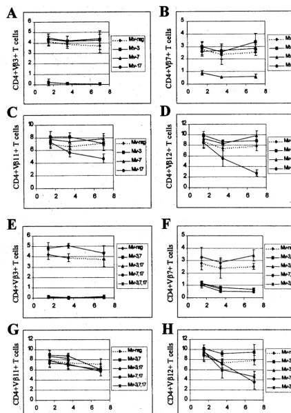
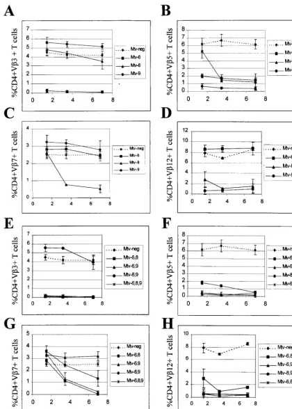
[image:3.612.55.292.613.685.2]FIG. 3. Kinetics of T-cell deletion in N1mice from the C58/J⫻PERA cross. Percentages of CD4⫹V3⫹(A and E), CD4⫹V7⫹(B and F), CD4⫹V11⫹(C and
G), and CD4⫹V12⫹(D and H) T cells were determined. All values were compared to the percentage of T cells inMtv-negative mice from the same cross. Each point represents the average of values from 3 to 12 mice, except that two animals withMtv-7alone were tested at 3.5 months. No N1animals withMtv-3, -7, and -17were
tested at 7 months. Standard deviations are represented by vertical bars spanning each point.
VOL. 73, 1999 MMTV sagmRNA EXPRESSION CORRELATES WITH T-CELL LOSS 6637
on November 9, 2019 by guest
http://jvi.asm.org/
11% deletion of CD4⫹V11⫹and 35% of CD4⫹V12⫹cells in Mtv-17-only mice, but the age of the mice tested was not reported (43). These results suggested that deletion of T cells specific for Mtv-7andMtv-3Sag occurred intrathymically. In contrast,Mtv-17Sag-specific cells were deleted in the periph-eral immune system.
To confirm and extend these observations, we also tested Sag-specific deletion of CD4⫹T cells in backcross animals that expressed multiple endogenous MMTVs (Fig. 3E to H). As anticipated, all mice that hadMtv-3orMtv-7also had deletion of V3- or V7-specific T cells, respectively (Fig. 3E and F). In addition, all animals with theMtv-17provirus and eitherMtv-3 or Mtv-7 showed slow kinetics of V12-specific deletion of CD4⫹ cells (Fig. 3H). Because deletion of T cells bearing V12 was not detectable in very young mice, these data sup-port the idea that peripheral, but not intrathymic, deletion is mediated byMtv-17Sag. In contrast to results observed with mice containing single MMTV proviruses, deletion of V 11-specific cells was not detected in mice withMtv-17in combi-nation with other endogenous MMTVs (Fig. 3G); this may be due to compensatory changes in the percentage of T-cell de-letion caused bysagexpression from other proviruses (43).
We also attempted to analyze deletion of V12⫹T cells in Mtv-17-only mice by testing whether deletion of these cells was detectable in the peripheral CD8⫹ subset. If deletion of V12⫹cells is initiated intrathymically when the majority of cells are CD4⫹ CD8⫹, then deletion of V12-specific cells should be detectable in both CD4 and CD8 single-positive subsets released from the thymus (23). Although there was a small difference between the levels of CD8⫹V12⫹T cells in Mtv-17-positive mice at 4 to 6 months of age and those in mice that lack MMTV proviruses, this was not statistically significant (P ⫽ 0.12) (Table 2); this result could be due to the low number of animals tested. As expected,Mtv-7-only mice de-leted V7⫹T cells (80% compared toMtv-negative animals) in the CD8⫹ subset (Table 2). These data and results from Scherer et al. (43) are consistent with the idea that Mtv-17 causes peripheral deletion of CD4⫹V12⫹T cells.
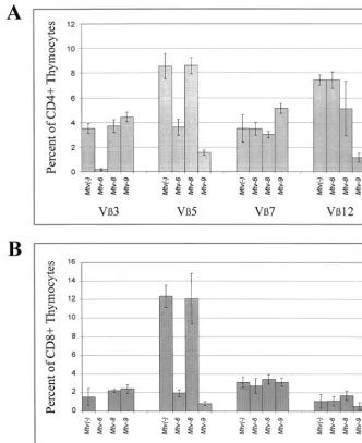
Analysis of (BALB/cJ ⴛ PERA)ⴛPERA backcross mice. Other endogenous MMTVs, such as Mtv-8, also have been reported to cause partial deletion of specific T-cell subsets (17, 43); however, the kinetics of this deletion have not been re-ported. Therefore, we analyzed N1progeny from a backcross
between the BALB/c strain (containingMtv-6, -8, and -9) and Mtv-negative PERA/Ei mice (Fig. 1). To determine the provi-ral content of these mice, we again used PCR assays that could discriminate among the three endogenous MMTVs of BALB/c mice. The primer pair used to detectMtv-6proviruses was the same as that used forMtv-3 in the C58/J cross, whereas the virtually identical Mtv-8 and Mtv-9 proviruses were distin-guished with a 5⬘ primer within thesaghypervariable region
and a 3⬘ primer derived from the cellular flanking region. Using optimal conditions, PCR products of 265, 488, and 537 bp were obtained for Mtv-6, -8, and -9, respectively, with BALB/c DNA (Fig. 2, lanes 3 to 5).
Backcross mice containing single MMTVs were examined for deletion of CD4⫹T cells expressing V3, -5, -7, and -12 (Fig. 4A to D). As expected from earlier experiments (48), mice containing onlyMtv-6 showed deletion of V3-specific and V5-specific T cells, respectively, at the earliest time point tested. Mice containing onlyMtv-9deleted 90% of V 5-spe-cific T cells by 6 weeks of age (Fig. 4B); these T-cell levels were lower than those achieved by theMtv-6orMtv-8proviruses (68 and 79% deletions, respectively, by 7 months). Mtv-9 also caused very efficient deletion of V12⫹ cells in 6-week-old mice (Fig. 4D). Interestingly,Mtv-8showed different kinetics of deletion for different T-cell subsets. Deletion of V5- and V7-specific T cells was not detectable in 6-week-old mice, but deletion of 79% of V5⫹ cells and 70% of V7⫹ cells was observed by 3.5 months. However, an average of 65% of V12⫹cells were deleted in 6-week-old mice withMtv-8only, but this was variable, and these levels declined slightly in the subsequent months (Fig. 4D). Therefore, the data indicate that Mtv-8causes relatively rapid deletion of certain T-cell subsets (V12⫹cells), yet the same provirus causes much slower de-letion of V5⫹ and V7⫹ cells in the peripheral lymphoid system.
BALB/c backcross mice with multiple MMTV proviruses also were analyzed (Fig. 4E to H). Again, all mice with the Mtv-6provirus deleted CD4⫹V3⫹T cells, whereas mice with any of the three endogenous MMTVs of BALB/c mice deleted V5⫹T cells. Mice containing bothMtv-6andMtv-8appeared to delete CD4⫹V5⫹ cells with slightly slower kinetics than mice having other proviral combinations (Fig. 4F). As ex-pected, mice withMtv-8in combination with other endogenous MMTVs had slow deletion of V7⫹ T cells; however, the presence ofMtv-9, in the absence ofMtv-6, appeared to slow deletion even further (Fig. 4G). This result may be due to compensatory changes in the percentage of V7⫹ T cells caused by deletion of other T-cell subsets by theMtv-9provirus (43). Rapid kinetics of V12⫹ T-cell deletion was observed again withMtv-8⫹mice, but the deletion appeared to be more complete and less variable in the presence of theMtv-9 provi-rus, which also deletes V12⫹T cells (Fig. 4H).
Intrathymic deletion of Sag-reactive thymocytes in mice containing single MMTV proviruses.To test directly whether individual MMTV proviruses caused intrathymic deletion, we analyzed thymocytes fromMtvsingle-positive mice. In Mtv-3 single-positive mice from the C58/J cross, we observed com-plete deletion of V3⫹T cells in both the CD4 and the CD8 single-positive populations (Fig. 5). However, in Mtv-7-only mice, there was 55% deletion of CD4⫹V7⫹thymocytes and 63% deletion of CD8⫹V7⫹thymocytes. This is in agreement with the observation that there is partial deletion of CD4⫹T cells in the periphery ofMtv-7 single-positive mice, and this deletion changes little with time (Fig. 3B).Mtv-17-only mice showed no significant deletion of thymocytes in either the CD4⫹or CD8⫹subset. Thus, the Sag proteins encoded by the Mtv-3andMtv-7proviruses appear to cause intrathymic dele-tion of specific T-cell subsets as reported previously (16, 48), whereasMtv-17Sag does not (Fig. 5).
[image:5.612.53.294.92.155.2]We also analyzed Sag-specific deletion of thymocytes in mice derived from the BALB/c backcross (Fig. 6).Mtv-6-only mice showed 95% or greater deletion of V3⫹thymocytes in both the CD4⫹and CD8⫹populations; these mice also deleted 58% of CD4⫹V5⫹ and 85% of CD8⫹V5⫹ thymocytes. Mtv-9 -only mice deleted 82% of CD4⫹V5⫹and 94% of CD8⫹V5⫹ TABLE 2. Deletion of CD8⫹T cells in mouse strains with single
MMTV proviruses
Mouse straina % of CD8
⫹lymph node cellsb
V3⫹ V7⫹ V11⫹ V12⫹
Mtv(⫺) 2.5⫾0.2 3.5⫾0.3 7.5⫾0.8 1.9⫾0.4 Mtv-7 3.3⫾0.3 0.7⫾0.2 8.7⫾1.1 2.7⫾0.9 Mtv-17 2.4⫾0.3 2.9⫾0.4 7.5⫾1.4 1.3⫾0.4
aFour animals were tested from each strain.
bAverage percentage⫾standard deviation in animals tested at 4 to 6 months
of age. Animals could not be tested at older ages because many of them devel-oped leukemia.
on November 9, 2019 by guest
http://jvi.asm.org/
FIG. 4. Kinetics of T-cell deletion in N1mice from the BALB/cJ⫻PERA cross. Percentages of CD4⫹V3⫹(A and E), CD4⫹V5⫹(B and F), CD4⫹V7⫹(C
and G), and CD4⫹V12⫹(D and H) T cells were determined. Each point represents the average of values from 3 to 11 mice, except that two animals withMtv-8alone were tested at 3.5 months; two animals with bothMtv-6andMtv-8were tested at 3.5 and 7 months; two animals withMtv-6, -8, and -9were tested at 7 months; and one animal withMtv-8and -9was tested at 3.5 months. All values were compared to the percentage of T cells inMtv-negative mice from the same cross. Standard deviations are given by vertical bars spanning each point.
VOL. 73, 1999 MMTV sagmRNA EXPRESSION CORRELATES WITH T-CELL LOSS 6639
on November 9, 2019 by guest
http://jvi.asm.org/
thymocytes as well as 84 and 51% of CD4⫹V12⫹ and CD8⫹V12⫹thymocytes, respectively. InMtv-8-only mice, we observed approximately 31% deletion of V12⫹CD4⫹T cells; this partial deletion was not statistically significant (P⫽0.13), largely because it was variable among different animals. The partial deletion of V12⫹cells was not observed in the CD8⫹ thymocytes. Therefore, as concluded from kinetic analysis of peripheral lymphocyte populations, it appears thatMtv-6and Mtv-9 cause intrathymic deletion of T-cell subsets, whereas Mtv-8causes variable and partial deletion of CD4⫹V12⫹cells intrathymically.
Although we also analyzed deletion of specific T-cell subsets in CD4⫹CD8⫹thymocytes fromMtvsingle-positive mice, the percentages were too low for us to reliably assess their signif-icance (data not shown).
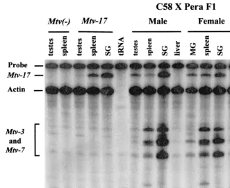
Expression of Mtv-17.Sags from two endogenous MMTV proviruses,Mtv-8andMtv-17, appear to cause peripheral de-letion of their cognate T cells. At least one report has
[image:7.612.134.471.72.495.2]sug-gested that theMtv-17provirus is expressed poorly in lymphoid tissues, particularly thymus (43). In addition, sequencing of the Mtv-173⬘LTR indicated that there is a mutation in the binding site for the transcription factor NF-1, and this mutation may greatly reduce transcriptional activity of the provirus (34). To examine the tissue distribution ofMtv-17expression, we used RNase protection assays in conjunction with a riboprobe span-ning the sag hypervariable region and the promoter of the Mtv-17provirus. As expected, hybridization of the Mtv-17 ri-boprobe to RNA extracted from the testes or spleen of an Mtv-negative backcross animal showed no specific protection of the probe from RNase digestion (Fig. 7, lanes 1 and 2). However, RNA obtained from the testes, spleen, and salivary glands of a male backcross mouse harboring only theMtv-17 provirus protected 340 nucleotides from digestion, consistent withMtv-17expression in all of these tissues (lanes 3 to 5). Each of the samples showed similar expression of the control FIG. 5. Sag-specific T-cell deletion in CD4⫹and CD8⫹thymocytes from mice containingMtv-3,Mtv-7, andMtv-17only. Percentages of CD4⫹(A) and CD8⫹(B) thymocytes in mice containing single MMTV proviruses were compared to those inMtv-negative mice derived from the same cross. Most animals tested (three to six mice from each strain) were 4 weeks old.
on November 9, 2019 by guest
http://jvi.asm.org/
actin gene. Therefore, theMtv-17promoter does not appear to be generally defective for transcription.
To determine whetherMtv-17RNA expression was affected significantly by the presence of other proviruses, we also ex-amined RNA extracted from male and female tissues of (C58/ J ⫻ PERA)F1 mice (Fig. 7). Again, Mtv-17 expression was
highest in the salivary gland of a male animal (lane 9), and this expression was comparable to that observed for the Mtv-3 and/orMtv-7proviruses (bands migrating faster than actin). A virgin female animal showed significant expression ofMtv-17in salivary gland, spleen, and mammary gland (lanes 11 to 13), although the expression of the Mtv-3 and -7proviruses was diminished considerably in virgin mammary gland (lane 11). Expression of Mtv-17RNA was enhanced in lactating com-pared to virgin mammary gland (data not shown), suggesting that proviral transcription is hormone inducible. Thus,Mtv-17 expression was easily detectable and similar to other endoge-nous MMTV proviruses in certain tissues, notably salivary gland.
[image:8.612.136.468.75.481.2]Four promoters have been described for MMTVsaggene expression (6, 18, 25, 47, 51), and at least two of these pro-moters are used by the Mtv-17 provirus for transcription of splicedsagmRNAs (56). Because RNase protection assays are not sufficiently sensitive to detect saggene expression and to discriminate among differentsagpromoters, we used RT-PCR assays to detect splicedMtv-17 sagmRNA in the thymus (Fig. 8). RT-PCR was performed with RNA from the salivary glands, spleens, and thymi ofMtv-negative,Mtv-3-only, or Mtv-17-only mice. To increase the sensitivity for detection ofsag mRNAs, PCR products then were subjected to Southern blot-ting followed by hybridization with an MMTV LTR probe. Strikingly, the splicedsagmRNAs initiated from the LTR were much more abundant than those initiated from the intragenic envelope promoter originally described by Elliott et al. (18); transcripts from the LTR promoters could be detected after 20 PCR cycles (Fig. 8A), whereas the envelope promoter tran-scripts could not (data not shown). Expression of both Mtv-3 and -7 sagRNAs was detected from the LTR promoters as well FIG. 6. Sag-specific T-cell deletion in CD4⫹and CD8⫹thymocytes from mice containingMtv-6,Mtv-8, andMtv-9only. Percentages of CD4⫹(A) and CD8⫹(B) thymocytes in mice containing single MMTV proviruses were compared to those inMtv-negative mice derived from the same cross. Animals tested (three mice from each strain) were 4 to 8 weeks old.
VOL. 73, 1999 MMTV sagmRNA EXPRESSION CORRELATES WITH T-CELL LOSS 6641
on November 9, 2019 by guest
http://jvi.asm.org/
as the envelope promoter (Fig. 8A and B; data not shown). Levels ofMtv-3expression appeared to be highest in the sali-vary gland, intermediate in spleen, and lowest in the thymus for both sag mRNAs. Mtv-3 had higher levels of LTR-directed transcripts in the thymus than did theMtv-7provirus. As pre-viously reported,Mtv-17 sagmRNAs were detected from both LTR and envelope promoters (56). However, by comparison to Mtv-3,Mtv-17appeared to have lower levels ofsagmRNA in salivary gland and spleen, and no expression was observed in the thymus (Fig. 8A and B, lanes 7 to 9). Additional PCR assays indicated that the Mtv-3 sag-specific RNAs from the envelope promoter were at least 100- to 500-fold more abun-dant in thymus than were transcripts from theMtv-17provirus (data not shown). Levels ofsagtranscripts from LTR promot-ers were approximately 10- to 50-fold higher in thymi from Mtv-7-only mice than in thymi fromMtv-17-only mice (data not shown).
DISCUSSION
We have investigated the kinetics of T-cell deletion resulting from the expression of Sags from six different endogenous MMTV proviruses,Mtv-3, -6, -7, -8, -9, and -17, in mice. Pre-vious data have described the T-cell subsets deleted due to Sag expression from each of these proviruses (3, 43, 44). However, the kinetics of the deletion induced has not been addressed for many MMTV proviruses.
Expression from four of these proviruses,Mtv-3, -6, -7, and -9, gave 50% or more deletion of the reactive T-cell subsets tested, and this deletion was apparent intrathymically and in peripheral T cells of animals that were 6 weeks old. This is
consistent with previous data that show intrathymic deletion of cognate T cells for theMtv-6andMtv-7proviruses (40, 48). In contrast, deletion of reactive T cells by the Sag proteins en-coded by theMtv-8and Mtv-17proviruses largely was unde-tectable in thymocytes or in peripheral T cells by 6 weeks but could be detected in peripheral T cells over a period of 7 months. Our data confirmed previous experiments indicating that Sags from Mtv-3, -6, -7, and -9 caused T-cell deletion during negative selection in the thymus of MHC class III-E⫹ mice (19, 21, 22, 27, 44, 52).
Deletion of T cells reactive withMtv-17Sag.Expression of Sags fromMtv-17resulted in slow kinetics of T-cell deletion that was detectable in the peripheral lymphoid system. Dele-tion of thymocytes reactive withMtv-17Sag was not detected. In agreement with this and data published by Scherer et al. (43), deletion of V12⫹T cells due toMtv-17Sag was detected only in peripheral CD4⫹T cells, not in the CD8⫹subset. Thus, presentation of Mtv-17Sag by MHC class II on antigen-pre-senting cells in the periphery will stimulate and delete mature CD4 single-positive cells.
[image:9.612.135.466.73.344.2]What are the factors that influence the kinetics of Sag-mediated T-cell deletion? One of the key factors influencing early deletion of T-cell subsets is the ability of MMTV provi-ruses to be transcribed in the thymus. RT-PCR analysis showed thatMtv-17-only mice had no detectable expression in the thymus as previously reported (55), whereas Mtv-3 and Mtv-7 both produced detectable expression of spliced sag mRNA from the LTR and envelope promoters (Fig. 8 and data not shown). The results of Scherer et al. suggested that the Mtv-17 provirus was expressed at low levels in the thymus; however, their assay did not distinguish between total (spliced FIG. 7. RNase protection assays for the expression of endogenous MMTVs. Total RNA (40g) was hybridized to a riboprobe containing theSau3A fragment of theMtv-17LTR (24), including the polymorphic region of thesaggene. An actin probe (Ambion, Austin, Tex.) was used as a control for the quality of the RNA. Hybridizations contained RNA from the following sources: tissues from an MMTV-negative backcross mouse (lanes 1 and 2), tissues from a backcross animal containing Mtv-17only (lanes 3 to 5), yeast RNA (lane 6), tissues from a male (C58/J⫻PERA)F1animal (lanes 7 to 10), and tissues from a female (C58/J⫻PERA)F1animal
(lanes 11 to 14). Abbreviations: SG, salivary gland; MG, mammary gland. The faster-migrating bands in these lanes are due to partial protection of the riboprobe by Mtv-3- andMtv-7-specific RNA transcripts. TheMtv-3andMtv-7transcripts cannot be distinguished in this assay.
on November 9, 2019 by guest
http://jvi.asm.org/
and unspliced) MMTV RNA andsag-specific transcripts (43). Greater sensitivity for detection ofsagmRNAs in our assays was achieved by Southern blotting of RT-PCR products, but the blotting did not allow detection ofMtv-17 sagtranscripts in the thymus. Thus, it appears likely that negative selection of Mtv-17 Sag-reactive T cells cannot take place in the thymus because the appropriate transcripts or a threshold level of these transcripts is not synthesized (40). In support of this, Morishima et al. showed that low expression of total Mtv-1 RNA in the thymus led to peripheral deletion of V3⫹T cells, whereas eightfold-higher expression of the relatedMtv-6 pro-virus in the thymus gave intrathymic deletion of V3⫹T cells (40). Waanders et al. also suggested that the levels of sag mRNA in the thymus correlated with intrathymic deletion of Sag-reactive T cells (48).
Deletion of T cells reactive withMtv-8Sag.CD4⫹V12⫹T cells inMtv-8-only mice showed partial and variable deletion intrathymically (Fig. 6A). Deletion of CD8⫹V12⫹T cells was not detected in thymocytes (Fig. 6B). Preliminary results from a 7-month-old mouse with the Mtv-8 provirus indicate that deletion of V12⫹ cells, but not of other T-cell subsets, is detectable in peripheral CD8 single-positive cells (data not shown) as well as in the CD4⫹subset (Fig. 4D). These data are consistent with a previous report thatMtv-8expression deleted V12⫹T cells in the CD4⫹and CD8⫹subsets (43). Although the ages of mice tested were not reported, theMtv-8-only mice deleted V5⫹, V7⫹, and V11⫹ T cells only in the CD4⫹ subset (43).
The results ofMtv-8expression on T-cell deletion are more complex than those observed for Mtv-17. We have not been able to test this directly because of breeding problems with the Mtv-8-only mice. However, we expect that Mtv-8 will be ex-pressed in the thymus because this provirus caused deletion of
peripheral CD4⫹V12⫹T cells at the earliest time point tested and variable deletion of CD4⫹V12⫹ thymocytes. However, other T-cell subsets (V5⫹and V7⫹cells) were not deleted in the thymus. Such cells were deleted with slow kinetics in the periphery, consistent with extrathymic deletion of reactive T cells. Therefore, it is likely thatMtv-8is expressed at very low levels in the thymus and that this level is close to the threshold required for elimination of cells expressing self-reactive anti-gens. TheMtv-8provirus is located in the Vlocus on mouse chromosome 6 (57) and appears to be expressed in B cells following certain light chain rearrangements that allow close proximity of the Mtv-8provirus and the Venhancers (58). Thus, expression ofMtv-8in certain mature B cells likely leads to the peripheral deletion observed for the V5⫹and V7⫹ T-cell subsets. In contrast, reaction of a limited amount of Mtv-8Sag on the surface of thymic antigen-presenting cells may allow negative selection of thymocytes if there is high affinity of this Sag for a given TCR (e.g., V12). Similarly, experiments by Chervonsky et al. indicated that expression of sag-specific mRNA in the thymus was sufficient to get periph-eral, but not intrathymic, deletion of V14⫹ T cells in mice expressing low amounts of the C3H MMTV transgene (9). Therefore, complete intrathymic deletion of self-reactive T cells will be determined both by the level of Sag expression and by the affinity for TCR (19). It will be interesting to determine the level ofMtv-8 sag-specific expression in the thymus since it may help to define the minimum level of expression needed for intrathymic deletion. Based on results withMtv-3 sagmRNA with a combination of RT-PCR and Southern blotting (Fig. 8) and complete intrathymic deletion of cells reactive withMtv-3 Sag, negative selection of thymocytes appears to be an exquis-itely sensitive process.
[image:10.612.123.487.72.295.2]BothMtv-8andMtv-17cause peripheral deletion of T cells. FIG. 8. Detection ofsag-specific spliced transcripts from the LTR and envelope promoters. RT-PCRs were performed with RNA extracted from the salivary gland (SG) (lanes 1, 4, and 7), spleen (lanes 2, 5, and 8), and thymus (lanes 3, 6, 9, and 10). RNA samples were derived fromMtv-negative animals (lanes 1 to 3),Mtv-3-only animals (lanes 4 to 6),Mtv-17-only animals (lanes 7 to 9), orMtv-7-only animals (lane 10). RNAs were derived from pools of organs from 6-week-old mice (three to five animals). Each cDNA was used in three separate PCRs containing primers for the LTR promoters (A), the envelope promoter (B), and GAPDH (C). The PCRs in panel A were performed for 20 cycles, and the PCRs in panel B were performed for 35 cycles. RT-PCR mixtures (one-third of the reaction mixture) were analyzed on 2% NuSieve agarose gels. After electrophoresis, DNA was transferred to nylon membranes, hybridized to an MMTV LTR probe (A and B), and subjected overnight to autoradiography. Longer exposures of the autoradiogram in panel A showMtv-7expression in the thymus.Mtv-17expression in the thymus was not detected after 35 PCR cycles with primers for the LTR promoters or long exposures of the autoradiograms. GAPDH reaction mixtures were stained with ethidium bromide after electrophoresis as a control for the integrity of cDNAs.
VOL. 73, 1999 MMTV sagmRNA EXPRESSION CORRELATES WITH T-CELL LOSS 6643
on November 9, 2019 by guest
http://jvi.asm.org/
What is the antigen-presenting cell used for peripheral elimi-nation of Sag-reactive T cells? Although we have presented no definitive data here, previous experiments indicate that B cells expressMtv-8(30, 58). Moreover, our RT-PCR experiments suggest that expression ofMtv-17 sagmRNA is detectable in spleen, a tissue rich in mature B cells (42), but not thymus. Previous experiments also have detectedMtv-17transcripts in gut-associated lymphocytes (55). Since MMTV expression in B cells is a requirement for exogenous MMTV transmission (7) and B cells are expanded during exposure of adult mice to Mtv-7-expressing spleen cells (49), B cells are likely candidates for presentation of Mtv-8and -17Sag in the peripheral im-mune system. However, CD8⫹ T cells also may cause Sag-specific T-cell deletion in the periphery (49). As expected, B cells are not required for deletion of cognate T cells by endog-enousMtv-7andMtv-9, two proviruses which cause intrathymic T-cell deletion (7).
Selection for endogenous MMTVs with Sag function. Previ-ous experiments have shown that expression of milk-borne MMTV Sag in transgenic animals leads to the intrathymic deletion of cognate T cells. Such transgenic animals are resis-tant to infection by milk-borne MMTVs that express the same Sag proteins (23). Thus, it has been proposed that endogenous MMTVs that express Sag have been retained by most mouse strains because these MMTVs protect against milk-borne virus infections (23, 28, 36). The existence of endogenous MMTVs that largely cause slow kinetics of T-cell deletion appears to contradict this proposal. Our data would suggest that mice that haveMtv-8and/orMtv-17would be susceptible to exogenous MMTVs with similar Sag proteins since T cells reactive with Mtv-8 and -17 Sags would be present during the neonatal period when milk-borne infection occurs. Moreover, these mice would be susceptible to tumorigenesis by certain recom-binants between different endogenous MMTVs (e.g., Mtv-17 recombinants with Mtv-2are integrated in GR tumors) (24). Alternatively, endogenous viruses such as Mtv-8and -17may provide limited protection against milk-borne MMTV infec-tion by anergizing Sag-reactive T cells. However, these provi-ruses may provide better resistance to bacterial or other viral infections that may occur later in life and that require a re-sponse from particular T-cell subsets (29, 37).
ACKNOWLEDGMENTS
We thank Susan Ross for useful discussions and for comments on the manuscript.
This work was supported by grants CA34780 and CA52646 from the National Institutes of Health. F.M. is a recipient of an NRSA award from the National Institutes of Health.
REFERENCES
1.Abe, R., M. Foo-Phillips, L. G. Granger, and O. Kanagawa.1992. Charac-terization of theMlsfsystem. I. A novel “polymorphism” of endogenous
superantigens. J. Immunol.149:3429–3439.
2.Abe, R., O. Kanagawa, M. A. Sheard, B. Malissen, and M. Foo-Phillips. 1991. Characterization of a new minor lymphocyte stimulatory system. I. Cluster of self antigens recognized by “I-E-reactive” Vs, V5, V11, and V12 T cell receptors for antigen. J. Immunol.147:739–749.
3.Acha-Orbea, H., W. Held, G. A. Waanders, A. N. Shakhov, L. Scarpellino, R. K. Lees, and H. R. MacDonald.1993. Exogenous and endogenous mouse mammary tumor virus superantigens. Immunol. Rev.131:5–25.
4.Acha-Orbea, H., and H. R. MacDonald.1995. Superantigens of mouse mam-mary tumor virus. Annu. Rev. Immunol.13:459–486.
5.Acha-Orbea, H., A. N. Shakhov, L. Scarpellino, E. Kolb, V. Muller, A. Vessaz-Shaw, R. Fuchs, K. Blochlinger, P. Rollini, J. Billotte, et al.1991. Clonal deletion of V14-bearing T cells in mice transgenic for mammary tumour virus. Nature350:207–211.
6.Arroyo, J., E. Winchester, B. S. McLellan, and B. T. Huber.1997. Shared promoter elements between a viral superantigen and the major histocom-patibility complex class II-associated invariant chain. J. Virol.71:1237–1245. 7.Beutner, U., E. Kraus, D. Kitamura, K. Rajewsky, and B. T. Huber.1994. B
cells are essential for murine mammary tumor virus transmission, but not for presentation of endogenous superantigens. J. Exp. Med.179:1457–1466. 8.Brandt-Carlson, C., J. S. Butel, and D. Wheeler.1993. Phylogenetic and
structural analyses of MMTV LTR ORF sequences of exogenous and en-dogenous origins. Virology193:171–185.
9.Chervonsky, A. V., T. V. Golovkina, S. R. Ross, and C. A. Janeway, Jr.1995. Differences in the avidity of TCR interactions with a superantigenic ligand affect negative selection but do not allow positive selection. J. Immunol. 155:5115–5123.
10. Choi, Y., J. W. Kappler, and P. Marrack.1991. A superantigen encoded in the open reading frame of the 3⬘long terminal repeat of mouse mammary tumour virus. Nature350:203–207.
11. Choi, Y. W., D. Henrard, I. Lee, and S. R. Ross.1987. The mouse mammary tumor virus long terminal repeat directs expression in epithelial and lym-phoid cells of different tissues in transgenic mice. J. Virol.61:3013–3019. 12. Cohen, J. C., and H. E. Varmus.1979. Endogenous mammary tumour virus
DNA varies among wild mice and segregates during inbreeding. Nature 278:418–423.
13. Dudley, J., and R. Risser.1984. Amplification and novel locations of endog-enous mouse mammary tumor virus genomes in mouse T-cell lymphomas. J. Virol.49:92–101.
14. Dudley, J. P.1988. Mouse mammary tumor proviruses from a T-cell lym-phoma are associated with the retroposon L1Md. J. Virol.62:472–478. 15. Dudley, J. P., A. Arfsten, C. L. Hsu, C. Kozak, and R. Risser.1986.
Molec-ular cloning and characterization of mouse mammary tumor proviruses from a T-cell lymphoma. J. Virol.57:385–388.
16. Dyson, P. J., J. Elliott, A. N. Antoniou, and K. T. Corley.1998. Efficient presentation of endogenous superantigen by H-2Aq. Eur. J. Immunol.28:
1034–1039.
17. Dyson, P. J., A. M. Knight, S. Fairchild, E. Simpson, and K. Tomonari.1991. Genes encoding ligands for deletion of V11 T cells cosegregate with mam-mary tumour virus genomes. Nature349:531–532.
18. Elliott, J. F., B. Pohajdak, D. J. Talbot, J. Shaw, and V. Paetkau.1988. Phorbol diester-inducible, cyclosporine-suppressible transcription from a novel promoter within the mouse mammary tumor virusenvgene. J. Virol. 62:1373–1380.
19. Fink, P. J., C. A. Fang, and G. L. Turk.1994. The induction of peripheral tolerance by the chronic activation and deletion of CD4⫹V5⫹cells. J. Im-munol.152:4270–4281.
20. Foo-Phillips, M., C. A. Kozak, M. A. Principato, and R. Abe.1992. Charac-terization of theMlsfsystem. II. Identification of mouse mammary tumor
virus proviruses involved in the clonal deletion of self-Mlsf-reactive T cells.
J. Immunol.149:3440–3447.
21.Frankel, W. N., C. Rudy, J. M. Coffin, and B. T. Huber.1991. Linkage ofMls genes to endogenous mammary tumour viruses of inbred mice. Nature349: 526–528.
22.Gollob, K. J., and E. Palmer.1992. Divergent viral superantigens delete V5⫹T lymphocytes. Proc. Natl. Acad. Sci. USA89:5138–5141. 23. Golovkina, T. V., A. Chervonsky, J. P. Dudley, and S. R. Ross.1992.
Trans-genic mouse mammary tumor virus superantigen expression prevents viral infection. Cell69:637–645.
24. Golovkina, T. V., O. Prakash, and S. R. Ross.1996. Endogenous mouse mammary tumor virusMtv-17is involved inMtv-2-induced tumorigenesis in GR mice. Virology218:14–22.
25. Gunzburg, W. H., F. Heinemann, S. Wintersperger, T. Miethke, H. Wagner, V. Erfle, and B. Salmons.1993. Endogenous superantigen expression con-trolled by a novel promoter in the MMTV long terminal repeat. Nature 364:154–158.
26. Held, W., A. N. Shakhov, S. Izui, G. A. Waanders, L. Scarpellino, H. R. MacDonald, and H. Acha-Orbea.1993. Superantigen-reactive CD4⫹T cells are required to stimulate B cells after infection with mouse mammary tumor virus. J. Exp. Med.177:359–366.
27. Held, W., A. N. Shakhov, G. Waanders, L. Scarpellino, R. Luethy, J. P. Kraehenbuhl, H. R. MacDonald, and H. Acha-Orbea.1992. An exogenous mouse mammary tumor virus with properties ofMls-1a(Mtv-7). J. Exp. Med.
175:1623–1633.
28.Held, W., G. A. Waanders, A. N. Shakhov, L. Scarpellino, H. Acha-Orbea, and H. R. MacDonald.1993. Superantigen-induced immune stimulation amplifies mouse mammary tumor virus infection and allows virus transmis-sion. Cell74:529–540.
29. Huber, B. T., P. N. Hsu, and N. Sutkowski.1996. Virus-encoded superanti-gens. Microbiol. Rev.60:473–482.
30. Jarvis, C. D., R. N. Germain, G. L. Hager, M. Damschroder, and L. A. Matis. 1994. Tissue-specific expression of messenger RNAs encoding endogenous viral superantigens. J. Immunol.152:1032–1038.
31. Kang, J., E. Ido, J. Pawling, U. Beutner, B. T. Huber, and N. Hozumi.1994. Expression ofMtv-7 sag genein vivousing a retroviral vector results in selective inactivation of superantigen reactive T cells. J. Immunol.152:1039– 1046.
32. Karapetian, O., A. N. Shakhov, J. P. Kraehenbuhl, and H. Acha-Orbea. 1994. Retroviral infection of neonatal Peyer’s patch lymphocytes: the mouse mammary tumor virus model. J. Exp. Med.180:1511–1516.
on November 9, 2019 by guest
http://jvi.asm.org/
33.Korman, A. J., P. Bourgarel, T. Meo, and G. E. Rieckhof.1992. The mouse mammary tumour virus long terminal repeat encodes a type II transmem-brane glycoprotein. EMBO J.11:1901–1905.
34.Kuo, W. L., L. R. Vilander, M. Huang, and D. O. Peterson.1988. A tran-scriptionally defective long terminal repeat within an endogenous copy of mouse mammary tumor virus proviral DNA. J. Virol.62:2394–2402. 35.Lund, F. E., T. D. Randall, D. L. Woodland, and R. B. Corley.1993. MHC
class II limits the functional expression of endogenous superantigens in B cells. J. Immunol.150:78–86.
36.Marrack, P., E. Kushnir, and J. Kappler. 1991. A maternally inherited superantigen encoded by a mammary tumour virus. Nature349:524–526. 37. Marrack, P., G. M. Winslow, Y. Choi, M. Scherer, A. Pullen, J. White, and
J. W. Kappler.1993. The bacterial and mouse mammary tumor virus supe-rantigens; two different families of proteins with the same functions. Immu-nol. Rev.131:79–92.
38. McMahon, C. W., B. Traxler, M. E. Grigg, and A. M. Pullen.1998. Trans-poson-mediated random insertions and site-directed mutagenesis prevent the trafficking of a mouse mammary tumor virus superantigen. Virology 243:354–365.
39. Moore, N. C., G. Anderson, D. E. McLoughlin, J. J. Owen, and E. J. Jen-kinson.1994. Differential expression ofMtvloci in MHC class II-positive thymic stromal cells. J. Immunol.152:4826–4831.
40. Morishima, C., C. Norby-Slycord, K. R. McConnell, R. J. Finch, A. J. Nelson, A. G. Farr, and A. M. Pullen.1994. Expression of two structurally identical viral superantigens results in thymic elimination at distinct developmental stages. J. Immunol.153:5091–5103.
41. Palmiter, R. D.1974. Magnesium precipitation of ribonucleoprotein com-plexes. Expedient techniques for the isolation of undergraded polysomes and messenger ribonucleic acid. Biochemistry13:3606–3615.
42. Picker, L. J., and M. H. Siegelman.1993. Lymphoid tissues and organs, p. 145–197.InW. E. Paul (ed.), Fundamental immunology. Raven Press, New York, N.Y.
43. Scherer, M. T., L. Ignatowicz, A. Pullen, J. Kappler, and P. Marrack.1995. The use of mammary tumor virus (Mtv)-negative and single-Mtvmice to evaluate the effects of endogenous viral superantigens on the T cell reper-toire. J. Exp. Med.182:1493–1504.
44. Simpson, E., P. J. Dyson, A. M. Knight, P. J. Robinson, J. I. Elliott, and D. M. Altmann.1993. T-cell receptor repertoire selection by mouse mam-mary tumor viruses and MHC molecules. Immunol. Rev.131:93–115. 45. Subramanyam, M., N. Mohan, D. Mottershead, U. Beutner, B. McLellan, E.
Kraus, and B. T. Huber.1993.Mls-1superantigen: molecular characteriza-tion and funccharacteriza-tional analysis. Immunol. Rev.131:117–130.
46. Vacchio, M. S., J. J. Ryan, and R. J. Hodes.1990. Characterization of the ligand(s) responsible for negative selection of V11- and V12-expressing T
cells: effects of a newMlsdeterminant. J. Exp. Med.172:807–813. 47. van Ooyen, A. J., R. J. Michalides, and R. Nusse.1983. Structural analysis of
a 1.7-kilobase mouse mammary tumor virus-specific RNA. J. Virol.46:362– 370.
48. Waanders, G. A., R. K. Lees, W. Held, and H. R. MacDonald.1995. Quan-titation of endogenous mouse mammary tumor virus superantigen expres-sion by lymphocyte subsets. Eur. J. Immunol.25:2632–2637.
49. Waanders, G. A., A. N. Shakhov, W. Held, O. Karapetian, H. Acha-Orbea, and H. R. MacDonald.1993. Peripheral T cell activation and deletion in-duced by transfer of lymphocyte subsets expressing endogenous or exoge-nous mouse mammary tumor virus. J. Exp. Med.177:1359–1366. 50. Weber, G. F., S. Abromson-Leeman, and H. Cantor.1995. A signaling
path-way coupled to T cell receptor ligation by MMTV superantigen leading to transient activation and programmed cell death. Immunity2:363–372. 51. Wheeler, D. A., J. S. Butel, D. Medina, R. D. Cardiff, and G. L. Hager.1983.
Transcription of mouse mammary tumor virus: identification of a candidate mRNA for the long terminal repeat gene product. J. Virol.46:42–49. 52. Woodland, D. L., M. P. Happ, K. J. Gollob, and E. Palmer. 1991. An
endogenous retrovirus mediating deletion of␣T cells? Nature349:529– 530.
53. Wrona, T., and J. P. Dudley.1996. Major histocompatibility complex class II I-E-independent transmission of C3H mouse mammary tumor virus. J. Virol. 70:1246–1249.
54. Wrona, T. J., M. Lozano, A. A. Binhazim, and J. P. Dudley.1998. Mutational and functional analysis of the C-terminal region of the C3H mouse mam-mary tumor virus superantigen. J. Virol.72:4746–4755.
55. Xu, L., T. J. Wrona, and J. P. Dudley.1996. Exogenous mouse mammary tumor virus (MMTV) infection induces endogenous MMTVsagexpression. Virology215:113–123.
56. Xu, L., T. J. Wrona, and J. P. Dudley.1997. Strain-specific expression of spliced MMTV RNAs containing the superantigen gene. Virology236:54– 65.
57. Yang, J. N., R. T. Boyd, P. D. Gottlieb, and J. P. Dudley.1987. The endog-enous retrovirusMtv-8on mouse chromosome 6 maps near several kappa light chain markers. Immunogenetics25:222–227.
58. Yang, J. N., and J. Dudley.1992. EndogenousMtv-8or a closely linked sequence stimulates rearrangement of the downstream V9 gene. J. Immu-nol.149:1242–1251.
58a.Yang, J.-N., and J. Dudley.Unpublished data.
59. Yazdanbakhsh, K., C. G. Park, G. M. Winslow, and Y. Choi.1993. Direct evidence for the role of COOH terminus of mouse mammary tumor virus superantigen in determining T cell receptor V specificity. J. Exp. Med. 178:737–741.
VOL. 73, 1999 MMTV sagmRNA EXPRESSION CORRELATES WITH T-CELL LOSS 6645