VALIDITY OF COLD IRRIGATION TEST (CIT)
USED IN TERTIARY CARE FOR DIAGNOSING
VALIDITY OF COLD IRRIGATION TEST (CIT)
USED IN TERTIARY CARE FOR DIAGNOSING
VESTIBULAR DYSFUNCTION
A dissertation submitted in partial fulfillment of the rules and
regulation for the
M.S. (Branch IV) Otorhinolaryngology
CERTIFICATE
This is to certify that the dissertation entitled “Validity of cold irrigation test
(CIT) used in tertiary care for diagnosing vestibular dysfunction” is a bonafide original work of Dr. Ramesh Menon U submitted in partial fulfillment of the rules and regulation for the M.S. (Branch IV) Otorhinolaryngologyexamination of the Tamil Nadu Dr. M.G.R Medical University to be held in March 2008.
Dr. Rupa Vedantam MS., DLO Professor & Head
Dept. of ENT
CERTIFICATE
This is to certify that the dissertation entitled entitled “Validity of cold
irrigation test (CIT) used in tertiary care for diagnosing vestibulardysfunction” is a bonafide original work of Dr. Ramesh Menon U submitted in partial fulfillment of the rules and regulation for the M.S. (Branch IV)
Otorhinolaryngology examination of the Tamil Nadu Dr. M.G.R Medical University to be held in March 2008.
Dr. Achamma Balraj MS., DLO Professor & Guide
Dept. of ENT
ACKNOWLEDGEMENTS
I wish to thank Dr Achamma Balraj Professor of ENT and my guide for this
thesis for all the help and guidance in the preparation of the thesis.
I also wish to thank the other ENT professors Dr Rupa Vedantham, Dr
Anand Job and Dr John Mathew for all the support and encouragement.
I wish to thank the staff of Audiology and Vestibulometry especially
Mr. Khan who went beyond the call of duty in order to help in the thesis by
testing normal volunteers.
I wish to thank my fellow post graduates who also helped in the testing of
normal volunteers.
I thank all the brave men and women who participated in the study as
normal volunteers.
I thank Fluid research committee for funding this study.
CONTENTS
Page No.
1. INTRODUCTION 1
2. AIMS AND OBJECTIVES 2
3. REVIEW OF LITERATURE 3
4. MATERIALS AND METHODS 37
5. RESULTS 41
6. DISCUSSION 48
7. CONCLUSION 51
8. REFERENCES 52
9. ANNEXURE:
1. Proforma for screening evaluation
INTRODUCTION
Disorders of balance cause the patient to present in different clinics like ENT, cardiology, neurology and geriatric medicine, and thus different specialities develop different protocols for evaluation and treatment in their own areas of expertise. Thus focused ways of treatment while having some advantages often cause doctors to overlook signs and symptoms other than their own speciality and further causes problems like unnecessary expensive investigations being done. This puts a strain on the patient’s scarce resources in developing countries and also burdens health care systems in developed countries that have to bear the cost of repeated referrals and expensive investigations often with no clear diagnosis.
A disorder of balance also puts severe restrictions on patient’s ability to work and the patient risks loosing his job due to continuous absence is estimated that patients with balance disorders constitutes nearly twenty percent of the patient load in an ENT clinic (Luxon 2002).
Minor pathologies in vestibular system can cause major disability. Hence management strategies should include more of disability and handicap alleviation rather than on pathology alone.
AIMS AND OBJECTIVES
Aim
To find out if cold irrigation test can be used as a screening test to identify patients with abnormality of the vestibular system.
Null hypothesis:
Cold irrigation test can be used as a screening test to detect abnormality of the vestibular system in patients with symptomatic vertigo
Objectives
1) Describe the range of values from cold caloric test in normal persons (asymptomatic)
2) Describe the range of values from cold caloric test among symptomatic patients
REVIEW OF LITERATURE
DEVELOPMENT OF THE INNER EAR:
At about the 22 to 23 day of gestation when the human embryo reaches the seventh somite stage an ectodermal thickening forms close to the neural tube that forms the brain and cranial nerves. This thickening is the otic plocade that deepens and sinks below the surface to form the otic pit which later forms the otocyst. Associated with the otocyst are neural crest cells that later separate into facial (Geniculate), auditory (Spiral) and vestibular (Scarpas) ganglions. A series of spectacular changes in the otocyst result in the adult membranous labyrinth at 25 weeks of gestation (Glasscock Gulya 2003).
Within one to two days of the formation of the otic cyst, two divisions the endolymphatic division and the utriculosaccular division become visible, the former gives rise to the utricle and the semicircular canal and latter to the saccule and cochlea.
The semicircular canal develops at around 35 days and the superior semicircular canal is the first to develop by about six weeks.
The sensory cells of the three cristae and two maculae develop around seven weeks of gestation (Lysakowski 2005). The hair cells of the vestibular end organs are fully developed by the ninth week. The maculae of otoliths are fully developed by about the 14 to 16 weeks; cristae of the semicircular canal by about 23 weeks and organ of Corti by about the 15 weeks (Gulya 2003).
The membranous labyrinth is housed within the bony labyrinth in the petrous portion of the temporal bone (Figure 1), where it is secured by connective tissue and is bathed in perilymph. Endolymph is contained within the membranous structure, where the specialized sensory neuro-epithelium is located. The vestibular apparatus consists of two groups of specialized sensory receptors.
Three semicircular canals – lateral, posterior, and superior, each of which originates from the utricle and terminates in a dilated, end (ampulla) that also opens into utricle.
Two otolith organs – the utricular and the saccular macula, are located in the vestibule.
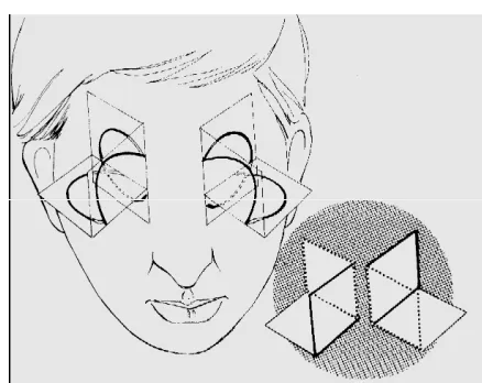
The semicircular canals are oriented in approximately orthogonal planes to the other ipsilateral canals. Although the two horizontal canals are in parallel planes, the two superior and the posterior canals are in planes approximately orthogonal to each other. The canals are organized into functional pairs. The two members of each pair are in parallel planes of orientation.
The otolith organs also function in a paired format, with the two utricular maculae in approximately the horizontal plane and the two saccular maculae in the vertical plane, with an approximate 300 angulation inward to the midsagittal
plane.
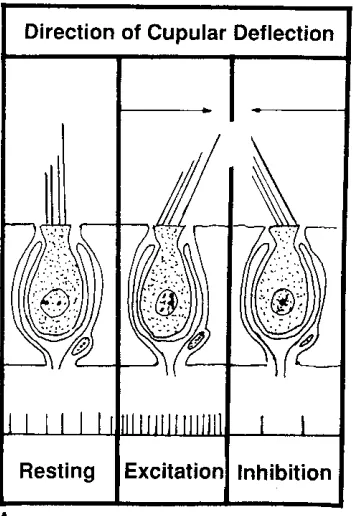
The crista ampullaris is shaped like a small hill and contains hair cells from which protrude many stereocillia and a single kinocilium embedded in a gelatinous substance called the cupula. The kinocilium in the horizontal semicircular canal is located towards the utricle and the stereocilia are away from the utricle. In the posterior and superior semicircular canals, however, the kinocilium is located away and the stereocilia are located towards the utricle (Figure 2)
Deflection of the kinocilium toward the utricle in horizontal, superior, and posterior canals result in utricolpetal (ampulopetal) deviation. Utriculopetal deviation is associated with increased electrical activity in the horizontal
semicircular canal and decreased electrical activity in the superior and posterior semicircular canals. Deflection of kinocilium away from the utricle in the
horizontal, superior and posterior canals results in utricofugal (ampullofugal) deviation. This deviation is associated with decreased electrical output in the horizontal semicircular canal and increased electrical activity in superior and posterior semicircular canals (Figure 3)
The maculae have hair cells and supporting cells. The stereocillia of the hair cells are embedded in a gelatinous material. On the surface of the gelatinous layer are otoconia, composed of calcium carbonate, which transmit the effects of gravity to the underlying hair cells, thereby making the hair cells more sensitive to linear acceleration.
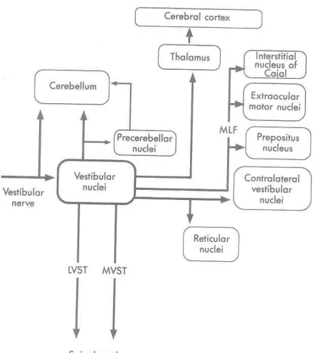
The vestibular branch of the vestibulocochlear nerve innervates the five vestibular sense organs: the cristae ampullaris of the three semicircular canals and the maculae of utricle and saccule. The first order vestibular neurons that are bipolar and situated in the Scarpa’s ganglion terminate in the vestibular nuclei in the lower brainstem (figure 4).
Figure 4: The vestibular organ
[image:14.612.143.363.349.597.2]The lateral semicircular canal is the most commonly tested because of its proximity to the middle ear. The lateral semicircular canal is excitatory to the contra lateral rectus and ipsilateral medial rectus and inhibitory to the contra lateral medial rectus and ipsilateral lateral rectus (Figure 6). The posterior semicircular canal is excitatory to the contra lateral inferior rectus and ipsilateral superior oblique and inhibitory to the contra lateral superior oblique and ipsilateral inferior oblique. The superior semicircular canal is excitatory to the contra lateral inferior oblique and ipsilateral superior rectus and inhibitory to the contra lateral superior oblique and ipsilateral inferior rectus. This is how the vestibulo ocular reflex is served.
INNER EAR FLUIDS
Endolymph: Among the extra cellular fluids endolymph has a unique ionic combination as it has a low sodium 5-25m mol and high potassium 150-160m mol causing it to resemble an intracellular fluid (Annico 1986, Smith 1954).
Perilymph: The site of production of perilymph may be as ultra filtrate of blood or CSF or both also known as Dual theory of origin (Kellerhals B 1979). CSF can reach the vestibule by means of the aqueduct or perivascular or perineural channels.
VESTIBULAR OCULAR REFLEX
The vestibulo-ocular reflex (VOR) represents one mechanism by means of which humans stabilize gaze, the clinical measurement of the oculomotor
control. The smooth pursuit system permits tracking of a visual target with a smooth continuous movement of the eye thereby providing a stable image projection to the fovea of the retina, the most sensitive area therefore greater clarity of image. The vestibulo-cerebellum (flocculus, nodulus, and posterior vermis) plays a dominant role in smooth pursuit (Carey, Santiana 2003)
The saccadic system of eye movement control provides the fast component during the production of jerk nystagmus. The function of the saccadic system is to reposition a visual target of interest onto the fovea with a single rapid eye motion(Leigh et al 1999). When a target of interest is moving outside the operating parameters of the smooth pursuit system, the saccade system facilitates the tracking ability by superimposing jerk movements onto the smooth movements. The difference between the position of the target on the retina and the desired position on the fovea is known as retinal slip (Neile et al 2000)
The optokinetic response is a combination of smooth pursuit and saccade mechanisms; the main purpose of this system is to provide clear visual images during sustained head movements. One system of oculomotar control is visual fixation. This is the active process of maintaining a fixed gaze on a target of interest. Although this system shares neurologic substrate with the smooth pursuit system, evidence suggests that it is a separate control system (Leigh 1999).
History and neuro-otological examination form the cornerstone of evaluation of a patient with giddiness. An appropriate history will help in making the all-important differentiation of a peripheral cause as opposed to a central cause. History also will differentiate from other systemic causes of instability like cardiovascular and metabolic. A good and comprehensive history must include the following (Hullar 2005).
• Does the patient have vertigo which is defined as hallucination of movement and indicates a lesion in the vestibular system
• Are symptoms episodic or continuous most vestibulopathies cause episodic symptoms
• Do symptoms indicate involvement of semicircular canals or otoliths sudden sensation of tilt and drop attacks are due to otolith dysfunction
• Thyroid disease diabetes anaemia autoimmunity hypo perfusion of brain, medications and arrhythmias all cause dizziness and vertigo
• Psychogenic causes like hyperventilation can cause episodic vertigo like symptoms
• Precipitating causes like position in BPPV sound in tullio phenomenon certain foods in migraine
• Exacerbation by head movements e.g. osscilopsia due to head movements indicate vestibular hypo function or vascular compromise of 8th nerve complex
EXAMINATION OF BALANCE SYSTEM :
Examination of patients with dizziness includes examination of the ear including otoscopy, tuning fork tests, examination of nose, nasopharynx, general physical examination, cranial nerves, and vestibulospinal and vestibulo-occular systems.
VESTIBULOSPINAL Static imbalance
Romberg’s: Patient stands with his feet together, eyes opened and eyes closed. If the patient sways it indicates proprioceptive loss or acute unilateral Labyrinthine dysfunction (Baloh 1995).
Walking: Patient is asked to walk on a straight line first with eyes open and then with eyes closed. Falls when tandem walking with hands stretched and eyes closed indicate ipsilateral horizontal canal dysfunction (Baloh 1995).
Unterbergers test – Patient is asked to stand in one place with hands out stretched and to march on the spot with eyes closed. If the patient moves more than 70 degrees to 100 degrees to one side, it indicates a paretic lesion on that side (Fitzgerald 1997).
Dynamic vestibulo-spinal function
2.VESTIBULOOCCULAR SYSTEM Nystagmus
Theseareinvoluntary rhythmic repetitive movements of eyeball.
Mechanism - Spontaneous nystagmus results from an imbalance of tonic signals arriving at the occulomotor neurons. Because vestibular system is the main source of occulomotor tonus, it is the driving force of most types of spontaneous nystagmus. The site of lesion may be in the peripheral vestibular pathway
(labyrinths and vestibular nerve till root-entry zone) or in the central vestibular pathways
The pathology may be located either in the peripheral vestibular system (sensory cell to the vestibular nuclei in the brainstem or central vestibular pathways
Lesions of peripheral vestibular system (labyrinth & 8th nerve) typically interrupt tonic afferent signals originating from all of the receptors of one labyrinth so that the resulting nystagmus has combined torsional horizontal and vertical components. The horizontal component dominates because the tonic activity from the intact vertical canals and otoliths partially cancel out. Gaze in the direction of the fast component increases the frequency and amplitude. Gaze in the opposite direction has the reverse effect.(Alexander’s Law). The slow phase is linear resulting in a saw-toothed wave-form. Peripheral vestibular nystagmus (PVN) is strongly inhibited by fixation. Unless seen within a few days of the acute episode, spontaneous nystagmus will not be present when fixation is permitted.
Central type of nystagmus
Congenital spontaneous nystagmus:
This is a purely horizontal nystagmus which disappears / markedly decreases with loss of fixation as well as during convergence.
Periodic Alternating nystagmus
This nystagmus changes direction at regular intervals (1 to 6 minutes) with null periods (where nystagmus is minimal or absent) between each half cycle varying between 2 to 20 secs. It may be found in with various conditions such as encephalitis, brainstem ischemia, syringobulbia, syphilis, trauma and as a congenital disorder
Inspection of spontaneous nystagmus:
The eyes are observed with the and without Frenzel’s lens when the eyes are looking straight. Frenzel’s Lens (+20 dioptre lens) is used to differentiate spontaneous nystagmus of peripheral origin (enhances without optic fixation) from central vestibular nystagmus which does not enhance on removal of optic fixation.
Table:1 DIFFERENTIATION BETWEEN SPONTANEOUS NYSTAGMUS OF PERIPHERAL AND CENTRAL ORIGIN ( Baloh et al 1990)
peripheral Central
appearance Combined/torsional/horizontal Often pure-horizontal/vertical/ torsional
fixation Inhibited Usually little effect
gaze Unidirectional(Alexander’s law) May change direction mechanism Asymmetric loss of peripheral
vestibular tone Imbalance in the central occulo-motor tone; may be OKN /pursuit* location Labyrinthine or vestibular nerve CNS/brainstem/
cerebellum
Gaze-holding nystagmus (Baloh 1990).
Patient is asked to maintain eccentric gaze for 30 sec from central orientation (Drift up to 15-sec normal). Presence of gaze-holding nystagmus is a hallmark of cerebellar floccular and medial vestibular lesion.
[image:22.612.49.553.79.343.2]The alternate cover test / Madrox rod (Baloh 1990).
Eyes of the patient are alternatively covered with a card looking for a vertical or horizontal corrective movements as an index of misalignment.
Saccades:
Patient is asked to alternatively fixate (with head still) on the examiner’s nose and finger, which is moved to different locations 150 from primary position. Parameters like velocity, accuracy, and initiation time are looked for.
Smooth pursuit
Patient is asked to follow a slowly moving target no faster than 200 per sec. Asymmetries in horizontal tracking as represented by the presence of more corrective saccades in one direction than other is looked for.
Head shaking nystagmus (Sawovaros 1999).
With Frenzel’s lens in place, patient is asked to shake head about 30 times horizontally with chin placed about 30 degrees downwards. On stopping the head shaking abruptly presence of any nystagmus is looked for. This test checks dynamic vestibular function
Head thrust test ( Halmagvi1988)
Patient is asked to look carefully on examiners nose. A brief high acceleration horizontal head thrust is done.
In unilateral vestibular failure a slow phase of abnormally low amplitude nystagmus will be evoked in response to head thrust towards a point of fixation. Positioning testing (Alford 1972)
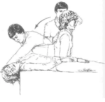
Dix – Hallpike manoeuvre (figure 7)
Patient sits upright on examination table and turns chin 45 degrees towards right shoulder. Patient is brought straight back rapidly into a right head hanging position. This position is maintained for at least for 30 sec.
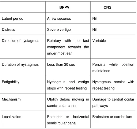
Table:2 COMPARISON OF THE POSITIONAL NYSTAGMUS OF BENIGN PAROXYSMAL POSITIONAL VERTIGO (BPPV) WITH THAT OF CERTAIN LESIONS OF THE CENTRAL NERVOUS SYSTEM (Baloh 1990 and AG Kerr et al 1997)
BPPV CNS
Latent period A few seconds Nil
Distress Severe vertigo Nil
Direction of nystagmus Rotatory with the fast component towards the under most ear
Variable
Duration of nystagmus Less than 30 sec Persists while position maintained
Fatigability Nystagmus and vertigo stops with repeat testing
Nystagmus persist with repeat testing
Mechanism Otolith debris moving in semicircular canal
Damage to central ocular pathways
Localization Posterior or horizontal semicircular canal
[image:25.612.84.531.193.614.2]Valsalva induced nystagmus (Baloh 1990)
The patient is asked to do Valsalva’s manoeuvre with Frenzel’s lens in position and nystagmus is looked for. It is seen in cranio-cervical junction anomalies, perilymphatic fistulas, other abnormalities of ossicles, oval window, semicircular canals and otoliths.
Hyperventilation
of vestibular nerve e.g. acoustic tumour, compression by a small blood vessel, multiple sclerosis may show hyperventilation induced nystagmus (Leigh et al 1999). The metabolic consequence of hyperventilation (alkalosis, changes in ionized calcium) may provoke nystagmus either by alternating conduction in peripheral (e.g. vestibular schwannoma) or central (e.g. multiple sclerosis) vestibular pathways, or by altering central compensation. For example, a vestibular schwannoma may cause focal demyelination of the vestibular nerve, reducing conduction across this segment. Central mechanism compensate for the resulting vestibular imbalance (minimal or absent spontaneous nystagmus). Hyperventilation, however, improves conduction, increasing input from the lesioned side and making the central compensation no longer appropriate. The result is a nystagmus with slow phase directed away from the side of the lesion (Walkerand and zee 2000)
Fistula test
This test is done by tragal compression or insufflation using Siegel’s speculum. Nystagmus is seen in otitic syphilis, perilymph fistula, and dehiscence of the semicircular canals caused by cholesteatoma or congenital anomalies. CLINICAL EVALUATION OF HEARING :
Hearing disorders are classified as conductive, sensorineural, and central based on the anatomic site of lesion. (Beagley 1981silverman 1978)
CONDUCTIVE HEARING LOSS:
The causes of conductive hearing loss are impacted wax, otitis media (suppurative otitis or serous otitis), otosclerosis, perforations of tympanic membrane, trauma, congenital malformation of the external and middle ears, and tumours of the temporal bone.
SENORINEURAL HEARING LOSS:
Sensory neural hearing loss results from lesions of the cochlea and/or the auditory division of the eighth cranial nerve. The cochlea analyze the frequency content of the sound, the high frequency sounds stimulate the sensory cells of the basal turn whereas for low frequency sounds maximum stimulation occurs at the apex. The common cause of acute unilateral hearing loss is infection of inner ear (labyrinthitis). It can either be viral (measles, mumps, infectious mononucleosis) or bacterial. The other causes are trauma and vascular occlusive disease. Relapsing unilateral sensorineural hearing loss associated with tinnitus, fullness in ear, and vertigo is typical of Meniere’s disease. Ototoxic drugs produce a subacute hearing loss. Acoustic neuromas characteristically produce a slowly progressive unilateral sensory neural hearing loss. The progressive bilateral hearing loss associated with advanced age is called as presbyacusis. CENTRAL HEARING DISORDER:
AUDIOMETRIC TESTS:
The audiologist is an integral member of the team, evaluating patients with dizziness. The audiologists role in to do a comprehensive audiologic evaluation which includes pure tone testing, speech audiometry, and immittance testing. The audiogram is performed in a sound proof room with calibrated audiometric equipment
Air conduction testing (AC): A series of frequency specific pure tones (250 – 8000 Hz) is presented via ear phones, asking the patient to respond each time he hears the stimulus. The audiologist finds and maps the threshold, defined as the softest intensity level at which a patient can hear the tone at least 50% of the time. The ears are tested individually, and the threshold is obtained in dB hl at 250,500, 1000, 2000, 4000 and 8000 Hz.
Bone conduction testing (BC): The second part of comprehensive audiologic evaluation is testing bone conduction. This involves placement of a bone conduction vibrator on each mastoid individually and finding thresholds at various frequencies. Bone conduction testing differs from air conduction in its mode of presentation; in addition it by passes the outer and middle ear, delivering the stimuli directly to the cochlea of the inner ear. In certain cases (e.g. with conductive loss and asymmetry between ears), the stimulus (tonal / speech) in the test ear can travel through the skull and around the head to be perceived by the non-test ear. To obtain valid test results in the test ear, the audiologist may find it necessary to present a noise to the non-test ear, preventing it from participating in the test. This is called masking (Valente 2001)
Caloric test:
roll over. Flourens further observed that the hearing was preserved in spite of lesion on the semicircular canals and that direction of movement was same as that of the canal divided. (Lustig 2000)
Nobel prize winning scientist Robert Barany introduced into clinical examination caloric testing and also explained different types of nystagmus and their central causes.
In 1870 Goltz concluded that the semicircular canals were responsible for balance only. Barany in 1906 used 10 to 20 ml of ice water to cause nystagmus. This caused past-pointing and drift (walking towards irrigated side) also along with autonomic symptoms of nausea and vomiting (Nelson 1969).
Kobrak in 1920 in order to minimize patient discomfort used smaller volumes of water at temperatures closer to body temperature. He used 5 to 10ml of water at 27 degrees and if no response was found lowered the temperature stepwise to 20 degrees. This required a temperature controlled water source was time consuming but reduced patient discomfort significantly
Hallpike and Fitzgerald in 1942 (Nelson 1969) introduced the accepted Bithermal Caloric test in which the ears were irrigated using water for forty seconds using temperatures 7 degrees above and below body temperatures. This test caused only moderate discomfort was very sensitive and specific and evaluates both central and peripheral vestibular system.
Caloric test is useful in determining the responsiveness of the labyrinth and is one of the few tests that allow the single labyrinth to be tested independently. Caloric test relies on alternatively heating and cooling the labyrinth by using water or air. Being closest to the plain of the temperature gradient it is the horizontal canals which is tested.
Caloric stimulus of the labyrinth cause a response in two ways. The first is a convective component with a temperature gradient causing a density difference. When the horizontal canal is oriented in plane of gravity either by raising 30 degrees or lowering 60 degrees from the upright position the gravity allows denser fluid to go lower and less denser to go higher this movement deflects cupula and results in nystagmus. A nonconvective component has been proved by demonstration of caloric effect in space where there is microgravity environment and no convective effect. This may be due direct caloric effect on hair cells or cupular displacement due to pressure changes in membranous duct. (John Stahle 1990)
Water at 30 and 44 degrees are used and irrigation is for 60 seconds cerumen to be removed water to be avoided in perforation of tympanic membrane mental arithmetic is done for concentration optic fixation is removed with frenzels glasses ( Luxon 1997)
canal paresis based on Jongkees formula compared to air at 27 degrees. This study showed that the minimal ice-cold irrigation study could be used safely as a screening test
In another study (Becker GD 1979) to assess the screening value of monothermal caloric tests the following observations were made
A valid screening caloric test must decrease examination time increase patient comfort and maintain high degree of sensitivity in predicting Bithermal caloric results.
Comparing monothermal warm and cold irrigation false negative results were obtained in 14 and 25 % of irrigations and false positive irrigations in 22 and 15 % of irrigations. This lead to some patients not getting Bithermal tests and others getting unnecessary Bithermal tests.
This lack of sensitivity limits the use of monothermal tests as screening tests.
In a study (Nelson 1969) Linthicum method was explored further in normal and vertiginous patients by not only measuring the amount of ice water needed but also the duration and intensity of response. This was to evaluate the overall stimulus strength and to assess the usefulness of the method as a screening test.
right ear and 130.8 +/-14.7 for the left. This 30 second duration difference was found to be statistically significant by student T test (p=<0.001). Even in vertiginous patients stimulus with .2 cc evoked lesser response than 30 degree 30 second stimulation . Thus .2 cc stimulus is a weaker stimulus by duration criterion than 30 degree stimulus.
Since the strength of stimulus is more represented by the slow phase velocity, the measurement of slow phase velocity showed it to be 12.7 for 30 degree and 3.2 for .2 cc stimulation. But the discomfort to the patient in terms of subjective vertigo was less with.2cc than 30-degree stimulus. A further conclusion made was when a marked asymmetry was noticed in the two ears with .2 cc stimulus the phenomenon of directional preponderance should be considered and when different amounts of water was needed for a response for example .2cc on right and .8cc on left then the phenomenon of canal paresis should be thought of. In this study the minimal ice-cold irrigation was found to need >.4 cc in cases of acoustic neuroma in 85% of proven cases.
Hence it was concluded that minimal ice cold irrigation test is a useful screening test.
Electronystagmography
This test has been used for clinical testing for nearly forty years. The test consists of a battery of tests collectively known as Electronystagmometry (Sheperd 2000).It is one of the standard investigations used in the assessment of patients suffering from vertigo and equilibrium disorders.
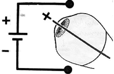
Principle of ENG
This potential is in microvolt range. A movement of eyeball causes a movement of the electrical field and the currents so created can be picked up by electrodes applied to skin around the eyeballs. The voltage measured is proportional to the amplitude of the eye movement.
The potentials are greatly amplified and recorded directly on a running strip of paper similar to ECG (usually a AC amplifier is used)
By convention, movements of the eye to right and up are recorded as an upward deflection, left and down as downward deflection.
The ENG Machine
It can be single channel - records conjugate horizontal eye movement Double channel - can record horizontal movement of each eye
separately
Or a multichannel - which can record both horizontal and vertical eye movements
The more recent machines have been computerized so that all parameters are computed and calculations available immediately.
The most recent machines make use of an infrared camera, which picks up the nystagmus and records into a computer and parameters calculated and made available instantly.
Test conditions
Patient undergoes a full ENT and examination prior to the test to exclude any ear wax/perforations/mastoid cavity/infections etc
Test should be done in a room with no electrical disturbances/interference and must have an even lighting with ability to darken surrounding.
Patient should be seated in a comfortable positioning chair for head to be positioned at 30 degrees above horizontal
Preparation of patient
Skin is cleaned well with spirit and electrode jelly applied and electrodes fitted on both the outer canthi of the eyes with the neutral electrode on the fore head.
A soft rubber catheter is applied to each deep meatal wall under vision
Calibration is done in two ways electrical Bio-calibration (pendular test)
Electrical calibration
An impulse of known voltage is fed in and machine so adjusted that a 200 microvolt signal produces 10 mm deflection of the recording needle. (Ambient electrical disturbances can produce movements of recording needle but these usually have amplitude of less than 20 micro volts. Therefore any beat less than one mm should not be considered as nystagmus). This is already done by the manufactures.
Bio calibration done by using a calibration bar - Done in order to interpret ENG
- A standard angle of eye deviation is represented by known amplitude of pen deflection.
Therefore 1mm of pen deflection is 2 degrees of eye movement
This test called pendular test can be used to calculate amplitude and slow phase velocity of nystagmus
It also gives information about proper fixing of electrodes and correctness of polarity of fixed electrodes
ENG test battery
Spontaneous nystagmus
Definition - Nystagmus present with head upright and eyes centered.
Technique - The patient is seated upright and eyes cantered. Recording is taken with eyes open for one minute and closed for one minute.(Latter 30 secs of each minute taken for calculation)
Gaze- evoked nystagmus
Nystagmus which appears on gaze deviation in one or more directions and is not present in imposition
Technique
Patient is asked to look 30 degrees to right and to left, up and down. Recording of eye movements is done in each gaze position for one minute.
- Pathological if amplitude >4 degrees
Types of nystagmus
Symmetric - equal amplitude to right and left
- also seen in pts with myasthenia gravis, multiple sclerosis and cerebellar atrophy
Asymmetric - always indicates structural brain lesion
- In Brun’s nystagmus (found in large c-p angle lesion which causes compression of brainstem) the larger amplitude nystagmus is usually directed towards side of the lesion
Rebound - nystagmus that disappears or reverses direction as the lateral gaze position is held
- occurs in patients with cerebellar atrophy, and focal structural lesions of cerebellum (only variety of nystagmus thought to be specific for cerebellar involvement)
Dissociated-results in lesions of medial longitudinal fasciculus( MLF). On adducting eye lags behind and develops a low amplitude nystagmus while abducting eye overshoots the target and develops a large amplitude nystagmus. E.g. demyelinating diseases, unilateral -usually vascular diseases of brainstem
Caloric testing (Baloh 1990)
Patient is laid supine, with head end elevated by 30 degrees from the horizontal position.20 ml of water at 44 degrees and 30 degrees are used for irrigation in the following order. Right 44 left 44 right 30 left 30.
An interval of 8 minutes is given between each successive irrigation.
Patient is kept alert during procedure using simple arithmetic (eg:subtracting 7 from 700).
2. Maximum slow phase velocity- true representative of vestibular activity There is a large intersubject variability. Therefore the intrasubject measurement is more useful clinically
The formula to calculate vestibular paresis (R30+R44)-(L30+L44)
--- x 100 % Vestibular Paresis (VP) R30+R44+L30+L44
For directional Preponderance the formula is (R30+L44)-(R44+L30)
--- x100 % Directional Preponderance (DP) (R30+L44+R44+L30)
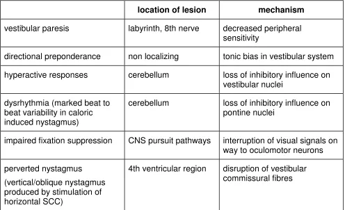
Table 3: Interpreting the result of Bithermal caloric testing
location of lesion mechanism vestibular paresis labyrinth, 8th nerve decreased peripheral
sensitivity
directional preponderance non localizing tonic bias in vestibular system hyperactive responses cerebellum loss of inhibitory influence on
vestibular nuclei dysrhythmia (marked beat to
beat variability in caloric induced nystagmus)
cerebellum loss of inhibitory influence on pontine nuclei
impaired fixation suppression CNS pursuit pathways interruption of visual signals on way to oculomotor neurons perverted nystagmus
(vertical/oblique nystagmus produced by stimulation of horizontal SCC)
4th ventricular region disruption of vestibular commissural fibres
Limitations include that it does not test full dynamic range of the vestibular system. Measures the function of the lateral semicircular canal only, and it is considered to be a very low frequency test for vestibular function.
Vestibular evoked myogenic potential are considered a test of the otolith as this response is shown to arise from the saccule. A burst of activity in the ipsilateral sternomastoid has been recorded using a electromyography to sound at 95 db spl.
MATERIALS AND METHODS
This is a descriptive, cross-sectional study of normal volunteers (controls) and patients with rotatory vertigo (symptomatic patients) as the study arm to determine the usefulness of cold caloric test as a screening test for patients with vertigo.
A sample size of 30 normal and 30 cases with vertigo will be sufficient to provide results for a test with a Sensitivity of 87% (95% CI: 73- 95) and a
Specificity of 50% (95% CI: 27 - 72), calculated for a type I error of 5% and a power of 80%.
The normals (controls) were volunteers selected from the staff and students of Christian Medical College. A standard questionaire was administered to them to ensure they were eligible to be included (Annexure 1). They were also asked to consent for undergoing tests and given an information sheet (Annexure 2), prior to the test.
Inclusion and exclusion criteria for selecting the controls and symptomatic patients are shown below.
Inclusion criteria:
For Normals: Persons 18 yrs to 65 years, with no history of ear-related
complaints (hearing loss, tinnitus, giddiness,) with normal hearing on pure tone audiogram.
Exclusion Criteria: For Normals
History of medication with potentially ototoxic drugs, exposure to excessive noise, history of ear discharge/ head trauma, systemic illnesses like diabetes, hypertension, hypothyroidism.
For symptomatic patients
Tympanic Membrane with central perforation, active otitis externa and patients on labyrinthine sedatives less than 48 hrs before the tests.
All controls and patients underwent a detailed history and otoneurological examination including examination of ears to rule out presence of wax or tympanic perforation. Their hearing status was established with pure tone audiometry and impedance audiometry done in a sound proof room.
Those who fitted into the inclusion and exclusion criteria underwent the minimal cold irrigation test (Baloh et al) and Bithermal caloric test as described by Fitzgerald and Hallpike (Nelson 1969).
Minimal cold irrigation test.
The test was done by a third person, not involved in recruiting or interpreting results. The patient was made to lie down supine, with the head flexed at an angle of 30 degrees to the horizontal, so as to place the horizontal semi circular canal in a vertical position. Under direct visualization of the ear drum, two cc of ice water (obtained by adding ice cubes to tap water until it reached 4 deg C ) was infused into the ear canal against the tympanic
observed through Frenzel’s lens (+20 diopter lens) and its duration measured using a stop watch. Moderate lighting was used to prevent optic fixation .
Bithermal caloric test
The patient was made to lie down supine with head flexed at a 30 degree angle to the horizontal so as to place the horizontal semi circular canal in a vertical position. Each ear was infused with water at 30 deg C and 44 deg C for 30 seconds, using a soft red rubber catheter fixed in such a way that the irrigation would impinge on the tympanic membrane. The patient was asked to count backward in twos, from 300 (to keep the patient alert). The duration of nystagmus was recorded by means of an infrared camera of the video nystagmography machine. The same was repeated after 8 minutes in the opposite ear. Both the ears were sequentially irrigated with warm water at 44 deg C, with an interval of 8 minutes by a third person who was not involved in recruiting or interpreting results. The 44 deg C irrigation was done in order to calculate canal paresis and directional preponderance using Jongkees formula.
(R30+R44)- (L30+L44) x 100 = % Vestibular Paresis (VP) (R30+R44+L30+L44)
(R30+L44)-(L30+R44) x 100 = % Directional Preponderance (DP) (R30+R44+L30+L44)
Recruitment was stopped as soon as the sample size was reached in each group. Data was entered on an excel spreadsheet and analysed using SPSS v 12 .The normal values for cold caloric test was taken as +/- 2 standard deviations (SD) of the mean values of the 30 normal volunteers.
RESULTS
There were 30 normal volunteers and 30 symptomatic patients; 15 (50%) normals were females and 15 (50%) were males while in the symptomatic patients, 12 (40%) were females and 18 (60%) were males.
Figure 9 shows their age distribution. Most volunteers were in the age group 25 to 34 years while symptomatic patients were more in the older age groups.
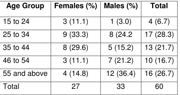
Table 4 shows the age and sex distribution in the study groups. Age
[image:47.612.162.455.405.560.2]groups 25 to 34 and 55 or more had the largest numbers. Only a small proportion (6.7%) were younger than 25. There were more males than females in this study.
Table 4. Age and sex distribution of normal and symptomatic groups
Age Group Females (%) Males (%) Total 15 to 24 3 (11.1) 1 (3.0) 4 (6.7) 25 to 34 9 (33.3) 8 (24.2 17 (28.3) 35 to 44 8 (29.6) 5 (15.2) 13 (21.7) 46 to 54 3 (11.1) 7 (21.2) 10 (16.7) 55 and above 4 (14.8) 12 (36.4) 16 (26.7)
Total 27 33 60
Table 5 shows the mean duration of nystagmus with cold caloric test among normals, 2 SD and mean ± 2 SD in the right and left ears.
[image:48.612.158.455.215.347.2]The mean duration of nystagmus for both right and left was more than 1½ minutes.
Table 5. Cold caloric test results in normal volunteers.
Right ear Left ear
Mean duration: 100.7 Mean duration: 103.2 2 SD: 54.75 2 SD: 49.0 Mean – 2 SD: 45.9 Mean – 2 SD: 54.2 Mean + 2SD: 155.4 Mean + 2SD: 152.2
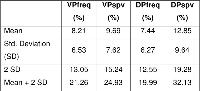
Table 6 shows the results of Bithermal caloric tests among normal
[image:48.612.144.471.530.678.2]volunteers, standard deviation and mean +/- 2 standard deviations The mean +/- 2 SD for VP frequency was 21.6; for VP spv was 24.93; for DP frequency was 19.99 and for DP spv was 32.13
Table 6. Results of Bithermal caloric tests among normal volunteers
VPfreq (%)
VPspv (%)
DPfreq (%)
DPspv (%)
Mean 8.21 9.69 7.44 12.85
Std. Deviation
(SD) 6.53 7.62 6.27 9.64
2 SD 13.05 15.24 12.55 19.28
Table 7 shows the clinical diagnosis of patients presenting with giddiness. The largest group were neurological with migrainous vertigo being the
[image:49.612.127.487.236.447.2]commonest diagnosis. Metabolic abnormalities were found in 8 out of the 30 patients which may have contributed to the symptom. Endolymphatic hydrops was next commonest with 5 patients.
Table 7 - Clinical diagnosis of symptomatic patients
Clinical diagnosis number
BPPV 3
Neurological (Migrainous vertigo / meningitis) 10
Endolymphatic Hydrops 5
Cardiovascular (postural hypotension / VBI ) Metabolic abnormalities (DM/ dyslipidaemia/ hypothyroidism)
8
Ototoxicity 1
Cochlear Otosclerosis 1
Figure9: Age distribution of normal volunteers and symptomatic patients
Age distribution of normals and symptomatics
3
16
7
1
3
1 1
6
9
13
0 2 4 6 8 10 12 14 16 18
15 to 24 25 to 34 35 to 44 45 to 54 more than 55
Age group
N
um
be
r
Table 8A. Symptomatics and of canal paresis (frequency) (mean ± 2 SDSD of the normals)
Value
(frequency) Number Percent <21.27 25 83.3 >21.26 5 16.7
[image:51.612.206.407.344.453.2]Total 30 100.0
Table 8B. Symptomatics and frequency of canal paresis (mean ± 2 SD of the normals)
Value
(SPV) Number Percent <24.94 12 40.0 >24.93 18 60.0
Total 30 100.0
Tables 8A and 8B show the number of symptomatic patients with canal paresis based on frequency of nystagmus and slow phase velocity respectively (during cumulative period) using values generated from normals. Of the 23 patients with canal paresis, 18 were detected by SPV and 5 were detected by frequency parameter.
Table 9A. Symptomatics and frequency directional preponderance Value
(Frequency) Number Percent
<20.0 24 80
>19.99 6 20
[image:52.612.206.407.262.372.2]Total 30 100
Table 9B. Frequency directional preponderance (SPV) among symptomatics.
Value
(SPV) Number Percent <32.14 22 73.3
>32.13 8 26.7
Total 30 100.0
Table 10 shows a comparison of results from cold caloric and Bithermal caloric tests among symptomatic patients. Sensitivity of the cold caloric test was 3/24= 12.5% and specificity was 5/6=83.3%
Table 10. A comparison of results from cold caloric and Bithermal caloric tests among symptomatic patients.
Bithermal test positive
Bithermal test negative
Total
cold caloric
test positive 3 1 4
cold caloric
test negative 21 5 26
24 6 30
[image:52.612.84.449.512.665.2]Table 11 shows a comparison of results obtained from structured questionaire and the Bithermal caloric tests among 30 normals and 30 symptomatics.
Table 11. Comparison of results obtained from structured questionaire and the Bithermal caloric tests among normals and symptomatics.
Bithermal test positive
(%)
Bithermal test negative
(%)
Total
Symptomatics (questionnaire)
positive
24 (40) 6(10) 30
Normals (asymptomatic) (questionnaire)
3 (5%) 27(45%) 30
DISCUSSION
This study was undertaken to evaluate the usefulness of a frequently used test i.e. cold irrigation test as a screening procedure for detecting patients with giddiness who may need further evaluation or to reassure the patient with giddiness that there was no abnormality with the vestibular system. The cold irrigation was evaluated against the available gold standard test which is the Bithermal caloric test.
Controls were used to establish the normal values in the
population. As can be seen from the Figure 1, the control group age group was considerably younger than the symptomatics. This was because most controls were student volunteers. The symptomatics as expected were older, with risk factors that predispose a person to develop giddiness.
Male predominance in the study group may be attributed to the pattern of hospital visit to a tertiary care centre. The duration of nystagmus after cold irrigation in normal volunteers were very similar in both ears and may be related to the fact that symmetrical activity occurs in both the labyrinths in a normal person.
This study (Table 6) had VP frequency, VP spv, DP frequency, DP spv similar to observations in other centres (Biswas 2006)
The clinical diagnosis reflects the types of patients who come to the vertigo clinic in a tertiary clinic. Migrainous vertigo is one of the commonest causes of giddiness followed by metabolic abnormalities like diabetes mellitus and dyslipidaemia.
since it showed that a significant proportion of symptomatics were identified as positive by this test.
In our study, we found that the sensitivity of cold caloric test was 12.5% and the specificity was 5/6=83.3%. Keith et al (1991) in their study to re-evaluate the monothermal caloric test by examining the correlations between unilateral weakness derived from Bithermal caloric stimulation compared to monothermal caloric results, found that while predicting normal or greater than 20% unilateral weakness, both warm and cool monothermal calorics have greater than 85% efficiency, with specificity greater than 94% and sensitivity greater than 64%. However, the false-negative rate is 29% for warm and 36% for cold caloric test. They concluded that the high rate of false-negative findings indicates that screening tests have no place in a diagnostic battery, especially in view of the implications for missing significant pathology.
Our study also shows that the cold caloric test has a poor sensitivity compared to Bithermal caloric test and so is not a useful test to screen patients with vertigo with vestibular pathology. Even though the specificity of cold caloric test is 83% it may still miss those patients with significant pathology by this test alone
A questionnaire takes about 40 minutes and identifies the aetiology while Bithermal caloric test takes 100 minutes and costs Rs 600 per test in CMC and measures the function of the labyrinth and its location and the side of the lesion. These being complementary, are both needed for final management and follow up of the patient.
pathology, a specificity of 83% is not adequate since one would like a test with close to a specificity of 100% for ruling out a pathology.
Based on these findings we recommend that all patients coming to the clinic are evaluated using the structured questionnaire, followed by
CONCLUSION
REFERENCES
1) Alford B. Meniere’s disease: Criteria for diagnoses and evaluation of therapy for reporting. Report of subcommittee on equilibrium and measurement. Trans. Am. Acad. Ophthalmol Otolaryngology 1972; 76: 1462.
2) Anniko M, Worblewski R; Ionic environment of cochlear hair cells , Hear Res 22: 279-293 1986
3) Anthony Wright Scott Brown otolaryngology Sixth edition 1997 volume 1 basic sciences chapter 1 Author Anthony wright general editor Allan G Kerr volume editor Micheal Gleeson
4) Baloh RW and Honrubia V. Clinical evaluation of hearing. In clinical Neurophysiology of the Vestibular System. Ed 2,Philadelphia, F.A. Davis p186,1990.
5) Baloh RW and Honrubia V. Electronystagmography. In Clinical Neurophysiology of the Vestibular system, Ed 2, Philadelphia, F.A.Davis p 137& 141,1990.
6) Baloh RW and Honrubia V. The history of the Dizzy patient – Psycho physiologic dizziness in Clinical Neurophysiology of the vestibular system, Philadelphia: FA Davis p. 98 1990.
7) Baloh RW and Honrubia V. The history of the Dizzy patient – Psycho physiologic dizziness in Clinical Neurophysiology of the vestibular system, Philadelphia: FA Davis p. 98 1990.
8) Baloh RW, Honrubia V. The History in the Dizzy patient and Bedside Examination of the vestibular system in, Clinical Neurophysiology of the Vestibular system, 2nd Ed. F.A. Davis, Philadelphia p 89-129, 1990.
10) Beagley HA (Ed): Audiology and Audiologic medicine. Oxford university press, Newyork, 1981.
11) Becker GD: LARYNGOSCOPE 1979 Feb 89: 311-4
12) Biswas 2006 An introduction to Neuro-otology Editor Anirban Biswas: 2006, second edition chapter 1 pages 1-19 Bhalani publishing house 13) Davis H and Silverman SR (Ed): Hearing and Deafness, ed.4, Holt,
Rinehart, and Winston, Newyork, 1978.
14) Drachman DA and Hart CW An approach to the dizzy patient. Neurology 1972; 22: 323.
15) Ewald 1892:Physiologische untersuchungen uber des end organ des nervus octavus Bergen: Wiesbaden
16) G.Michael Halmagyi, Ian S Curthoys A clinical sign of Canal Paresis. Arch Neurol 1988; Vol 45: 737-39.
17) Gacek 1997 Scott Brown otolaryngology Sixth edition 1997 volume 3 chapter 5 general editor Allan G Kerr volume editor John Booth
18) Goldberg JM: Afferent diversity and organization of central vestibular pathway Exp Brain Res 130; 277-97 2000
19) Gotch F, Meyer JS and Yasuyuki T Cerebral effect of hyperventilation in man. Arch Neurol 1965; 12: 410.
20) Gullya 2003 Shambough Surgery of the ear fifth edition 2005 chapter 1 Author Aina Juliana Gulya Editor Michel Glascock and Aina Julliana Gulya Elsilver publications
23) Journal of vestibular research 2005: 215-224
24) Kellerhals B:Perilymph production and cochlear blood flow, Acta otolaryngol (stockh) 87; 370 –374 1979
25) Keith RW, Pensak ML, Katbamna B. Prediction of Bithermal caloric response from monothermal stimulation. Otolaryngol Head Neck Surg. 1991 Apr;104(4):499-502.
26) Leigh RJ, Zee DS. The Neurology of Eye movements. Ed 3 Philadelphia, FA Davis, 1999.
27) Lustig 2000 Neuro otology second edition Chapter 1 Author Lawrence R Lustig Editor Robert K Jackler And Derald E Brackmann
28) Luxon 1997 Scott Brown otolaryngology Sixth edition 1997 volume 2 Adult audiology chapter 19 Author Linda luxon general editor Allan G Kerr volume editor Dafydd stephens
29) Luxon 2002 Diseases of the ear edited by Harold Ludman and Tony wright 2002 sixth edition chapter 14 Author Linda Luxon Arnold publishers
30) Lysakowski Cummings Text book of otolaryngology fourth edition 2005 Charles W Cummings volume four chapter138 Author Anna Lysakowski pages 3089 –3111 Elsilver Mosby
31) Mark F.Walkerand, David S. Zee. Bedside vestibular examination, Otolaryngologic Clinics of North America 2000; Vol.33. Number3: 495-506.
32) Maureen Valente and Michael Valente Audiometric Tests, In Practical management of the Dizzy patient, edited by Joel A. Goebel, Chapter 15, Lippincott Williams and Wilkins, Philadelphia, p182, 2001.
35) Pompeiano o, Brodal A: The origin of vestibulo spinal fibres in the cat, with comments on the descending medial longitudinal fasciculus Arch Ital Biol 95,166-195,1957
36) Saowaros Asawaviahiangianda, Massaki Fujimoto, Mabel Mai, Heather Desroches and John Ruthka Significance of Head- Shaking nystagmus in the evaluation of the Dizzy patient Acta otolaryngol (Stockh) 1999: Suppl 540:27-33.
37) Sheperd 2000 Practical manage management of dizzy patient 2001 Author Joel A Goebel chapter 12 pages 113-126 Arnold publishers 38) Smith ca, Lowry OH: The electrolytes of the labrynthine fluids ,
Laryngoscope: 64: 144-153 1954
39) Sterkerso: origin of endolymph and electro composition of endolymph in the cochlea Drescher DG editor auditory bio chemistry Spring field Illinois 1985 Charles c Thomas
40) Thalmann R: metabolic features of auditory and vestibular systems, Laryngoscope.81: 1245-1260 1971
ANNEXURE – I
Vertigo clinic evaluation form
Name Age Sex Hospital no Address
Presenting complaint: vertigo /lightheadedness / imbalance /others______________________
1.VERTIGO (An illusion of movement (Rotatory /linear displacement)
Duration of vertigo hours days months years
Type of vertigo: episodic continuous uncertain
Present at rest at rest and movement on movement only Head rotating surrounding rotating uncertain
Description of first attack: same as present attacks yes/no different from present attacks
How? ---
Description of a typical attack:
Duration of each attack seconds minutes hours days
weeks (Max Min )
Date of last attack Frequency of each attack
Periods in between attacks free of symptoms not free of symptoms
Warning signs before attack fullness in ear/aura/ others /nil
Clustering of attacks yes no
Aggravating/precipitating factors: nil/coughing/sneezing/loud sounds/specific food/ specific head positions
Standing from sitting position/turning in bed /raising hands/ others
Relieving factors nil/yes (specify__________________________________________)
Difficulty walking in the dark/streets/open spaces yes no
Any URI/fever before attack yes no Any barotrauma (swim/fly) yes no
Fullness/pressure sensation in the ear yes no
Tinnitus rt (yes/no) left (yes/no) annoyance / continuous / intermittent
hearing loss rt(yes/no) left(yes/no) fluctuating (rt/left)/nil phonophobia; better/ worse in noisy
surroundings/ rapidly progressing hearing loss/ sudden in onset
Other ENT complaints no yes(ear discharge ,others )
Neurological complaints no yes (specify dysarthria / diplopia/ headache/ loc / blackouts / pins
and needles or tingles in hand and feet / facial pain or numbness / spots before eyes/ seizures / others )
Headache: no yes severity side/site associatednausea/vomiting/scotomas/aura/others
Cervical pain no yes loss of balance on walking no yes
Cardiovascular disorders no yes (past h/o MI/palpitations/ chest pain/leg pain on walking or at
rest /ccf/others )
Medical problems no yes (thyroid / DM / anemia /polycythemia/ autoimmune/ TB /
smoking /alcohol / /loss of wt / appetite / blood in stools /diarrhea / food intolerance/ indigestion / bleeding disorders /macroglobinaemia/ others )
Eye problems no yes(loss of vision /pain /discharge or tearing /
glaucoma/diplopia/refractory errors/new glasses/others )
Head injury/ any trauma no yes ( )
Ototoxic drugs/other medications no yes (Immunosupressents/steroids/others )
Medicines taken/ taking (and duration)
Psychiatric no yes (Insomnia / depression / conversion reactions/agrophobia/others )
Any known allergy no yes( )
Family h/o giddiness/Psych dis no yes( )
Hyperventilation- anxiety, stress, panic attacks
Decreased cardiac output –arrhythmia, valvular disease, heart failure
If 2 H/so orthostatic hypotension
Postural hypotension no yes on any hypotensive drugs no yes
Antidepressants no yes major tranquilizers no yes
Any h/o autonomic dysfunction no yes
(Bladder / bowel dysfunction/ periph neuropathy)
H/so vasovagal attack no yes
(Associated with prolonged standing/ severe pain/ severe vertigo preceding) no yes
H/so hyperventilation no yes
(anxiety/stress/panic attacks /associated symptom -frequent sighing, air hunger/tightness in chest/ perioral numbness/paresthesia of extremities)
H/so reduced cardiac output no yes
(Arrhythmia / valvular disease/heart failure)
3. PSYCHOPHYSIOLOGICAL DIZZINESS - Sensation of floating/swimming /rocking/spinning inside
head (not associated with an illusion of movement of environment)
mechanism–impaired central integration of sensory signals
If 3. Constant in attacks
? Associated with tension headache /palpitations / fatigue / weakness no yes ? Precipitating factors social situations / driving in open spaces no yes
4. MULTISENSORY DIZZINESS mechanism–partial loss of multiple sensory system function
Any systemic diseases no/yes(peripheral neuropathy/decreased visual acuity/hearing impairment/vestibular impairment sec to ototoxic drugs/others )
5. PHYSIOLOGICAL OVERLOAD
mechanism- sensory conflict due to unusual combination of sensory signals
Any h/o air travel/sea travel no yes H/o motion sickness no yes
6.IMBALANCE / DYSEQUILIBRIUM-(only on standing/walking and not related to abnormal head
Imbalance/dysequilibrium (algorithm for localizing the lesion) fig 1
yes no
no yes yes
Yes no
Yes
If 4.
Worse in dark no yes oscillopsia no yes
Numbness / weakness / bladder bowel dysfunction no yes Slow loss of associated no yes Movement dysfunction; incordination / dysarthia no yes
5. OCULAR DIZZINESS mechanism- visual-vestibular mismatch due to impaired vision
Algorithm for localizing lesions in patients complaining of oscillopsia (fig 2)
Yes yes no yes no yes no no
no yes
Imbalance Worse in dark Oscillopsia,
hearing loss Numbness, weakness, bowel, bladder dysfunction slow, loss of
associated movement
bilateral
vestibular loss proprioceptive loss
central VOR head movement
induced persists after head
movement stops bilateral peripheral cerebellar visual association cortex OSCILLOPS IA Spontaneous
nystagmus Transient with
Unilateral persistant Frontal lobe --- Basal Incoordination,
NEURO-OTOLOGICAL EXAMINATION
Systemic Pallor y / n BP lying standing
Bruits carotids no / yes rt / lft
Peripheral pulses dorsalis rt/lft radial rt/lft
Ears
TM
rt
lft
TFT Rennie rt lft Webbers rt lft ABC rt lft
Cranial nerves corneal rt lft
Eye movements
normal /abnormal
ptosis
no yes
7th 9th 10th 11th 12th
Neurological system
deep tendon reflexes
normal /abnormal
Babinsky
normal /abnormal
muscle strength
normal /abnormal
sensation
-
face
normal /abnormal
limbs
normal
/abnormal
EXAMINATION OF BALANCE SYSTEM
1
) VESTIBULOSPINAL
STATIC IMBALANCE
Ronberg's standing test Walking-tandom
eyes open eyes closed eyes open eyes closed eyes open eyes closed
Untenbergers test Fukadas writing test eyes open eyes closed
DYNAMIC VESTIBULO-SPINAL FUNCTION
EXAMINATION OF GAIT
2)VESTIBULO-OCCULAR SYSTEM
ABNORMAL EYE MOVEMENTS
Opsoclonus yes no ocular bobbing yes no ocular flutter yes no
Ocular myoclonus yes no
Inspection of spontaneous nystagmus
Using Frenzel Lens no/yes [horiz(rt/ lft) rotat(clockwise/anticlw) vert (up/dn)
Skew deviation and ocular tilt reaction
The alternate cover test/Madrox rod horiz no / yes vertical no / yes
Saccades no / yes
Smooth pursuit normal / abnormal {corrective saccades no yes (number )}
Head shaking nystagmus no yes (horizontal rt/left vertical no / yes)
Head -thrust test
no
yes rt / lft
Positioning testing Dix- Hallpike maneuver
.
Positional nystagmus sitting HS HR HL HE HF HP
supine HS HR HL HE HF HP
Dynamic visual nystagmus no yes Valsalva induced nystagmus no yes
Hyperventilation dizziness no yes nystagmus no yes
Vertigo clinic evaluation form
Name Age Sex Hospital no Address
DIAGNOSIS AFTER HISTORY AND OTONEUROLOGICAL EXAMINATION
1)_____________________________ 2)____________________________3)___________________________
INVESTIGATION ASKED FOR AND RESULTS
AUDIO PTA right_________________left______________________
IMPEDENCE right__________left_________SPEECH TESTS right__________left_________
ENG PENDULAR SPONTANEOUS NYSTAGMUS
GAZE NYSTAGMUS CALORIC
BLOODS_________________________________________________________________________________
IMAGING________________________________________________________________________________
GLYCEROL TEST right__________left_________ OTHERS_________________________________
REFERAL ASKED FOR AND THEIR IMPRESSION
__________________________________________________________________________________________ __________________________________________________________________________________________ _________________________________________________________________________________________
VERTIGO SYMPTOM SCALE vertigo severity________somatic anxiety_____________
FINAL
DIAGNOSIS______________________________________________________________________________ __________________________________________________________________________________________
MANAGEMENT PLAN