OCULAR INJURIES IN ROAD TRAFFIC
ACCIDENTS - A STUDY
Dissertation submitted to
THE TAMILNADU DR.M.G.R.MEDICAL UNIVERSITY
CHENNAI – 600 032.
In partial fulfillment of the regulations
for the award of the degree of
M.S.DEGREE - OPHTHALMOLOGY
STANLEY MEDICAL COLLEGE,
CHENNAI – 600 001.
CERTIFICATE
This is to certify that the dissertation entitled “OCULAR INJURIES
IN ROAD TRAFFIC ACCIDENTS - A STUDY” presented here is the
bonafide original work done by Dr.T.KAVITHA, in the Department of
Ophthalmology, Government Stanley Medical College, Chennai – 600 001, in
partial fulfillment of the regulations for the M.S. DEGREE
OPHTHALMOLOGY Examination of the Tamil Nadu Dr.MGR Medical
University to be held in April 2012.
DEAN
Govt. Stanley Medical College Chennai – 1.
HOD
Department of Ophthalmology
DECLARATION
I, Dr.T.KAVITHA, solemnly declare that this dissertation entitled,
“OCULAR INJURIES IN ROAD TRAFFIC ACCIDENTS - A STUDY” is a
bonafide work done by me, at Government Stanley Medical College, Chennai
between 2009 – 2012, under the guidance and supervision of
Prof.Dr.K.Basker, M.S.,D.O., Head of the Department, Department of
Ophthalmology.
This dissertation is submitted to Tamilnadu Dr.M.G.R. Medical
University, towards partial fulfillment of regulation for the award of
M.S.Degree Ophthalmology.
Place: Chennai
ACKNOWLEDGEMENT
I express my sincere thanks to Dr.S.Geethalakhsmi, M.D., Ph.D.,
Dean, Stanley Medical College for permitting me to conduct this study.
I express my profound gratitude to Prof.Dr.K.Basker, M.S., D.O.,
Head of Department of Ophthalmology for assigning me this interesting topic,
guiding me with this vast knowledge and experience and providing me all the
necessary facilities.
I am very grateful to Prof.K.Kanmani, M.S., D.O., Prof.Thangarani,
M.S., D.O., for their continuous support and guidance.
I am very grateful to my Assistant Professors Dr.B.Meenakshi, M.S.,
Dr.P.Geetha, M.S., D.O., Dr.Vinayagamoorthy, M.S., Dr.Nandhini M.S.,
Dr.Venkatesh, M.S., for rendering their valuable advice and guidance for the
study.
I wish to express my sincere thanks to all my colleagues who had helped
me in bringing out this study.
Finally, I am deeply indebted to all my patients for their sincere
CONTENTS
Sl.No. Title Page No.
1. INTRODUCTION 1
2. REVIEW OF LITERATURE 3
3. AIM OF THE STUDY 45
4. MATERIALS AND METHODS 46
5. OBSERVATION AND DISCUSSION 48
6. RESULTS 69
7. CONCLUSION 72
8. BIBILIOGRAPHY 1
9. PROFORMA
10. MASTER CHART
INTRODUCTION
ROAD TRAFFIC ACCIDENTS are common occurrence every day.
With ever increasing new drivers and new vehicles, RTAs keep on increasing
causing mild to severe injuries to eyes, the special organ designed for vision.
Eyeball is anatomically well protected inside bony orbit socket and
orbital margin, covered anteriorly by eye lids with lashes, embedded in cushion
of retro bulbar pad of fat behind. Inspite of all these protection it is vulnerable
to injuries.
Ocular trauma is single most important cause of monocular blindness
Worldwide and second leading cause of visual impairment and blindness in all
age groups.
Eye injuries not only results in defective or loss of vision but creates
enormous loss to both victim and society productivity
Eye trauma is an evolving sub specialty of its own. The recent
breakthrough in understanding mechanisms of injury has dramatically
improved the management of eye injuries.
A lot of research has been done in the past, regarding blunt or
regarding road traffic accidents and ocular injuries even though it is a common
occurrence in daily outpatient department in a multi specialty hospital like our
hospital.
So it is of great relevance to create a data regarding RTA and ocular
injuries and its effects in a multi specialty hospital like Stanley medical college
and hospital located in a heavily populated place like north madras surrounded
REVIEW OF LITERATURE
OCULAR TRAUMA.
THE BIRMINGHAM EYE TRAUMA TERMINOLOGY (BETT)
OPEN GLOBE-full thickness eye wall wound present
CLOSEDGLOBE-no full thickness eye wall defect present
LACERATING INJURY –open globe resulting from sharp force
PENETRATING INJURY-entrance wound, no exit wound
PERFORATING INJURY-entrance and exit wound
INTRA OCULAR FOREIGN BODY-entrance wound with foreign
body in eye
CONTUSION INJURY-blunt injury, no open globe
LAMELLAR LACERATION –sharp force, partial thickness eye wall
Eye Injury
Contusion 123455267
5284629ABC7
6629AB555284629
Laceration Rupture
Penetrating
IOFB Perforating
Closed Globe Open Globe
Injury
SYSTEM FOR CLASSIFYING OPEN GLOBE INJURY:
TYPE: A. rupture
B. penetrating
C. Intra ocular foreign body
D. perforating
E. mixed
GRADE: (VISUAL ACUITY): A.>/=20/40
B.20/50 to 20/100
C.19/100 to 5/200
D.4/200 to PL
E.NO PL
PUPIL: (RAPD) Present
Absent
ZONE: 1. Isolated to cornea.
SYSTEM FOR CLASSIFING CLOSED GLOBE INJURY
TYPE (mechanism of injury): A.Contusion
B.Lamellar laceration
C. Superficial foreign body
D. Mixed
GRADE (presenting visual acuity):A.>20/40
B.20/40-20/100
C.19/100-5/200
D.4/200- light perception
E. no light perception
PUPIL (RAPD): Present
Absent
ZONE: 1. External (bulbar conjunctiva, sclera, cornea)
2. Anterior (all structures in relation with anterior chamber & pars plicata)
CALCULATING THE OCULAR TRAUMA SCORE
STEP1: IDENTIFY RELEVANT VARIABLE:
VARIABLE RAW POINT
INITIAL VISION
No PL 60
PL/ HM 70
1/200-19/200 80
20/200-20/50 90
>20/40 100
RUPTURE -23
ENDOPHTHALMITIS -17
PERFORATING INJURY -14
RETINAL DETACHMENT -11
APD -10
STEP 2: CALCULATE THE SUM OF RAW POINTS=
RAW
SUM OTS
NO
PL PL/HM 1/200-19/200
20/200-20/50 >20/40
0-44 1 74% 15% 7% 3% 1%
45-65 2 27% 26% 18% 15% 15%
66-80 3 2% 11% 15% 31% 41%
81-91 4 1% 2% 3% 22% 73%
92-100 5 0% 1% 1% 5% 94%
GENERAL EVALUATION OF TRAUMATIC EYE
An accurate and thorough evaluation is important in eye injury. There should not be any rigid approach but overall examination is adjusted according to history from patient or witness. The status of fellow eye should be noted.
Appropriate sedation, adequate analgesia and protection from further injuries are important during examination. Associated life threatening injuries should be detected and treated accordingly.
SYSTEMIC EVALUATION:
HISTORY:
Injuries sustained in high velocity such as motor vehicle accidents are associated with extra ocular injuries which demand immediate attention.
The details of events preceding and leading to injury, description of mechanism of injury, presence of safety glasses, seatbelts, air bags, obtained.
Time of accident and time of coming to hospital are important in both evaluation and treatment of ocular trauma and chemical injuries.
Current and previous medical history medications allergies, past ocular history are obtained. Status of tetanus immunization determined.
Involvement of blunt force may indicate presence of ocular rupture. Place of occurrence of injury affect the outcome of injury.
History of previous ocular surgery may affect the surgical approach such as laceration or rupture that extends near scleral buckle or glaucoma filtering device. Patients who have significant glaucomatous optic atrophy tolerate the post operative pressure elevation poorly. Patients on immune suppression, diabetes or vitamin or protein deficiency may exhibit poor wound healing.
INITIAL OCULAR EXAMINATION:
VISUAL ACUITY:
Initial visual acuity is the best predictor of final visual acuity. Pinhole acuity is obtained if possible by Snellen chart or ETDRS chart. Tumbling E chart for illiterates and if vision (vn) is very less, counting fingers, hand movements, light perception and projection of rays checked.
CONFRONTATIONAL VISUAL FIELDS:
Static finger counting in each quadrant is gross way of accessing fields. Complete visual fields done after the patient becomes stable.
PUPILLARY EXAMINATION:
Pupillary size, shape, irregularities of margin, directs and indirect light reflex is noted.
‘SWINGING FLASH” test may demonstrate abnormal pupillary reaction. Neutral density filter may detect subtle RAPD. Anisocoria if any should be noted.
COLOUR VISION:
EXTRAOCULAR MOTILITY:
Extra ocular motility abnormality is most likely to occur with orbital injuries where muscle entrapment or injury, orbital hemorrhage or cranial nerve injuries result in decreased motility.
Testing of ocular motility should be performed, but it may be complicated in periorbital edema, pain with examination or poor patient co-operation. Forced duction test can differentiate between neuropathy or muscle injury and muscle entrapment.
ANTERIOR SEGMENT EVALUATION:
First the orbit and surrounding Periorbital tissues are evaluated for laceration, ecchymosis, edema, lid abnormalities, enophthalmos, obvious bony deformities,
PALPATION:
In fracture orbital wall, irregularity of orbital rim, infraorbital hypoesthesia, crepitus and obvious foreign bodies if any should be noted.
EYELIDS:
Partial thickness lacerations are meticulously repaired with interrupted or horizontal mattress suture. Full thickness lid injuries are sutured in layers with slight eversion of edges to prevent eye lid notch with 6-0silk.
CONJUNCTIVA:
The conjunctival surface should be inspected for evidence of ocular laceration or rupture with uveal prolapse or visible scleral defects. Large areas of dense bloody chemosis increase the likelihood of underlying ocular rupture.
Conjunctival hemorrhage may be due to fracture of the orbital wall or the base of skull. In this case, posterior limit of the hemorrhage cannot be made out while anteriorly it may not reach the limbus and the color is purplish. The blood gets absorbed from one to three weeks without treatment.
Scleral entrance wounds are more likely to be associated with ocular perforation in foreign body injuries.
CORNEA:
The corneal epithelium should be evaluated for the presence of staining defects; superficial foreign bodies are safely removed at slit lamp.
Deep stromal foreign bodies are removed in theatre under microscope.
Corneal Edema:
This is seen after severe concussion injuries due to temporary disturbance of the cells of corneal endothelium, their permeability being altered, so that aqueous gets free access to the corneal tissues. Mostly it clears up without specific treatment.
Corneal abrasions:
Superficial abrasions are commonly found in many cases .most of them are small and heal easily by itself.
Lacerations of the Cornea:
Any corneal laceration should be closely examined to determine depth and stability, using slit lamp inspection and seidel testing.
Corneal perforations can be non perforating corneal lacerations or full thickness corneal laceration.
Folds in Corneal tissues:
Blood staining of the cornea:
It results from a contusion injury that is associated with a massive hyphema and raised intraocular pressure. The damaged corneal endothelium permits erythrocyte fragments to enter the corneal stroma.
The affected area is stained at first a rusty brown or a greenish black, which gradually changes to greenish yellow or gray. It may take the form of a central ring (Manschot 1947) or disc (Schousboc and Morard 1935). The resultant blood staining clears spontaneously but it may require a considerable amount of time. The central cornea clears last.
Surgical intervention to remove blood from the anterior chamber and reduce IOP is indicated on earliest sign of corneal blood staining. Earlier intervention is done in patients with compromised endothelial function..
Corneo scleral laceration:
It is a rare event that occurs only on the impact of severe force directly upon eye. The sclera usually bursts at its weakest point near the cornea scleral margin. The presence of old cicatrix may increase the tendency.
Infectious microbial keratitis:
Ocular trauma is a frequent precipitating cause of infective microbial keratitis. The treatment is according to history, clinical findings and microscopic examination of corneal smear.
ANTERIOR CHAMBER:
The anterior chamber depth should be evaluated by slit lamp. There can be shallow anterior chamber due to corneal full thickness defect or anterior lens subluxation. An increase in depth of anterior chamber is seen in posterior dislocation of lens or depth is irregular in subluxation of crystalline lens.
Presence of flare cells, whether inflammatory, pigmented or RBCs should be noted. Any foreign body should be described. In a patient who has intact globe, gonioscopy should be performed looking for an IOFB in the angle.
Blunt trauma can produce traumatic iritis with photophobia and eye pain resulting from ciliary spasm. Slit lamp examination may show perilimbal injection with cells and flare.
TRAUMATIC HYPHAEMA:
Hyphema is graded by the amount of blood in AC.
Grade 3: one half to ¾ of visible volume of AC.
Grade 4: complete filling of visible AC.
Eight-ball hyphema refers to completely filled anterior chamber with black colored clots .gonioscopy is contra indicated since it may cause re-bleeding.
IOP should be monitored regularly.
Medical treatment:
Miotics, cycloplegics, fibrinolytics, estrogen and corticosteroids are useful.
Surgical intervention:
>50 mm Hg for 5 days or >35 mm Hg for 7 days to avoid optic nerve damage is the criteria.
Paracentesis and anterior chamber wash, clot expression and limbal delivery, automated hyphemectomy using cutting /aspiration instruments are various options. When pupillary block glaucoma in large hyphema extending into posterior chamber, occasionally peripheral iridectomy is also done.
ANGLE RECESSION:
reveal unevenness in width of ciliary body band or band greater than trabacular meshwork in width. If more than 180 degree is involved there is 10% chance of developing glaucoma.
Angle Recession Grade:
Grade I: - Ciliary body band appear darker and wider, scleral spur appear whiter than the fellow eye owing to tearing of the uveal meshwork.
Grade II: - (Moderate angle tears) angle is deeper than the fellow eye owing to tears in the face of the ciliary body.
Grade III: - (Deep angle tears) a deep furrow extends into the ciliary body, the apex of the fissure cannot be identified gonioscopically. Deep tears can be visualized by using anterior segment ultrasound bio-microscope.
Angle recession glaucoma was found to be the second most common mechanism of unilateral glaucoma. Glaucoma may develop months or years after injury. Eyes with less than 180 degrees of recession are unlikely to develop late glaucoma.
CYCLODIALYSIS CLEFT:
Cyclodialysis cleft is separation of ciliary body attachment to scleral spur. Gonioscopy reveal white area below the scleral spur. Ultrasound biomicroscopy (UBM) may be helpful.
IRIS:
Iris should be evaluated closely for injuries .Iris tear may be visible within stroma or at the sphincter or gross separation of peripheral iris from the eye wall may be seen indicating Iridodialysis or cyclodialysis.
Iris stroma should be evaluated for transilluminating defects, best seen in retro illumination, may show penetrating injury in RTA.
An incarcerated non prolapsing iris should rarely be removed. In peripheral iris incarceration and a well formed AC, acetyl choline in anterior chamber may constrict and release the iris.
If the incarceration is central, epinephrine dilates pupil and may release the iris.
Prolonged prolapsed iris leads to ischemia and subsequent necrosis. Repositioning should be made only when the exposition is for small time. Iris is abscissed if it is macerated, necrosed and exposed for more than 24 hours.
The lens capsule should be inspected for areas of decreased lucency or obvious penetration. The lens body should be inspected for signs of penetrating injuries such as cataract, foreign body, or obvious disruption.
Pigment deposition on the anterior lens capsule (vossius ring), instability of the crystalline lens (phacodonesis) and displacement of crystalline lens with/without the presence of vitreous in anterior chamber(zonular dehiscence, luxation, or subluxation) may be present with blunt trauma involved in RTA. Pseudophakic patients may show instability, displacement or dislocation of implanted lens.
Posterior capsular status is importment in planning surgery, is by slit lamp or B-scan.
Subluxation and Dislocation of the Lens:
When more than 25 percent of the fibers are broken the lens is said to be subluxated, it may remain in the posterior fossa retained by its attachment to the vitreous.
A dislocated lens is a lens that is completely detached from its zonular and vitreous attachments.
Miotics are used to minimize diplopia and astigmatism produced by traumatic minimally subluxated lens. Mydriatics is used to enlarge Aphakic portion, so that Aphakic correction is given.
The lens may be mobile in the vitreous(Lens natans) and by changing the position of the patient from the prone to supine through the dilated pupil the lens travel from the vitreous into the anterior chamber(wandering lens) (Wolte 1945) but eventually organized membrane tend to anchor it. Very rarely it may be present subconjunctivaly.
Surgical approach:
Anterior segment methods:
Cataract with intact posterior capsule, no displacement, no vitreous in anterior chamber is removed by anterior approach.
A hard subluxated lens with extensive zonular damage if associated with extensive zonulardamageshouldbe extracted using intra capsular approach with scleral fixated lens or anterior chamber IOL implantation.
Posterior segment method:
Pars plana lensectomy with vitrectomy is useful in significant posterior capsular disruption or posterior dislocation of lens and vitreous prolapsed into the anterior chamber and management of associated vitreous hemorrhage, intraocular foreign body, retinal detatchment, etc
As a rarity the lens may slip through a retinal tear into inter retinal space or it may lie between the sclera and ciliary body (sub scleral Luxation).
Concussion Cataract:
Mechanism:
When a sudden force strikes the eye, a wave of pressure thrusts the aqueous and iris, forcibly against the lens and pushes it backwards on the vitreous, on its rebound the lens curls itself against the iris. In the fluid contents of the globe the force is transmitted in all directions, so that capsule and its epithelium as well as lenticular substances itself are concussed.
Tears occur at the thin portion of the capsule covering the posterior pole of the lens. Sometimes they are covered by the iris, so rapidly sealed by fibrin and sub capsular epithelium, which secretes a new capsule. In these cases entrance of aqueous is stopped. So opacity in the lens may remain stationary. If the tear remains open the opacification progresses to involve the entire lens.
In surgical removal of traumatic cataract with intact posterior capsule, no displacement, no vitreous in anterior chamber or lens dislocation into AC, anterior approach via limbus is preferred.
A posterior pars plana approach for removing cataract with posterior capsule ruptured, lens dislocated or subluxated into vitreous posteriorly.
Lens induced intraocular inflammation may occur secondary to phaco anaphylactic uveitis.
Phacolytic glaucoma should be treated with corticosteroids and anti glaucoma medications and lensectomy. phacomorphic glaucoma can be treated by lens removal.
ANTERIOR VITREOUS:
The anterior vitreous should be examined for presence of IOFBs, particulate matter and or cells which could indicate infection.
POSTERIOR SEGMENT EVALUATION:
Posterior segment evaluation is must in all cases of trauma. It is done through direct and indirect ophthalmoscope.
Eyes with smaller, more stable wounds or blunt trauma without rupture may be safely dilated and examined to document the extent of injury and plan for surgical repair.
FUNDUS:
BERLIN’S EDEMA:
Commotio retinae, a cloudy swelling giving grey appearance involving temporal fundus, subsequent post traumatic macular changes like progressive RPE degeneration and macular hole should be looked for. Typically the edema is opposite the site of injury.
The edema may be localised confined to macula, posterior pole or peripapillary region. If the entire posterior pole is involved, a pseudo-cherry red spot may occur, mimicking central retinal artery occlusion. All this may subside in few weeks.
RUPTURE OF CHOROID:
Choroidal rupture may be direct, involving anterior retina at the site of impact running parallel with oraserrata or indirect rupture involving area opposite to the site of impact after a severe concussion injury.
RETINAL CHANGES:
Retinal breaks, holes retinal hemorrhage, retinal dialysis and retinal detachment should be looked for and managed accordingly. A traumatic retinal tear from blunt trauma is by transmission of external force to globe.
Avulsion of vitreous base from the peripheral retina is pathognomonic of ocular trauma, because the vitreous base is normally firmly attached to the retinal periphery and pars plana.
Retinal dialysis with or without retinal detachment is more common in supero temporal area. Traumatic retinal tears with or without retinal detachments can occur.
A giant retinal tear involves tears of more than one quadrant of retina.
Prevalence of retinal dialysis in traumatic retinal detachment according to a study is 84%,gaint retinal tears is 8%,horse shoe tear is 3% and round hole is5%.
Retinal tears with blunt trauma can be treated with photocoagulation or cryopexy. Retinal detachments are treated with scleral buckling, pneumatic retinopexy and vitrectomy.
TRAUMATIC OPTIC NEUROPATHY:
Damage to optic nerve may be due to direct injury by fractured bone pressing over nerve or compression by hemorrhage. It may occur at intraocular, infraorbital, intracanalicular or chiasmal level.
Indirect optic nerve trauma refers to optic neuropathies that follow closed head trauma. The posterior indirect optic nerve traumas, typically damage the intracanalicular segment. The 10mm intra canalicular portion together with its meninges, the ophthalmic artery and sympathetic nerves are crowded tightly with in bony canal.
The damage could be due to shearing force on optic nerve at optic canal where it is tethered to dural sheath or transmission of shock waves through orbit.
CT scan of orbit and head is the initial study of choice. Decompression of optic canal is by craniotomy or through paranasal sinuses.
After ruling out fracture at optic canal, a course of high dose steroids, inj methyl prednisolone1000mg /day in divided doses for three days followed by tablet prednisolone 1 to 1.5 mg/kg for 11 days is given according to ONTT trial.
OPTIC NERVE AVULSION:
extreme rotation or anterior displacement of globe. Prognoses depend on whether the avulsion is complete or incomplete.
Purtcher’s retinopathy:
Purtcher’s retinopathy is a traumatic angiopathy, commonly caused by head and chest trauma with bilateral retinal signs including white ischemic infarcts with blot, preretinal and flame shaped hemorrhages.
OPEN GLOBE RUPTURE:
Scleral rupture can be occult,hidden under the conjunctiva, tenons capsule or recti muscle. Chemosis or Subconjunctival swelling suggest the presence of occult rupture. Slit lamp biomicroscopy and B-scan are useful. CT scan is useful in demonstrating rupture and radio opaque foreign body.
In many cases of penetrating injury it is necessary to repair a corneo -scleral laceration immediately. Following that some require pars plana vitrectomy. Even then the chance of endophthalmitis in open globe injury is 6.8%.
Traumatic Hypermetropia:
A posterior dislocation of the lens with an increased depth of anterior chamber will alter the refraction in the direction of hypermetropia.
Traumatic Myopia:
This is the commonest refractive change following a concussion to the globe. As a rule an increase in myopic refraction of the eye ranges from ID to 6D and disappear within a week or two.
Ciliary spasm accounts for majority of cases due to irritation of the muscle fibers themselves or irritation of the third nerve or paresis of cervical sympathetic chain. This account for 1D to 4D myopia. It disappears with atropine. There will be a spastic miosis.
Subluxation may produce about 5D or 6D myopia (Moller). Higher degrees may be produced by anterior dislocation of lens.
INTRAOCULAR PRESSURE TESTING:
IOP is tested using applanation tonometer. If any globe rupture is suspected, non contact tonometers are used.
IOP testing is deferred in open globe injuries. Patients with low IOP can have ciliary body dysfunction or occult ocular penetration.
ORBITAL FRACTURES:
Orbital wall blow out fracture is of two types:
1. Pure blow out fracture which does not involve orbital rim.
2. Impure blow out fracture involves the orbital rim and adjacent facial bones.
A blow out fracture is caused by increase in orbital pressure greater than 5cm, such as fist or tennis ball. The most common fracture is floor of orbit along with thin bone covering the infra orbital canal. Medial orbital wall may also be fractured. The lateral wall and roof is usually able to withstand such trauma.
Mid facial fractures are classified by LeFort classification.
LeFort 1 is not associated with orbital fractures.
LeFort 11 is associated with pyramidal maxillary fracture extending to medial floor and wall of orbit.
BLOW OUT ORBITAL FLOOR FRACTURE(#):
Orbital floor fractures are classified into direct and indirect.
Direct Forceful trauma on inferior rim result in zygomatic fractures involving zygomatic arch or its posterior articulation in addition to its lateral and inferior orbital rim. This three part zygomatic fracture is referred as tripod
fracture.
1. Trap door type-large fragment of medial portion of floor of orbit is fractured but remains attached to laminar bar resemble a trap door.
2. Medial blow out-disruption of floor of orbit occurs between infraorbital nerve and laminar bar.
3. Lateral blow out-there is communition from the laminar bar to lateral wall.
SIGNS:
1. Periocular signs-Ecchymosis, edema,subcutaneous emphysema.
2. Infra orbital anesthesia due to involvement of infra orbital nerve.
3. Diplopia-due to
b. Mechanical entrapment of inferior rectus or inferior oblique in the fracture.
c. Direct injury of extra ocular muscle associated with negative forced duction test.
1. Enophthalmos
2. Hyphema, angle recession, retinal dialysis.
X-RAY PARANASAL SINUS:
Classic tear drop sign seen.
Caused by prolapsed of orbital contents into the maxillary sinus.
Corresponding maxillary sinus is hazy.
Hemosinus is common.
CT SCAN:
Coronal view is useful in evaluating fracture and prolapsed orbital tissues.
When non-displaced, these fractures do not require fixation. Direct inferior rim fractures and tripod fractures rarely affect ocular motility.
Surgery is indicated in patients with
- diplopia in primary gaze with muscle entrapment
- large floor fractures with enophthalmos>2mm
- progressive numbness in infraorbital areas
- severe -hypophthalmos.
Though various approaches including trans-lid-infra orbital rim, sub ciliary, Caldwell-Luc, Trans conjunctival incision, periostium elevated, entrapped tissues freed, fracture stabilized with autologous /alloplastic material.
Timing of surgical intervention is easier in terms of scaring and fibrosis if performed within first 10 days of injury. But it is better to wait for a few days for some resolution of soft tissue swelling and hemorrhage. If acute intervention is not warranted wait for 6 weeks.
BLOW OUT FRACTURE OF MEDIAL WALL:
Most medial wall fractures are associated with floor fractures.
TYPES:
TYPES 1: pure medial wall fracture
TYPE 3: medial wall, floor and trimalar fracture
TYPE 4: medial, floor, maxillary nasoorbital and frontal bones
In Type 1-fracture of medial wall orbit is by assault. Other types are by RTA.
Visual disturbance common in type 1, 2, 3, but not in type 4.
Eye ball injury is more common in type2.
Diplopia and enophthalmos is more common intype2.
Displacement of orbital wall and prolapsed of orbital contents uncommon in type1, 2, and 4 but common in type 3.
SIGNS:
1. Periorbitalhematoma, sub cutaneous emphysema-after blowing nose
2. Defective abduction, adduction
CT SCAN:
To know the extent of damage.
FRACTURE LATERAL ORBITAL WALL:
1. Itis associated with fracture of zygoma and malar complex.
2. This fracture is common in adults.
3. This fracture should be suspected in severe fascial injuries.
FRACTURE ROOF OF ORBIT:
This type of fracture is quite rare.
Associated with falling on sharp objects or blow to brow /fore head.
Signs:
1. Hematoma of upper eye lid and peri ocular ecchymosis.
2. Inferior or axial displacement of globe.
3. Transmission of CSF pulsations.
Roof # is treated conservatively with IV antibiotics, surgical intervention only in the presence of complications like fracture of optic canal or frontal bone displacement.
INTRA OCULAR FOREIGN BODIES:
Retained IOFB is a true emergency that can lead to severe vision loss due to endophthalmitis, retinal detachment, ciliary body dysfunction, ocular metallosis and even loss of eye since IOFB injuries are costly, both economically and personally.
Most of the eyes with retained IOFBs maintain good vision with appropriate treatment. Visual prognosis is best when IOFB is removed during the initial wound repair surgery.
MECHANISM OF INJURY:
Majority of IOFBs are small sharp projectiles produced from hammering metal or stone. Up to 90% of IOFBs are metallic and 55-80% of these are magnetic. Since they travel at high velocity they are sterile. They penetrate and cause ` ocular disruption. Motor vehicle accidents are important cause of IOFB. Other causes are assault, insect stings, explosion and use of machine tools.
Common IOFBs:
Iron,lead,copper,zinc,silver,gold,platinum,nickel,plastic,woodand glass. Majority of patients with IOFB are male.
Route of entry and location of foreign body:
also be located within the anterior chamber (15%), retina (14%), and lens (8%) or sub retinal space (5%).
Investigating intra ocular foreign body:
The diagnosis of an IOFB begins with a thorough history and suspicion for its presence. History should include circumstance of trauma, the elapsed time since injury, the use of safety glasses, and any exposure to hammering, grinding, drilling, or an explosion.
History of trauma and ocular signs such as localized lenticular opacity, a self sealing corneal or scleral wound, mild intraocular pressure asymmetry, a minor change in shape of the pupil, or mild iris heterochromia, may be all that suggest the presence of an IOFB.
Important information to document at the initial examination includes baseline visual acuity, papillary reaction, intraocular pressure, external examination, slit lamp biomicroscopy, assessment of media clarity, extent and location of wound, iris color, lens status and presence of retinal tears and detachment.
Prognosis and complication:
When an intra ocular foreign body is established, prognosis is always guarded.
Complications
Siderosis:
This is a late occurring syndrome caused by retained foreign body. Initial effects are mechanical and contusive. It is uncommon to have infections as the heat sterilizes the metal.
Iron interacts with intraocular tissues by electrolytic dissociation causing gradual disappearance of foreign body and spread of ferric ions throughout the globe.
In Siderosis,ferrous pigment causes rusty coloration of cornea, iris or lens. The predilection of copper for deposition on limiting membranes is the presence of electric current. Copper interaction with intra ocular fluids and tissues causes ionization and its distribution on limiting membranes by electrostatic factors.
Chalcosis:
Copper and its common alloy bronze and brass may induce Chalcosis. If the copper content is high more severe reaction occur including hypopyon and localized scleral abscesses.
Acute Chalcosis mimic pyogenic Endophthalmitis.
A K-F ring in descemets and a sunflower cataract in anterior capsule are manifestations of Chalcosis which resemble Wilson’s disease.
Anterior chamber has retractile particles, iris becomes green and sluggish in reaction .Retina may have shining particles along vessels.
Sometimes retained foreign body may cause destruction of neurons and vision loss.
REMOVAL OF INTRAOCULAR FOREIGN BODY:
SUPERFICIAL FOREIGN BODIES:
Foreign bodies lodged in cornea and conjunctiva does not produce any serious damage.
A foreign body in bulbar conjunctiva is removed by cotton tipped applicator without anesthesia and in tarsal conjunctiva is removed after double eversion of upper eye lid.
are most difficult ones to remove.Vegetable foreign bodies are associated with infection.
ANTERIOR SEGMENT FOREIGN BODIES:
Foreign body’s entering and remaining in anterior segment of eye can be removed through wound of entry at primary surgical repair.
Depending on size and nature of foreign body larger incision at limbus can be made for foreign body at angle.
POSTERIOR SEGMENT FOREIGN BODIES:
Giant magnet, permanent hand magnet, hand electro magnet and Bronson-magnion instrument were previously used for foreign body removal.
Non-magnetic foreign bodies:
Surgical maneuvers are difficult. Usually direct Trans scleral extraction is done to minimize trauma sclerotomy must directly over the foreign body.
If the foreign body lays intra vitreally then use forceps or other devices to grasp the foreign body through parsplana route.
Posterior segment foreign bodies obscured by opaque media, too large for pars plana route require pars plana vitrectomy to ensure a minimally traumatic extraction.
Intra orbital foreign bodies:
It may remain without symptoms and signs. It may produce granuloma, orbital cellulites, periostitis, orbital abscesses, osteomyelitis, and draining fistulas.
If it strikes orbital apex, blind, anesthetic and immobile globe result.
INVESTIGATIONS:
PLAIN FILMS:
Plain X-rays are valuable screening tool for the evaluation of orbital fractures and intraocular and intra orbital foreign bodies. With the advent of CT and MRI, the plain film is used often for purposes other than rapid screen.
IMAGING:
ULTRASOUND:
Ultra sound means high frequency sound waves. These are above the audible range.
In ophthalmology a-scan and b-scan are used for various diagnostic purposes.
1. It does not generate heat in the ocular tissues.
2. It does not use high power sound energy.
3. Its frequency is much higher in the range of megahertz.
Higher frequency of 10 MHZ is commonly used in ophthalmic ultrasound.
FOREIGN BODIES IN ULTRASONOGRAM:
Foreign bodies in the eye present some unique artificial echoes. In fact, these artifacts are quite useful in making an evaluation of what the material might be.
The artifacts created by foreign bodies may be grouped into two categories-those with:
- Extra echoes generated
- Lack of echoes posterior.
It can also demonstrate ocular abnormalities including retinal and choroidal detachments, vitreous hemorrhage and exit wounds.
Higher frequency ultra sound is useful for more accurate description of IOFBs shape size or relative position. This is used in anterior chamber IOFB.
It can differentiate a small metallic foreign body from air bubble introduced by trauma.
Though ultra sound can be done over lid in open globe injuries, it should be performed cautiously.
Ultrasound is usually not done in open globe injuries to avoid prolapsing of ocular contents. But it can be done intra operatively, after the entrance wound is closed.
COMPUTERISED TOMOGRAPHY (CT SCAN):
However, the intraocular structures are less well imaged than with ultrasound. The scanning technique does not require contact with the ocular surface and so may be used in patients with open globe injuries.
CT scan can suggest an intra ocular foreign body’s density. But CT scan may not detect fragments that are smaller than 0.7mm in dimension, composed of wood or lying undetected in sclera.
CT scan findings indicative of open globe injury include the presence of intraocular air or foreign body, deformity of eye wall, and the presence of intraocular hemorrhage.
CT scan artifacts may be minimized by appropriate technique if the radiologist is notified of a possible foreign body near the posterior eye wall, increasing the accuracy of intraocular vs. extra ocular localization.
CT scanning is less expensive than MRI and, unlike MRI, may be safely used to image metallic foreign bodies. CT scan is generally contraindicated in pregnant patients.
MAGNETIC RESONANCE IMAGING (MRI):
But MRI is more expensive and less available. It produces motion artifacts. It cannot be used in patients with pacemakers or implanted metallic hardware. MRI is not a screening tool and used after CT scan has excluded metallic foreign body.
It employs strong magnetic forces and can produce movement of magnetic foreign body and cause further ocular and neurological damage.
In cases of acute trauma, CT scan is preferred to MRI because of its lower cost ,more rapid results, and ability to image metallic foreign bodies
ULTRASOUND BIOMICROSCOPY (UBM):
UBM is high frequency ultrasound (35-50MHZ) with axial resolution as small as30 microns in imaging anterior segment of eye. The penetration is poor.
Anterior segment trauma may be associated with hyphema, where it is difficult to visualize iris and lens.UBM is useful to study the lens position, status of iris, ciliary body and configuration of angle. Angle recession and cyclodialysis cleft can also be evaluated.
ELECTROPHYSIOLOGY:
when decision making is centered on enucleation /evisceration Vs secondary reconstruction.
7
7
AIM OF THE STUDY
MATERIALS AND METHODS:
This is a study carried out in the patients who suffered RTA and treated as outpatients and inpatients in Stanley medical college and hospital during a period of 2 Years patients were taken for study from October 2009 to September 2011 and follow up for each case is a minimum 6 months. This is a randomly conducted study and demographic data and details of injuries obtained.
The cases that came to the ophthalmology department were taken for the study.175 cases that came to outpatients were included in random fashion and entered in a pre structured profoma. The sample also includes those who were grossly injured and with limitation of vision.
Information regarding time, location, type and mechanism of injury use of spectacles, car safety belts and helmets were obtained.
Vision, eye findings, diagnostic tests, medical and surgical treatment were recorded. Tests were all done free of cost at Stanley hospital.
Whenever necessary, X-rays, ultrasonography, CT-scan which the patients had were utilized for the study.
INCLUSION CRITERIA:
1. Patients who suffered road traffic accidents within 7days.
2. Age group: >10 to <60 yrs.
3. Patients with no previous head injuries.
4. Minimum follow-up for 6 months.
EXCLUSION CRITERIA:
1. Head injuries and ocular trauma by modes other than RTA.
2. Age<1oyrs and>60 yrs.
3. Previous history of head and ocular injuries.
OBSERVATION AND DISCUSSION
The observations made during the study are as follows.It is compared with various studies of trauma and with a few studies of ocular trauma and road traffic accidents.
Out of 175 patients,23 patients did not report back for the 6 month follow up due to various reasons and 152 patients were finally included in the study.The obsevations made are as follows.
[image:53.612.153.482.328.700.2]OCULAR INJURY DUE TO RTA IN MALES AND FEMALES:
Table 1:
Cause of injury Male Percentage Female
Percentage
RTA 138 90.78 14 9.2
There were about 138 males and 14 females in the study. This clearly
shows male preponderance in road traffic accidents, as shown in other studies
also. This may be due to the fact that males are more exposed to the highway
traffic when compared to females. In various studies it varies from 10:1 to 7:1.
Here in our study about 90% of the study group was males, i.e. 10:1.In a study
by KAMATH.S.J in200727 the ratio is 10:1 as in this study.
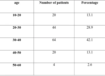
AGE AND OCULAR INJURY:
Table 2:
age Number of patients Percentage
10-20 20 13.1
20-30 44 28.9
30-40 64 42.1
40-50 20 13.1
[image:54.612.117.478.327.590.2]Figure 2:
About 20 (13%) people were of age between10-20, mainly teen agers.
Majority of those involved were from 20-30(28.9%) and from 30-40 yrs
(42%).A very few were above 50 yrs. At Tripoli study, mean age of patients
was 32.5% and they included all age groups in the study24. In various recent
studies the mean age is less than 40 years. In urban study at Delhi the mean age
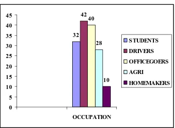
OCCUPATION AND RTA: Figure 3: 32 42 40 28 10 0 5 10 15 20 25 30 35 40 45 OCCUPATION STUDENTS DRIVERS OFFICEGOERS AGRI HOMEMAKERS
As already seen more young people of age 20 are involved in road
traffic accidents and hence about 32 people (21.05%) were students.
Many people involved in trauma were bread winners, mostly drivers and
office goers. Among them were 42 drivers (27.63%) and 40 office goers
(26.3%).
28 people (18.4%) were agricultural workers mainly from areas around
Chennai and some were referred from other hospitals for their other ailments.
Those who were riding 2 wheelers were mainly students, drivers and
office goers. This indirectly shows more increasing number of 2 wheelers and
their use by younger population. Those driving 4 wheelers were persons
involved in professional driving. Privately owned cars were less involved in
accidents.
[image:57.612.122.509.294.602.2]EYES INVOLVED IN RTA:
Figure 4:
Eyes involved were mainly right about 80(52.6%). About 34(22.3%)
were left eyes. Both eyes were involved in 18(11.8%). the Tripoli study on
Time of injury
: The following table shows accidents during different [image:58.612.129.506.174.711.2]time of a day.
Table 3:
Time of accident Number of
patients Percentage
Morning 46 30.2
Afternoon 12 7.8
Evening 30 19.7
[image:58.612.136.499.185.345.2]Night 64 42.1
Time of injury affect trauma in many ways. The above chart clearly shows that trauma especially involving eye occur mostly at night. Almost more than one third occurred at night (42.1%).
The reason could be poor light, poor roads, traffic pattern and consumption of alcohol especially during night after a day’s tiring work may be the reasons. During afternoon, when the movement of vehicles is less, accidents are less.
RTA-What vehicle was involved
: [image:59.612.136.501.393.647.2]The different types of vehicles involved by patients in RTA have been described in the following diagram.
Figure 6:
102 10
6
34
2WHEELER 3WHEELER 4WHEELER
cases were pedestrians hit by 2 or 3 wheelers. Just 3% of the patients were riding 4 wheelers.
The vehicles which caused the accidents were mainly Lorries, motor bikes and cars. Autos and vans were involved. Many of these vehicles did not have proper head lights and rear lights, which were also the reason for accidents at night.
Some accidents occurred due to the carelessness of the drivers. Stray dogs were responsible for many accidents in and out of the city.
The study of eye injuries in Tripoli recorded that road traffic accidents were mainly due to 4 wheelers.24
[image:60.612.137.496.401.667.2]RTA – Two Wheeler Accident and Helmet Use:
The above chart shows that among 152 patients, 102 used 2 wheelers. Of them, 86 patients did not use helmet. But only 16 of them used helmet which is only 15%. This attributes to the severe head and eye injury sustained by them. The use of helmet is seen as disturbance and a cause of more sweating among 2 wheeler drivers.
[image:61.612.124.513.252.691.2]Influence of Alcohol on RTA
:Table 4:
RTA - Influence of
Alcohol
Yes No
90 62
Out of 152 patients, 90 patients (59.3%) were under the influence of
alcohol while driving vehicles or walking along the road irrespective of age
group .62 patients (40.7%) had not taken alcohol during the accident. This has
a major influence on the occurrence of the accident. Those accidents which
occurred during night were mainly under the influence of alcohol. Alcohol
intake is assessed by history, breath analyzer test and the condition of the
Common ocular signs in Road Traffic accident:
Anterior segment findings:
The following table shows the findings in the trauma patient’s anterior
segment involved in the study. Various findings are as follows.
Table 5:
Road Traffic Accident Number of injuries
Percentage
Periorbital edema 133 87.5 Periorbital ecchymosis 72 47.36
Orbital fracture 13 8.5 Eyelid edema 133 87.5 Eyelid laceration 21 13.8 Conjunctival chemosis 46 30.2 Subconjunctival hemorrhage 141 92.76
Corneal edema 6 3.9
Corneal abrasion 14 9.2 Corneal laceration 6 3.9 Traumatic iridocyclitis 21 13.8
Iris prolapse 4 2.6 Iridodialysis 5 3.2 pupillary sphincter tear 13 8.5 Traumatic mydriasis 10 6.5 Traumatic miosis 4 2.6 Anterior chamber hyphema 5 3.2
EYE LID:
Table 6:
Eyelid tear
IN RTA
RTA STUDY URBAN
STUDY*
PERCENTAGE 13.8 8.2%
* EPIDEMIOLOGICAL STUDY OF OCULAR TRAUMA IN
URBAN SLUM POPULATION IN DELHI BY S.VATS
AIIMS-2007.23
A common sign in RTA associated trauma is eye lid edema along with peri-orbital edema (87.5%). Tripoli study has recorded Periorbital edema of 35.5%.
Eye lid laceration was found in 21 patients (13.8%). 8 mild partial thickness tears were present. Those 13 patients with severe lid tears required suturing. Mechanical ptosis noted in 23 patients and1 case of aponeurotic ptosis noted.
CONJUNCTIVA:
Table 7:
SCH
IN RTA
RTA STUDY HEAD
INJURYSTUDY*
PERCENTAGE 92.76% 19%
* OCULAR MANIFESTATION OF HEAD INJURY BY KULKARNI-EYE, 2004
From the above tables it is evident that sub conjunctival hemorrhage (92.7%) is the most common eye sign associated with RTA in eye. Dense Subconjunctival hemorrhage should be looked carefully with slit lamp since it may mask a scleral laceration.
CORNEA:
6 patients had corneal edema. 3 patients had severe corneal lacerations and were repaired at OT. 3 patients had mild lacerations. Corneal abrasion of varying degree is a common sign in cornea. Some healed completely without scar, while some healed with corneal scar.
CORNEAL
LACERATIONS RTA STUDY
HEAD INJURY STUDY*
ANTETIOR CHAMBER (AC):
Traumatic iridocyclitis was found in 21 patients.5 patients had developed hyphema. Out of 5 patients, 2 were of grade 4 completely filing the AC and 1of them were of grade 2.2 patients were of grade 1 occupying less than quarter volume of AC.
HYPHAEMA:
HYPHAEMA RTA STUDY URBAN
STUDY*
HEAD INJURY STUDY**
PERCENTAGE 3.2% 0.6% 0.5%
* EPIDEMIOLOGICAL STUDY OF OCULAR TRAUMA IN
URBAN SLUM POPULATION IN DELHI BY S.VATS
AIIMS-2007.23
** OCULAR MANIFESTATION OF HEAD INJURY BY
KULKARNI-EYE, 200425
PUPILLARY INVOLVEMENT:
PUPILLARY INVOLVEMENT
RTA STUDY HEAD INJURY STUDY**
PERCENTAGE 8.5 6.5
Pupillary involvement is more in RTA when compared to ocular injury associated with head injury due to other causes too.
IRIS:
4 patients with corneal and scleral tear had iris prolapse. Pupillary margin is inspected for sphincter tear.13 patients had pupillary sphincter tear.10 patients had traumatic mydriasis and 4 patients had traumatic miosis.6 patients showed relative afferent pupillary defect in the side of affected eye.
LENS:
6 of the 152 patients had traumatic cataract.4 of them had the posterior capsule was intact found using B-scan and in 2 cases vitreous was disturbed and vitreous found in AC. 8 cases had subluxated lens.
TRAUMATIC
CATARACT RTA STUDY URBAN STUDY**
PERCENTAGE 3.9 19.6
** EPIDEMIOLOGICAL STUDY OF OCULAR TRAUMA IN URBAN SLUM POPULATION IN DELHI BY S.VATS
AIIMS-2007. Traumatic cataract is only 3.95% in RTA when compared to
INTRAOCULAR AND INTRA ORBITAL FOREIGN BODY:
[image:68.612.128.504.363.630.2]Out of 152 patients, 6 patients were found to have intra ocular foreign body in this study 3.9% had intra ocular foreign body, while according to other studies it could be up to 40% with penetrating injuries. Intra orbital foreign bodies were found in 4 patients only accounting to 2.6% of total cases.
Posterior segment findings:
The table shows different findings of posterior segment of the eyes following injury.
Table 8:
Posterior segment
findings No of patients percentage
Vitreous hemorrhage 6 3.9
Choroidal rupture 2 1.3
Retinal edema 4 2.6
Retinal break 6 3.9
Retinal detachment 4 2.6
neovascularisation after 6 months 6 patients had retinal tears and 4 developed retinal detachments.
6 cases of vitreous hemorrhage noted. Various types of retinal hemorrhages were also found in various patients. Multiple Pre retinal hemorrhages were associated with vitreous hemorrhage.
VITREOUS HEMORRHAGE:
RTA STUDY HEAD INJURY STUDY*
VITREOUS
HEMORRHAGE 3.9% 0.5%
* OCULAR MANIFESTATIONSOF HEAD INJURY BY DR KULKARNI A.R.EYE-200425
This shows that vitreous hemorrhage is more in RTA than with other ocular trauma.
RETINAL DETATCHMENT:
According to a study ROLE OF USG IN OCULAR
RTA STUDY URBAN STUDY*
RETINAL
DETATCHMENT
2.6% 1.3%
* EPIDEMIOLOGICAL STUDY OF OCULAR TRAUMA IN
URBAN SLUM POPULATION IN DELHI BY S.VATS
AIIMS-2007.23This shows that retinal detachment incidence is higher in trauma
due to RTA.
[image:70.612.125.512.75.174.2]TRAUMATIC OPTIC NEUROPATHY:
Table 9:
TRAUMATIC OPTIC NEUROPATHY
RTA STUDY URBAN
STUDY *
PERCENTAGE 3.9%(6) 0.6%
There were 6 cases of RAPD with severe limitation of vision. Those
patients did not have pallor of disc immediately following injury. But they
developed pallor later.
According to this study, traumatic optic neuropathy occur in 3.9%,
slum population in Delhi by S.VATS-AIIMS, 2007.23This shows that optic
neuropathy is more with RTA than with general ocular trauma.
Methyl prednisolone 500 mg IV bd for 3 days followed by
T.prednisolone 1mg/kg for 11 days is usually given medical therapy.
In extended regimen, nowadays, injection methyl prednisolone is given
for 5 days followed by T.prednisolone for 11 days. 4 patients who had PL +
and one with PR defective did not improve in vision, while 1 of them improved
from 2/60 to 6/60 and another improved from 3/60 to 6/60 with high dose of
systemic steroids given immediately within 24 hours of trauma.
* Indirect optic nerve injury involvement in 2 wheeler riders in north east
India shows that methyl prednisolone does not improve the outcome of
indirect traumatic optic neuropathy.in our study 2 cases improved in
vision while 4 cases did not improve.11
* Indirect optic nerve injury in 2 wheeler riders in north east India
IJO, 2008, BY HARSHA BHATACHARJEE.11
VARIOUS TYPES OF ORBITAL WALL FRACTURES IN RTA:
The various orbital wall fractures found in the study are enumerated
Table 10:
TYPE OF FRACTURES NO OF PATIENTS
SUPERIOR WALL 1
MEDIAL WALL 3
LATERAL WALL 5
INFERIOR WALL 4
Out of 152 patients, 13(8%) patients sustained orbital wall fractures. Lateral wall fracture being the most common of all. Multiple fractures involving facial bones occurred in 5 patients.
Those with medial wall fractures were associated with crepitus. Inferior wall fractures were associated with maxillary wall fractures and hemosinus of maxilla.
One patient had superior wall fracture with head injury and was treated for it.
Figure 9:
Especially due to fall on the sides from 2 wheelers without wearing helmets
produced fracture of lateral wall, while in all other ways in which eye injuries occur like blunt injury by fist, medial wall fractures are common.
RTA study &* orbital fracture study27
Floor 30% 19.6%
Lateral wall 38% 13.7%
Roof 7% 2.9%
RESULTS
1. Out of 152 patients, 138 were males and14 were females which is about 10:1 ratio.
2. Regarding age distribution, the maximum numbers of patients were in the age group between 20 to 30 and 30 to 40, about 108 which accounts for 42.1%.
3. Occupation of the patients shows that the risky group in the study were professional drivers and office goers followed by students. Women were the least involved.
4. In about 52.6% patients right eye was involved and 22.3% only left eye was involved. In 11.8% both eyes were involved.
5. Time of injury when studied in detail showed that most of the accidents about 42% occurred in night and 30% occurred in morning hours. The remaining 25% occurred in afternoon and evening hours.
6. In this study those involved in RTA were mainly 2 wheelers followed by pedestrians though they were hit mainly by 4 wheelers and 2 wheelers. A few were travelling in autos and 4 wheelers.
8. 60% of people involved in RTA were under the influence of alcohol irrespective of the time of incident though at night it is at the maximum.
9. Regarding fractures involving orbital rim, lateral wall fractures were more common followed by medial, inferior and superior wall fractures. As such about 8% cases had some sort of orbital wall fractures.
10. Among the various periocular signs involved in trauma due to RTA, the most common sign was SCH and Periocular edema, followed by ecchymosis. Various other complications of anterior segment like corneal partial thickness lacerations and full thickness lacerations were observed in the study. Scleral tear, hyphema of various degree, iris prolapse, iridodialysis, traumatic iridocyclitis, pupillary sphincter tear, traumatic cataract, subluxated lens into anterior and posterior chamber were noticed.
11. Posterior segment findings include vitreous hemorrhage, retinal edema, retinal hemorrhages, choroidal rupture, retinal breaks, and retinal detachment. Certain patients after injury may not show any sign but may later develop disc pallor. Patients who develop traumatic optic neuropathy after trauma show severe deterioration of vision with limited posterior segment findings initially. 2 of them improved with steroids while 4 of them did not improve much.
13. Grievous injuries:
Corneal lacerartion-3.9%
Traumatic cataract-3.9%
Vitreous hemmorage-3.9%
Retinal detatchment-2.6%
Traumatic optic neuropathy-3.9%
CONCLUSION
According to this RTA and ocular trauma study conducted at Stanley medical college, ophthalmology department, the following conclusions were made.
The incidence of road traffic accidents is increasing with the increase in vehicle population.
The higher incidence of trauma in men is attributed to the fact that men in our society are exposed to higher level of risk due to RTA since they are more exposed to highway traffic in a heavily populated industrial area like North Chennai.
The age group involved in RTA is becoming younger since younger age groups are using more vehicles especially 2 wheelers.
More RTAs were found to occur during night time when the visibility is poor, awake fullness is less and alcohol abuse is more.
Fractures of orbital rim injuries occur in motorists with severe external injuries. They do occur even with less severe external injuries. Hence there should be a severe suspicion of fracture in all patients with severe pain and defective ocular movements.
Those with severe loss of vision should be investigated for direct and indirect causes of traumatic optic neuropathy and those with indirect traumatic optic neuropathy should be given high dose systemic steroids.
Use of helmet:
Of the people driving 2 wheelers, 15% were only wearing helmets, while rests of the 85% patients were not wearing any protective head gears. According to the study patients who were not wearing helmets were more prone for grievous injuries mentioned above.
Almost none of the 3 wheeler and 4 wheeler drivers were wearing seat belts.
Precautionary measures:
Based on the conclusions of the study certain precautionary measures can be undertaken to avoid motor vehicle accidents.
Accidents occur whenever vehicles are more and traffic violations are more. With ever increasing population growth and migration of people to metro cities, government should look into things to decongest the population by creating satellite town ships and creating good job opportunities in sub urban areas.
Government should take steps to maintain street lights, reduce power cuts at night, have wider roads with road barricades ,wide medians, separate lanes for different vehicles, more pedestrian crossings, good maintenance of sub ways, maintenance of roads ,filling up of potholes, good water drainage system, creating highways with speed limits, bye-passes and ring roads. Speed monitors are to be introduced in all main roads.
Road lanes should be painted with florescent paints for good visibility during night. Sign boards should be painted with florescent paints.
Stray dogs and cows are main cause of road traffic accidents next to bad roads and use of alcohol. This problem should be addressed along with the blue cross and vetenary department.
Screening the drivers for ocular ailments like refractory errors, diabetes, hypertension, hearing defects should be done periodically especially after 40 years. Use of reflective coated stickers at the back of the vehicles, specific type of horns for specific type vehicles, stickers at the centre of head lamp to avoid glare are to be strictly followed.
Road traffic rules should be a part of the curriculum and school students should be educated to spread the message. The powerful media can be utilized to spread the message. Wearing proper helmets, safety goggles and seat belts should again become compulsory.
breakers. Monitoring traffic at night should be done .Drunken driving should be strictly monitored.
PICTURES
2345647589A13B3C91
DEFE11
A5B1A9349856 1
CORNEAL LACERATION-SUTURED
CORNEAL LACERATION WITH IRIS PROLAPSE-(SUTURED)
IRIDODIALYSIS
TOTAL HYPHAEMA
VITREOUS HEMORRHAGE (B SCAN)
LATERAL WALL OF ORBIT FRACTURE
FLOOR OF ORBIT FRACTURE (TEAR DROP SIGN)
849!C98511628513!46298"#1
43859A1B3898"C38
43859A1B3898"C381
BIBLIOGRAPHY
1. A.K.Gupta - current topics in ophthalmology pg. -545-606
2. Bradford J. Shingleton and Peter S. Herch- EYE trauma pg-63-78,104-116,117-125
3. Duane’s clinical ophthalmology vol 3
4. Duke Elder S. system of ophthalmology, vol xiv, part I, mechanical injuries
5. Journal of American academy of ophthalmology 1997, a system of classifying mechanical injuries of the eye.
6. Jack J kanski- clinical ophthalmology, sixth edition. Pg-847-868
7. Samuel Boyd by Modern management of ocular trauma. Pg-2,5,6,10-12,13,14,18-20,21,22.
8. Parson’s disease of the eye, 18th Edison, Churchill Livingston international student edition. pg-361-378
9. Stallard’s eye surgery, seventh edition1989, chapter11-traumatic surgery.
11. Indirect optic nerve injuries in 2 wheeler riders in north-east India, by Harsha battacharjee, IJO, 2008
12. Structural changes in ocular tra