Dissertation on
STUDY ABOUT THE EFFECTIVENESS OF
SERIAL STRETCHING IN POST BURN
ELBOW AND KNEE FLEXION
CONTRACTURE
M.Ch. DEGREE
BRANCH - III - PLASTIC SURGERY
DEPARTMENT OF PLASTIC SURGERY
KILPAUK MEDICAL COLLEGE
CHENNAI - 600 010
THE TAMILNADU
DR. M.G.R. MEDICAL UNIVERSITY
CHENNAI.
CERTIFICATE
This to certify that this dissertation entitled “STUDY ABOUT THE EFFECTIVENESS OF SERIAL STRETCHING IN POST BURN ELBOW AND KNEE FLEXION CONTRACTURE” is a
bonafide work done by Dr.S.Ahamed Rafeeq Meeran, under our
guidance and Supervision in the Department of Burns, Plastic & Reconstructive Surgery, Government Kilpauk Medical College, Chennai - 10, submitted for the M.Ch. (Plastic Surgery) Branch III examination, to be held in August 2009, by The Tamilnadu Dr.M.G.R. Medical University, Chennai.
Prof. Dr. V. Kanagasabai., M.D.,
Dean,
Kilpauk Medical College & Hospital, Chennai-10.
Prof.S.R.Vijayalakshmi., M.S., M.Ch., (Plastic Surgery)
Professor & Head Of the Department,
Department of Burns, Plastic & Reconstructive Surgery, Kilpauk Medical College & Hospital,
Chennai - 10.
DECLARATION
I Solemnly declare that the dissertation titled “Study about the effectiveness of serial stretching in post burn elbow and knee flexion contracture” was done by me at Govt. Kilpauk Medical College & Hospital, Chennai-10 during November 2006 to March 2009 under the guidance and supervision of Prof.S.R.Vijayalakshmi., M.S., M.Ch., (Plastic Surgery).,
The dissertation is submitted to The Tamilnadu
Dr.M.G.R.Medical University towards partial fulfillment of the requirement for the award of M.Ch., Degree (Branch-III) in Plastic
Surgery.
Place : Chennai ( Dr.S.AHAMED RAFEEQ MEERAN )
Date :
ACKNOWLEDGEMENT
I express my sincere thanks and gratitude to
Prof. V. Kanagasabai., M.D., Dean, Kilpauk Medical College & Hospital, Chennai, for permitting me to utilize the clinical materials of this hospital.
I have great pleasure in thanking my
Prof. Dr. S. R. Vijayalakshmi. M.S., M.Ch., (Plastic Surgery), Professor and Head of the Department, Department of Burns, Plastic and Reconstructive Surgery, Kilpauk Medical College & Hospital Chennai, for her valuable support in the conduct of the study and for her valuable guidance, suggestion and supervision throughout my career and my period of study. I thank my professor for being helpful in successfully completing this dissertation.
I am extremely grateful to Prof. T.Mathivanan., M.S., M.Ch.,
(Plastic Surgery) who has given his full support and guidance for this study.
I thank all my professors, and Assistant professors who have helped me in this study. I am thankful to all my colleagues for their valuable help.
Finally, I would like to place on record my sincere thanks to all my patients for their immense co-operation without which this study would not have been possible.
CONTENTS
Page No.
1. INTRODUCTION 1
2. AIM OF THE STUDY 4
3. REVIEW OF LITERATURE 5
4. MATERIALS AND METHODS 43
5 RESULTS 46
6. DISCUSSION 51
7. CONCLUSION 58
8. PROFORMA
9. BIBLIOGRAPHY
INTRODUCTION
Human beings are unique creation of god, as they have an upper
limb which is distinctly different from the lower limb. Evolution of
human race has allowed us to have a complex amount of movements in
the limbs. The hands are the eyes of the blind, the tongue of a dumb and
the aid of the deaf to communicate. The upper limbs have to extend bend
and hold. The lower limbs have to be straight, strong and move.
Burn injury is a systemic illness and its severity is usually assessed,
if not by patient’s survival, by the consequence of the burn injury i.e. scar
hypertrophy, contracture and structural deformities due to loss of body
components.
Body deformity is closely related to the magnitude of the injuries
i.e. extend and depth of injury, mode of intervention, physiotherapy and
follow-up care.
Formation of Scar tissue at the wound site and contraction of the
scar tissue are the normal consequence of an injury. Although the exact
mechanism accounting for the sequential change in wound healing and
scar formation remain incompletely understood, wounds with infection
and or allowed to heal spontaneously tend to form scar that are thickened
The thickened and contracted scar tissues, changes that are normal
and expected consequence of wound healing process are microscopically
composed of collagen, arranged in whorls and nodules. The changes may
be observed as early as 3 to 4 weeks following the injury and they are
cosmetically unsightly and functionally disturbing
Scarring secondary to burns leads to a multitude of adverse medical
consequences including loss of function, restriction of joint mobility,
restriction of growth, altered appearance and adverse psychological effect
When the upper limb is at rest, it relaxes the muscles, tendons and
joint capsules and when prolonged the rest, leads to contracture and
deformity.
The hands are no longer capable of full function and the mobile
part of man becomes an unaesthetic prong from the body. Similarly when
the lower limb is bend, the person is unable to be on his feet. Early
mobilization and splinting does miracles, by not only restoring the
anatomical shape and size but also retains the function.
The upper limbs which tried to rescue a burning victim needs the
supportive care. Proper and timely care of the scar prevent, the formation
This study is an effect to find out the effectiveness of stretching the
scar both in the upper and lower limb at the level of elbow and knee.
Though the act of stretching and splinting is tender, the results
AIM OF THE STUDY
To study about the effectiveness of serial stretching in post burn
elbow and knee flexion contracture.
Objective:
1. To study about the amenability of the post burn scar to stretching.
2. Average time needed for full extension
3. Relationship between age of scar and time needed for full
extension
4. Complications of Stretching
5. Effectiveness as an adjunct procedure in a patient with multiple
contracture, while the more important areas are getting surgical
REVIEW OF LITERATURE
Biophysics of thermal injury:
Burns can lead to pyrolysis, disruption and oxidation of tissues.
Among the various cell types present in the skin some are more likely
sensitive to killing by elevated temperature than others. The degree of
temperature elevation at the given site in the skin depends on the rate of
heat transfer with in the tissue. The thermal conductivity of Dermis is
greater than the subcutis, since fat is a good insulator. Perhaps for this
reason thermal injury often leads to necrosis of the entire dermis with
little cell death in the Subcutis.
Hair follicle in some sites typically extends well beyond the
dermis, into the adipose tissue of the upper subcutis and eccrine sweat
glands are also seen in the subcutaneous fat. Despite the presence of
adipose tissue around them, often hair follicles are entirely destroyed
even though there is little or no apparent necrosis in the upper subcutis. In
severe burns the entire subcutis may become necrotic and cell death may
occur in the underlying fascia and skeletal muscle or even in the
NORMAL SKIN
First degree or superficial injury of skin
Superficial burns are those in which part or all of the epidermis is
lost but the epidermal basal lamina remains intact and the dermis is
uninjured. In these areas epidermal regeneration is required.
Second degree or partial thickness injury
In partial thickness wound, the entire epidermis and the upper part
of the dermis become necrosed. The deep portion of the hair follicle
remain viable and the keratinocytes lining the hair follicle become
migratory, undergo mitosis behind the migrating cells, eventually
covering the new epidermis. In severe cases loss of hair follicle may lead
to insufficient regenerative activity to cover the surface.
Third degree or full thickness injury
Full thickness burns extend deep enough to destroy the entire hair
follicle including the root and some subcutaneous tissue. In this case
regeneration from the hair follicle is not possible and the wound can
develop an epidermal covering only slowly, as the epidermis lateral to the
wound spreads out over the wound surface. During this time the necrotic
tissue in the wound bed is at risk of infection and extensive activity of
tissue macrophage is required to remove it. Granulation tissue form
the eschar formed by the dead tissue leading to restoration of the
epidermis and production of dermal connective tissue in the form of thin
scar.
Wound heals through three over lapping phases:
1. Inflammatory phase or Reactive phase
Coagulation system activation and formation of thrombus aids in
haemostasis. Compliment system activation occurs, resulting in formation
of inflammatory cytokines, recruitment of neutrophils, macrophages,
resulting in removal of necrotic tissues.
2. Proliferative phase or Regenerative phase
This stage is characterized by the formation of granulation tissue,
with massive proliferation of fibroblast and endothelial cells. These cells
secrete fine fibers of collagen and ground substance formed of, by
multiple mucopolysacchride and proteoglycan that make up the
connective tissue matrix. Many peptides like TGF β and FGF and VEGF
are involved in the formation of this connective tissue matrix.
3. Maturation phase or remodelling phase
Scar remodelling and contraction predominate during this phase.
collagen gets reorganised. Although the collagen content in a healed
wound is maximum at 21 days, scar tissue is rearranged and replaced by
more organised thicker fibers with more cross linking, to obtain a wound
that has approximately 80% tensile strength of normal dermis by 6
months. The deposition of ECM and its component is regulated by MMPs
and their counter regulatory inhibitors, tissue inhibitors of
metalloproteinase.
Scar remodelling continue to occur for upto 12 months following
the initial injury, with scar becoming soft, less vascular and less
indurated. Scar tissue never achieves the tensile strength of normal skin.
Fibroblast become myofibroblast which produces scar contraction.
When there is a tissue loss, wound heals by secondary intention. It
is characterized by granulation tissue formation and wound contraction.
Wound contraction pulls the nearby tissues towards the centre of the
wound resulting in contracture.
MOLECULAR AND CELLULAR BASIS OF HYPERTROPHIC SCARRING
Chemical composition and organization of the extra cellular matrix
Collagen
tensile strength for the tissue. However collagen constitutes a smaller
proportion, about 30% less of the dry weight of hypertrophic scars
because there are greater increase in other components such as
proteoglycan and glycoprotein.
The major genetic form of collagen in skin and scar is type I, which
assembles into thick fibrils, fiber and fiber bundles. In normal dermis
there are smaller amounts of type III collagen, 15 % of the total, and very
small amount of type V and type VI collagens. Pure type III & V
collagens are assembled in vitro into thin fibrils but are found in tissues
mainly in heterotypic fibrils mixed with larger amounts of type I
collagen. Both type III & V collagen are considered to reduce the
diameter of the collagen fibers of which they form a part.
Hypertrophic scar generally contains thinner collagen fibrils than
normal dermis. The difference might be explained by the higher
proportion of type III & V collagen, reported to be about 33% and 10%
respectively. Type III collagen appears in healing wounds within few
days after injury and its persistence in hypertrophic scars is probably a
reflection of their biological immaturity. Type VI collagen does not
assemble into fibrils but into thin beaded filaments, 5 to 20 nm wide, that
In the light microscope, collagen in hypertrophic scars is arranged
in whorls or nodules rather than thick fibers or fiber bundles that are
oriented parallel to the surface in normal dermis. In some specimens there
are extensive regions of almost hyaline appearance where little
organization of the fine fibered collagen is seen. In electron microscopy
the narrow collagen fibrils in these region are seen to be more widely
spaced than in normal dermis or mature scar and found to be ovoid or
irregular in cross section. The interfibrillar space in fibrous connective
tissue is occupied mainly by molecules of two other classes, the
proteoglycan and glycoprotein.
Proteoglycan and glycoprotein
Proteoglycans influence physical properties of connective tissues
such as turgor, resilence and resistance to compression, while
glycoproteins such as fibronectin and tenascin are involved in cell-matrix
adhesion, and have effects on cell behavior mainly through this
mechanism. Proteoglycan also influence cellular activity, through a
variety of mechanism including both positive and negative modulation of
growth factor activity. The morphology of collagen fibers and their
organization is affected by the nature and the amount of proteoglycans
Proteoglycans consists of one or more glycosaminoglycan chains,
which are linear polymers of anionic disaccharides, covalently attached to
a protein core. In glycosaminoglycans like, dermatan sulfate, Chondrotin
sulphate, heparan sulfate and hyaluronic acid, one unit of the repeating
disaccharide is an uronic acid.
Chemical analysis of the hypertrophic scar shows elevated levels of
uronic acid and hence glycosaminoglycans. Since it is an anionic
polysaccharide, glycosaminoglycans are mainly responsible for the water
holding capacity of the connective tissues. So hypertrophic scars are
hyper-hydrated relative to normal dermis or mature scars. However the
increase in glycosaminoglycan content is disproportionately high relative
to the increase in water content. Since the collagen fiber normally restrict
swelling of connective tissues, the high concentration glycosaminoglycan
in hypertrophic scar is responsible for their enhanced turgor.
Hypertrophic scars contain on an average only 25% of the total
amount of small dermatan sulfate proteoglycan, decorin, the major
proteoglycan in normal dermis and six fold higher concentration of large
proteoglycan resembling versican. This versican is normally present in
the proliferating zone of the epidermis, and is in association with elastin
in the dermis. Decorin and versican detected by immunohistochemistry
show a striking inverse distribution in the nodules. Decorin is implicated
fibrils into fibers and fiber bundles. Another proteoglycan, biglycan
present in lesser amounts than decorin in normal dermis is found at
elevated levels in hypertrophic scars. Normally biglycan is found close to
the cell surface but in hypertrophic scars it is associated with collagen in
the extra cellular matrix.
The difference in proteoglycan proportions and distributions
between normal dermis and hypertrophic scars could result from altered
biosynthesis or altered dehydration, the former mechanism more likely.
As hypertrophic scars mature, the collagen fiber become coarser and
better organized and there is an increase in immunohistochemically
detectable decorin. At about 12 months after injury many scars start to
resolve spontaneously, with increase in number of cells expressing
decorin suggesting that this proteoglycan may play an active role in the
resolution. Mature scars show contents of collagen, proteoglycan and
water that are indistinguishable from those in normal dermis.
Chemical analysis of hexose and sialic acid contents showed that
hypertrophic scars contain elevated concentrations of glycoproteins, part
of which is fibronectin. This extra cellular matrix molecule has effects on
cell attachment and its activity, that is important in development and
Myofibroblast and delayed apoptosis in Hypertrophic scars
Myofibroblasts are identified by their positive staining for alpha
smooth muscle actin and is considered pathognomonic for fibrous tissue,
that is prone to undergo contracture. The reduction in cellularity that
accompanies maturation of scar is associated with induction of apoptosis.
The prevalence of myofibroblast in hypertrophic scar may actually a sign
of delay in normal onset of apoptosis and this delay is responsible for the
hypercellularity.
The extend and strength of interactions between the attachment
dependent cells such as fibroblast and epithelial cells and the underlying
extra cellular matrix are important factors determining cell survival.
Detachment of cells often leads to apoptosis. In the anchored lattice,
which resists deformation in all, but the short vertical dimension,
fibroblasts continue to proliferate, while in floating or stress relaxed
collagen lattice that are rapidly reorganized, they are triggered to undergo
apoptosis. Reorganization of matrix may be impaired as a result of its
chemical stabilization by cross linking, by tissue transglutaminase, an
enzyme expressed in greater amounts in hypertrophic scar than by normal
dermal fibroblasts.
TGF-β stimulates the proliferation of fibroblast and synthesis of
muscle actin in fibroblast and may delay the onset of apoptosis. Like
TGF-β, insulin like growth factor is increased in response to tissue injury.
It is mitogenic for fibroblast and endothelial cells, stimulates collagen
production by dermal fibroblast. Normally expression of IGF-1 in
uninjured skin is restricted to epidermis, sweat and sebaceous gland, but
in healing burnt tissues these elements are destructed and epithelial cells
migrate to the wound surface could possibly secrete IGF-1 in proximity
to fibroblasts and affect their activity.
Histology of hypertropic scar
Abnormal hypertrophic scar shows distinct difference from
uncomplicated scar. The striking difference is the presence of rounded
whorls of immature collagen that consist of delicate thin collagen fibrils
rich in type III collagen, Small blood vessels and plentiful of
mucopolysaccharide. These nodules are sharply demarcated from the scar
tissue, which may be composed of similar materials or of mature thick
collagen fiber, that are oriented parallel to each other and to the wound
surface, typical of mature scar. Although they are clearly seen with
routine h & e staining, these dermal nodules are distinctly seen with the
movat stain which stain mucopolysaccharides, blue green and mature
The nodule of hypertrophic scar vary in size from 0.5mm to 1cm in
diameter and appear to be sometimes spherical, ovoid or cylindrical in
shape. The abnormal dermal tissue is very firm almost like a cartilage in
firmness and cutting properties. Both normal and hypertrophic scar are
characterized by lack of elastin, which is also visible during movat stain.
However there are often residual elastin fiber in the deepest part of the
dermis below the zone of hypertrophic scarring.
In some hypertrophic scarring cases, very hard hypereosinophilic
collagen fibers oriented parallel to each other but at varying angle with
the skin surface are seen. In some cases such broad dense fibers dominate
the wound. Generally they are surrounded by whorls of circularly
oriented immature collagen, typical of hypertrophic scar.
Hypertrophic scar contains more of type III collagen and
hyaluronic acid than normal flat scar. They appear more vascular and
contain more T-cells, macrophages, langerhans and mast cell with high
levels of circulating IG-E.
Immunohistochemical staining shows presence of smooth muscle
actin, a contractile protein within fibrocystic cells in collagen nodules of
hypertrophic scar. The sulfated proteoglycan of hypertrophic scar shows
and also shows presence of large number of small nerve fibers. Recent
studies show presence of bone marrow derived stem cells.
Hypertrophic scar fibroblast behaves differently from mature
fibroblast. They secrete more collagen than normal fibroblast when
stimulated with cytokines. Moreover there is more of TGF-β secretion in
hypertrophic scar and normal apoptosis is delayed, shown by differential
expression of apoptosis modulating protein, i.e. increase of antiapoptosis
protein such as P-53.
Genetic analysis shows forty-four genes over expressed and 124
genes under expressed. Some notable gene over expressed include genes
coding for collagen type X and XVI, thrombospondin-4 and matrix
metalloprotein-16. Cultured hypertrophic fibroblast shows reduced
response to IL-6 compared to normal fibroblast.
Contracture
The functional disability due to scar contraction is called
contracture.
Causes:
1. Soft tissue loss, which heals by secondary indention and scarring
3. Prolonged immobilisation without splinting
4. Congenital
5. Spastic paralysis
6. Arthritis
7. Myositis.
Burn injuries regardless of etiology, rarely involve a joint.
However joint function is often impaired in burns due to inactivity,
combined with limitation of joint movement due to scar contracture.
Holding bodily joints in flexion, so called position of comfort, a
characteristic body posture is seen commonly in distressed individual.
Although the exact mechanism is not clear, contracture of muscle fiber at
rest and the contractile force difference between flexion and extension
muscles may play a role in the genesis of this body posture. Prolonged
period of inactivity associated with burn treatment and the scar tissue
contraction around the joint structure, further impedes joint mobility.
The regimen of burn management, especially during the period
immediately following the injury, seldom include plan for care of the
joint. Instead the treatment is focused on the resuscitation, to ensure fluid
The functional consequence of the joint dysfunction is usually left for
later reconstruction.
The efficacy of splinting the joint reduces the incidence of
contracture. Splints have to be given for a minimum period of 6 months
to one year depending on the quality of scar and its maturation.
In response to changes in mechanical environment, chondrocytes
alter their pattern of gene expression to remodel the extra cellular matrix.
Excessive or insufficient mechanical joint stimulation can lead to
cartilage degeneration. Dynamic loading of articular cartilage leads to
enhanced synthesis of proteoglycan and collagen, while reduced loading
and immobility leads to reduced synthesis of proteoglycan contents.
Deprivation of mechanical joint stimulation leads to cartilage destruction
as seen in paraplegic patient. Mechanical stimulation is necessary for
metabolic activity of chondrocytes and maintenance of cartilage structure
and its function.
The pathologic loss of ECM is produced by metalloprotease
enzyme which degrades type II collagen and aggrecan, mediated by
TNF-α and IL-1. Our body responds by increasing the protective proteins
and reduce the rate of degeneration. CH13L1 is a protective protein
coworkers have shown increased expression of CH13L1, the protective
protein when the joint is immobilized.
Every body joint is susceptible to change. Of all the body joints,
major joint contractures are common. Elbow contracture is the
commonest and hip contracture, the least involved.
PREVENTION OF CONTRACTURE
1) BODILY POSITION AND JOINT SPLINTING
Proper bodily positioning and splinting of joint structure must be
incorporated into the regimen of burns treatment and it should be
implemented as soon as the patient’s condition becomes stable.
a) Acute Phase
Supine position is preferred, but patients can also be placed in
lateral position. The head should be placed in neutral position with the
head slightly extended, by placing a small pillow between the scapulae.
Neck brace may be used if the patient is nursed in any other position.
SHOULDER JOINT
Shoulder joint is kept at 90-120° of abduction and 15 -20° of
flexion. This position helps in protecting the brachial plexus from traction
This position is best kept, with the use of either a foam wedge or air plane
splint. Figure of eight wrapping over a pad around the axilla during the
intermediate period of recovery is effective in maintaining shoulder
abduction and preventing shoulder flexion.
ELBOW JOINT
Elbow joint flexion is common if left unattended and maintaining
elbow joint in extension is essential. An extension brace or three point
extension splint across the joint is effective.
KNEE JOINT
Flexion of knee joint is another position commonly assumed by a
burn victim and uncontrolled flexion of the knee joint will lead to
exposure of joint structure. Maintenance of full extension of the knee
joint is necessary to prevent it, and is maintained by knee brace or three
point extension splint.
ANKLE JOINT
Planter flexion of ankle joint is common after burns and ankle
should be maintained at 90° or neutral position by applying a posterior
HAND
Hand management during the acute phase consists of elevation of
upper limb to decrease edema and pain. Dorsal skin has great propensity
for swelling due great laxity and edema, which drives the MCP joint into
extension. If hand is maintained in this position for too long, the collateral
ligaments gets shorter and tight.
Once the hand gets stiff with MCP in extension, the contractual
forces of the shortened collateral ligament is difficult to overcome. With
MCP in extension, increased tension on the intrinsic and extrinsic tendons
drives the IP joint into flexion, thus hand assumes a position of MCP in
extension and IP joints in flexion during the course of scar contracture
and healing. If unchecked burnt hand contracture result in deformity
called claw hand deformity, defined by wrist flexion, MCP
hyperextension, boutonniere deformity with PIP in flexion and DIP in
extension, and thumb adduction contracture.
Active and passive range of motion exercise is begun as soon as
possible. Splinting should be done with hand in protective position, with
collateral ligament of MCP stretched to 70 -90° MCP flexion. Wrist is
maintained in 30° extension and IP joints in full extension and thumb
kept abducted and opposed. Most hand therapy programs advise splint
Postoperatively hand is splinted with k-wires or skeletal traction
using Steinman pin through distal radius and attaching hayrake or banjo
splint. When dorsum of hand is grafted maximum surface area is
achieved in fist position with all joints flexed. While majority favour
protective position, the fist position can also be used without adverse
effect for the initial immobilization during immediate post grafting
period, with resumption of standard rehabilitation after graft take.
b) Intermediate Phase Of Recovery
The period from the second month to fourth month following
recovery is considered the intermediate period of recovery in burns
injury. The burns victim typically has full physiological function and
integumental integrity restored at this time. The cicatrial processes around
the injury site are still physiologically active, though healing of the
wound is considered satisfactory. At this time the rate of collagen
synthesis is maximum and also there is a steady rise in the fibroblast,
which produces contraction of scar tissue. Continuous use of splinting
and pressure to support the joint and burned site is essential to control
changes caused by the scar formation and contraction.
Joint splinting and body positioning are similar during acute phase
of burn recovery. The shoulder kept in 15-20° flexion, 80-120° abduction.
2) EXERCISE
Primary goal of exercise is to maintain joint functional integrity
and muscle strength, attained by manually moving the joint actively or
passively.
The need for therapeutic exercise to enhance mobility of the burn
patient begins during the acute phase itself and continues through out the
months of healing. Even though painful and extensive, exercise therapy is
required during the long rehabilitation process. The results are for the
most part depends on the patient and their families understanding, about
the importance of exercise therapy, their involvement and dedication to
the treatment.
The frequency and intensity of exercise regimen depends on the
magnitude of the injury, and the extend of the joint involvement. The
treatment if possible should be intensive and as frequent as possible.
The goals of the therapeutic exercise in burn rehabilitation are to.
1. Reduce the edema and immobilization stiffness
2. Maintain functional joint motion and muscle strength.
3. Stretch the scar tissue
3) PRESSURE DRESSING
Pressure dressing helps in reducing the tissue swelling, in
promoting softening of the scar and provides mechanical support to the
joint. Compression of the burn wound, even though healing is still in
progress, is most easily achieved by means of wrapping the extremity in
elasticized dressing. Wrapping should begin at the hand or feet. The
bandage is moved cephaloid in a criss cross fashion. The splint is
reapplied over the bandage. It is important to rewrap the extremity 3 or 4
times daily. Wrapping an extremity with elasticized bandage can produce
a pressure gradient of 10 25 mm Hg, and it should be continued for 12
-18 months.
Mechanism of Action
Kischer and Co-workers, reported a reduction in fibroblast content,
total chondroitin-4-sulfate and cohesiveness of collagen fibres in pressure
treated hypertrophic scars. Under electron microscopy they noted more
rapid disappearance of the collagen nodules that are normally found in
hypertrophic scar and reorientation of the collagen bundles parallel to the
skin surface. Furthermore, large bundles of collagen fibers convert to
smaller groups of fibers arranged in a less compact fashion. The reduction
in size and thickness of the hypertrophic scar may also be related to a
reduction in histamine production. Most of these changes are postulated
to be due to local tissue hypoxia caused by occlusion of the
microvasculature.
Baur et al., presented a different hypothesis and proposed that the
decrease in capillary blood flow secondary to the pressure, and increased
collagenase mediated collagen breakdown. They also found a diminished
number of myofibroblasts in pressure treated hypertrophic scars. In
addition, compression produces a reduction in tissue oedema with less
ground substance production.
Beranek et al. on the other hand state that compression devices
neutralize local venous hypertension by preventing a leakage of plasma
proteins and by a subsequent amelioration of tissue oxygenation. Pressure
is also said to accelerate maturation, decrease erythema, thickness and
firmness of the scar.
More recently Krieger et al., proposed that the mechanism of
pressure therapy is related to an elevation in skin temperature in the range
of 1-3°C caused by the blockage of skin surface heat loss.
a) Elastic garment
Pressure therapy is applied by elasticated garment acting on skin
made of 80 -85% polyurethane materials is commonly used. Pressure
therapy should be started early, within 2 weeks after burn wound or skin
grafting area has healed. (Larson et al, 1974, Thomson et al, 1974, Leung
et al, 1980, and Robertson et al, 1980). The garment should be worn 24
hours a day with short periods for needs of hygienic measures, and should
be continued for at least 9 -12 months or till the scar fully matures.
Pressure of atleast 24mmHg was considered necessary for effective
therapy (Larson et al, 1974; Baur et al, 1976).
Pressure therapy has been demonstrated to be effective in inducing
earlier remodeling of hypertrophic scar both clinically (Fugimori et al,
1971, Kischer et al.,1975, Baur et al 1976, Tully et al, 1980)
bio-chemically, and mechanically (Tully, 1980). The effective external
pressure is taken around arterial capillary pressure of 25mmHg (Larson et
al, 1974, Baur et al, 1976).
Clinical improvements can be seen with 5 – 15mmHg. Recent
studies have shown that pressure within safety margin of 35 -40 mm Hg,
induce more rapid maturation of hypertrophic scar.
Variation with different sites of body relates to the compliance of
the underlying tissue and the geometry of that area. Interface pressure
tends be very high over bony prominence, in contrast to soft abdomen
Geometrically pressure over a convex area with small radius of
curvature is higher than an area with large radius, although the garment
tension appears same. Concave surface like presternal area, abdomen
surface, inter scapular or sacral area, where there is change of surface
from convex to concave tends to show zero pressure, no matter how tight
the garment is stretched.
This observation is explained by Laplace law which states,
Pressure=Tension/ Radius Of Curvature. Concave surface has negative
radius of curvature and therefore will not be able to produce any interface
pressure despite higher tension provided by the garment.
Continuous fall of pressure at garment scar interface is the result of
marked viscoelastic property of the garment material and appears to be
unavoidable, unless different material with more favourable
extensibility properties are used.
Pressure treatment can be enhanced by
1. Regular check on the pressure under the garment using pressure transducer
2. Follow up of clinical response
3. Tailoring adjustment of garment done regularly when found
4. New set of garment every month. It was found, 40% drop in
the initial pressure after 4 weeks. Every patient is given 2
sets of garments, with 12 hourly change to minimize time
dependent drop in pressure
5. Garments are worn 24 hours a day, except for hygienic
measures and it should be continued for at least one year or
until the scar fully matures.
6. Appropriate pressure padding to increase the effective
convexity of the scarred area. For concave area pressure
padding is given to change the effective radius of curvature
from negative to positive convex one.
Nilufer Yidiz advices to mark rectangle is on the garment. The
stretch needed to obtain the desired compression, of around 25mmHg,
should also be marked, a new technique to give adequate pressure, by
pressure garments.
Materials available for pressure padding are, soft materials like
polyurethane foam (Fujimori et al, 1968) harder material like plastozote,
sansplint and orthoplast (Larson et al, 1971, Thomson, 1974, Tolhurst,
1977) are all useful. For even scarred surface elastomer works well by
Theoretically pressure that exceed capillary pressure of 24mmHg is
required. However, good clinical results are reported with levels as low as
5 -15 mmHg. Reid and co-workers stated that 15 mmHg is required to
accelerate maturation process, and the effects of pressure below 10mmHg
are minimal. Pressures above 40mm Hg may produce maceration and
paraesthesia .
Undesirable side effects of lycra material include,
1. Anisotropic property resulting from unidirectional lay of the
elastic element producing different elastic properties at right angles,
2. Non linear response to stress
3. Time dependent fabric elastic deterioration
4. Abrasive and non absorbing nature of nylon kit component
causes intolerance, blistering problem in certain patient,
especially in hot humid weather.
b) Silicones
Silicones are synthetic polymers based on dimethyl siloxane
monomer. These contain repeated units of (Sio(CH3)2). They therefore
have a silica derived backbone and organic groups as SiOC chains
In wound care and rehabilitation three types of silicones are used.
1. Silicone fluids-short unbound ,straight PDMS chains.
2. Silicone gel –lightly crosslinked PDMS chains usually formed in the presence of catalyst.
3. Silicone elastomer –long and strong cross-linked PDMS chains also formed in the presence of a catalyst, usually silica.
Depending on the amount and type of catalyst, the final product
can differ in physical and chemical properties.
In burn wound care the history of use of silicone dates from early
sixties, where silicone fluids were used as immersion treatment for burns
patient. This method promoted complete separation of eschar, early
formation of granulation tissue and early joint motion of a spontaneous
healing or grafted burn wound. This method was particularly useful in
hand. Unfortunately their use was stopped, when impure industrial grades
were used to augment soft tissues, and was banned.
Silicone can be applied without use of pressure, or with pressure as
pressure or position device. Pressure pads are individually made using
elastomer, putty, or foam and is fitted directly on the patient. They are
worn in combination with classical pressure garments, masks and splints.
Currently these insert can be custom made based on an imprint of the
Remarkable evolution during the last decade was its use as drug
delivery medium. This applies either in treating hypertrophic scar with
vitamin-E or even burns wound with topical antimicrobial agents. Now
silicone coated polyamide dressing are used for skin graft fixation.
Mechanism of Action:
Various mechanism of action has been proposed. In 1985,Quinn et
al found no effects with regard to pressure, change in scar temperature
and difference in oxygen tension within scar. They found that hydration
of the skin was altered with the sheets, as water vapour loss was of one
half of normal skin and they concluded that the stratum corneum
provided a reservoir for fluid. Scars have greater trans-epidermal loss of
water than normal skin. Reduction in water vapour loss has been
postulated to decrease capillary activity, reducing collagen deposition and
scar hypertrophy. Beranek suggested that hydration of horny layer results
in increased permeability of water soluble compounds, permitting a
diffusion of components of inflammation towards the skin surface, which
helps in scar maturation.
In 1987, Quinn thought release of low molecular weight silicone
would enter stratum corneum and affect the scar maturity. This was
supported by the work of Shigeki et al . Sawada suggested hydration and
presence of silicone is not essential for obtaining clinical benefits. This
opinion was supported by the work of Chang and co-workers, who found
hydration significantly inhibited the fibroblast proliferation in vitro.
Ricketts et al., found molecular evidence for extensive connective
tissue remodelling occuring during occlusive dressing therapy. James
et al., demonstrated short-term topical application of tap water on the skin
significantly influence its properties, especially of the epidermis.
Hydration should also benefit joint motion when used over a burn
wound contracture. This beneficial effect may be due to diminished stress
on the tissue.
Hirshowitz et al., proposed silicone sheets produce a static electric
field from friction, and this field was responsible for the scar maturity. As
hypertrophic scars are associated with increased mast cell, it was
suggested that electric stimulation reduces both vascularity and mast
cells, thus reducing scarring.
Skin temperature elevation occurs due to blocking skin heat loss
and it may also play a role in scar tissue metabolism
Minor complications such as rash, ulcer, erythemia and pruritis do
however occur more regularly in children, especially when the dressing
complications usually disappear when the therapy is stopped temporarily
or the duration of the therapy is reduced or when hygienic measures are
taken.
Most publications support the fact that hydration and occlusion
improve both clinical parameter (colour, pliability and thickness) and
subjective symptoms such as pain and itching. This is probably also the
reason why other occlusive dressing like hydrocolloid, glycerin based gel
and occlusive tapes have been introduced for treatment for hypertrophic
scar.
c) Combination modalities
The most common combination is that of silicon silastic sheet,
gelsheet or pad with a classical pressure garment, using the previously
mentioned manipulation technique. Silicone orthosis and garments with
varying degree of stiffness or rigidity can be made. The latest
development in this regard is that of inflatable silicone insert to treat scars
(ISIS) in which the pressure on the scar can be adjusted by means of a
pump.
The main advantage of this treatment is that two therapeutic
The working mechanism of the combined therapy is evident in that
the presumed working mechanism of the individual modalities, pressure,
hydration, occlusion and static electricity may combine and reinforce
each other.
Eric van den kerckhove and co-workers have suggested that the
key to success of the therapy, is by ensuring adequate hygienic
precautions. They recommend initial duration of 12 hours treatment per
day, particularly when combined with pressure, on children or in warm
weather or climate. Further strict guidelines are followed for cleaning and
possible disinfection of both the product and the skin to avoid irritation of
healthy skin.
4) Burns Scar Massage
Once the scar has matured enough to tolerate sheering forces, scar
massage can be started. It aids in softening and remodeling of the scar
tissue, to become more elastic and stretchy, thus improving joint
mobility. Initially non frictional massage by applying pressure to blanch
the scar and mobilization of the skin surface, without friction is done. As
the patient begins to tolerate frictional massage, scar tissue can be
manipulated with, rotatory, parallel and perpendicular motions using a
can be used for desensitizing purpose. Massage should be done 3 -5 times
for 5- 10 minutes.
Other method for scar management is by heat therapy. Heat relaxes
tissues and makes them pliable and prepares it for mobilization. Heat
modalities include hot packs, paraffin wax, fluid therapy and ultrasound.
Heat therapy is rarely used in burns rehabilitation.
TREATMENT OF CONTRACTURE
Elbow and Knee contracture
Flexion deformity is the most common deformity encountered in
these joints. The scar formed across the cubital fossa and popliteal fossa
frequently aggravates the contracture problem in these joints. The
following techniques are frequently used before surgery, to obtain joint
movement and joint extension.
1) Serial Casting
Serial casting is frequently used in correction of significant
contractures. Joints with over 30° of contracture respond well to casting.
The applied cast provides total contact with circumferential and evenly
distributed pressure. Casting is a relatively simple, fast and painless
intervention and provides an alternative to dynamic splinting, when
Plaster casts are inexpensive, light weight, easy to fabricate and
allows for ventilation down to the skin surface, as plaster is porous when
cured, thus avoiding skin and wound maceration. Disadvantages of this
technique include, decreased resistance to water and will break if not
constructed strong enough to withstand patients own muscle strength.
Fiberglass casts are alternative to plaster cast and are fast setting
when reacting with water, light weight and are stronger. Because of the
fiber abrasive properties, therapist should wear gloves prior to handling it.
Recently non-latex polyester materials such as deltacast, are used as
alternatives to plaster of paris and fiberglass, as they are light weight and
conform well because of their elastic properties. When casting is applied
the patient should feel a gentle stretch. The first cast should be removed
at approximately 24 hours and thereafter could be applied up to a week
time.
2) Splinting
The prolonged gentle stretch aids in tissue elongation without
causing micro trauma. Low-torque and long-duration repeated stretching
leads to a greater restoration of range of movement with more normal
mechanical properties compared to high-torque and short duration
Mechanical properties of elasticity and percent extension or strain
were thought appropriate to ascertain any scar maturation. However
normal skin is anisotrophic and viscoelastic, thus its properties are
directional dependant and load strain rate dependant. The load extension
curves of normal skin display an initial compliant phase, during which
large extension was produced by applying a low load, less than 100g/cm
width, followed by progressive stiffening with increased extension. The
time dependent initial behaviour is associated with the response of the
elastin content of the skin, whereas stiffening is the result of the collagen
content resisting stretching after an initial realignment phase of these
fibers. High grade scar subjected to smallest load stiffen abruptly
displaying a very different response to that of normal skin, suggesting a
different fiber arrangement.-J.A.CLARK et al.
Dynamic extension or flexion splint may be utilized to provide
prolonged gentle, sustained stretch for correction of contracture.
Three point extension splint is assembled like prothetic or orthotic
device. It has two-side bars hinged at the middle with the bracing trough
at the end. A cap pad is attached at the midsection of the sidebar to fit
over the elbow or the kneecap. The splint is placed across the antecubital
Extending the amount of movement of the joint is determined by
the extend of the preexisting joint stiffness. The magnitude of extension is
controlled by tightening the olecranon or patella pad. It is increased
gradually as the joint gains its mobility. Problems are uncommon,
however breakdown of the skin can occur.
3) Lateral Suspension And Skeletal Traction
Reports of Larson and Evans describe the use of lateral suspension
for positioning and for extremity elevation and skeletal traction for
prevention and treatment of contractures. Harner and Youel report deals
mainly with the management of burns with skeletaly anchored digital
traction splint like, banjo, halo and hayrake external fixator. Traction is
altered by changing the traction weights. In the suspension mode the
distal weight should be such as to maintain the desired position of the
extremity, when the patient is sleeping or inactive, but is not so great to
prevent active motion. In the traction mode, weight must be sufficient
enough to correct a contracture or to maintain the surgically gained
positioning.
Skeletal traction requires percutaneous insertion of Steinman pin
through the radius, for elbow joint and through the tibia for knee joint.
The pin is inserted through both the cortex at the junction of proximal
mobilized by the continuous and constant pull on the long bone by a
gravitational force generated by a 10 -15 pound weight placed on a
pulley device.
For a flexion deformity of the elbow, the patient is placed in the
supine position and the pulley traction device will provide a horizontal
and then vertically downward pull. For knee contracture, patient is placed
prone and weight placed around the ankle.
SURGICAL TREATMENT
Pre-Surgical Evaluation
The following features are assessed
1. Extent of contracture
2. Magnitude of scarring and scar thickness
3. Location and size of the uninjured skin
4. Point and axis of joint rotation
1) Incisional Release
The incisional release is in alignment with the axis of joint
movement. The contracted joint is freed by making an incision in the scar
across the joint surface. Prior infiltration with lignocaine containing
and nerves. In rare instances scarring could involve capsule and
reconstruction may be necessary. After contracture release wound is
covered by the following techniques,
1. Partial thickness skin graft
2. An interposition skin flap mobilized from adjacent area.
3. Combination of interposition skin flap and split skin grafting
4. Muscle or fasciocutaneous or myocutaneous flap mobilized from adjacent area
5. Free Skin flap or fasciocutaneous or myocutaneous flap harvested from a distant site and transferred via microsurgical technique.
Skin Grafting technique
Skin grafting is technically simple and results in minimal
morbidity. A partial thickness graft of 15/1000 to 20/1000 inch thickness
is harvested from an unburned area. Thighs, leg and scalp are the
common donor sites. Full thickness skin graft can be harvested from
inguinal region, post auricular area or supraclavicular area, without
leaving unsightly scar. The donor defect is closed primarily. The graft is
cut to fit into the defect, and the edges are anchored with sutures. The
suture ends are left long to tie over a bolster, to immobilize the graft.
the graft against the bed. Haemostasis around the recipient site is essential
as haematoma found underneath the graft will hinder its take.
The bolster is removed on the 4th or 5th day after the procedure.
Bodily fluid or blood accumulated under the graft i.e. seroma or
haematoma should be evacuated, by making a small nick in the graft. The
fluid is rolled out with a cotton tip applicator. The joint is immobilized
immediately and the pressure dressing is used to minimize the
consequence of contracture. Physical exercise is resumed 3 weeks after
the surgery.
Good post operative care is given to the grafted area and also to the
donor site. Liquid paraffin massage is given to the grafted skin to prevent
dryness of the graft and it also helps in graft maturation. Grafted area has
to be splinted till the graft matures, to prevent recurrence of contracture.
2) Ilizarov External Distraction:
Ilizarov distraction technique is very useful in severe contracture
were surgical release and cover is not possible. In this method Illizarov
external fixation frames are built around the elbow joint contracture or
knee joint contracture. The frames are fixed perpendicular to the
diaphyseal axis of each segment. Olive wires are used for stabilization
interconnected by lateral and medial hinges, taking into account the
correct axis for the movements of the joints.
Additional threaded long push rods are placed on the posterior
aspect of the rings in case of knee joint contracture, and in the anterior
aspect in case of elbow joint contracture. These rods help in gradual
distraction of the contracture scar.
This method does not add scars as do distant flaps, and the
bulkiness typical to the flaps in the recipient area is also avoided. Caution
is needed while introducing the pins as neurovascular damage can occur.
Slow distraction helps in regaining full extension of the elbow and
MATERIALS AND METHOD
From the patients admitted, or attending the out patient department,
detailed history about the following are taken,
1. Information about the nature of the injury
2. Date of the injury
3. Treatment history of the wound
4. Previously done Surgical procedure
5. Whether splinting was done while wound was healing and after wound has healed and,
6. Follow up care
Local examination of the joint include assessing the,
1. Extent of the scar
2. Maturity of the scar.
3. Presence of blister, raw area, ulceration or scar breakdown, if present is noted.
4. Degree of Contracture.
5. Active and passive range of joint mobility
PRE-STRETCHING POST - STRETCHING
POST - STRETCHING WITH STATIC SPLINT
INTER THIGH ADHESION
Investigations like, X-ray of the joint is taken to assess the joint
condition and to rule out heterotopic calcification. Other routine blood
and urine investigations are done to assess the general condition of the
patient.
After ruling out heterotopic calcification and joint abnormality,
informed consent about the therapeutic procedure is obtained. Gentle
massage to the scar is started using liquid paraffin and the joint is
mobilized passively, by fully flexing and extending the joint and the
maximum extent of the extension is noted. Dry absorbable dressing with
good padding is given to the scar and firmly bandaged. Plaster of paris
splint is given and the joint held in maximum extendable limit till the
plaster of paris hardens. Distal part should be looked for any neuro
vascular compromise. Maximum extendable limit of the joint is noted by
the pain tolerability and by the extension initially obtained passively,
before the application of the splint.
Paracetamol tablet is given to give symptomatic relief to the
patient. After seven days or when the patient is comfortable without pain,
the dressings are removed and any complications like blistering, scar
break down, if present are noted. Blister if present should be allowed to
settle on its own by puncturing with a sterile needle and letting out the
PRE-STRETCHING POST-STRETCHING
RAW AREA RIGHT - FOREARM SKIN GRAFTING
heals. Mercurochrome is applied to the wound. Liquid paraffin massage
is given to other areas of the scar. During the next visit, or when the scar
break down has healed in case of scar breakdown, plaster of paris splint is
changed, after giving liquid paraffin massage to the scar and mobilizing
and further extending the joint. These procedures are repeated till the
patient attains full extension.
When full extension was obtained, plaster of paris splint is
removed and pressure garment with static extension splint is given and
the patient is advised to continue it for one year. Both the Pressure
garment and the splint are removed once in 4 hours and massage to the
scar is given with liquid paraffin. Gradual flexion of the joint actively
and passively is done, and both the splint and the pressure garment are
reapplied. When full extension and flexion was obtained, patient is
advised to use the joint for full range of movement with the pressure
garments on. Splints are applied during the night time. This splinting and
pressure garments are continued for one year or till the scar fully matures.
Patient is asked to attend the out patient department for regular
follow up, once in 15 days for first 3 months and then once a month after
3 month to ensure they are regularly using the splint and no recurrence of
RESULTS
Total of 23 cases were selected for the study during the period,
November 2006 – March 2009. All the 23 patients were corrected by
serial stretching
1. Average time at which the patients report to the hospital, after developing contracture was 4.31 months, and it ranges from 20 days to 10 months
2. Flame burn was the commonest cause of burns
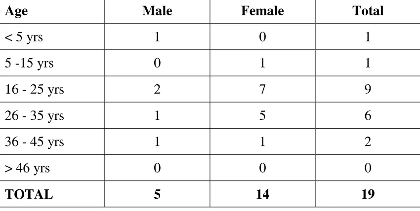
[image:55.595.96.521.512.726.2]3. Female gender was commonly affected and the age group was 16 - 25 years in Elbow contracture and 5 - 15 years in knee contracture.
TABLE -1
ELBOW CONTRACTURE - AGE / SEX DISTRIBUTION
Age Male Female Total
< 5 yrs 1 0 1
5 -15 yrs 0 1 1
16 - 25 yrs 2 7 9
26 - 35 yrs 1 5 6
36 - 45 yrs 1 1 2
> 46 yrs 0 0 0
0 1 2 3 4 5 6 7
< 5 yrs 5 -15 yrs 16 - 25 yrs
26 - 35 yrs
36 - 45 yrs
> 46 yrs
NO. O
F PA
T
IENTS
[image:56.612.120.523.80.679.2]AGE GROUP
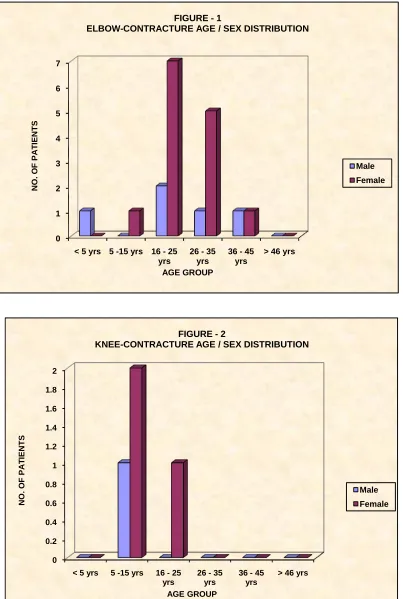
FIGURE - 1
ELBOW-CONTRACTURE AGE / SEX DISTRIBUTION
Male Female 0 0.2 0.4 0.6 0.8 1 1.2 1.4 1.6 1.8 2
< 5 yrs 5 -15 yrs 16 - 25 yrs
26 - 35 yrs
36 - 45 yrs
> 46 yrs
NO. O
F PA
T
IENTS
FIGURE - 2
KNEE-CONTRACTURE AGE / SEX DISTRIBUTION
TABLE -2
KNEE CONTRACTURE - AGE / SEX DISTRIBUTION
Age Male Female Total
< 5 yrs 0 0 0
5 -15 yrs 1 2 3
16 - 25 yrs 0 1 1
26 - 35 yrs 0 0 0
36 - 45 yrs 0 0 0
> 46 yrs 0 0 0
TOTAL 1 3 4
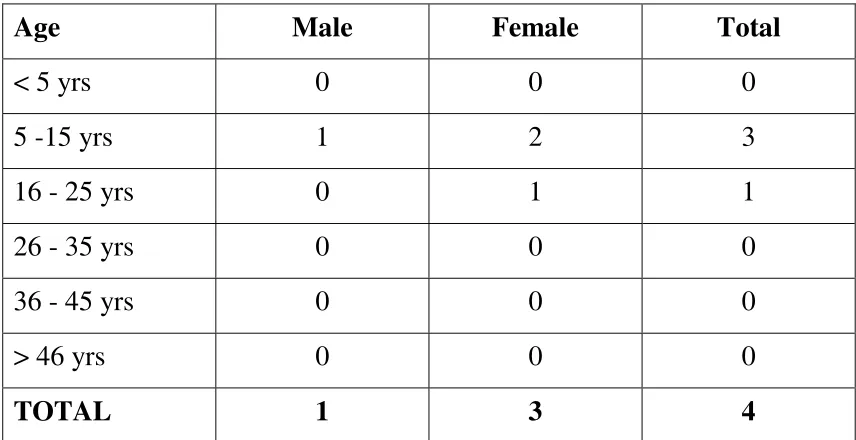
4. Elbow contracture being the commonest one, account for 82.6%
of the total contracture
5. Degree of contracture commonly reported was, more than 60°
for the elbow joint and 30-60° for the knee joint.
TABLE - 3
DEGREE OF CONTRACTURE - ELBOW
Degree Male Female Total
< 30° 3 3 6
30-60° 0 6 6
[image:57.595.95.523.602.744.2]82.6% 17.4%
FIGURE - 3
PERCENTAGE DISTRIBUTION
ELBOW
CONTRACTURE KNEE
CONTRACTURE
0 2 4 6 8 10 12 14
[image:58.612.120.516.388.692.2]Elbow Joint Knee Joint FIGURE - 4
ASSOCIATED DEFORMITY CORRECTION
Associated Deformity
TABLE - 4
DEGREE OF CONTRACTURE - KNEE
Degree Male Female Total
< 30° 0 1 1
30-60° 0 2 2
> 60° 1 0 1
6. All patients had full correction of flexion deformity
7. Average time taken for full correction of flexion deformity was
37.94 days for elbow contracture and 47.25 days for knee
contracture.
TABLE - 5
ASSOCIATED DEFORMITY CORRECTION
Elbow Joint Contracture
Knee Joint Contracture
Total
Associated Deformity 13 4 17
[image:59.595.95.512.565.694.2]0 0.5 1 1.5 2 2.5 3 3.5 4
SCAR BREAKDOWN BLISTER NEURO-VASCULAR COMPROMISE
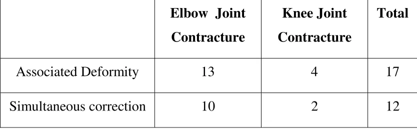
[image:60.612.117.523.79.677.2]RECURRENCE OF CONTRACTURE FIGURE - 5
COMPLICATIONS - ELBOW CONTRACTURE
MALE FEMALE 0 0.2 0.4 0.6 0.8 1 1.2 1.4 1.6 1.8 2
SCAR BREAKDOWN BLISTER NEURO-VASCULAR COMPROMISE
RECURRENCE OF CONTRACTURE
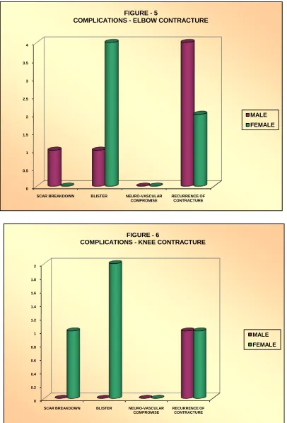
FIGURE - 6
COMPLICATIONS - KNEE CONTRACTURE
8. 13 patients amounting to, 68.4% of the total elbow contracture
patients and all the patients with knee contracture had
associated deformity. 10 patients with elbow contracture and 2
patients with knee contracture had simultaneous correction of
the associated deformity.
TABLES - 6
COMPLICATIONS - ELBOW CONTRACTURE
COMPLICATIONS MALE FEMALE TOTAL
Scar Breakdown 1 0 1
Blister 1 4 5
Neuro-Vascular Compromise 0 0 0
Recurrence of Contracture 4 2 6
TABLES - 7
COMPLICATIONS - KNEE CONTRACTURE
COMPLICATIONS MALE FEMALE TOTAL
Scar Breakdown 0 1 1
Blister 0 2 2
Neuro-Vascular Compromise 0 0 0
9. 5 patients with elbow contracture and 2 patients with knee
contracture had developed blister. One patient with elbow
contracture and one patient with knee contracture had scar break
down. All of them settled with conservative management
10. 6 patients with elbow contracture and 2 patient with knee
contracture had discontinued the splint and had developed
recurrence of contracture after correction by serial stretching,
PRE-STRETCHING
HAND CONTRACTURE
AXILLARY CONTRACTURE CONTRACTURE
RELEASE WITH Z PLASTY CONTRACTURE
RELEASE WITH SSG POST - STRETCHING
DISCUSSION
Burn injuries, regardless of the etiology, rarely involve the joint.
However, the joint function is often impaired because of burns. The joint
problems and joint deformities noted in burn patients are mostly due to
physical inactivity combined with limitation of joint movement because
of scar contracture.
Initial management of burns during the acute stage is concentrated
in the resuscitation of the patient from the burns shock ,with intravenous
fluids, analgesics and antibiotics. Management of the burn wound
depends on the depth of the injury. Collagen dressing is given to the
superficial wound. Wound that does not heal by twenty one days needs
surgical intervention, to prevent the wound from healing by secondary
intention and contracture deformity. Wound that needs more than 2
weeks to heal have very high chance of developing hypertrophic scar, so
pressure garments has to be given to prevent hypertrophic scarring, till
the scar matures.
Limb that has burnt is elevated during the acute stage to lessen the
edema formation and is splinted in the appropriate functional position.
Later active and passive mobilization of the joints are done to prevent
PRE-STRETCHING POST-STRETCHING
RIGHT ELBOW
FLEXION CONTRACTURE CONTRACTURE RELEASEWITH SKIN GRAFTING
COMBINED INTERVENTION
Most of our patients have very poor general condition and
nutritional status. They are very reluctant for mobilization and these are
the patients who are very prone to develop contracture, if physiotherapy
is not initiated at the proper time.
Nutritional status of most of our patients is very low, moreover
the food intake is also very poor. Both of these factors prolong the wound
healing process. Apart from the smelly discharge from the wound, pain
associated with the dressing change and mobilization, these patients are
very much depressed, which all add to their woes, making them reluctant
to mobilization, splinting and to have adequate food intake. When these
patients recover, contracture and other deformity like hypertrophic
scarring has already developed.
Holding bodily joints in flexion, so called posture of comfort, a
characteristic body posture is commonly seen in distressed individual.
Although the exact reasons are not entirely clear, contraction of muscle
fibers at rest and the contractile force difference between the flexor
muscles and the extensor muscles may play an important role in the
genesis of this body posture. The magnitude of joint flexion, furthermore,
increases as the individual loses voluntary control of muscle movement,
as frequently seen in burn victim. Prolonged periods of physical
PRE-STRETCHING POST - STRETCHING
POST - STRETCHING WITH STATIC SPLINT
INTER THIGH ADHESION
around the joint structure, as the recovery ensues, further impedes the
joint mobility.
Analysis of our study shows flame burn was the commonest cause
of burns. Female gender was commonly affected by burns, 17 out of the
total 23 patients included in our study were female, and the age group
commonly involved was between 16-25 years in elbow contracture. Knee
contracture was commonly seen in 5 - 15 years.
Elbow joint was commonly affected by contracture in our study,
accounting for 82.6% of the total cases. Literature review also shows
elbow joint being commonly involved in post burn contracture.
Degree of contracture commonly reported was more than 60° for
elbow joint and 30-60° for knee joint. The average time taken for full
correction of elbow contracture was 37.94 days and 47.25 days for knee
contracture.
13 patients amounting to 68.4% of total elbow contracture and all
the knee contracture patients involved in the study had associated other
deformity.
When multiple joints get deformed, important areas get priority in
contracture correction. Neck contracture is initially treated, as a secure
ASSOCIATED DEFORMITY
PRE-STRETCHING POST-STRETCHING