A STUDY OF INJURIES TO NECK STRUCTURES
IN CASES OF HANGING
Dissertation submitted to
THE TAMILNADU DR. M.G.R. MEDICAL UNIVERSITY
in partial fulfillment for the award of the degree of
DOCTOR OF MEDICINE
IN
FORENSIC MEDICINE
INSTITUTE OF FORENSIC MEDICINE MADRAS MEDICAL COLLEGE
CHENNAI - 600 003.
CERTIFICATE
This is to certify that the dissertation entitled, “A STUDY OF
INJURIES TO NECK STRUCTURES IN CASES OF HANGING”
submitted by Dr. N. VIJAYAKUMARI, in partial fulfillment for the award
of the degree of Doctor of Medicine in Forensic Medicine by the
Tamilnadu Dr.M.G.R. Medical University, Chennai is a bonafide record
of the work done by her in the Institute of Forensic Medicine, Madras
Medical College, during the academic year 2007– 2010.
DEAN DIRECTOR AND PROFESSOR,
MADRAS MEDICAL COLLEGE & INSTITUTE OF FORENSIC MEDICINE, GOVT. GENERAL HOSPITAL, MADRAS MEDICAL COLLEGE,
ACKNOWLEDGEMENT
It is an immense pleasure for me to be privileged to express my
sincere thanks and gratitude to my beloved professor
Dr.R.Vallinayagam. M.D., Director and Professor, Institute of Forensic
Medicine, Madras Medical College, Chennai-3 who has been the main
guiding force from the inception of this dissertation and throughout,
without his help this dissertation would not have been completed.
I offer my sincere thanks to Professor Dr.R.Selva Kumar M.D.,
Professor, Institute of Forensic Medicine, Madras Medical College,
Chennai-3 for his untiring help and advice rendered in bringing out this
study.
I am deeply indebted to Dr.V.Satyamurthy. M.D., Assistant
Professsor, Madras Medical College, Chennai for his attitude and sound
advice given to me, and sparing his valuable time in bringing out this
study.
I am extremely thankful to Dr. J. Mohana sundaram M.D., Ph.D.,
DNB., Dean, Govt. General Hospital and Madras Medical College,
I am also thankful to all my Assistant Professors, colleagues,
Technicians of the Institute of Forensic Medicine and of the Morticians of
Government General Hospital, Chennai.
Last but not the least, I am extremely thankful to my Husband
Dr.K.Kesavalingam, for his support and loving co-operation at all stages
CONTENTS
ACKNOWLEDGEMENT
1. INTRODUCTION
2. AIM OF THE STUDY
3. MATERIALS AND METHODS
4. REVIEW OF LITERATURE
5. RESULTS
6. DISCUSSION
7. CONCLUSION
8. RECOMMENDATION
BIBILIOGRAPHY
INTRODUCTION
Hanging remains to be one of the common methods of committing
suicide. Homicidal and accidental hanging are rare. Hence all cases of
hanging are considered as suicidal until the contrary is proved. Because
of the above, postmortem suspension of the body may be resorted to
mask the crime. So, a careful forensic examination is of great
importance, with the aim of ascertaining the antemortem character of the
lesion and also to exclude the possibility of murder dissimulation.
There is no specific gold standard to distinguish between
Antemortem hanging and Postmortem hanging. However presence of :
1) Vertical salivary dribble mark from the dependant angle of mouth
2) The phenomenon of Lefacies sympathetique
3) Presence of Petechial haemorrhages
4) Hyperaemia and ecchymosis of margins of ligature mark are
considered as features of antemortem hanging.
But obvious salivary dribble mark could be detected only in 56% of
cases. A meticulous examination of the body right at the scene of
hanging, that too, before removing the clothes and apparels can give the
1% as observed in different studies. Although petechial haemorrhages
commonly occur in cases of antemortem hanging, they are not
diagnostic of antemortem hanging and they occur in various asphyxial
and nonasphyxial deaths. And it is also equally important to note that
petechiae may be absent in rapid death due to vagal stimulation in
hanging. So, petechial haemorrhages cannot be taken as a specific
feature of antemortem hanging. Furthermore, the ligature mark, which is
considered as the principal external sign of hanging, is mainly a
postmortem phenomenon.
So, it is very much necessary to look for any injury to inner neck
structures, that is quiet frequent than the above, which cannot be
artificially produced and which also indicates ligature mark intravitality to
establish the antemortem hanging.
With this point in view I have chosen this topic of “A STUDY OF
INJURIES TO NECK STRUCTURES IN CASES OF HANGING”, to find
out which is the most common and most reliable criteria of neck injury to
AIM
The aim of this prospective study, done during the period of
August 2008 to July 2009, at the Department of Forensic Medicine,
Government General Hospital, Madras Medical College, Chennai-3, in
cases of deaths due to hanging, is to determine the prevalence of neck
injuries like,
1. Rupture/contusion of sternomastoid and strap musles of neck
2. Carotid intimal tear
3. Fracture of hyoid bone
4. Fracture of thyroid cartilage
5. Fracture of cricoid cartilage
6. Fracture and dislocation of cervical vertebra and
To find out, the most common and most reliable criteria of neck injury, to
MATERIAL AND METHODS
This prospective study was conducted in the Institute of Forensic
Medicine, at Madras Medical college and Government General Hospital,
Chennai-3, from August 2008 to july 2009. Only cases in which the
history and scene of crime examination report given by police and
relatives of the deceased are suggestive of Antemortem hanging were
included. 63 cases of deaths due to hanging, which was subjected to
Medicolegal Autopsy, were the subjects of this study.
All cases with,
1. External neck injuries other than the ligature mark,
2. Other external injuries suggestive of homicide, and
3. Cases with postmortem interval of more than 24hrs to avoid
artifacts of decomposition were excluded.
The information regarding identification of the deceased, reason
for committing suicide, place of hanging, material used, position of the
knot, type of knot, whether it was a complete or partial hanging, any
blood stains or disturbance, etc., at the scene of crime, to rule out the
possibility of homicide, history of any illness or drug intake, alcoholism,
from the police and detailed interviews of the relatives of the deceased.
Irrespective of the information gathered from the police records and
accompanying relatives of the deceased, in all cases, both external and
internal findings were observed meticulously during postmortem
examination to rule out homicidal hanging or any other cause of death.
Complete perusal of all the records done prior to Medicolegal Autopsy,
which is a routine protocol in all cases.
After identification of the body, a careful search for any external
injuries, dribbling of saliva, signs of asphyxia like bluish discoloration of
fingernails, petechial haemorrhages, signs of sphincter relaxation,
Le-facie symphathetic, pattern and also any distribution of hypostasis ,
extent of rigor mortis developed, etc are looked for. A detailed study of
the Ligature mark done. Finally a meticulous dissection of neck is done
by a step-by-step layer wise reflection of the tissues after the thoracic
organs and the brain have been removed. This allows the blood in the
neck to drain away, providing for a cleaner dissection field.
DISSECTION OF NECK:
The neck is extended by keeping a wooden block under the
shoulder. With a midline incision the skin and subcutaneous tissue is
fascial plane. The manubrium sternum is left intact at the beginning of
the autopsy when the rib cage is removed so that the inferior
attachments of the anterior cervical strap muscles remain unaltered.
After cutting the inferior attachments of the muscles, each muscle is
examined anteriorly and posteriorly for contusion or rupture and then
reflected superiorly. Now the carotid sheeth is identified and opened to
view the internal jugular vein and the carotid artery. With gentle
dissection carotid artery is separated and dissected out on both sides
from its origin till high up in the neck or few centimeters above its
bifurcation. Then, it is opened by cutting it longitudinally with small
scissors with blunt tips from below upwards and examined for transverse
carotid intimal tears, extravasation of blood or ruptures. Now the thyroid
glands are examined in situ and then removed to study the underlying
tracheal rings. Reflection of the trachea towards the face will allow for
the visualization of trauma to the prevertebral musculature and fascia.
Next the tongue, larynx and upper trachea are removed as a whole by
inserting the scalpel blade over the body of the hyoid bone and into the
floor of the mouth. Then the scalpel blade is directed downward along
each greater horn of the hyoid bone, cutting the pharyngeal tissues until
the anterior surface of the cervical vertebrae is seen. Then gentle
traction is applied to the larynx while dissecting it from rest of the neck
separated from ligaments and soft tissues and examined for areas of
blood extravasation and fracture. Cervical vertebra are examined for any
fractures, dislocations with areas of extravasation. All the positive and
negative findings are documented and photographed.
Blood samples taken from the left chamber of the heart and tissue
samples were collected for systemic toxicological analysis according to
regular procedures. All samples were sent to the Forensic Science
Laboratory, Mylapore, Chennai, to determine/rule out use of drugs,
poison and ethanol.
The data so obtained from detailed history, postmortem
examination and chemical analysis was statistically analysed and
ANATOMY OF NECK
Neck is the region of the body that lies between lower margin of
the mandible, mastoid process of temporal bone and superior nuchal
line of occipital bone above and upper border of clavicle and
suprasternal notch below. It is the major conduit between head and trunk
and limbs. Many important structures are crowded together in the neck
such as muscles, vessels, glands, nerves, trachea, oesophagus, larynx,
vertebrae etc. Some of these structures are very important to life like
thyroid gland, trachea, jugular vein, vagus nerve, vertebral arteries
etc.41,43 ‘Cervix’ or ‘Collum’ are the Latin words for neck.21
BONES OF NECK
There are seven cervical vertebrae and hyoid bone in the neck.
Typical cervical vertebrae have small broad body, triangular vertebral
foramen and bifid spine. The characteristic feature of cervical vertebrae
is foramen transversarium on the transverse process which transmits
vertebral artery. Third to Sixth vertebrae are typical cervical vertebrae.
The first cervical vertebra is called Atlas. It is a ring shaped one without
a body and spine. The second one is called Axis, having a strong tooth
occupying the position of body of Atlas. The seventh vertebra is called
vertebra prominence, has a long spinous process, which is not bifid and
a large transverse process with a small foramen.15, 21
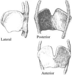
HYOID BONE
The word hyoid came from Greek word ‘hyoides’ which means
“shaped like letter upsilon”. It is a U shaped bone. It lies between
mandible and thyroid cartilage at the level of third cervical vertebra. It is
suspended by muscles connecting it to mandible, styloid process,
thyroid cartilage, manubrium and scapulae and does not articulate with
any other bone. Functionally hyoid bone serves as an attachment for
anterior muscles of neck and a prop to keep the airway clear. It has a
body and greater and lesser horns. Body of hyoid is 2.5cm wide and
1cm thick. Each end of the body is united with greater horn that projects
postero-superiorly. This union is by fibro cartilage in young people and
by bone in old people. The lesser horn is a small bony projection from
superior part of the body near its union with greater horn. It is connected
Fig. No. 1 Hyoid Bone 43
Hyoid bone is developed from the cartilages of second and
third visceral arches; lesser cornua from second arch greater horn from
third arch and body is formed from the fused ventral ends of both.
Chondrification of hyoid bone starts in the fifth week and completes by
third and fourth month of intra uterine life. There are six secondary
centres of ossification, two for the body and one for each cornu. Centre
for greater horn starts at the end of intra uterine life, for body before or
shortly after birth and for lesser horn around puberty. Apices of greater
horn remain cartilaginous until third decade.15
JOINTS OF NECK
Typical cervical joints are between lower six cervical vertebrae.
Here the supraspinous ligament is replaced by ligamentum nuchae
which is triangular in shape. Its apex is at the spine of seventh cervical
vertebra, base at external occipital crest, anterior border to cervical
spines and posterior border is free.6
Atlanto- occipital joints are between occipital condyles and
superior articular facets of atlas. They are synovial joints of ellipsoid
variety. 5Atlanto- axial joints are a pair of plane type of lateral atlanto
and a pivot type of median atlanto axial joint between the dens and the
anterior arch and transverse ligament of atlas.5
FASCIA OF NECK
Superficial cervical fascia is a thin layer of subcutaneous
connective tissue between the dermis of skin and investing layer of deep
cervical fascia anterolaterally. It contains platysma.5, 21
Deep cervical fascia has three layers. Investing layer lies deep to
platysma and it encloses trapezius and sternocleidomastoid muscles
and parotid and submandibular salivary glands. Pretracheal fascia
encloses thyroid gland and forms its false capsule. Para vertebral fascia
lies in front of para vertebral muscles.5, 21
SUPERFICIAL MUSCLES OF NECK
Platysma, sternocleidomastoid and trapezius are the superficial
muscles of neck.
Platysma is a broad thin sheet of muscle in the subcutaneous
tissue of neck. Platysma helps to depress the mandible and draw the
corners of mouth inferiorly.
Sternocleidomastoid is a broad strap like muscle with two heads;
to the medial third of clavicle. It is attached superiorly to the mastoid
process of temporal bone. On acting bilaterally it flexes the neck and
unilaterally it flexes and rotates the head and neck.
Trapezius is a large, flat, triangular muscle which covers
posterolateral aspect of neck and thorax. It is attached superiorly to
external occipital protuberance, superior nuchal line and ligamentum
nuchae and inferiorly to lateral third of clavicle.15, 21.
TRIANGLES OF NECK
Sternocleidomastoid muscle divides the neck into two main
triangles, anterior triangle anterior to it and posterior triangle posterior
to it.
Posterior triangle is bounded by sternocleidomastoid, clavicle and
trapezius. It is formed by splenius capitis, levator scapulae and scalenius
medius. Inferior belly of omohyoid again divides the posterior triangle
into occipital triangle above, which contains occipital artery and
accessory nerve and supraclavicular triangle below, which contains
external jugular vein and suprascapular artery. 5, 21
Anterior triangle is bounded by sternocleidomastoid muscle,
midline of neck and mandible. It is again divided into four smaller
1. Submandibular triangle is bounded by mandible and anterior
and posterior bellies of digastric muscle. It is also called digastric triangle
and contains submandibular salivary gland and submandibular lymph
nodes.21
2. Submental triangle is the unpaired suprahyoid area which is
inferior to chin. It is bounded by hyoid bone and anterior bellies of both
digastric muscles. Its floor is formed by myelohyoid muscles which meet
in the median fibrous raphae and it contains submental lymph nodes.21
3. Carotid triangle is bounded by superior belly of omohyoid,
posterior belly of digastric and anterior border of sternocleidomastoid
muscles. It contains common carotid artery, internal jugular vein and
vagus nerve in a tubular fascial condensation known as carotid sheath.
The triangle is also called vascular triangle.21
4. Muscular triangle is bounded by superior belly of omohyoid and
sternocleidomastoid muscles and median plane of neck. It contains infra
MUSCLES IN THE ANTERIOR TRIANGLE
Supra hyoid and infra hyoid muscles are attached to hyoid bone.
Supra hyoid muscles attach hyoid to the skull. They are:
1. Mylohyoid muscle is attached to mandible, hyoid bone and
median raphae. It forms the floor of mouth and supports the tongue and
elevates it.
2. Geniohyoid muscle is attached to genial tubercle i.e. inferior
mental spine of mandible and hyoid bone.
3. Stylohyoid muscle is attached to styloid process and body of
hyoid bone.
4. Digastric muscle has two bellies, anterior belly arises from
digastric fosse of mandible and posterior belly from mastoid notch and
these two are joined by an intermediate tendon which is attached to
hyoid bone by a fibrous sling.15,21
Infrahyoid muscles anchor hyoid bone to sternum, clavicles and
scapulae and depress the hyoid bone and larynx during swallowing and
speaking. They are called strap muscles because of their ribbon like
appearance. Sternohyoid and omohyoid belong to superficial group and
1. Sternohyoid muscle is narrow and lies superficially, parallel and
adjacent to median line. It is attached to manubrium sterni, medial
end of clavicle and body of hyoid bone.
2. Omohyoid muscle has a superior belly which is attached to inferior
border of hyoid bone and an inferior belly attached to superior
border of scapula. Superior belly lies laterally to sternohyoid muscle.
These two bellies are united by an intermediate tendon which is
connected to clavicle by a fascial sling.
3. Sternothyroid muscle is attached to posterior surface of manubrium
sterni and oblique line of thyroid cartilage. It is wider than
sternohyoid and lies under this and covers the lateral lobe of thyroid
[image:24.612.157.466.448.691.2]gland
4. Thyrohyoid muscle appears as a continuation of sternothyroid
muscle, running superiorly from the oblique line of thyroid cartilage
to inferior border and greater horn of hyoid bone. 15,21
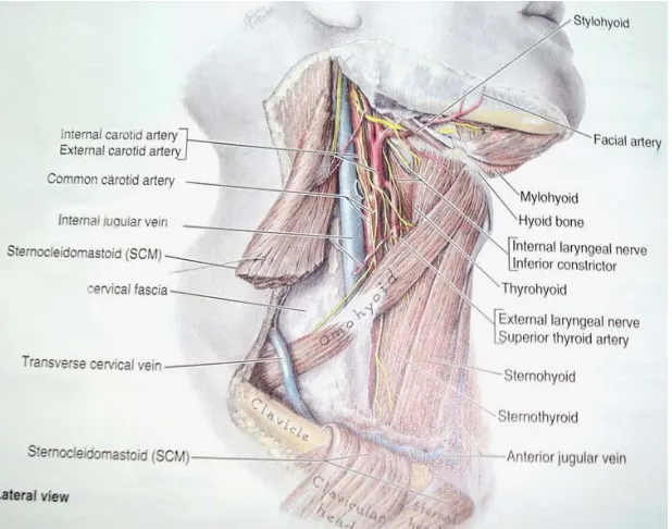
VESSELS OF NECK
Arteries of Neck: Common carotid artery is a branch of
brachiocephalic trunk on the right side and a branch of arch of aorta on
the left side. It ascends up in the carotid sheath and at the level of
superior border of thyroid cartilage, opposite to the level of inter vertebral
disc between third and fourth cervical vertebrae it divides into internal
and external carotid arteries. Internal carotid artery is a direct
continuation of common carotid artery and has no branches in the neck.
External carotid artery is the main branch supplying the head and neck
region. It has six branches, ascending pharyngeal, superior thyroid,
lingual, facial, occipital and posterior auricular arteries and two terminal
branches like maxillary and superficial temporal arteries. Carotid sinus is
a small dilatation involving the common carotid artery. Carotid sinus is a
baro receptor which reacts to changes in arterial blood pressure. Carotid
body is a small reddish brown mass of tissue lying on the medial side of
bifurcation of common carotid artery in close relation to carotid sinus. It
is stimulated by low levels of oxygen and initiates a reflex which
increases the rate and depth of respiration, cardiac rate and blood
pressure. Both carotid sinus and carotid body are innervated by
Glossopharyngeal and Vagus respectively.15, 21
Vertebral artery arises from first part of subclavian artery and it
ascends up through the foramen of transverse process of sixth cervical
vertebra. The cervical part of vertebral artery then passes up in the
foramen transversarium of cervical vertebrae up to the level of first
cervical vertebra where it grooves the posterior arch of atlas and enters
the cranial cavity through foramen magnum. The vertebral arteries on
both sides join to the basilar artery and participate in the formation of
cerebral arterial circle of Willis and supplies posterior part of brain.15, 21
VEINS OF NECK
External jugular vein is formed by the union of posterior division of
retromandibular vein with posterior auricular vein at the level of
mandibular angle, it crosses sternocleidomastoid muscle and perforates
deep fascia to enter the subclavian vein. It is covered by platysma,
superficial fascia and skin. Its tributaries are posterior jugular vein which
drains upper and posterior part of neck and anterior jugular vein which
drains the submandibular region. Anterior jugular veins of both sides are
Internal jugular vein is a direct continuation of sigmoid sinus at the
level of jugular foramen and behind the sternal end of clavicle it unites
with subclavian vein to form brachiocephalic vein. It drains blood from
brain and deep muscles of neck.15, 21
NERVES OF NECK
Vagus nerve is the tenth cranial nerve and is composed of both
sensory and motor fibres. It originates in the medulla oblongata and
leaves through middle of jugular foramen. It passes vertically downward
within the carotid sheath between common carotid artery and internal
jugular vein. Branches of vagal nerve in the neck are superior laryngeal
nerve, recurrent laryngeal nerve, pharyngeal branches and two to three
cardiac branches which accompany the sympathetic trunk and end in
cardiac plexus in thorax.41
Phrenic nerve is formed by anterior primary rami of third, fourth
and fifth cervical spinal nerves. In addition to the sensory innervations it
is the only motor supply to diaphragm.21
Sympathetic trunk receives white rami communicants in the neck
and associates with superior, middle and inferior cervical sympathetic
ganglia by way of grey rami communicants. These ganglia receive
synaptic fibres to splanchnic nerves. Superior ganglion is the largest one
and gives branches to external carotid artery. Inferior ganglion gives
branches to deep cardiac plexus. Middle ganglion is some times
absent.21
DEEP STRUCTURES OF NECK
Anterior vertebral muscles
1. Longus colli muscle on the anterior surface of vertebral column
extends between atlas and third thoracic vertebra.
2. Longus capitis muscle is attached to the anterior tubercles of third
to sixth cervical vertebrae and basilar part of occipital bone.
3. Rectus capitis anterior muscle attached to the anterior surface of
lateral mass of atlas to jugular process of occipital bone.
4. Rectus capitis lateralis muscle attached to transverse process of
atlas and to jugular process of occipital bone.16
LATERAL MUSCLES OF NECK
1. Scalenus anterior muscle extends between anterior tubercles of
transverse processes of third to sixth cervical vertebrae and
scalane tubercle of first rib. It lies behind sternocleidomastoid
2. Scalenus medius muscle is the largest and longest scalene
muscle. It is attached to posterior tubercle of third to seventh
cervical vertebrae, transverse process of atlas and axis to upper
border of first rib.
3. Scalenus posterior muscle is attached to posterior tubercle of
fourth to sixth cervical vertebrae and outer surface of second rib.
4. Levator scapulae muscle is attached to transverse process of first
to fourth cervical vertebrae and medial border of scapula.15
VISCERA OF NECK
The viscera of neck is deposited in three layers, superficial to deep
are endocrine layer formed of thyroid and parathyroid glands, respiratory
layer formed of larynx and trachea and alimentary layer formed of
pharynx and oesophagus.21
ENDOCRINE LAYER
Thyroid gland: It is deep to sternohyoid and sternothyroid muscles
and against fifth, sixth and seventh cervical and first thoracic vertebrae.
Parathyroid glands: Superior and inferior glands on either side lie
RESPIRATORY LAYER
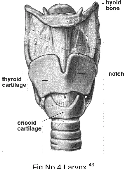
Larynx is the organ of voice production. It connects oropharynx
with trachea between the levels of third to sixth cervical vertebrae. It is
composed of nine cartilages, of which thyroid, cricoid and epiglottic
cartilages are single, and aretynoid, corniculate and cuneiform are
paired.
Thyroid cartilage (Fig.No.3) is the largest cartilage. Inferior two
thirds of its plate like laminae fuses anteriorly in the median plane to
form laryngeal prominence and superior part of it diverges to form
superior thyroid notch. The shallow indentation in the middle of inferior
border is known as inferior thyroid notch. Posterior border of each
lamina projects superiorly as superior horn and inferiorly as inferior horn.
Superior horns and borders are attached to hyoid bone by thyrohyoid
membrane. Inferior horns articulate with lateral surface of cricoid at
Fig.No.3 Thyroid cartilage43
Cricoid cartilage is a signet ring shaped thick and strong bone with
its band facing anteriorly and a lamina at the posterior part.
Epiglottic cartilage is a heart shaped elastic cartilage situated
posterior to root of tongue and hyoid bone and its broad superior end is
free. Aretynoid cartilages are a pair of pyramid shaped cartilages
articulating with lateral parts of superior border of cricoid cartilage
lamina. Corniculate and cuneiform cartilages are small nodules in the
posterior part of aryepiglottic folds. Triticeal cartilage is small
cartilaginous nodule found in the lateral thyroid ligament.54
Laryngeal muscles are extrinsic muscles and intrinsic muscles.
Extrinsic muscles are supra hyoid muscles which elevates larynx and
Posterior Lateral
infra hyoid muscles which depresses larynx. Intrinsic muscles are
cricothyroid, vocalis, thyroarytenoid, posterior cricoarytenoid, lateral
thyroarytenoid and transverse and oblique arytenoid muscles.
Cricothyroid muscle is the only extrinsic muscle which can be seen
externally. These intrinsic muscles make alterations in the length and
tension of vocal folds.15, 21
Trachea is a fibro-cartilaginous tube supported by incomplete
tracheal rings which are deficient posteriorly where trachea is adjacent
to oesophagus. It extends between the lower border of cricoid cartilage
opposite sixth cervical vertebra and sternal angle opposite to inter
vertebral disc between fourth and fifth thoracic vertebrae.5, 15, 21
(Fig.No.4)
ALIMENTARY LAYER
Pharynx extends from base of the skull to the inferior border of
cricoid cartilage opposite to lower border of sixth cervical vertebra.
Oesophagus is a muscular tube extending down from the inferior border
of cricoid cartilage opposite to lower border of sixth cervical vertebra as
Fig No.4 Larynx 43
CONTENTS OF VERTEBRAL CANAL
Vertebral canal contains from without inwards epidural space,
duramater, subdural space, arachnoid mater, subarchnoid space with
cerebrospinal fluid, spinal cord and spinal medulla.5
Medulla oblongata is the lowest part of brain stem, extending from
lower border of pons to a plane just above the first cervical spinal nerve.
Medulla contains respiratory and vasomotor centre.5Spinal cord is the
lowest part of central nervous system, extending from lower border of
ASPHYXIA
The term ‘asphyxia’ has originated from a Greek word ‘asphuxia’
meaning loss of heart beat.53 ‘Asphyxia’ literally means ‘defective
aeration of blood’ and etymologically, it implies ‘pulselessness’.23
Adelson defined asphyxia or anoxia as ‘the physiologic and chemical
state in a living organism in which acute lack of oxygen available for the
cell metabolism is associated with inability to eliminate excess of carbon
dioxide’.23 Anoxic anoxia can occur due to interference with respiration
by any mechanical obstruction in the air passages by constriction of air
passages. 29 Taylor suggested the word ‘apnoea’ as more appropriate
instead of asphyxia.52 Under the broad categorization of asphyxia, death
by constricting force on neck structures, include any form of pressure as
in hanging and strangulation.
CHARACTERISTIC FEATURES
i.
Well defined scattered tiny round pin point petechial haemorrhages
noticed over the visceral surface of pleurae, under conjunctivae,
over pericardium, epicardium and endocardium. They are also
seen on the connective tissue of aorta and oesophagus.42 They
are the result of venous stasis leading to capillary congestion and
rupture.38 They occur due to a) increased capillary pressure, b)
negative pressure in the chest due to inspiratory efforts made by
subject in an attempt to overcome the obstruction of air passage.
Petechial haemorrhages beneath the pleura and pericardium were
first described by French Police Surgeon Auguste Ambroise
Tardieu (1866) as pathognomonic of death by asphyxia and
became known as Tardieu spots.24,40,52
ii.
Cyanosis is a feature of anoxia. The word ‘cyanosis’ derived from
the Greek, means ‘dark blue.’ Cyanosis indicates blue colour of
skin, mucous membrane and internal organs.23 When the airway is
blocked, the oxygenation in the lungs is reduced which in turn
leads to diminution in the oxygen content of the arterial blood. This
will lead to darkening of all organs and tissues.22 It become more
pronounced in skin of lips, finger tips, ear lobules and also in
organs.29
iii. Intense visceral congestion as a result of obstructed venous
return.22
iv. Increased capillary permeability results from vasodilatation stasis
and suboxia; will in turn cause transudation of fluid from capillaries
into tissue spaces with oedema of organs and collection of excess
v. Postmortem fluidity of blood may occur. Various theories have
been postulated. This is due to fibrinolysins liberated from the
vascular endothelium. Fibrinolysin activation occurs due to release
of plasminogen activator, through the receptors on the vascular
wall for vasoactive materials that increase during agonal period.
Fibrinolysis is also activated by the leakage of plasminogen
activator due to increased permeability, and to degeneration and
necrosis of cell membrane, as a result of excessive acidosis after
death.39 Mole in 1948 demonstrated fibrinolysins in more than 90%
samples of fluid and incoagulable blood.28
Vogel and Cordier attributed this to be due to disturbance in the
plasma calcium. According to Oki the fluidity is because of the coagulum
formed from conversion of fibrinogen to fibrin which is not persisting as it
gets dissolved by proteolytic enzyme. Lenggenhagger opines that this
dissolution of fibrin is due to acidosis.28
HANGING
Hanging is a form of violent asphyxial death produced by
suspending the body with a ligature round the neck, the constricting
force being the weight of the body or a part of body weight.12, 27
When the point of suspension is over the centre of the occiput, the
vessels of neck become occluded to the maximum, and this is called
typical hanging. If point of suspension is any where else, it is called
atypical hanging. In the study by Shrabana Kumar Naik at Pune 7.4%
were typical hanging and 92.4% were atypical hanging.31
In hanging from high point of suspension, the victim is fully
suspended with his feet clear off the ground and it is called complete
hanging. Partial hanging is hanging from a low point of suspension; the
bodies are either partially suspended or where the bodies are found
sitting, kneeling, squatting or reclining, prone or supine position with feet,
heel or knee touching the ground. Hence complete suspension is not
essential to cause death by hanging, so also the constricting force need
not be the weight of the whole body. Only a slight degree of constriction
of neck may even cause eventual death.49 In a study at Pune 93.5%
were complete hanging and 6.5% were partial hanging.31 In another
study of 100 cases of hanging at Medical College Thiruvananthapuram
81% of cases were complete hanging and 19% were partial hanging.
Judicial Hanging is that form of hanging, when execution is carried
out by hanging by neck until death, following judicial decree. The
Procedure, is carried out with hanging by neck till death for more than
hundred years.
A well twisted Manilla rope of one inch in diameter having a length
of 19 feet already tested with one and a half times the weight of the
prisoner shall be used for executions. It should be sufficiently strong to
bear a strain of 280 lbs. (127Kg) with a 7 foot drop. The knot should be
placed one and half inches to the right or left of the middle line usually
under the angle of jaw. The "drop" is the length of the rope from a point
on the rope opposite the angle of the lower jaw of the criminal as he
stands on the scaffold, to the point where the rope is embraced in the
noose after allowing for the constriction of the neck that takes place in
hanging. Height of drop proportioned to the weight of the prisoner. 11
The sudden stoppage of the moving body associated with the
position of the knot causes the head to be jerked violently. This cause
fracture dislocation of second and third or third and fourth cervical
vertebrae.39 The body shall remain hanging for one hour and should not
be taken down till the medical officer declares life to be extinct.
Sex associated asphyxia involve the males exclusively. Reduction
in the blood supply to brain stimulates and heightens the sexual
suspended body of a male clothed in female attire, with complex
arrangement of ligature involving the genitals and presence of padding
between the ligature and neck indicate accidental hanging.38
Lynching is a method of homicidal hanging practiced in South
America.29 Lynch means ‘to put a person to death by mob action for an
alleged offence without legal trial’. 38
MECHANISM OF DEATH BY HANGING
A number of anatomical and physiologic factors are there in the
effect of constriction on neck due to hanging. The closure of airway is
not an essential element of hanging. Rein both (1895) has reported a
case of suicidal hanging in a man who had undergone tracheostomy and
the ligature was above the level of tracheostomy.23
CAUSES OF DEATH CAN BE ANY OF THE FOLLOWING.
1. Occlusion of airway or Asphyxia:
It may occur either due to direct pressure over larynx or cricoid
cartilage or trachea or the base of the tongue and epiglottis forced
upwards and backwards against the posterior pharyngeal wall and thus
obstructs airway. Asphyxiation appears to be the common
effective to close the trachea.22,27,51 Actual compression of airway by the
noose in hanging cases is not as common as generally believed.51
2. Occlusion of the neck veins or Venous Congestion or apoplexy
or congestive suboxia:
As the ligature presses upon the neck, the jugular veins are
compressed; as a result the return of blood from the brain is interfered
with, when carotid blood flow remains more or less patent. This results in
congestion and stasis of venous circulation of brain. Jugular veins will be
closed by a tension of 2 Kg (4.4 Lbs).38 Classical sign of congestion such
as cyanosis, congestion and oedema will be markedly pronounced.
3. Occlusion of arteries or Cerebral ischaemia or Syncope or Acute
Arterial Suboxia
The constricting force of the ligature may be sufficient to cause
compression of the carotid arteries and thus will prevent blood flow to
the brain, resulting in rapid unconsciousness from cerebral anemia.
Carotid arteries are blocked by a tension of 5 Kg (11Lbs) 23, 38 or 31/2
Kg14 and vertebral arteries by that of 30 Kg (66Lbs) 23, 38 or 16Kg14 on the
4. Effect of pressure on nerves of neck:
The pressure of the ligature, specially when it is a thin cord which
sinks deeply into tissues, may press upon the vagal sheath or carotid
bodies to result in reflex vagal inhibition, when the collapse will be
sudden, with little chance for suboxia to develop.29, 38 Stimulation of
nerve endings in the carotid sinus or sheath may be effected by direct
pressure from the hands or ligature where the death may ensue
immediately.23
5. Fracture Dislocation of cervical vertebrae
This mechanism usually occurs in the case of “Judicial hanging” or
“hanging with a long drop” where death is instantaneous from the effect
of fracture dislocation of second, third or fourth cervical vertebrae with
compression or laceration of the spinal cord.29
6. Remotely Stimulated Cardiac Dysfunction (RSCD)
RSCD is a recent expression denoting impairment of cardiac
function brought about by any remote neural or humeral mechanism, or
by mixture of the two either due to hypoxia or due to pressure upon
7. Combination of above factors:
When both air tubes and the blood vessels are compressed, even
though the pressure on air tube is partial, death will occur from
combined causes of obstructive asphyxia and impaired cerebral
circulation. This is taken to be the commonest cause.27 Death by
hanging usually results from arrest of the arterial supply to the brain or
obstruction of the venous return from it.51 The cause of death in hanging,
in most instances, is the compression of cervical vasculature and not
asphyxia by air way obstruction.36
SIGNS AND SYMPTOMS
When there is obstruction or interference of the process of
respiration the course of events passes through following distinct
phases.18
A. Dyspnoeic phase with expiratory dyspnoea showing increased
respiratory rate, cyanosis and tachycardia which last for 60 to 80
seconds.
B. Convulsive phase with loss of consciousness, depressed respiratory
movements, facial congestion, bradycardia and convulsion which
C. Pre terminal phase with respiratory pause, tachycardia and systemic
hypertension which lasts for 60 to 120 seconds.
D. Gasping for breath due to primitive respiratory reflexes.
E. Finally loss of movements, absence of reflexes and dilatation of
pupils which lasts for 1 to 4 minutes.
FATAL PERIOD
Fatal Period usually depend upon the mode of death. Hanging
causes instantaneous death, if cervical vertebrae are fractured as in
judicial hanging27 due to pressure of the dislodged bony process on vital
nerve center of the brain controlling respiration and circulation.51 If the
mode of death is pure asphyxia, death may occur very rapidly. In case of
partial obstruction of air passage, death may occur with in 5-10 minutes.
Death will occur least rapidly i.e. within 10-15 minutes when venous
congestion is the mode of death. If vagal inhibition occurs, death will be
very rapid, if not instantaneous.39
Death delayed for several days is rare. Delayed death occur due
to aspiration pneumonia, infections, oedema of lungs, oedema of
larynx, hypoxic encephalopathy, infarction of brain and abscess of
POST MORTEM APPEARANCES
The Post mortem findings can be divided into
A. General external appearances
B. Local external findings
C. Internal neck findings
D. Internal appearances.
A. GENERAL EXTERNAL APPEARANCE
a. Face will be pale, and not much congested in hanging.
b. Cyanosis will be noticed in hanging, when the suspension is from a
low point, ligature is deeply set, or when the ligature has broken
between the knot and the point of suspension.29 Features of
asphyxia are minimal in case of hanging compared to strangulation.
c. The eyes may be closed or partly open. Pupils are usually dilated or
mid dilated. If the ligature knot presses upon the cervical
sympathetic, the eye on the same side remain open and its pupil is
dilated. It indicates ante mortem hanging.27 Etienne Martin (1950)
has described this state as “Le facies sympathique”.38 Lopes C
Portugal Medico believes this to be due to unequal tension over the
structures of neck.38 In a study of fifty cases of hanging from Medical
d. Tongue may remain with in the teeth line or it may even be
protruded and get bitten, due to base of the tongue being forced
upwards by the ligature.38 Protruding part of the tongue is commonly
dark brown or even black as a result of drying.51 Some times the
tongue may be caught between teeth.27
e. Saliva will be found dribbling from the angle of mouth, down the chin
on the chest in straight lines, opposite to the side of knot in the
ligature. This is due to the increased salivation before death due to
the sympathetic stimulation of salivary glands by the ligature.
Salivation is increased by brain hypoxia or by stimulation of
pterygopalatine ganglion.39 The secretion of saliva is a vital act due
to stimulation of salivary gland and is indicative of suspension during
life, for the secretion ceases after the cessation of circulation.
Evidence of dried salivary dribble marks from one of the angles of
mouth is a sure sign of ante mortem hanging, but its absence alone
will not suggest that the body was suspended after death.23,27,29
f. Blood stained frothy fluid and mucous may escape from mouth and
g. Penis may be found engorged with blood due to hypostasis, it may
be found in semi erect position with or without evidence of emission
of seminal fluid in hanging.23,42
h. Postmortem staining will be well marked in the dependent parts of
the body; in hanging if the body has been suspended for some time
postmortem staining will be seen on lower part of the arms and legs
of the body i.e. glove and stocking distribution.27, 13 If the body is cut
down after death, before the fixation of postmortem staining and is
placed in supine position, postmortem lividity in the extremities will
fade and new areas of lividity will appear along the back and
buttocks.51
Usually fixation of postmortem staining occurs in 6 to 12 hours due
to postmortem coagulation of blood in capillaries. In asphyxial deaths
fixation of postmortem staining is delayed due to postmortem fluidity of
blood.
B. LOCAL EXTERNAL FINDINGS
On the neck, ligature material and the ligature mark is an important
LIGATURE MATERIAL
Consistency of the material
Any substance available at hand is used as ligature. Articles
commonly used as ligature may be soft materials like “dhothie”, “Saree”,
“Turban”, “Bed-sheet”, “Sacred thread”, “handkerchief”, ”neck tie”, “cord
of pyjama”, “boot lace”, hard and pliable materials like “Electric cord”,
“Belt”, “wire”, “Leather strap”, “metallic chain” and materials producing
patterns like “Rope” made of cotton, coconut fibre or jute, etc or may be
any thing handy and available near the place of occurrence.
Dr.Shrabana noticed soft material in 54.7% cases of hanging and
hard material in 28.6% hanging cases in his study.31 Prisoners tear
their sheets into strips and use as ligature as well as they use T- shirts,
under shots as ligature.1 Rarely couples hang themselves together with
the same rope.7
TYPE OF KNOT
Different types of knots were described.34 The usual choice is a
simple slipknot to produce a running noose or a loose loop or a fixed
loop produced by non slipping granny or reef knot. Occasionally a simple
loop may be used without any knot.29 A fixed noose is one in which the
passed through the loop made from the other end.35 Artificer’s knot is
used in the case described by Tardieu.38
POSITION OF KNOT
The knot is usually situated over the side of the neck over the
angle of mandible, mastoid region or occiput; at times it may be situated
below the chin. Suspension by a knot below the chin is rare and
described by Tardieu.38 After suspension, the knot remains at a higher
level than the remainder of the ligature, movement of the knot being due
to the act of suspension. The position of the knot determines the force
exerted on the neck by the ligature and it will be on the side opposite the
point of suspension.
LIGATURE MARK
Mark of ligature on the neck is the most out standing and
characteristic sign of death from hanging. The situation of the mark will
be largely influenced by the method of application and movement of the
ligature. The more tightly the ligature is applied, the deeper will be
ligature mark.
The mark in hanging is usually situated above the level of thyroid
between larynx and chin and is directed obliquely upwards towards the
irregular impression of a knot. The depth of the ligature mark will be
more on the side of the neck opposite the knot.23, 29, 38 Mark may be on or
below the level of suspension in case of partial hanging.13 The mark may
be circular or oblique if a ligature is passed round the neck more than
once.27 In hanging, the ligature mark extend upwards and forms an
inverted “V” in the back of neck. The inverted “V” represents where the
knot of the noose was located.9
Considering the level of constriction represented by ligature mark,
it was found to be situated on or above the level of laryngeal prominence
in most cases of suicidal hanging and all cases of homicidal hangings.
Ligature mark was found below the level of laryngeal prominence in 12
(4.76%) cases of suicidal hanging most of which were partial hangings.32
Considering discontinuity along the course of ligature mark, it was
found as a common feature in most of the suicidal as well as homicidal
hangings and discontinuity in the mark could not be detected in 11.11%
cases of suicidal hanging and 33.33% cases of homicidal hanging.32
Reddy KSN has mentioned that mark of hanging is situated above
the level of thyroid cartilage, between larynx and chin in 80% cases. It
below the level of thyroid cartilage in about 5% cases, especially in
partial suspension.30, 39
Pattern of ligature material impressed on neck and characteristic
diagonal marks of strands of the rope may be seen. This is known as
mirror image phenomenon.35 Ligature pattern is better appreciated by
examining under oblique lighting and using magnifying lens.39 The mark
is superficial and broad if a cloth or soft material is used. The wide band
of cloth when used as a ligature on the bare skin may cause narrow
ligature mark due to tension lines in the stretched cloth. Ligature mark is
well defined, narrow and deep if a firm string is used.
There may be two ligature marks on neck in hanging, one being
oblique, non continuous and high up in the neck and the other being
circular and low down in the neck due to slipping of noose from the lower
to higher position for long suspension.29
The fibres can be lifted off by sticking thin transparent adhesive
tape 5cm long around the ligature mark and its surrounding. These
tapes later transferred into clean microscopic slides, can be examined
directly under microscope for fibres. Inspection of the neck with oblique
light may show the pattern produced by ligature. Examination under UV
adhesive cello tapes from palms of the deceased can be examined for
fibres if he / she have handled the ligature material. If fibres are detected
it can be compared with those of the ligature with the help of a
comparison microscope. This test is called as cellophane test or fibre
test.53
B. INTERNAL EXAMINATION OF NECK STRUCTURES
a. Injury to subcutaneous tissue and muscles of neck
The subcutaneous tissue underneath the ligature mark on
dissection will be compressed, dry, white, shiny, glistening and leathery
in nature in hanging.
Injuries to neck musculature are rare in hanging.51 Bruising and
rupture of muscle fibres of platysma and sternocleidomastoid especially
at their sternal attachments can be seen especially in long drops or
complete hanging.23,35 This is seen in 2-10% of cases.12 Dr.S.Sivasuthan
noted rupture of lower attachment of sternomastoid muscle in 62% of
cases.50 In a retrospective study of 175 cases of suicidal hanging over a
five- year period, the most frequent injury was injury to
sternocleidomastoid muscle.3
When a ligature around the neck suspends a body, the ligature
of the body. Among the straps muscles of neck Sternomastoid(s) are
strong and bulky muscles, the lower attachments of which can easily be
dissected and examined. When weight of the trunk is born by these
muscles during suspension, there is every chance for rupture of the
lower attachment of the muscles. In the case of hanging, death occurs in
five to ten minutes except in judicial hanging where it is almost
instantaneous, but the heart continues to beat for 15 to 20 minutes.
Hence if the rupture of the muscles occurs, there will be definite
infiltration of blood at the site. Such an infiltration will not occur if a dead
body is suspended (post mortem hanging)
b. Injury to vessels of neck
Carotids may show transverse tear of the intima, with adjacent
haemorrhage or with extravasations of blood in the vessel wall under the
groove above the point of bifurcation in their walls as a result of friction
of opposite sides due to pressure exerted by the ligature i.e. traction
associated with hanging.23,51 Direct compression of the carotid artery
between the ligature and the cervical vertebra can lead to intimal tear.
Even minimal trauma has been associated with carotid intimal tear or
dissection and some relate such dissections to a minor traumatic event
including excessive cervical rotation and hyperextension either on a
intima of carotid arteries is seen in 5 to 10% of hanging cases.38, 39 Dr.
Sivasuthan has noted carotid intimal tear in 1% of cases.40 Dr. Madan
Mohan noted intimal tear in 4% of cases.26
c. Injury to bones of neck
Regarding the fractures, fracture of hyoid bone is an exception
rather than a rule in hanging. The hyoid bone fractures were divided by
Weintraub into (a) those caused by inward or side wise compression, (b)
those due to anteroposterior compression, (c) traction or avulsion
fractures.23, 38 Hanging has produced both inward and outward fractures
of hyoid bone.38
Hyoid bone is usually intact in 80% of cases of hanging.30 In his
study of one hundred hyoid bones; Renjith found hanging as major
cause of hyoid bone fracture. He found an incidence of 28.5% of hyoid
bone fracture, out of 21 cases of hanging, all of them were males.40 It
may get fractured involving the greater horns at the junction of outer
2/3rd and inner 1/3rd.8There may be traction fracture through the pressure
by the ligature on the thyrohyoid ligament. Smith and Fades remarked
that ‘the hyoid bone is practically never injured’.23 It is known that larynx
remain cartilaginous and joints of hyoid remain mobile up to 40 years.29
The fracture is more frequent in persons over 40 years.8,23 In a review of
cartilages were found in 9% of cases and were more common in older
individuals. In this study, genders, height of suspension and ligature type
do not seem to be of predictive value.Dr.Shrabana has noted that out of
his 257 cases of hanging not a single victim had hyoid bone fracture. 31
X-ray examination is more helpful to disclose the fracture in
suspected case of fracture hyoid.3, 12
Fracture or dislocation of upper cervical vertebrae with
compression or laceration of the cord or its transection or separation
from its junction with medulla, may be noticed in judicial hanging with a
long drop and also in suicidal hanging where the victim jumps from a
height and its fall is arrested by sudden jerk of a ligature.27 Cervical
vertebral fractures have been reported in all cases of judicial hanging in
an analysis of six cases.48
d. Injury to cartilages of neck
Regarding the fracture of cartilages, fracture of thyroid cartilage or
other cartilages are exception rather than a rule in hanging. Such
fractures are more likely to occur in hanging if elderly individuals who
jump to their death and remain suspended after a long drop, due to
calcification of cartilage and fragility of bony structures.51 DiMaio in his
there are no injuries. Only ten cases (12%) showed fractures, of which
nine were thyroid cartilage fracture, specifically the superior horns; one a
fracture to cervical spine and none were hyoid bone fracture. Of the nine
cases of thyroid cartilage fractures, seven were unilateral and two were
bilateral.7
Polson and Gee found fracture of superior horn/ horns of thyroid
cartilage in 50% of cases of hanging.23Fracture of cricoid cartilage is
rather less common. Fracture dislocation of cervical vertebrae is not at
all common, seen only in judicial hanging.29
All other organs will be congested with petechiae on the surface of
brain, heart and lungs.8 Lungs were found congested and oedematous
with numerous sub pleural petechial haemorrhages.29 Typical asphyxial
TABLE – 1
AGE WISE DISTRIBUTION OF DEATHS DUE TO HANGING
AGE(YRS) NUMBER OF
CASES PERCENTAGE
<20 9 14%
20-30 24 38%
30-40 18 29%
40-50 6 10%
>=50 6 10%
Total 63 100%
0 5 10 15 20 25 30
<20 20-30 30-40 40-50 >=50
P e rc e n ta g e
Age in Years
AGE WISE DISTRIBUTION OF DEATHS DUE
TO HANGING
[image:57.612.82.514.310.630.2]TABLE - II
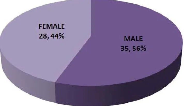
GENDER WISE DISTRIBUTION OF DEATHS DUE TO HANGING
GENDER NUMBER OF CASES PERCENTAGE
MALE 35 56%
[image:58.612.146.502.311.516.2]TABLE – I: Shows age wise distribution of 63 cases of deaths due to
hanging. The age ranged from 13yrs to 72yrs. The age
group, 20-30years, accounted for the maximum number of
cases, 24cases (38%), followed by the age group 30-40yrs,
18cases (29%). Extremes of age, i.e., age less than 20yrs,
comprised 9 cases (14%) and more than 60yrs, 2cases
(3%) of victims each.
TABLE – II: Shows gender wise distribution of 63 cases of deaths due
to hanging. The incidence among males was 56% which
comprised 35 cases and among females was 44% which
TABLE - III
LIGATURE MATERIAL USED
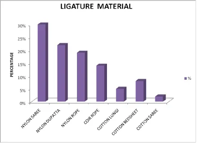
MATERIAL USED NUMBER OF CASES PERCENTAGE
NYLON SAREE 19 30%
NYLON DUPATTA 14 22%
NYLON ROPE 12 19%
COIR ROPE 9 14%
COTTON LUNGI 3 5%
COTTON BEDSHEET 5 8%
[image:60.612.104.511.338.636.2]TABLE – III:Shows the incidence of ligature material used in 63 deaths
due to hanging. Nylon saree is the most common ligature
material that is used in 19cases (39%), next common being
the nylon dupatta in 14cases (22%) and nylon rope in 12
cases (19%). Rarely used material was cotton lungi in
3cases (5%) and cotton saree in only one case (2%). So
most commonly used ligature material is the commonly
available household soft material, i.e., nylon saree, dupatta
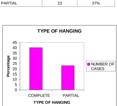
TABLE - IV TYPE OF HANGING
TYPE OF HANGING NUMBER OF
CASES PERCENTAGE
COMPLETE 40 63%
PARTIAL 23 37%
TYPE OF HANGING
0 5 10 15 20 25 30 35 40 45
COMPLETE PARTIAL
TYPE OF HANGING
P
e
rcen
ta
ge
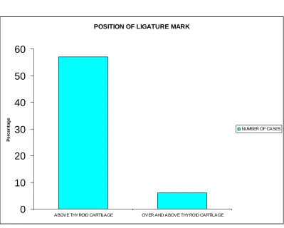
TABLE - V
POSITION OF LIGATURE MARK
POSITION OF LIGATURE MARK
NUMBER OF
CASES PERCENTAGE
ABOVE THYROID CARTILAGE 57 90%
OVER AND ABOVE THYROID
CARTILAGE 6 10%
POSITION OF LIGATURE MARK
0
10
20
30
40
50
60
ABOVE THYROID CARTILAGE OVER AND ABOVE THYROID CARTILAGE
P
e
rcen
ta
g
e
TABLE - VI
POSITION OF KNOT
POSITION OF KNOT NUMBER OF
CASES PERCENTAGE
LEFT SIDE OF THE NECK 36 57%
BACK OF NECK 19 30%
RIGHT SIDE OF NECK 7 11%
BELOW THE CHIN 1 2%
POSITION OF KNOT
57% 30%
11% 2%
LEFT SIDE OF THE NECK
BACK OF NECK
RIGHT SIDE OF NECK
TABLE – IV: Shows incidence of complete and partial hanging in this
study of 63 deaths due to hanging. Overall, 40 cases (63%)
were complete hanging and 23 cases (37%) were partial
hanging. So incidence of, complete hanging is twice as that
of partial hanging.
TABLE – V: Shows the incidence of, ligature mark position over the
neck in this study. In 57cases (90%) it was seen above the
thyroid cartilage and in only 6cases (10%) it was seen over
and above the thyroid cartilage.
TABLE – VI:Shows the incidence of position of the knot in this study. 19
cases (30%) were typical hanging and 44 cases (70%) were
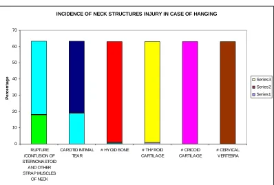
TABLE - VII
INCIDENCE OF NECK STRUCTURES INJURY IN CASE OF HANGING
INJURIES Present Absent
No. % No. %
RUPTURE /CONTUSION OF STERNOMASTOID AND OTHER STRAP MUSCLES
OF NECK 18 29% 45 71%
CAROTID INTIMAL TEAR 19 30% 44 70%
# HYOID BONE 1 2% 62 98%
# THYROID CARTILAGE 1 2% 62 98%
# CRICOID CARTILAGE 0 0% 63 100%
# CERVICAL VERTEBRA 0 0% 63 100%
INCIDENCE OF NECK STRUCTURES INJURY IN CASE OF HANGING
0 10 20 30 40 50 60 70 RUPTURE /CONTUSION OF STERNOMASTOID AND OTHER STRAP MUSCLES OF NECK CAROTID INTIMAL TEAR
[image:66.612.98.496.360.629.2]TABLE – VII :Shows incidence of internal neck structure injuries in case
63 deaths due to hanging. Out of 63 cases, carotid intimal
tear and rupture/contusion to sternomastoid and other strap
muscles was the commonest internal neck structure injury
that was seen in 19cases (30%) and in 18cases (29%)
respectively. Hyoid bone fracture and thyroid cartilage
fracture was seen in only one case (2%). Fracture of cricoid
cartilage and cervical vertebral fracture was not seen in any
TABLE - VIII
AGE WISE DISTRIBUTION OF INJURIES TO NECK STRUCTURES
1. RUPTURE/CONTUSION OF STERNOMASTOID AND OTHER STRAP MUSCLES OF NECK
Present Absent
No. % No. %
<20 3 33% 6 67%
20-30 6 25% 18 75%
30-40 4 22% 14 78%
40-50 4 67% 2 33%
>=50 1 17% 5 83%
2. CAROTID INTIMAL TEAR
Present Absent
No. % No. %
<20 1 11% 8 89%
20-30 7 29% 17 71%
30-40 5 28% 13 72%
40-50 4 67% 2 33%
3. FRACTURE OF HYOID BONE
Present Absent
No. % No. %
<20 0 0% 9 100%
20-30 0 0% 24 100%
30-40 0 0% 18 100%
40-50 1 17% 5 83%
>=50 0 0% 6 100%
4. FRACTURE OF THYROID CARTILAGE
Present Absent
No. % No. %
<20 0 0% 9 100%
20-30 0 0% 24 100%
30-40 0 0% 18 100%
40-50 1 17% 5 83%
6. FRACTURE OF CERVICAL VERTEBRA
Present Absent
No. % No. %
<20 0 0% 9 100%
20-30 0 0% 24 100%
30-40 0 0% 18 100%
40-50 0 0% 6 100%
>=50 0 0% 6 100%
5. FRACTURE OF CRICOID CARTILAGE
Present Absent
No. % No. %
<20 0 0% 9 100%
20-30 0 0% 24 100%
30-40 0 0% 18 100%
40-50 0 0% 6 100%
TABLE – VIII
1. RUPTURE/CONTUSION OF STERNOMASTOID AND OTHER
STRAP MUSCLES OF NECK:
In this study, rupture/contusion of sternomastoid and other strap
muscles of neck are more common in the age group of 40-50yrs, seen in
4cases (67%), lowest being in age group above 50yrs, seen in 1case
(17%). In other age groups it is around 22-33%.
2. CAROTID INTIMAL TEAR:
Out of 63 cases, carotid intimal tear was commonly seen in the
age group of 40-50yrs, i.e., around 67% of cases. The lowest being in
the age group below 20yrs (11%).
3. FRACTURE OF HYOID BONE, THYROID CARTILAGE, CRICOID
CARTILAGE, AND CERVICAL VERTEBRA:
In the present study, hyoid bone fracture and thyroid cartilage
fracture was seen in only one case (17%) in the age group of 40-50yrs.
Cricoid cartilage and cervical vertebra fracture was not seen in any
TABLE – IX
DISTRIBUTION OF NECK STRUCTURE INJURIES IN DIFFERENT TYPES OF HANGING
1. RUPTURE/CONTUSION OF STERNOMASTOID AND OTHER STRAP MUSCLES OF NECK
Present Absent
No. % No. %
COMPLETE 15 38% 25 63%
PARTIAL 3 13% 20 87%
2. CAROTID INTIMAL TEAR
Present Absent
No. % No. %
COMPLETE 14 35% 26 65%
PARTIAL 5 22% 18 78%
3. FRACTURE OF HYOID BONE
Present Absent
No. % No. %
COMPLETE 1 3% 39 98%
4. FRACTURE OF THYROID CARTILAGE
Present Absent
No. % No. %
COMPLETE 1 3% 39 98%
PARTIAL 0 0% 23 100%
5. FRACTIRE OF CRICOID CARTILAGE AND CERVICAL VERTEBRA
Present Absent
No. % No. %
COMPLETE 0 0% 40 100%
TABLE – IX
DISTRIBUTION OF NECK STRUCTURE INJURIES IN DIFFERENT
TYPES OF HANGING
1. RUPTURE/CONTUSION OF STERNOMASTOID AND OTHER
STRAP MUSCLES OF NECK:
This table shows incidence of rupture/contusion of sternomastoid
and other neck muscles in different types of hanging. Out of 63 cases of
death due to hanging injury to sternomastoid and other neck muscles
was found to be more common in complete hanging, 15cases (38%). In
partial hanging it was seen only in 3cases (13%).
2. CAROTID INTIMAL TEAR:
Carotid intimal tear was more common in complete hanging,
14cases (35%). In partial hanging it was seen in only 5 cases (22%).
3. FRACTIRE OF HYOID BONE, THYROID CARTILAGE, CRICOID
CARTILAGE AND CERVICAL VERTEBRA:
Fracture of hyoid bone and thyroid cartilage was seen in only
complete hanging, 1case (3%). Fracture of cricoid cartilage and cervical
TABLE – X
RELATIONSHIP OF POSITION OF KNOT WITH INJURIES
1.STERNOMASTOID RUPTURE
Present Absent
No. % No. %
BACK OF NECK 7 37% 12 63%
BELOW THE CHIN 0 0% 1 100%
LEFT SIDE OF THE NECK 10 28% 26 72%
RIGHT SIDE OF NECK 1 14% 6 86%
2. CAROTID INTIMAL TEAR
Present Absent
No. % No. %
BACK OF NECK 8 42% 11 58%
BELOW THE CHIN 1 100% 0 0%
LEFT SIDE OF THE NECK 9 25% 27 75%
RIGHT SIDE OF NECK 1 14% 6 86%
3. FRACTURE OF HYOID BONE
Present Absent
No. % No. %
BACK OF NECK 1 5% 18 95%
BELOW THE CHIN 0 0% 1 100%
LEFT SIDE OF THE NECK 0 0% 36 100%
4. FRACTURE OF THYROID CARTILAGE
Present Absent
No. % No. %
BACK OF NECK 1 5% 19 100%
BELOW THE CHIN 0 0% 1 100%
LEFT SIDE OF THE NECK 0 0% 36 100%
RIGHT SIDE OF NECK 0 0% 7 100%
5. FRACTURE OF CRICOID CARTILAGE AND CERVICAL VERTEBRA
Present Absent
No. % No. %
BACK OF NECK 0 0% 19 100%
BELOW THE CHIN 0 0% 1 100%
LEFT SIDE OF THE NECK 0 0% 36 100%