J
OURNAL OFV
IROLOGY,
0022-538X/00/$04.00⫹0
May 2000, p. 4273–4283
Vol. 74, No. 9
Copyright © 2000, American Society for Microbiology. All Rights Reserved.
Rescue of Multiple Viral Functions by a Second-Site Suppressor
of a Human Immunodeficiency Virus Type 1
Nucleocapsid Mutation
ANDREA CIMARELLI,
1SARA SANDIN,
2STEFAN HO
¨ GLUND,
2ANDJEREMY LUBAN
1,3*
Departments of Microbiology
1and Medicine,
3Columbia University College of Physicians and Surgeons, New York,
New York 10032, and Department of Biochemistry, Biomedical Center, Uppsala, Sweden
2Received 30 November 1999/Accepted 25 January 2000
Human immunodeficiency type 1 (HIV-1) bearing the nucleocapsid (NC) mutation R10A/K11A is replication
defective. After serial passage of the mutant virus in tissue culture, we isolated a revertant that retained the
original mutation. It had acquired, in addition, a new mutation (E21K) that was formally demonstrated to be
sufficient for restoration of viral replication. Detailed analysis of the replication defect of R10A/K11A revealed
a threefold reduction in virion yield and a fivefold reduction in packaging of viral genomic RNA. Real-time PCR
was then used to quantitate viral DNA synthesis following infection of Jurkat T cells. After adjustment for the
assembly and packaging defects, a minor (twofold) reduction in synthesis of either strong-stop, full-length
linear DNA or 2-LTR circles was observed with R10A/K11A virions, indicating that reverse transcription and
nuclear transport of the viral genome were largely intact. However, after adjustment for the amounts of
full-length or 2-LTR circles produced, R10A/K11A virions were at least 10-fold less infectious than wild type,
indicating that viral DNA produced by the R10A/K11A mutant failed to integrate. Each of the above-mentioned
defects was corrected by introduction of the second-site compensatory mutation E21K. These results
demon-strate that the replication defect of mutant R10A/K11A can be explained by impairment at multiple steps in
the viral life cycle, most important among them being integration and RNA packaging. The E21K mutation is
predicted to restore positive charge to the face of the R10A/K11A mutant NC protein that interacts with the
HIV-1 SL3 RNA stem-loop, emphasizing the importance of NC basic residues for HIV-1 replication.
Retroviral nucleocapsid (NC) proteins are expressed as part
of a Gag polyprotein precursor which is cleaved by the
virus-encoded protease during virion maturation (reviewed in
refer-ences 29 and 47). With the exception of spumaviruses, NC
proteins encoded by different retroviruses share two structural
characteristics: the presence of either one or two Cys-His box
motifs (Cys-X
2-Cys-X
4-His-X
4-Cys) and a large number of
ba-sic residues distributed throughout the protein (reviewed in
references 5 and 63).
NC plays roles in nearly all steps of the viral life cycle. As a
domain within the Gag polyprotein, NC specifically binds and
incorporates viral genomic RNA into virions (reviewed in
ref-erences 5 and 63) and drives virion assembly by promoting
interaction among Gag polyproteins (4, 11, 14, 17a, 28, 34, 42,
54, 67). Within the nascent virion, after cleavage from the Gag
polyprotein, NC coats viral genomic RNA and promotes its
maturation into a more stable dimeric form (26, 27, 30, 31).
Upon infection of a susceptible target cell, NC contributes to
reverse transcription (RT) (1, 2, 39, 49, 59, 62, 68, 71). NC may
also facilitate integration of viral DNA into host cell
chromo-somal DNA, either by facilitating the integrase-mediated
strand transfer or by relieving DNA secondary structure, as
suggested by in vitro studies (15, 16, 48). It has been difficult to
confirm the in vitro effects of NC on integration in vivo since
many NC mutations decrease RNA packaging or directly
in-hibit the efficiency of RT. The effect of these mutations is to
limit the yield of viral DNA synthesized after infection to levels
too low for meaningful analysis of subsequent events.
Re-cently, however, Moloney murine leukemia virus (M-MuLV)
NC mutations have been shown to block a step in the
replica-tion cycle that follows nuclear entry of viral DNA, suggesting
that NC plays a role in integration in vivo (36).
All of NC’s varied functions appear to depend on its ability
to bind RNA (for reviews, see references 5, 19, and 63). Both
Cys-His boxes and basic residues are determinants of NC’s
interaction with RNA, the former providing specificity for
in-teraction with viral genomic RNA and the latter providing
nonspecific association with nucleic acid (21). Though NC
Cys-His boxes have received a great deal of attention, the basic
residues, through their nonspecific RNA-binding activity,
me-diate many of NC’s functions, as mutation of human
immuno-deficiency virus type 1 (HIV-1) NC basic residues can disrupt
RNA packaging (6, 17a, 58, 60), virion assembly (17a, 20), and
RT (6, 40).
In this study, we report the isolation of a viral revertant of a
replication-defective mutant in which two basic residues at the
N terminus of HIV-1 NC are replaced by alanine (R10A/
K11A). We show that the phenotypic reversion is due to the
presence of a second-site compensatory mutation (E21K).
De-tailed characterization of the R10A/K11A mutant shows that
there are multiple defects throughout the viral life cycle,
rang-ing from genomic RNA packagrang-ing to integration of viral DNA.
Each of the defects is corrected to a considerable extent by the
presence of the E21K mutation.
MATERIALS AND METHODS
Plasmid DNAs.The HIV-1 proviral construct R10A/K11A is described
else-where (17a). This construct, as well as all the proviral constructs used in this study, are chimeric proviral DNAs in which anSphI/EcoRV fragment in NL4-3 that spans the NC coding sequence has been replaced by the corresponding fragment of HXB-2 (nucleotides 1443 to 2977, according to reference 57). Mu-tation E21K was introduced de novo into mutant and wild-type proviral DNAs by
* Corresponding author. Mailing address: Departments of
Microbi-ology and Medicine, Columbia University College of Physicians and
Surgeons, 701 W. 168th St., New York, NY 10032. Phone: (212)
305-8706. Fax: (212) 305-0333. E-mail: [email protected].
4273
on November 9, 2019 by guest
http://jvi.asm.org/
mutagenic PCR, according to standard procedures and using oligonucleotides 5⬘-CAATTGTGGCAAAAAAGGCCACACAGCCAG-3⬘(nucleotides 1965 to 1994) and 5⬘-CTGGCTGTGTGGCCTTTTTTGCCACAATTG-3⬘(nucleotides 1994 to 1965). The products obtained after mutagenic PCR were digested with
SphI/ApaI (nucleotides 1443 to 2001) and used to replace the corresponding fragments of R10A/K11A or wild-type proviral DNAs. Fragment sequences were confirmed by dideoxy sequencing.
Cell lines.The human T-lymphocyte cell line Jurkat (69) was maintained in
RPMI 1640 supplemented with 10% fetal bovine serum (FBS). Human 293T and HeLa fibroblasts were maintained in Dulbecco modified Eagle medium (DMEM) supplemented with 10% FBS. HeLa-CD4-LTR--gal (obtained through the AIDS Research and Reference Reagent Program; catalog no. 1470) were maintained in DMEM supplemented with 10% FBS, 0.2 mg of G418 per ml, and 0.1 mg of hygromycin B per ml; this cell line expresses CD4 and contains a-galactosidase (-Gal) gene under the control of HIV-1 long terminal repeat (LTR) (45).
Viral replication assay.Viral infections were initiated in 106Jurkat cells by
DEAE-dextran (250g/ml; Pharmacia Biotech Inc., Piscataway, N.J.), using 2g of proviral DNA in 1 ml of serum-free RPMI 1640 for 20 min at room temper-ature. Cells were then washed in serum-free medium and resuspended in 3 ml of conditioned medium. Every 2 days supernatant was harvested and frozen, and cells were passaged. At the conclusion of the experiment, the stored samples were analyzed for exogenous RT activity as described below.
Exogenous RT assay.Cell culture supernatant (10l) was added to 50l of
RT cocktail {60 mM Tris-HCl (pH 8.0), 180 mM KCl, 6 mM MgCl2, 6 mM dithiothreitol (DTT), 0.6 mM EGTA, 0.12% Triton X-100, 6g of oligo(dT) and 12g of poly(rA) per ml, 0.05 mM [␣-32P]dTTP (800 Ci/mmol)} for 1 h at 37°C; 2l was spotted onto DEAE-81 paper and washed three times with 2⫻SSC (1⫻
SSC is 0.15 M NaCl plus 0.015 M sodium citrate) (64). A PhosphorImager (Molecular Dynamics, Sunnyvale, Calif.) was used to quantify the radioactivity incorporated.
Molecular cloning of an R10A/K11A revertant.RNA was extracted from 100
l of supernatant containing revertant virions at the peak of infection, using an RNAzol B isolation kit (Tel-Test Inc., Friendswood, Tex.) as instructed by the manufacturer. RNA was reverse transcribed for 1 h at 37°C using 200 ng of random primers (Stratagene, La Jolla, Calif.), 20 U of RNAsin inhibitor, 40 U of M-MuLV reverse transcriptase (Gibco-BRL, Rockville, Md.), 50 mM Tris-Cl (pH 8.3), 40 mM KCl, 6 mM MgCl2, 1 mM DTT, and 260M deoxynucleoside triphosphates (dNTPs) in a total volume of 30l. One-tenth of the RT reaction was amplified using PfuDNA polymerase (Stratagene, La Jolla, Calif.) and primers specific for thegagregion: 5⬘-ATGGGTGCGAGAGCGTCGG-3⬘ (nu-cleotides 788 to 806) and 5⬘-CTTTATTGTGACGAGGGGTCGC-3⬘ (nucleo-tides 2291 to 2270). PCR products were blunt cloned into pBluescript that had been linearized withEcoRV.
Metabolic labeling and immunoprecipitation.HeLa cells in 35-mm-diameter
plates were transfected with proviral DNAs by using calcium phosphate as previously described (17a). Forty-eight hours posttransfection, cells were incu-bated for 1 h at 37°C with 2 ml of DMEM lacking methionine and cysteine prior to a 45-min pulse with 100Ci of [35S]Met/Cys (Translabel; ICN) in 500l. Cells were washed with phosphate-buffered saline (PBS), incubated with complete DMEM, and lysed 0, 1, 3, and 6 h later in radioimmunoprecipitation assay (RIPA) buffer (150 mM NaCl, 1% NP-40, 0.5% deoxycholate, 0.1% sodium dodecyl sulfate [SDS], 50 mM Tris-Cl [pH 8.0]). Virions were purified from the supernatant by ultracentrifugation for 2 h at 80,000⫻gthrough a cushion of 25% sucrose (wt/vol); the pellet was resuspended in RIPA buffer. Cell lysate- and virion-associated fractions were incubated with 100l of protein A-Sepharose beads (Sigma; 10% slurry in RIPA buffer) for 1 h at 4°C. Supernatant was removed from the beads and incubated with 25g of total immunoglobulin from an HIV-1-infected individual (serum obtained from the AIDS Research and Reference Reagent Program; catalog no. 3957) for 2 h at 4°C. Protein A-Sepharose beads (100l) were then added for 1 h at 4°C. Beads were washed three times, and proteins bound to the beads were analyzed by SDS-polyacryl-amide gel electrophoresis (PAGE) and PhosphorImager quantification.
Analysis of HIV-1 virion morphology by electron microscopy.Jurkat cells were
transfected with HIV-1 proviral DNAs, and the infections were allowed to proceed for 14 days. Cells were then fixed with freshly made 2.5% glutaraldehyde in phosphate buffer (pH 7.0). Cells were postfixed in 1% osmium tetroxide and then embedded in Epon. Poststaining was done with 1% uranyl acetate. Sections were cut approximately 60 nm thick to accommodate the volume of the core structure parallel to the section plane. Specimens were analyzed with a Zeiss CEM 902 electron microscope, equipped with a spectrometer to enhance image contrast, at an accelerating voltage of 80 kV. A liquid nitrogen-cooling trap of the specimen holder was used throughout. For each mutant, a series of electron micrographs was used for the statistical evaluation of the classes of different morphology present in the sample; 90 to 300 particles were evaluated for each sample.
Dot blot analysis.Dot blot analysis was performed as previously described
(17a). Briefly, virions produced by calcium phosphate transfection of 293T cells were purified by ultracentrifugation through 25% sucrose (wt/vol), resuspended, and normalized by exogenous RT activity. Virions were then transferred to a nylon membrane using a dot blot apparatus (Bio-Rad). The membrane was hybridized overnight at 42°C in 10% polyethylene glycol, 1.5⫻SSPE (1⫻SSPE
is 0.18 M NaCl, 10 mM NaH2PO4, and 1 mM EDTA [pH 7.7]), 7% SDS, 100g of salmon sperm DNA per ml with a 32P-end-labeled DNA oligonucleotide (5⬘-CTGACGCTCTCGCACCC-3⬘, antisense nucleotides 808 to 792 from pNL4-3) that hybridizes with HIV-1 genomic RNA (53). The membrane was washed in 0.1% SDS–0.2⫻SSC and analyzed with a PhosphorImager.
Endo-RT assay.Endogenous RT (endo-RT) reactions were performed as
previously described (35). Briefly, particles in supernatant obtained from trans-fection of four 293T cell plates (100-mm diameter) were purified by ultracen-trifugation through 25% sucrose at 80,000⫻gas previously described (17). Virions were resuspended in PBS for 12 to 18 h on ice and normalized by exogenous RT assay. Virions thus normalized were permeabilized by addition of 5 mM-octylglucoside for 10 min at room temperature. After the permeabili-zation step, the reaction mixture was made 50 mM Tris-HCl (pH 8.4), 2 mM DTT, 2 mM magnesium acetate, 0.1 mM each dATP, dGTP, and dCTP, and [32P]TTP (12 Ci/mmol) and incubated overnight at 37°C in a total volume of 100
l. Samples were then treated for 1 h at 55°C with 0.5% SDS, 25 mM EDTA, 100 mM NaCl, tRNA (50g/ml), and proteinase K (20g/ml), phenol extracted, and ethanol precipitated. Samples were denatured in 0.3 M NaOH for 30 min at 37°C and run on a 1% agarose gel. The gel was dried and exposed for PhosphorImager analysis.
Real-time PCR analysis.Infections were performed as previously described
(9). Jurkat cells (106) were infected in a total volume of 200l of RPMI 1640 for 1 h at 37°C. Medium, was added and cells were incubated 12 h at 37°C. Cell lysis for the isolation of low-molecular-weight DNA was performed as previously described (9, 40). A portion (1/15) of each DNA preparation was amplified in triplicate using 1⫻TaqMan buffer A (Perkin-Elmer, Norwalk, Conn.), 3.5 mM MgCl2, molecular beacon (0.4 pmol/l), primer (0.4 pmol/l), and 1.25 U of AmpliTaq Gold DNA polymerase (Perkin-Elmer) in a total volume of 50l. The sequence of molecular beacon (50) is 5⬘-FAM-GCGGGTTCTGAGGGATCTC TAGTTACCAGACCCGC-DABCYL-3⬘ (underlined sequence corresponding to nucleotides 9675 to 9653), where 6-carboxyfluorescein (FAM) serves as the reporter fluorochrome and 4-dimethylaminophenylazobenzoic acid (DABCYL) serves as the quencher. This molecular beacon recognizes both full-length pro-viral DNA and 2-LTR circles. One cycle of denaturation (95°C for 10 min) was followed by 45 cycles of amplification (95°C for 15 s, 60°C for 30 s, and 72°C for 30 s). PCR was carried out in a spectrofluorometric thermal cycler (ABI PRISM 7700; Applied Byosystem Inc.) that monitors changes in the fluorescence spec-trum of each reaction tube during the annealing phase while simultaneously carrying out programmed temperature cycles. PCR primer pairs used to specif-ically amplify full-length proviral DNA and 2-LTR circles were 5⬘-GCTAGTA CCAGTTGAGCCAGATAAG-3⬘(nucleotides 9215 to 9239) plus 5⬘-AGCAAG CCGAGTCCTGCGTC-3⬘(nucleotides 705 to 686) and 5⬘-GGTACTAGCTTG AAGCACCATCC-3⬘(nucleotides 149 to 127) plus 5⬘-GCCTCAATAAAGCTT GCCTTGAGTG-3⬘(nucleotides 9594 to 9618), respectively. Each sample was normalized as described above with primers and beacon specific for mitochon-drial DNA (accession no. J01415): 5⬘-CACAGCCACTTTCCACACAGACA T-3⬘ (nucleotides 257 to 280), 5⬘-GATGCGATTAGTAGTATGGGAGTGG G-3⬘(nucleotides 485 to 459), and 5⬘-FAM-GCGCGGTGGGGTTTGGCAG AGATGTGCCGCGC-DABCYL-3⬘(underlined sequence corresponding to nu-cleotides 357 to 337).
Single-round infectivity assay.HeLa-CD4-LTR--gal cells (45) were seeded
24 h before infection at 40,000 cells/well (12-well plate) in medium lacking drug for selection. Infection was performed in 500l with DEAE-dextran (15g/ml). Two hours postinfection, 1 ml of DMEM was added. After 2 days, cells were washed with PBS and fixed with a freshly made solution of 1% formaldehyde– 0.2% glutaraldehyde in PBS for 5 min at room temperature. Cells were again washed with PBS and incubated with 1 ml of 4 mM potassium ferrocyanide, 4 mM potassium ferricyanide, 1 mM MgCl2, and 0.4 mg of 5-bromo-4-chloro-3-indolyl--D-galactopyranoside (X-Gal) per ml in PBS for 50 min in a non-CO2 incubator. Cells were washed in PBS, and-Gal-positive cells were counted in an optic microscope.
Computer modeling of NC structure.Three-dimensional coordinates for the
nuclear magnetic resonance structure of NC complexed with the SL3-RNA (21) were retrieved from the National Center for Biotechnology Information data-base (PBD Id:1A1T). Graphical display of the data was generated by using the computer software program Insight II (Biosym/Molecular Simulation).
RESULTS
Isolation of a phenotypic revertant of the R10A/K11A
mu-tant.
Previous studies demonstrated that mutation of multiple
basic residues in HIV-1 NC generally impairs viral replication
in T-lymphocyte cell lines (17a, 60). In an attempt to obtain
phenotypic viral revertants of such mutants, five viral DNAs
bearing different NC mutations were each transfected
individ-ually into Jurkat T cells. These viral mutants ranged in severity
from 2 to 10 basic residues substituted with alanine (17a).
After 60 days of passage, RT activity was detected in the
supernatant of Jurkat cells that had been transfected with the
on November 9, 2019 by guest
http://jvi.asm.org/
R10A/K11A HIV-1 proviral DNA construct (Fig. 1). No other
culture yielded evidence of viral replication. To determine if
the increase in exogenous RT activity was due to the
emer-gence of a phenotypic revertant, supernatant from this cell
culture was used to infect fresh Jurkat cells. Upon retesting,
virus grew with wild-type kinetics, suggesting the presence of a
revertant virus (data not shown).
A second-site suppressor mutation (E21K) is sufficient to
rescue replication of the R10A/K11A mutant.
To determine
the genetic basis for phenotypic reversion, the entire
gag
cod-ing sequence was analyzed. Viral genomic RNA was extracted
from virions, reverse transcribed, and amplified by PCR using
primers that allowed amplification of the entire
gag
gene. The
PCR product was ligated into a plasmid, and the complete
gag
coding sequences of several individual clones were
deter-mined. All clones contained the original R10A/K11A NC
mu-tation, indicating that phenotypic reversion was not explained
by a reversion mutation or by contamination with wild-type
virus. One clone contained a silent mutation in CA (data not
shown). A second-site mutation in NC (E21K) was present in
all clones (Fig. 1), suggesting that we had identified the genetic
basis for phenotypic reversion.
To determine if the second-site mutation in NC was
respon-sible for the observed phenotypic reversion, the E21K
muta-tion was introduced de novo into both wild-type and R10A/
K11A mutant proviral DNA constructs. Jurkat T cells were
transfected with the proviral DNAs. Supernatant was collected
every 2 days, and exogenous RT activity was determined as an
indication of viral spread through the culture (Fig. 2). As
expected, no detectable exogenous RT activity was measured
in the supernatant of Jurkat cells transfected with the R10A/
K11A proviral DNA construct. In contrast, exogenous RT
ac-tivity accumulated with the same kinetics as the wild type when
cells were transfected with either R10A/K11A/E21K or E21K
proviral DNA. These results prove that the E21K mutation is
sufficient to correct the replication defect of mutant R10A/
K11A.
Analysis of virion assembly kinetics.
Since NC plays roles in
practically all steps of the retroviral life cycle, we examined the
effect of the R10A/K11A double mutation and of the R10A/
K11A/E21K triple mutation on each of these steps. The effect
of R10A/K11A on viral replication has been partially
charac-terized elsewhere (17a, 60). A decreased efficiency of virion
release and the accumulation of incompletely processed p25
(consisting of CA [capsid protein] fused to the spacer peptide)
has been found with R10A/K11A (17a). To determine if virion
assembly of R10A/K11A was rescued by the E21K mutation,
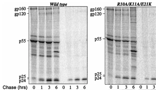
HeLa cells were transfected with wild-type and R10A/K11A/
E21K proviral DNAs and pulsed for 45 min with [
35S]Met/Cyst
(Fig. 3). Labeled proteins were chased for 0, 1, 3, and 6 h, and
cell-associated and virion-associated proteins were
immuno-precipitated with serum from an HIV-1-infected individual.
Mutant R10A/K11A/E21K assembled virions with kinetics
similar to that of the wild type (Fig. 3), with normal
accumu-lation of Gag-processing intermediates. The magnitude of
virion release was determined to be near normal when the
virion-associated CA signal at the 6-h time point was
normal-ized to the intensity of the cell-associated Gag polyprotein
signal at the 0-h time point (Table 1). These results
demon-strated that the defect in viral assembly of mutant R10A/K11A
was corrected by the second-site mutation E21K. Expression
and processing of
env
-encoded proteins was normal. Also,
pro-cessing of
gag
- and
pol
-encoded proteins at other sites
ap-peared normal, as judged by Western blot analysis (data not
shown).
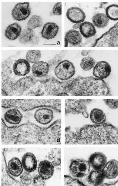
[image:3.612.125.472.83.199.2]Morphology of NC mutant virions.
Infection of Jurkat cells
was initiated by transfection of proviral DNAs; after 14 days,
cells were fixed and analyzed by electron microscopy. As
ex-pected, the majority (96%) of wild-type virions exhibited
ma-ture, cone-shaped core structures of high density (Fig. 4a); 3%
of wild-type particles exhibited immature morphology with a
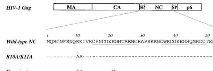
FIG. 1. Schematic representation of the major domains of the HIV-1 Gag polyprotein. Amino acid sequences of wild-type and mutant NC proteins are given below. Dashes indicate amino acid identity with the wild type. Cys-His boxes are underlined. MA, matrix; SP, spacer.FIG. 2. Replication of HIV-1 wild type and NC mutants following transfec-tion of proviral DNAs into the Jurkat T-cell line. The accumulatransfec-tion of RT activity in the cell culture supernatant (ordinate) is shown for the indicated day posttransfection (abscissa).
V
OL. 74, 2000
SUPPRESSION OF AN HIV-1 NUCLEOCAPSID MUTANT
4275
on November 9, 2019 by guest
http://jvi.asm.org/
[image:3.612.323.538.508.691.2]rim of high-density material inside the envelope, and 1%
ex-hibited an irregular core structure. The majority of particles
(79%) observed with the R10A/K11A mutant were immature,
with a characteristic rim of high-density material present inside
the envelope (Fig. 4b); 2% of these virions had a dense,
glob-ular core structure in the center.
Consistent with the restored replicative capacity conferred
by the second-site suppressor mutation, 85% of R10A/K11A/
E21K virions exhibited a mature morphology with cone-shaped
core structures of high density (Fig. 4d). In addition, 13% of
virion particles contained two globular core structures of high
density (Fig. 4c); 2% of the R10A/K11A/E21K virions
exhib-ited a dense, globular core structure in the center.
When virions were produced by provirus bearing only the
E21K mutation in an otherwise wild-type background, 89%
exhibited the morphology of normal, mature virions (Fig. 4f),
and 3% had a dense, globular core structure in the center (Fig.
4e). Interestingly, 8% had two or as many as four core
struc-tures of high density (Fig. 4g).
Effect of the second-site compensatory mutation on viral
genomic RNA incorporation into virions.
Viral genomic RNA
incorporation has been shown to be modestly impaired in
R10A/K11A virions ((17a, 60). To determine if the second-site
mutation restored viral genomic RNA incorporation to normal
levels, virions produced by transfection of 293T cells with
wild-type, R10A/K11A, or R10A/K11A/E21K proviral DNAs were
purified and normalized by exogenous RT activity. The
amount of viral genomic RNA incorporated was then
deter-mined by dot blot analysis as previously described (17a, 52).
Supernatant obtained from cells transfected with proviral
DNA coding for a myristylation-deficient Gag was used as a
negative control. The R10A/K11A mutation reduced RNA
packaging to levels 22% of that of the wild-type (Fig. 5 and
Table 1) (17a, 60). The presence of the second-site mutation
(R10A/K11A/E21K) increased the amount of viral genomic
RNA incorporated into virions by approximately threefold
compared to the R10A/K11A mutant (Fig. 5 and Table 1).
Effect of the second-site mutation on endo-RT.
To
deter-mine if impairment in viral replication was due to an intrinsic
defect in the RT reaction, endo-RT was examined next.
Puri-fied virions were permeabilized with 5 mM

-octylglucoside
and incubated for 24 h at 37°C with unlabeled dNTPs plus
[
32P]TTP. Virions were then treated with proteinase K, and
nucleic acid was extracted and precipitated. Samples were then
run on an agarose gel. A band corresponding to full-length
viral DNA was observed in wild-type and both mutant virions
(Fig. 6). We also observed a smear, containing degradation
products and incomplete forms of RT (8, 35, 44), constituting
up to 90% of the total radioactivity present in the lanes.
Be-cause of the smear, we considered quantitation of the results
relatively inaccurate, but a decrease (about threefold) in the
overall amount of endo-RT products was observed in R10A/
K11A virions relative to the wild type. For the R10A/K11A/
E21K mutant, the reduction in endo-RT product was slightly
less (about twofold). Similar results were obtained when virion
permeabilization was performed with 0.01% Triton X-100
(data not shown).
Quantitation of viral DNA synthesis after infection using
real-time PCR.
The effect of the NC mutations on the early
steps of the viral life cycle was studied by quantifying
full-length linear viral DNA and 2-LTR circles synthesized after a
single round of infection using real-time PCR. Virions
normal-ized by exogenous RT were used to infect Jurkat T cells.
Low-molecular-weight DNA was harvested 12 h postinfection.
PCR was performed with primer pairs that amplify full-length
linear DNA or 2-LTR circles. By exploiting nucleotide
differ-ences between the 5
⬘
and 3
⬘
LTRs of pNL4-3, the primers
amplify only newly synthesized viral DNA and not
contaminat-ing plasmid DNA carried over from the transfection in which
the virions were produced (9). To ensure that there were equal
amounts of sample DNA added to each real-time PCR,
mito-chondrial DNA was amplified with specific primers (9) and
quantitated as described below with a molecular beacon (see
Materials and Methods for details).
To quantify the viral DNA template copy number in each
sample, a molecular beacon was used in combination with
real-time PCR as previously described (46, 50). The molecular
beacon is an oligonucleotide with a fluorochrome at one end
and a quencher at the other. It is designed to form a stem-loop
structure that brings the quencher in close proximity to the
fluorochorome. As a result, little signal is emitted when the
beacon is in its folded conformation. The loop is designed to
FIG. 3. Pulse-chase analysis of HIV-1 NC wild type and R10A/K11A/E21K mutant. HeLa cells transfected with the indicated proviral DNAs were metabolically labeled with [35S]Met/Cys for 45 min and chased for 0, 1, 3, 6 h, as indicated. Virion-associated proteins were purified by ultracentrifugation through 25% sucrose. Virion- and cell-associated proteins were immunoprecipitated with sera from an HIV-1-infected individual and analyzed by SDS-PAGE. Positions of mobility of the envelope glycoprotein precursor (gp160), surface envelope protein (gp120), Pr55Gagprecursor (p55), incompletely processed Gag precursors (p41 and p25), and completely processed CA (p24) are indicated on the left.on November 9, 2019 by guest
http://jvi.asm.org/
[image:4.612.148.459.72.251.2]FIG. 4. Analysis of virion morphology by electron microscopy. Jurkat cells were transfected with proviral DNAs; 14 days later, cells were fixed, stained, embedded, and visualized by electron microscopy. (a) Mature wild-type virions showing a characteristic cone-shaped core structure of high density. (b) Virus particles of mutant R10A/K11A showing a rim of high-density material inside the envelope and occasionally a dense, globular core structure in the center. (c) Particles of mutant R10A/K11A/E21K showing a rim of high-density material inside the envelope (left) and two globular core structures (left, middle, and right). (d) Particles of mutant R10A/K11A/E21K showing a cone-shaped core structure of high density (left). (e) Particle of mutant E21K showing a dense, globular core structure in the center. (f) Mutant E21K virions showing a cone-shaped core structure of high density (left), a rim of high-density material inside the envelope (middle), and two core structures of high density (right). (g) Particles of mutant E21K showing two extended core structures of low density (right), two to four globular core structures of high density (left), and a rim of high-density material inside the envelope (middle). The bar indicates 100 nm.
4277
on November 9, 2019 by guest
http://jvi.asm.org/
hybridize with the amplified sequence, so that at each
anneal-ing step, the molecular beacon anneals to the amplified
se-quences. Consequently, with each PCR cycle increasing
fluo-rescence is emitted that is detected by a spectrofluorometric
thermal cycler (ABI PRISM 7700; Applied Biosystems Inc.).
For each experiment, a standard curve was generated by
experimentally determining the threshold cycle (
C
T) for known
template DNA copy number (in duplicate) ranging from 10 to
10
6molecules.
C
T
is the cycle number at which the mean
fluorescence rises 10 standard deviations above the baseline.
For either the full-length linear DNA (Fig. 7A) or the 2-LTR
circles (data not shown), the
C
Twas directly proportional to
the template input copy equivalents (Fig. 7A, inset).
Copy number for each experimental sample was calculated
by interpolation from the experimentally determined
C
Tstan-dard curve. At 12 h postinfection, the steady-state level of
full-length proviral DNA with mutant R10A/K11A was 10% of
the wild-type level (Fig. 7B and Table 1). Considering the
method that we used for normalizing virions, and correcting
for the fivefold reduction in RNA packaging, the actual defect
in RT with R10A/K11A was only twofold. An identical defect
was observed in the formation of 2-LTR circles by the R10A/
K11A mutant (Fig. 7C), suggesting that there was no
measur-able defect in nuclear import of the preintegration complex.
R10A/K11A/E21K accumulated nearly wild-type levels of both
full-length linear and 2-LTR DNAs (Fig. 7B and C).
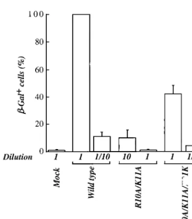
Effect of NC mutants in the MAGI assay.
An indicator cell
line, HeLa-CD4-LTR-

-gal, bearing a

-Gal gene under the
control of the HIV-1 LTR (45), was used to determine the
infectivity of mutant virions in a single round of infection.
Virions produced by transfection of 293T cells were purified by
ultracentrifugation through 25% sucrose, resuspended, and
normalized by exogenous RT activity. Normalized virions were
used to infect HeLa-CD4-LTR-

-gal cells in triplicate. Two
days postinfection, cells were washed and fixed in
formalde-hyde, and

-Gal-positive cells were counted.
Cells infected with different dilutions of virion stocks
indi-cated that the R10A/K11A mutant had 100-fold-lower titer
than the wild type (Fig. 8). A 100-fold relative decrease in titer
associated with the R10A/K11A mutation was observed
whether the experiment was performed with virus stocks
gen-erated by transfection with complete provirus (Fig. 8) or with
env
-deleted proviruses pseudotyped with vesicular stomatitis
virus G protein (VSV-G) (data not shown). This indicates that
the observed

-Gal activity was due to a single round of viral
infection. Similar results were obtained when, instead of
mea-suring

-Gal activity, we measured luciferase activity after
in-fection with a VSV-G-pseudotyped virus in which
env
had been
replaced with a luciferase gene cassette (data not shown).
Since full-length viral DNA and 2-LTR circles were decreased
10-fold with the R10A/K11A mutant, these results indicate
that there is at least an additional 10-fold reduction in
infec-tivity due to disruption of postnuclear import processes such as
integration. As judged by the MAGI assay, the second-site
mutation restored the infectivity of mutant R10A/K11A/E21K
virions almost to wild-type levels (42%).
DISCUSSION
[image:6.612.97.247.71.332.2]In this report we have described the detailed
characteriza-tion of HIV-1 NC mutant R10A/K11A and of a phenotypic
FIG. 5. Viral genomic RNA incorporation into wild-type and mutant virions.Virions produced by transfection of 293T cells with proviral DNAs were purified by ultracentrifugation through 25% sucrose, resuspended in PBS, and normal-ized by exogenous RT assay. Normalnormal-ized amount of virions were loaded onto a nylon membrane and probed with a32P-end-labeled DNA oligonucleotide spe-cific for viral genomic RNA. The signal obtained after hybridization was quan-tified with a PhosphorImager. Results are presented as percentage of wild-type virus activity. The bar graph presents results obtained from three independent experiments with standard errors of the mean (primary data from a representa-tive experiment are shown underneath).myr⫺indicates a virion preparation from cells transfected with a myristylation-deficient NL4-3.
FIG. 6. Endo-RT in wild-type and mutant virions. After purification through 25% sucrose and normalization by exogenous RT, virions produced by transient transfection of 293T cells were permeabilized with-octylglucoside and incu-bated with [32P]TTP and unlabeled dNTPs. The products of endo-RT were extracted with proteinase K, precipitated, and run on an agarose gel prior to PhosphorImager analysis.
on November 9, 2019 by guest
http://jvi.asm.org/
[image:6.612.378.480.424.677.2]revertant bearing a second-site suppressor mutation (R10A/
K11A/E21K). Given the many roles of NC in the retrovirus life
cycle, we examined the effect of our mutants on steps spanning
the entire replication cycle. By this approach, we demonstrated
that mutant R10A/K11A exhibits several, distinct defects, the
major ones being in integration and RNA packaging.
A defect in viral RNA packaging and virion assembly has
been previously reported for mutant R10A/K11A (17a, 60).
FIG. 7. Quantitation of steady-state viral DNA 12 h after infection of Jurkat T cells. (A) Representative standard curve for the quantitation of full-length viral DNA sequences using real-time PCR with a molecular beacon. Change in fluorescence (⌬Rn) as a function of cycle number is demonstrated for viral DNA plasmid copy numbers ranging from 10 to 106per reaction. TheCTis shown for duplicates of the standards used. The inset shows the relationship between known input DNA copy numbers and theCT. TheCTis directly proportional to the log of the input copy equivalents, as demonstrated by the standard curve generated (r2⫽0.992). (B and C) Low-molecular-weight DNA was isolated from Jurkat cells after infection with the indicated viruses (9, 40). Real-time PCR was performed using primers specific for full-length (B) or circular 2-LTR (C) viral DNA. Input copy number was determined as shown in panel A. Results are presented as percentage of wild-type viral DNA. The bar graph presents results obtained from two independent experiments with standard errors of the mean.V
OL. 74, 2000
SUPPRESSION OF AN HIV-1 NUCLEOCAPSID MUTANT
4279
on November 9, 2019 by guest
http://jvi.asm.org/
These defects were corrected in the revertant, although the
level of RNA packaging in R10A/K11A/E21K virions was still
only 60% of the wild-type level. In addition, a Gag-processing
defect present in mutant R10A/K11A (17a) was also corrected
in R10A/K11A/E21K virions. The biochemical analysis was
supported by electron microscopy analysis showing that R10A/
K11A/E21K virions had wild-type morphology, while R10A/
K11A virions showed lack of core condensation, also as
previ-ously reported (17a, 60).
Though the majority have immature morphology, R10A/
K11A mutant virions were still able to infect target cells and to
complete RT. The efficiency of RT was accurately quantitated
using real-time PCR and found to be 10-fold lower in mutant
R10A/K11A than in the wild type or R10A/K11A/E21K.
How-ever, the actual magnitude of the RT defect is much less, given
that R10A/K11A mutant virions are impaired for RNA
pack-aging and therefore contain less template for DNA synthesis.
Indeed, in our assays, virions were normalized for protein
content and do not account for differences in RNA packaging.
If one corrects the results obtained by real-time PCR for the
amount of viral genomic RNA packaged into mutant virions,
the reduction in DNA synthesis by mutant R10A/K11A is only
twofold compared to the wild-type level. Thus, mutant R10A/
K11A virions infect cells and reverse transcribe to
quasi-wild-type levels. This finding is in agreement with the observation
that endo-RT is essentially normal in our mutants, and it also
suggests that tRNA
3Lysplacement and dimeric RNA formation
are not affected in our NC mutant.
Formation of 2-LTR circles was found to be reduced to the
same extent as full-length viral DNA, suggesting that nuclear
import of the preintegration complex (PIC) occurs normally in
cells infected with the R10A/K11A mutant. 2-LTR circles are
not believed to be substrates for integration (10, 18, 23, 43, 51)
but are considered markers of successful nuclear important
because they result from ligation of full-length viral DNA by
cellular DNA ligases that localize in the nucleus (3, 24, 65, 66,
70). Our quantitation of 2-LTR circles was performed with
dividing Jurkat T cells, and so it remains possible that a nuclear
import defect would be detected after infection of a
nondivid-ing cell such as a macrophage.
Since RT and nuclear import in Jurkat T cells are hardly
affected in the R10A/K11A mutant, a defect in a subsequent
step must be invoked to explain the 100-fold reduction in
infectivity in MAGI assays (or at least a 10-fold reduction if
one considers the 5-fold packaging defect and 2-fold RT
de-fect). We believe this defective step to be integration. A role
for NC in integration has been proposed based on in vitro
assays (15, 16) and based on the finding that certain NC zinc
finger mutants exhibit a defect in vivo, after nuclear
translo-cation of viral DNA (36, 37).
How NC might affect integration is unclear, especially since
NC’s presence in purified PICs is not agreed on by all
inves-tigators (25, 32). NC might act directly on the integration
reaction, as recently proposed by others (16), by facilitating
DNA condensation and/or DNA melting, conditions that
pro-mote integration (7, 12, 56, 61). NC might maintain the
integ-rity of viral DNA; M-MuLV NC mutants have been shown to
synthesize 2-LTR circles containing large deletions and
inser-tions at the juncinser-tions (36). A limited analysis of the sequence
of the 2-LTR circle junctions produced by our R10A/K11A
mutant, however, showed no obvious differences from the wild
type (data not shown).
Alternatively, the effect of NC on integration might be
indi-rect, being exerted, for example, by regulating viral protease
processing of PIC components. The only evidence for a
pro-tease processing defect in mutant R10A/K11A is the
accumu-lation of incompletely processed CA, as processing at other
sites appears normal (17a). How this would affect integration is
unclear, since only traces amounts of CA are found in highly
purified PICs, questioning their relevance in the integration
process (55). Last, although we believe that mutant R10A/
K11A exhibits a defect in integration, it is also possible that our
mutant is affected at an as yet uncharacterized step that occurs
after nuclear translocation of viral DNA.
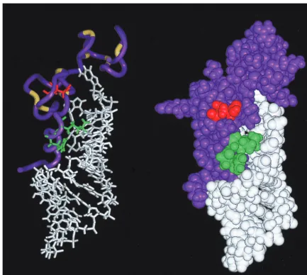
[image:8.612.74.271.71.295.2]The presence of the second-site change E21K compensates
for all of the defects that were observed with the R10A/K11A
mutation. In the solution structure of wild-type HIV-1 NC
complexed with the HIV-1 SL3 stem-loop RNA, NC residues
R10 and K11 lie on a 3
10helix and contact phosphodiester
groups on the RNA (21). Interestingly, residue E21 lies on the
same face of NC as R10 and K11, where it contacts residue
K14 and is believed to stabilize the first zinc finger (Fig. 9).
Requirement for this stabilization must not be absolute, since
residue E21 can be mutated in the context of otherwise
wild-type proviral DNA without an appreciable defect in virion
TABLE 1. Summary of results obtained with HIV-1 NC mutants
Mutant Replication
% of wild-type value
Virion assembly
Viral genomic
RNA packaginga
Single-round infectiona
PCR; full length MAGIassay
R10A/K11A
⫺
33
b22
10
1
R10A/K11A/E21K
⫹
68
62
98
42
[image:8.612.53.294.627.711.2]aAdjusted for virion assembly defect. bFrom reference 17a.
FIG. 8. Infectivity of HIV-1 wild-type and NC mutant virions after a single-round infection (MAGI assay). Virions produced by transfection of proviral DNAs into 293T cells were purified by ultracentrifugation through 25% sucrose. Virions were normalized by exogenous RT and used to infect HeLa-CD4-LTR--gal cells. Cells were infected with the indicated amounts of wild-type and mutant virion preparations. Infectious titers were determined by scoring the number of-Gal-positive cells 2 days postinfection. Results are presented as percentage of wild-type virus activity. The bar graph presents results obtained from three independent experiments with standard errors of the mean.
on November 9, 2019 by guest
http://jvi.asm.org/
replication, in agreement with a previous study (22). However,
a significant proportion (
⬃
10%) of E21K virions have multiple
core structures, and this may be related to a destabilizing effect
of the mutation on the first zinc-finger. How an NC mutation
would induce the production of virions with multiple cores is
unknown. Clearly though, NC is present when the core
assem-bly process is initiated and, interestingly, successful attempts to
induce conical core formation in vitro have utilized a Gag
fusion protein that encompasses CA and NC (13, 33, 38).
A complete understanding of how E21K rescues R10A/
K11A replication will require the determination of the
struc-ture of these mutants. The E21K mutation might rescue
rep-lication by restoring local positive charge, thus reestablishing
contacts with the RNA phosphodiester groups that had been
disrupted by the R10A/K11A mutation. Alternatively, the
ef-fects of E21K might be less specific and the mutation might
simply serve to restore the net positive charge of NC above a
critical threshold required for nonspecific RNA binding. The
latter possibility seems unlikely given that several HIV-1 NC
mutants in which two basic residues are mutated to alanine are
able to replicate to wild-type levels (60). In either case, the
identification of E21K as a second-site suppressor of R10A/
K11A underlines the importance of NC basic residues for
HIV-1 replication.
ACKNOWLEDGMENTS
We thank Anna Aldovini for generously providing plasmid DNA.
We thank Cagan Gurer for critical reading of the manuscript and
Douglas Brateen and Leondios Kostrykis for technical assistance with
the real-time PCR. We are indebted to Mohammed Asmal for graphic
work.
This work was supported by grant AI 41857 (J.L.) and by shared core
facilities of the Columbia-Rockefeller Center for AIDS Research (P30
AI42848), both from the National Institutes of Health, and by contract
975313 from the Swedish Cancer Foundation (S.H.).
REFERENCES
1.Allain, B., M. Lapadat-Tapolsky, C. Berlioz, and J. L. Darlix.1994.
Trans-activation of the minus-strand DNA transfer by nucleocapsid protein during reverse transcription of the retroviral genome. EMBO J.13:973–981.
2.Allain, B., J. B. Rascle, H. de Rocquigny, B. Roques, and J. L. Darlix.1998.
CIS elements and acting factors required for minus strand DNA trans-fer during reverse transcription of the genomic RNA of murine leukemia virus. J. Mol. Biol.277:225–235.
3.Ansari-Lari, M. A., L. A. Donehower, and R. A. Gibbs.1995. Analysis of
human immunodeficiency virus type 1 integrase mutants. Virology213:680.
[image:9.612.86.520.71.459.2]4.Bennett, R. P., T. D. Nelle, and J. W. Wills.1993. Functional chimeras of the
FIG. 9. Ribbon diagram (left) and space-filling image (right) of the HIV-1 NC-SL3-RNA complex (21). NC is shown in purple. Residues R10/K11 and E21 are shown in green and red, respectively. SL3 RNA is shown in white. Cysteine and histidine residues participating in the two Cys-His boxes are shown in yellow on the left.
V
OL. 74, 2000
SUPPRESSION OF AN HIV-1 NUCLEOCAPSID MUTANT
4281
on November 9, 2019 by guest
http://jvi.asm.org/
Rous sarcoma virus and human immunodeficiency virus Gag proteins. J. Vi-rol.67:6487–6498.
5.Berkowitz, R., J. Fisher, and S. P. Goff.1996. RNA packaging. Curr. Top.
Microbiol. Immunol.214:177–218.
6.Berthoux, L., C. Pechoux, M. Ottmann, G. Morel, and J. L. Darlix.1997.
Mutations in the N-terminal domain of human immunodeficiency virus type 1 nucleocapsid protein affect virion core structure and proviral DNA syn-thesis. J. Virol.71:6973–6981.
7.Bor, Y. C., F. D. Bushman, and L. E. Orgel.1995. In vitro integration of
human immunodeficiency virus type 1 cDNA into targets containing protein-induced bends. Proc. Natl. Acad. Sci. USA92:10334–10338.
8.Borroto-Esoda, K., and L. R. Boone.1991. Equine infectious anemia virus
and human immunodeficiency virus DNA synthesis in vitro: characterization of the endogenous reverse transcriptase reaction. J. Virol.65:1952–1959.
9.Braaten, D., E. K. Franke, and J. Luban.1996. Cyclophilin A is required for
an early step in the life cycle of human immunodeficiency virus type 1 prior to the initiation of reverse transcription. J. Virol.70:3551–3560.
10. Brown, P. O., B. Bowerman, H. E. Varmus, and J. M. Bishop.1987. Correct
integration of retroviral DNA in vitro. Cell49:347–356.
11. Burniston, M. T., A. Cimarelli, J. Colgan, S. P. Curtis, and J. Luban.1999.
Human immunodeficiency virus type 1 Gag polyprotein multimerization requires the nucleocapsid domain and RNA and is promoted by the capsid-dimer interface and the basic region of matrix protein. J. Virol.73:8527– 8540.
12. Bushman, F. D., and R. Craigie.1992. Integration of human
immunodefi-ciency virus DNA: adduct interference analysis of required DNA sites. Proc. Natl. Acad. Sci. USA89:3458–3462.
13. Campbell, S., and A. Rein.1999. In vitro assembly properties of human
immunodeficiency virus type 1 Gag protein lacking the p6 domain. J. Virol.
73:2270–2279.
14. Carriere, C., B. Gay, N. Chazal, N. Morin, and P. Boulanger.1995. Sequence
requirements for encapsidation of deletion mutants and chimeras of human immunodeficiency virus type 1 Gag precursor into retrovirus-like particles. J. Virol.69:2366–2377.
15. Carteau, S., S. C. Batson, L. Poljak, J. F. Mouscadet, H. de Rocquigny, J. L.
Darlix, B. P. Roques, E. Kas, and C. Auclair.1997. Human
immunodefi-ciency virus type 1 nucleocapsid protein specifically stimulates Mg2⫹ -depen-dent DNA integration in vitro. J. Virol.71:6225–6229.
16.Carteau, S., R. J. Gorelick, and F. D. Bushman.1999. Coupled integration
of human immunodeficiency virus type 1 cDNA ends by purified integrase in vitro: stimulation by the viral nucleocapsid protein. J. Virol.73:6670–6679.
17. Cimarelli, A., and J. Luban.1999. Translation elongation factor 1-alpha
interacts specifically with the human immunodeficiency virus type 1 Gag polyprotein. J. Virol.73:5388–5401.
17a.Cimarelli, A., S. Sandin, S. Ho¨glund, and J. Luban.2000. Basic residues in
human immunodeficiency virus type 1 nucleocapsid promote virion assembly via interaction with RNA. J. Virol.74:3046–3057.
18. Craigie, R., T. Fujiwara, and F. Bushman.1990. The IN protein of Moloney
murine leukemia virus processes the viral DNA ends and accomplishes their integration in vitro. Cell62:829–837.
19. Darlix, J. L., M. Lapadat-Tapolsky, H. de Rocquigny, and B. P. Roques.
1995. First glimpses at structure-function relationships of the nucleocapsid protein of retroviruses. J. Mol. Biol.254:523–537.
20. Dawson, L., and X. F. Yu.1998. The role of nucleocapsid of HIV-1 in virus
assembly. Virology251:141–157.
21. De Guzman, R. N., Z. R. Wu, C. C. Stalling, L. Pappalardo, P. N. Borer, and
M. F. Summers.1998. Structure of the HIV-1 nucleocapsid protein bound to
the SL3 psi-RNA recognition element. Science279:384–388.
22. Dorfman, T., J. Luban, S. P. Goff, W. A. Haseltine, and H. G. Gottlinger.
1993. Mapping of functionally important residues of a cysteine-histidine box in the human immunodeficiency virus type 1 nucleocapsid protein. J. Virol.
67:6159–6169.
23. Ellis, J., and A. Bernstein.1989. Retrovirus vectors containing an internal
attachment site: evidence that circles are not intermediates to murine ret-rovirus integration. J. Virol.63:2844–2846.
24. Engelman, A., G. Englund, J. M. Orenstein, M. A. Martin, and R. Craigie.
1995. Multiple effects of mutations in human immunodeficiency virus type 1 integrase on viral replication. J. Virol.69:2729–2736.
25. Farnet, C. M., and F. D. Bushman.1997. HIV-1 cDNA integration:
require-ment of HMG I(Y) protein for function of preintegration complexes in vitro. Cell88:483–492.
26. Feng, Y. X., S. Campbell, D. Harvin, B. Ehresmann, C. Ehresmann, and A.
Rein.1999. The human immunodeficiency virus type 1 Gag polyprotein has
nucleic acid chaperone activity: possible role in dimerization of genomic RNA and placement of tRNA on the primer binding site. J. Virol.73:4251– 4256.
27. Feng, Y. X., T. D. Copeland, L. E. Henderson, R. J. Gorelick, W. J. Bosche,
J. G. Levin, and A. Rein.1996. HIV-1 nucleocapsid protein induces
“matu-ration” of dimeric retroviral RNA in vitro. Proc. Natl. Acad. Sci. USA
93:7577–7581.
28. Franke, E. K., H. E. H. Yuan, K. L. Bossolt, S. P. Goff, and J. Luban.1994.
Specificity and sequence requirements for interactions between various
ret-roviral Gag proteins. J. Virol.68:5300–5305.
29. Freed, E. O.1998. HIV-1 gag proteins: diverse functions in the virus life
cycle. Virology251:1–15.
30. Fu, W., R. J. Gorelick, and A. Rein.1994. Characterization of human
im-munodeficiency virus type 1 dimeric RNA from wild-type and protease-defective virions. J. Virol.68:5013–5018.
31. Fu, W., and A. Rein.1993. Maturation of dimeric viral RNA of Moloney
murine leukemia virus. J. Virol.67:5443–5449.
32. Gallay, P., S. Swingler, J. Song, F. Bushman, and D. Trono.1995. HIV
nuclear import is governed by the phosphotyrosine-mediated binding of matrix to the core domain of integrase. Cell83:569–576.
33. Ganser, B. K., S. Li, V. Y. Klishko, J. T. Finch, and W. I. Sundquist.1999.
Assembly and analysis of conical models for the HIV-1 core. Science283:
80–83.
34. Gheysen, D., E. Jacobs, F. de Foresta, C. Thiriart, M. Francotte, D. Thines,
and M. DeWilde.1989. Assembly and release of HIV-1 precursor Pr55gag
virus-like particles from recombinant baculovirus-infected insect cells. Cell
59:103–112.
35. Goncalves, J., Y. Korin, J. Zack, and D. Gabuzda.1996. Role of Vif in
human immunodeficiency virus type 1 reverse transcription. J. Virol.70:
8701–8709.
36. Gorelick, R. J., W. Fu, T. D. Gagliardi, W. J. Bosche, A. Rein, L. E.
Hen-derson, and L. O. Arthur.1999. Characterization of the block in replication
of nucleocapsid protein zinc finger mutants from Moloney murine leukemia virus. J. Virol.73:8185–8195.
37. Gorelick, R. J., T. D. Gagliardi, W. J. Bosche, T. A. Wiltrout, L. V. Coren,
D. J. Chabot, J. D. Lifson, L. E. Henderson, and L. O. Arthur.1999. Strict
conservation of the retroviral nucleocapsid protein zinc finger is strongly influenced by its role in viral infection processes: characterization of HIV-1 particles containing mutant nucleocapsid zinc-coordinating sequences. Vi-rology256:92–104.
38. Gross, I., H. Hohenberg, and H. G. Krausslich.1997. In vitro assembly
properties of purified bacterially expressed capsid proteins of human immu-nodeficiency virus. Eur. J. Biochem.249:592–600.
39. Guo, J., L. E. Henderson, J. Bess, B. Kane, and J. G. Levin.1997. Human
immunodeficiency virus type 1 nucleocapsid protein promotes efficient strand transfer and specific viral DNA synthesis by inhibiting TAR-depen-dent self-priming from minus-strand strong-stop DNA. J. Virol.71:5178– 5188.
40. Hirt, B. 1967. Selective extraction of polyomavirus DNA from infected
mouse cell cultures. J. Mol. Biol.26:365–369.
41. Housset, V., H. De Rocquigny, B. P. Roques, and J. L. Darlix.1993. Basic
amino acids flanking the zinc finger of Moloney murine leukemia virus nucleocapsid protein NCp10 are critical for virus infectivity. J. Virol.67:
2537–2545.
42. Jowett, J. B., D. J. Hockley, M. V. Nermut, and I. M. Jones.1992. Distinct
signals in human immunodeficiency virus type 1 Pr55 necessary for RNA binding and particle formation. J. Gen. Virol.73:3079–3086.
43. Katz, R. A., G. Merkel, J. Kulkosky, J. Leis, and A. M. Skalka.1990. The
avian retroviral IN protein is both necessary and sufficient for integrative recombination in vitro. Cell63:87–95.
44. Kiernan, R. E., A. Ono, G. Englund, and E. O. Freed.1998. Role of matrix
in an early postentry step in the human immunodeficiency virus type 1 life cycle. J. Virol.72:4116–4126.
45. Kimpton, J., and M. Emerman.1992. Detection of replication-competent
and pseudotyped human immunodeficiency virus with a sensitive cell line on the basis of activation of an integrated-galactosidase gene. J. Virol.66:
2232–2239.
46. Kostrikis, L. G., S. Tyagi, M. M. Mhlanga, D. D. Ho, and F. R. Kramer.1998.
Spectral genotyping of human alleles. Science279:1228–1229.
47. Krausslich, H. G., and R. Welker.1996. Intracellular transport of retroviral
capsid components. Curr. Top. Microbiol. Immunol.214:25–63.
48. Lapadat-Tapolsky, M., H. De Rocquigny, D. Van Gent, B. Roques, R.
Plas-terk, and J. L. Darlix.1993. Interactions between HIV-1 nucleocapsid
pro-tein and viral DNA may have important functions in the viral life cycle. Nucleic Acids Res.21:831–839.
49. Lener, D., V. Tanchou, B. P. Roques, S. F. Le Grice, and J. L. Darlix.1998.
Involvement of HIV-I nucleocapsid protein in the recruitment of reverse transcriptase into nucleoprotein complexes formed in vitro. J. Biol. Chem.
273:33781–33786.
50. Lewin, S. R., M. Vesanen, L. Kostrikis, A. Hurley, M. Duran, L. Zhang, D. D.
Ho, and M. Markowitz.1999. Use of real-time PCR and molecular beacons
to detect virus replication in human immunodeficiency virus type 1-infected individuals on prolonged effective antiretroviral therapy. J. Virol.73:6099– 6103.
51. Lobel, L. I., J. E. Murphy, and S. P. Goff.1989. The palindromic LTR-LTR
junction of Moloney murine leukemia virus is not an efficient substrate for proviral integration. J. Virol.63:2629–2637.
52. Mak, J., M. Jiang, M. A. Wainberg, M. L. Hammarskjold, D. Rekosh, and L.
Kleiman.1994. Role of Pr160gag-polin mediating the selective incorporation
of tRNALysinto human immunodeficiency virus type 1 particles. J. Virol.
68:2065–2072.
on November 9, 2019 by guest
http://jvi.asm.org/
53.Mak, J., A. Khorchid, Q. Cao, Y. Huang, I. Lowy, M. A. Parniak, V. R.
Prasad, M. A. Wainberg, and L. Kleiman.1997. Effects of mutations in
Pr160gag-pol upon tRNA(Lys3) and Pr160gag-pol incorporation into HIV-1. J. Mol. Biol.265:419–431.
54. McDermott, J., L. Farrell, R. Ross, and E. Barklis.1996. Structural analysis
of human immunodeficiency virus type 1 Gag protein interactions, using cysteine-specific reagents. J. Virol.70:5106–5114.
55.Miller, M. D., C. M. Farnet, and F. D. Bushman.1997. Human
immunode-ficiency virus type 1 preintegration complexes: studies of organization and composition. J. Virol.71:5382–5390.
56. Muller, H. P., and H. E. Varmus.1994. DNA bending creates favored sites
for retroviral integration: an explanation for preferred insertion sites in nucleosomes. EMBO J.13:4704–4714.
57. Myers, G., B. Korber, S. Wain-Hobson, K.-T. Jeang, L. E. Henderson, and
G. N. Pavlakis.1994. Human retroviruses and AIDS. Los Alamos National
Laboratory, Los Alamos, N. Mex.
58. Ottmann, M., C. Gabus, and J. L. Darlix.1995. The central globular domain
of the nucleocapsid protein of human immunodeficiency virus type 1 is critical for virion structure and infectivity. J. Virol.69:1778–1784.
59. Peliska, J. A., S. Balasubramanian, D. P. Giedroc, and S. J. Benkovic.1994.
Recombinant HIV-1 nucleocapsid protein accelerates HIV-1 reverse tran-scriptase catalyzed DNA strand transfer reactions and modulates RNase H activity. Biochemistry33:13817–13823.
60. Poon, D. T., J. Wu, and A. Aldovini.1996. Charged amino acid residues of
human immunodeficiency virus type 1 nucleocapsid p7 protein involved in RNA packaging and infectivity. J. Virol.70:6607–6616.
61. Pruss, D., F. D. Bushman, and A. P. Wolffe.1994. Human immunodeficiency
virus integrase directs integration to sites of severe DNA distortion within the nucleosome core. Proc. Natl. Acad. Sci. USA91:5913–5917.
62. Rascle, J. B., D. Ficheux, and J. L. Darlix.1998. Possible roles of
nucleo-capsid protein of MoMuLV in the specificity of proviral DNA synthesis and
in the genetic variability of the virus. J. Mol. Biol.280:215–225.
63. Rein, A.1994. Retroviral RNA packaging: a review. Arch. Virol. Suppl.
9:513–522.
64. Sambrook, J., E. F. Fritsch, and T. Maniatis.1989. Molecular cloning: a
laboratory manual, 2nd ed. Cold Spring Harbor Laboratory Press, Cold Spring Harbor, N.Y.
65. Shank, P. R., and H. E. Varmus.1978. Virus-specific DNA in the cytoplasm
of avian sarcoma virus-infected cells is a precursor to covalently closed circular viral DNA in the nucleus. J. Virol.25:104–114.
66. Swanstrom, R., W. J. DeLorbe, J. M. Bishop, and H. E. Varmus.1981.
Nucleotide sequence of cloned unintegrated avian sarcoma virus DNA: viral DNA contains direct and inverted repeats similar to those in transposable elements. Proc. Natl. Acad. Sci. USA78:124–128.
67. Tanchou, V., C. Gabus, V. Rogemond, and J. L. Darlix.1995. Formation of
stable and functional HIV-1 nucleoprotein complexes in vitro. J. Mol. Biol.
252:563–571.
68. Tsuchihashi, Z., and P. O. Brown.1994. DNA strand exchange and selective
DNA annealing promoted by the human immunodeficiency virus type 1 nucleocapsid protein. J. Virol.68:5863–5870.
69. Weiss, A., R. Wiskocil, and J. Stobo.1984. The role of T3 surface molecules
in the activation of human T cells: a two stimulus requirement for IL-2 production reflects events occurring at a pretranslational level. J. Immunol.
133:123–128.
70. Wiskerchen, M., and M. A. Muesing.1995. Human immunodeficiency virus
type 1 integrase: effects of mutations on viral ability to integrate, direct viral gene expression from unintegrated viral DNA templates, and sustain viral propagation in primary cells. J. Virol.69:376–386.
71. You, J. C., and C. S. McHenry.1994. Human immunodeficiency virus
nu-cleocapsid protein accelerates strand transfer of the terminally redundant sequences involved in reverse transcription. J. Biol. Chem.269:31491–31495.
V
OL. 74, 2000
SUPPRESSION OF AN HIV-1 NUCLEOCAPSID MUTANT
4283
on November 9, 2019 by guest
http://jvi.asm.org/