JOURNAL OFVIROLOGY,
0022-538X/00/$04.00⫹0 May 2000, p. 4853–4859 Vol. 74, No. 10
Copyright © 2000, American Society for Microbiology. All Rights Reserved.
Cytoplasmic Trafficking of the Canine Parvovirus Capsid and Its
Role in Infection and Nuclear Transport
MAIJA VIHINEN-RANTA,† WEN YUAN,ANDCOLIN R. PARRISH* James A. Baker Institute for Animal Health, College of Veterinary Medicine,
Cornell University, Ithaca, New York 14853
Received 20 October 1999/Accepted 14 February 2000
To begin a successful infection, viruses must first cross the host cell plasma membrane, either by direct fusion with the membrane or by receptor-mediated endocytosis. After release into the cytoplasm those viruses that replicate in the nucleus must target their genome to that location. We examined the role of cytoplasmic transport of the canine parvovirus (CPV) capsid in productive infection by microinjecting two antibodies that recognize the intact CPV capsid into the cytoplasm of cells and also by using intracellular expression of variable domains of a neutralizing antibody fused to green fluorescence protein. The two antibodies tested and the expressed scFv all efficiently blocked virus infection, probably by binding to virus particles while they were in the cytoplasm and before entering the nucleus. The injected antibodies were able to block most infections even when injected 8 h after virus inoculation. In control studies, microinjected capsid antibodies did not interfere with CPV replication when they were coinjected with an infectious plasmid clone of CPV. Cytoplas-mically injected full and empty capsids were able to move through the cytosol towards the nuclear membrane in a process that could be blocked by nocodazole treatment of the cells. Nuclear transport of the capsids was slow, with significant amounts being found in the nucleus only 3 to 6 h after injection.
The infection of cells by viruses is a multistep process that requires that the virus pass through a series of barriers so as to deliver the genome into the appropriate region of the cell for replication. The early steps of virus entry into cells involve attachment to the cell surface, followed by penetration of the virion or its components into the cytoplasm either by direct fusion with the plasma membrane or after uptake into the endocytic pathway. During uptake in endosomes the release of enveloped virus nucleocapsids to the cytoplasm is in many cases associated with structural changes of viral proteins trig-gered by exposure to an acidic pH or binding to cellular re-ceptors. Fusion of the viral envelope releases the viral genome and other components into the cytoplasm (reviewed in refer-ence 19, 30, and 32 to 34).
Less is known about the mechanisms by which nonenveloped viruses enter a host cell, although many of those viruses use interactions between the hydrophobic portions of the outer capsid proteins or of lipid-conjugated proteins to allow the particle to penetrate the cellular membranes (33, 34). Nonen-veloped viruses may enter the cell through either pH-depen-dent pathways (e.g., picornaviruses, adenoviruses, reoviruses, and parvoviruses [6, 20, 36, 39, 40, 44, 45, 51]) or pH-indepen-dent pathways (papovaviruses or poliovirus [27, 47]), and some may also directly penetrate the plasma membrane (rotaviruses [25]).
Nuclear replicating viruses must pass through the cytoplasm to access the nuclear pore or the nucleus (28, 55). Although it has been assumed that this was a largely passive process, it is now clear that the intracytosolic movements of adenovirus and herpes simplex virus 1 capsids use the active transportation system of the cell, mediated by the microtubule cytoskeleton,
to move through the cytoplasm to the vicinity of the nuclear pore (49, 50). Polyomavirus virions are endocytosed via small monopinocytotic vesicles derived from the plasma membrane, and then it is suggested that they enter the nuclei via direct fusion of the vesicles with the nuclear membrane (21). The slow caveola-mediated entry of simian virus 40 (SV40) parti-cles into cells appears to be followed by targeting of virions to the endoplasmic reticulum, where the viruses induce the for-mation of interconnected tubular smooth membrane structures (2, 9, 27). However, mechanisms by which the virion or viral genome is translocated from the endoplasmic reticulum to the nucleus for replication are not known. It appears that the SV40 capsids enter the cytoplasm of cells during infection, as infec-tion can be blocked by intracytoplasmic injecinfec-tion of antibodies against VP1 or VP3 (38). Microinjected SV40 particles in the cytoplasm of cells were imported into the nucleus within 1 h, and they infected the cell, indicating that trafficking through the cytoplasm is part of the infectious pathway (10).
Before the genome of the incoming virion can replicate, it must be released from its capsid, and uncoating may occur at one or more of several sites in the cell, ranging from the cell surface to the nuclear matrix (19, 20, 22, 29). For viruses that replicate in the nucleus, the genome and possibly some asso-ciated proteins must enter that compartment. Most viruses utilize the nuclear import system of the cell, including the nuclear pore complex, receptors, and import factors (33, 55). The nuclear pore has an effective diameter of about 26 nm when Xenopus oocytes are injected with coated gold beads (13). Larger viruses appear to localize to the nuclear pore and then to release their DNA for transport to the nucleus associ-ated with specific viral proteins (18, 26, 49). SV40 particles within the nuclear pore appeared to be about 21 to 24 nm in diameter, while those seen within the nucleus had a diameter of 38.2 nm, suggesting that the capsid structure is distorted during nuclear import (56).
Autonomous parvoviruses have a nonenveloped 25-nm-diameter capsid that contains a single-stranded DNA genome. Canine parvovirus(CPV) is a member of the feline parvovirus * Corresponding author. Mailing address: James A. Baker Institute
for Animal Health, College of Veterinary Medicine, Cornell Univer-sity, Ithaca, NY 14853. Phone: (607) 256-5649. Fax: (607) 256-5608. E-mail: crp3@cornell.edu.
† Present address: Department of Biological and Environmental Science, University of Jyva¨skyla¨, FIN-40351 Jyva¨skyla¨, Finland.
4853
on November 9, 2019 by guest
http://jvi.asm.org/
subgroup, which includes several host range variants that infect carnivores. CPV enters cells by receptor-mediated endocytosis into clathrin-coated vesicles (42). CPV infection can be pre-vented by lysosomotropic bases, indicating that infection in-volves an acidic intracellular compartment (6), and tempera-ture- and microtubule-dependent transport steps are also required (53). CPV capsids associated with cells appear not to be degraded (54). However, the mechanisms of penetration of CPV from the endosomes into the cytoplasm and nuclear tar-geting of the virus or its genome prior to replication are still poorly understood. In previous studies only a small proportion of CPV capsids injected into the cytoplasm of cells were seen to enter the nucleus within 1 or 2 h (53, 54). CPV capsids added to cells are detected only in endosomes for several hours (6, 42, 53, 54). Adeno-associated virus (AAV) capsids are taken into cells in a dynamin-dependent endocytic process (12), and labeled capsids added to cells are seen in the nucleus (4, 5). Up to 50% of the input single-stranded DNA-AAV capsids may be filled in after uptake into some cells (14, 15). Here we examine the steps in CPV entry that occur between internalization via receptor-mediated endocytosis and nuclear replication of the virus. Both microinjection of monoclonal anti-capsid antibodies and intracellular expression of a single-chain antibody prevented CPV replication, showing that the capsid enters the cytosol during productive infection. Full or empty CPV capsids microinjected into the cytoplasm of cells accumulated in a perinuclear area and were mostly transported into the nucleus only after 3 to 6 h. Depolymerization of microtubules by nocodazole treatment of cells resulted in the capsids being found distributed throughout the cytoplasm, and this treatment also inhibited nuclear transport.
MATERIALS AND METHODS
Viruses and cells.CPV type 2 (CPV-d) was grown in NLFK cells, and aliquots were stored frozen at⫺70°C (43). Empty and full CPV particles were purified and quantified as previously described and stored in 10 mM Tris-HCl (pH 7.5) at 4°C (1). NLFK cells were grown and maintained in a 1:1 mixture of McCoy’s 5A medium and Leibovitz L15 medium with 5% fetal bovine serum (43). Virus-infected cells were detected by staining for the newly produced viral NS1 protein
using a monoclonal antibody (MAb), obtained from Caroline Astell (57), con-jugated to Texas red (TxR).
Antibodies and intracellular expression of single-chain antibody (scFv).MAb 8 and MAb 14, which both recognize the intact CPV capsid, were purified from hybridoma culture supernatant by chromatography on protein G (54). Purified mouse immunoglobulin G (IgG) was obtained from Sigma (St. Louis, Mo.).
The variable domain sequences of MAb 8 light and heavy chain were cloned and expressed as a single-chain antibody (scFv) in bacteria as detailed by Yuan and Parrish (58), and that purified protein bound and efficiently neutralized CPV capsids. That scFv was recloned into the vector pEGFP-N (Clontech, Palo Alto, Calif.) so that it was fused at its C terminus with the enhanced green fluorescent protein (EGFP). Cells transfected with the plasmid expressing the scFv-EGFP fusion protein or with pEGFP-N alone were inoculated 24 h later with CPV and then incubated for a further 24 h. Cells were then incubated with 3.7% (wt/vol) paraformaldehyde in phosphate-buffered saline for 15 min, then with 0.1% Triton X-100 in phosphate-buffered saline and 1% (wt/vol) bovine serum albu-min. The transfected cells were detected by EGFP expression, and the CPV-infected cells were detected by staining with anti-NS1-TxR antibody. After being washed, coverslips were mounted in Prolong medium (Molecular Probes, Eu-gene, Oreg.) and examined by fluorescence microscopy. The proportions of scFv-EGFP- or EGFP-expressing cells that became infected were compared to the infection of the nontransfected cells in the same culture.
Cytoplasmic microinjection of antibodies.Purified MAb 8 and Mab 14 IgGs were dialyzed against 10 mM Tris-HCl–120 mM KCl (pH 7.4). Cells at 7⫻103
per cm2on Microgrid coverslips (12-mm diameter, 175-m grid size; Eppendorf,
Hamburg, Germany) were injected with 0.1 to 0.5 pl of antibodies at 5 mg/ml using an Eppendorf 5246 Microinjector and Eppendorf 5171 Micromanipulator with Eppendorf capillaries. The cells were inoculated with CPV either before the time of microinjection or up to 12 h afterwards. The cells were then incubated for a total time after inoculation of 24 h before paraformaldehyde fixation. Fluores-cein isothiocyante-labeled goat anti-mouse IgG was used to detect injected cells, while virus infection was detected using TxR-conjugated anti-NS1.
In control studies MAb 8 at 5 mg/ml was microinjected along with an infec-tious plasmid clone of CPV (43) at a concentration of 200g/ml, and the cells were analyzed by staining for IgG and viral NS1. The colocalization of mouse IgG with viral capsids was visualized using an FITC-labeled goat anti-mouse IgG and rabbit polyclonal anticapsid antibody, followed by TxR-conjugated anti-rabbit secondary antibody. Cells were examined with a confocal fluorescence microscope (Bio-Rad Laboratories, Hercules, Calif.). The proportion of cells injected with MAb 8 and plasmid that became infected was compared to the proportion of control mouse IgG-injected cells and also to the noninjected cells in the same culture.
[image:2.612.86.521.71.284.2]Capsid microinjection and nocodazole treatment.To further examine the distribution and transport of capsids within the cell, we injected full or empty CPV capsids at 2.5 mg/ml into the cytoplasm of cells and then incubated the cells at 37°C for 0, 1, 3, 6, 12, or 24 h. The cells were then fixed and permeabilized as described above and stained with MAb 8 (to detect the intact capsids) and with TxR-anti-NS1, and the cells were then examined by confocal microscopy. In FIG. 1. Microinjection of cells with of MAb 8 or MAb 14, but not with control antibodies, resulted in the cells becoming resistant to infection. A) NLFK cells were injected with MAb 8 and then inoculated with CPV. The cells were dual stained for IgG (green) or for the viral NS1 protein (red). B) NLFK cells were injected with a control IgG and then inoculated with CPV. The cells were stained for IgG (green) or for the viral NS1 protein (red). C) The percentage of MAb 8- or MAb 14-injected cells infected in the various experiments compared to noninjected cells in the same dishes. Controls include NLFK cells injected with control mouse IgG.
on November 9, 2019 by guest
http://jvi.asm.org/
some experiments the cells were incubated in medium containing 20M no-codazole for 1 h before microinjection; then, the drug was maintained thereafter for 1, 6, or 12 h before fixation in 3.7% paraformaldehyde. Some nocodazole-treated cells injected with capsids were first incubated with drug for 6 h and then washed and incubated in normal medium for an additional 6 h before fixation and staining were carried out as described above. Control cells were stained with antitubulin antibody (Amersham, Little Chalfont, Buckinghamshire, United Kingdom) to confirm the nocodazole affect on the microtubule structure.
RESULTS
Anticapsid antibodies in the cytoplasm and the nucleus of
cells block CPV infection.We microinjected two MAbs against
assembled capsids into cells prior to CPV inoculation. The antibodies were seen throughout the cytoplasm and nucleus (Fig. 1A and B). In several independent experiments only between 0 and 0.35% of the MAb 8- or MAb 14-injected cells became infected and expressed NS1, while 32 to 45% of the noninjected cells in the same dishes became infected (Fig. 1C). The effect of the injected MAb 8 or MAb 14 was specific for the virus capsid and was not a nonspecific effect of microinjec-tion on the cells, as control IgG injected into the cells did not affect virus infection (Fig. 1B and C).
To determine whether the block to infection was due to a specific effect on the infecting capsid and not to a nonspecific effect on some later step in viral replication, we coinjected cells with MAb 8 and the infectious plasmid clone of CPV. In different experiments NS1 expression was seen in between 18 and 33% of the injected cells, not significantly different from values seen for plasmid microinjected alone in the same ex-periment (Fig. 2; results not shown). It is likely that the in-jected cells that did not show virus infection did not enter S phase during the time of incubation, preventing virus DNA replication. In MAb 8-injected and CPV-infected cells, the IgG appeared as small labeled aggregations which were either in the perinuclear cytoplasm or within the nucleus (Fig. 2A). Immunofluorescence staining using anti-mouse IgG to detect MAb 8 and polyclonal anticapsid antibodies confirmed that those spots represented aggregated viral capsids (Fig. 2B). In contrast, in noninfected cells diffuse immunofluorescence of the IgG was seen throughout the cytoplasm and the nucleus (Fig. 1 and 2).
MAb 8 expressed as an scFv fused to EGFP accumulated in both the nucleus and the cytoplasm of the transfected NLFK
cells. Cells which expressed the scFV-EGFP completely re-sisted infection (0% infection), while control cells expressing only EGFP showed normal levels of infection (20 to 30%) (Fig. 3).
Antibody injected after virus inoculation blocks infection.
The kinetics of the blockade of CPV infection by antibody was examined by inoculating the cells with virus, injecting MAb 8 into the cytoplasm at various times afterwards, and then de-termining the proportions of infected cells (Fig. 4). A large increase in the proportion of virus-infected cells was observed only after 8 h, and by 12 h there was little effect on the rate of the infection (Fig. 4).
Virus in the cytoplasm is transported into the nucleus after
a period of delay.Both full and empty particles were
distrib-uted throughout the cytoplasm immediately after microinjec-tion (Fig. 5). By 1 h of incubamicroinjec-tion at 37°C the capsids were more concentrated around the nucleus. Capsids appeared to be transported into the nucleus in two phases. Up to 3 h after injection, only small amounts of capsids were seen inside the nucleus, but by 6 h, 30 to 50% of the injected cells showed substantial nuclear localization of the capsids (Fig. 5). Twenty-four hours after injection of empty capsids, almost all of the capsids were seen inside the nucleus (results not shown), and those cells did not show viral infection detectable by staining for NS1. However, in cultures injected with full capsids many of the injected cells and cells surrounding them showed NS1 expression at 24 h but not at 1, 3, or 6 h (results not shown). To determine whether capsid transport through the cytosol towards the nuclear area depended on an intact microtubule network, we monitored the localization of full and empty cap-sids between 1 and 6 h after cytoplasmic injection in cells pretreated for 1 h with nocodazole. Immunofluorescence stain-ing of nocodazole-treated cells for tubulin showed extensive disruption of microtubules (Fig. 6C). At 1 and 6 h full and empty capsids were both distributed randomly throughout the cytoplasm with little perinuclear accumulation or nuclear lo-calization (Fig. 6A), and none of the cells was infected as seen by anti-NS1 antibody staining (results not shown). In some cases the capsid-injected cells treated with nocodazole were washed and incubated in normal medium for a further 6 h, in which case nuclear accumulation of capsids was observed in between 10 and 15% of cells, indicating the reversible nature of inhibition (Fig. 6A). Empty or full capsids injected into cells and incubated in the presence of the drug for 12 h appeared to be aggregated, but they showed little nuclear localization (Fig. 6B). Some of the cells injected with full capsids and incubated for 12 h with nocodazole became infected and showed NS1 expression (Fig. 6B), but this was not seen for the empty capsid-injected cells.
DISCUSSION
Intracellular antibodies show that capsids enter the
cyto-plasm during infection.Here we show that the intact
parvovi-rus capsid must enter the cytoplasm of the host cell during infectious entry, as infection could be almost completely blocked by intracytoplasmic injection of MAb 8 and 14, which only recognize assembled capsids, or by expression of the MAb 8 variable domain fused to EGFP. This effect was virus specific and not due to an indirect effect of the microinjection process, as injection of cells with nonspecific IgG did not affect infec-tion.
That the blocking effect was due to antibody binding to the capsid was confirmed by injection of infectious plasmid DNA, where the infection was not blocked by either antiviral MAb 8 or 14. In the MAb-injected cells which became infected, the
FIG. 2. CPV infection of cells microinjected simultaneously with MAb 8 (5 mg/ml) and the infectious plasmid clone of CPV (200 g/ml). (A) MAb 8 detected using FITC-labeled goat anti-mouse IgG (green) and viral infection visualized with TxR-labeled anti-NS1 antibody (red). The insert shows another cell subjected to the same treatment. (B) Capsids were present within the ag-gregates seen in the cells. Injected MAb 8 was detected using FITC-labeled goat anti-mouse IgG (green), while capsids were detected using rabbit polyclonal anti-capsid IgG and TxR-conjugated anti-rabbit antibody (red). The insert shows another cell subjected to the same treatment.
VOL. 74, 2000 CPV AND THE CYTOPLASM 4855
on November 9, 2019 by guest
http://jvi.asm.org/
capsid-antibody complexes were frequently observed in the cytoplasm. Although capsids normally accumulate in the nuclei of parvovirus-infected cells, neither the site of assembly nor any process of assembled capsid transport into the nucleus or cytoplasm has been defined (16, 48). These data suggest that the CPV capsids can assemble in the cytoplasm, that they can be transported there after assembly in the nucleus, or that both processes can occur.
After CPV inoculation most cells were still protected from infection by injection with MAb 8 up to 8 h later, indicating that separation of the viral DNA from the capsid occurred only slowly after cellular entry. The reasons for this delayed process are not clear. Slow trafficking of the capsids through the en-dosomal pathway has also been suggested by the finding that bafilomycin A1 added to cells 90 min after virus inoculation reduces infection by about 70% (42). However, there may also be a slow transport of the capsid through the cytoplasm or into the nucleus, or DNA release from the capsid may require cellular factors associated with S-phase. Several attempts to inject antibodies into isoleucine-deprived and aphidicolin-treated cells resulted in few cells surviving (results not shown), and so we have not been able to test the last hypothesis. The slow kinetics of infection are similar to those seen during the uptake of SV40, where infection could be blocked by injection
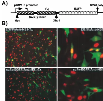
FIG. 3. (A) The insert in the plasmid expressing the variable domains of MAb 8 fused to EGFP. The light variable domain (VL) was linked to the heavy variable
domain (VH) with three repeats of GlyGlyGlyGlySer sequence (G4S)3and then fused in frame with EGFP. (B) Intracellular expression of the variable domain ScFv
[image:4.612.131.475.73.410.2]of MAb 8 and susceptibility to infection with CPV. Cells transfected with a plasmid expressing MAb 8 scFv fused to EGFP (scFv-EGFP) were inoculated with CPV. Control cells (EGFP) were transfected with a plasmid expressing EGFP alone. After a further 24 h of incubation, the cells were fixed, examined for GFP expression (green), and stained with a TxR-labeled anti-NS1 (red). Low- and high-magnification views of typical fields are shown in each case (to the left and right, respectively).
FIG. 4. Effect of the timing of MAb 8 injection on infection by CPV. Cells incubated with CPV for 1 h at 0°C were warmed to 37°C and then injected with MAb 8 at various times between 1 and 12 h later. The percentage of cells that became infected after injection at the various times after warming was deter-mined by staining for the injected MAb and for NS1 to indicate the virus infection. The percentage of infection of noninjected cells in the same cultures was used as a control.
on November 9, 2019 by guest
http://jvi.asm.org/
[image:4.612.314.541.507.666.2]of antibodies against nucleoporin up to 8 but not 12 h after virus inoculation (56).
The mechanism(s) of intracellular neutralization is not known for certain, but the antibodies would likely bind directly the incoming virus in the cytoplasm, interfering with capsid functions by masking structures required for interactions with cellular factors or for nuclear transport, or they may prevent essential conformational changes of the capsid required for infection or DNA release. Where sufficient virus was present the antibodies clearly aggregated the capsids, as seen in cells infected with CPV plasmids (Fig. 3).
In previous studies we did not observe infection by the in-jected capsids (53), while that was sometimes seen in these studies 12 or 24 h after we injected full virus capsids. We believe that this difference was due to small variations in the procedures used. It is not clear that capsid injection would recapitulate normal virus transport during infection; very large amounts of capsids were injected into the cytoplasm, and it is also likely that capsids were deposited onto the plasma mem-brane during the injection process.
Capsids are transported within the cytoplasm and enter the
nucleus slowly.When we injected CPV full or empty capsids
into normal cells, they rapidly became localized to a perinu-clear region. This suggests that capsid transport was controlled by microtubules, perhaps in a manner similar to that reported for the transport of adenovirus and herpesvirus capsids or their proteins (23, 49, 50). The association between the microtubules
and nuclear transport was also shown when the cells were preincubated with nocodazole to cause microtubule break-down, where the capsids became distributed throughout the cytoplasm (Fig. 6). The cytoskeleton is closely associated with the nuclear pore complex (17). It is therefore likely that capsid transport with the microtubular system could facilitate nuclear transport and that capsids would not otherwise be efficiently transported through the cytoplasm due to the high viscosity and other steric obstacles present (31).
We showed in previous studies that during a 1- to 2-h period after injection, most CPV capsids are found within the cyto-plasm, with only small amounts being transported to the nu-cleus (53, 54). Here we confirm those findings but also show that by 6 h after injection there was significant nuclear trans-port of capsids in between 30 and 50% of the cells. Nuclear transport required intact microtubules, as it was blocked for up to 12 h by incubation of the cells with nocodazole, while further incubation of the treated cells in normal medium restored nuclear transport (Fig. 6). The 26-nm diameter of the parvo-virus particle is close to the maximum size that is reported to pass through the nuclear pore, and it is not clear whether any structural change in the capsids occurs during the process. However, MAb 8 used to detect the virus recognizes only intact particles, so the intranuclear particles seen were largely intact. As nuclear transport of the capsids appears to require virus transport to the vicinity of the nuclear pore, the small amounts of capsid found within the nucleus immediately after
microin-FIG. 5. Localization of CPV capsids after microinjection into the cytoplasm of cells. (A) Differential interference contrast and fluorescence confocal image of cells 2 h after injection, showing the specificity of staining for the virus in the injected cells. (B) Full or empty CPV capsids injected into the cytoplasm of the cells that were either fixed immediately (0 h), or incubated for 1, 3, or 6 h at 37°C prior to fixation. The cells were stained for the capsid with MAb 8, followed by FITC-conjugated anti-mouse secondary antibody.
VOL. 74, 2000 CPV AND THE CYTOPLASM 4857
on November 9, 2019 by guest
http://jvi.asm.org/
[image:5.612.103.505.73.399.2]jection may be those that were deposited in the vicinity of the nucleus by the injection (Fig. 5A).
Other changes in either the capsids or the cells which may facilitate nuclear import of the capsids after 3 h include expo-sure of the apparent nuclear localization sequence in the VP1 N terminus (11, 52), removal of bound calcium ions from the capsid, modification of the capsid by cellular enzymes, or bind-ing of the virions to cellular components. As no differences were seen between the transport of injected full or empty capsids, the transport events examined here do not appear to be affected by the exposed N-terminal sequences of VP2 or by the viral DNA in full capsids.
The transport of infecting viral capsids or nucleocapsids within the cytoplasm of cells has been found to be a tightly regulated process in most cases that have been examined (7, 28, 35, 37, 55). The processes often involve multiple cellular factors, changes in the virus components or in the cell, and a variety of specific interactions between viral and host cell com-ponents. AAV capsids have been reported to be transported into the nucleus when added to cells in vivo (4, 5), and some of the viral DNA is converted to a double-stranded form in re-ceptive cells (14). The autonomous parvoviruses appear to differ in that most endocytosed capsids are retained within endosomes for long periods and neither the capsid nor the viral DNA appears to reach the nucleus at significant levels (42, 46, 53, 54). However, the infection process must be closely regu-lated and depend on specific cellular factors, as one or two amino acid sequence substitutions on the surface of autono-mous parvovirus capsids can alter the infectivity for specific
cells by over 105-fold, and the block to infection appears to occur after virus binding and endocytosis but before successful DNA replication in the nucleus (3, 8, 24, 41).
In our future studies we will examine the process of virus transport through the cytoplasm in detail and the role of the microtubule network, as well as the specific cellular and viral factors that control nuclear transport.
ACKNOWLEDGMENTS
Gail Sullivan and Wendy Weichert provided excellent technical as-sistance.
This work was supported by grants NR 64951 from the Academy of Finland and AI28385 from the National Institutes of Health (to C.R.P.).
REFERENCES
1.Agbandje, M., R. McKenna, M. G. Rossmann, M. L. Strassheim, and C. R. Parrish.1993. Structure determination of feline panleucopenia virus empty particles. Proteins16:155–171.
2.Anderson, H. A., Y. Chen, and L. C. Norkin.1996. Bound simian virus 40 translocates to caveolin-enriched domains, and its entry is inhibited by drugs that selectively disrupt caveolae. Mol. Biol. Cell7:1825–1834.
3.Ball-Goodrich, L. J., and P. Tattersall.1992. Two amino acid substitutions within the capsid are coordinately required for acquisition of fibrotropism by the lymphotropic strain of minute virus of mice. J. Virol.66:3415–3423. 4.Bartlett, J. S., and R. J. Samulski.1998. Fluorescent viral vectors: a new
technique for the pharmacological analysis of gene therapy. Nat. Biotechnol.
4:635–673.
5.Bartlett, J. S., R. J. Samulski, and T. J. McCown.1998. Selective and rapid uptake of adeno-associated virus type 2 in brain. Hum. Gene Ther.9:1181– 1186.
6.Basak, S., and H. Turner.1992. Infectious entry pathway for canine parvo-virus. Virology186:368–376.
FIG. 6. (A) Intracellular localization of capsids injected into cells in the presence of nocodazole. Full or empty capsids were injected into cells in the presence of nocodazole and then incubated for 1 h (1h ND) or for 6 h (6h ND). Other cells were injected in the presence of nocodazole, incubated for 6 h, washed, and incubated for a further 6 h in normal medium (6h ND⫹6h chase). (B) Cells injected with full or empty capsids in the presence of nocodazole and then incubated for 12 h with the drug (12h ND). The cells were then fixed and stained with MAb 8 or with TxR-labeled anti-NS1 to distinguish those that were infected. (C) Cells that were untreated (⫺ND) or incubated for 1 h with 20M nocodazole (ND) were fixed and then stained with an antibody against tubulin.
on November 9, 2019 by guest
http://jvi.asm.org/
[image:6.612.73.528.71.364.2]7.Bui, M., G. Whittaker, and A. Helenius.1996. Effect of M1 protein and low pH on nuclear transport of influenza virus ribonucleoproteins. J. Virol.
70:8391–8401.
8.Chang, S.-F., J.-Y. Sgro, and C. R. Parrish.1992. Multiple amino acids in the capsid structure of canine parvovirus coordinately determine the canine host range and specific antigenic and hemagglutination properties. J. Virol.66:
6858–6867.
9.Chen, Y., and L. C. Norkin.1999. Extracellular simian virus 40 transmits a signal that promotes virus enclosure within caveolae. Exp. Cell Res.246:83–90. 10.Clever, J., M. Yamada, and H. Kasamatsu.1991. Import of simian virus 40 virions through nuclear pore complexes. Proc. Natl. Acad. Sci. USA88:7333– 7337.
11. Cotmore, S. F., A. M. D’Abramo, C. M. Ticknor, and P. Tattersall.1999. Controlled conformational transitions in the MVM virion expose the VP1 N-terminus and viral genome without particle disassembly. Virology254:
169–181.
12. Duan, D., Q. Li, A. W. Kao, Y. Yue, J. E. Pessin, and J. F. Engelhardt.1999. Dynamin is required for recombinant adeno-associated virus type 2 infec-tion. J. Virol.73:10371–10376.
13. Dworetzky, S. I., R. E. Landford, and C. M. Feldherr.1988. The effects of variations in the number and sequence of targeting signals on nuclear up-take. J. Cell Biol.107:1279–1287.
14. Ferrari, F. K., T. Samulski, T. Shenk, and R. J. Samulski.1996. Second-strand synthesis is a rate-limiting step for efficient transduction by recombi-nant adeno-associated virus vectors. J. Virol.70:3227–3234.
15. Fisher, K. J., G. P. Gao, M. D. Weitzman, R. DeMatteo, J. F. Burda, and J. M. Wilson.1996. Transduction with recombinant adeno-associated virus for gene therapy is limited by leading-strand synthesis. J. Virol.70:520–532. 16. Fox, E., P. T. Moen, Jr., and J. W. Bodnar.1990. Replication of minute virus of mice DNA in adenovirus-infected or adenovirus-transformed cells. Virol-ogy176:403–412.
17. Gorlich, D., and I. W. Mattaj.1996. Nucleocytoplasmic transport. Science
271:1513–1518.
18. Greber, U., M. Suomalainen, R. P. Stidwill, K. Boucke, M. W. Ebersold, and A. Helenius.1997. The role of the nuclear pore complex in adenovirus DNA entry. EMBO J.16:5998–6007.
19. Greber, U. F., I. Singh, and A. Helenius.1994. Mechanisms of virus uncoat-ing. Trends Microbiol.2:52–56.
20. Greber, U. F., M. Willets, P. Webster, and A. Helenius.1993. Stepwise dismantling of adenovirus 2 during entry into cells. Cell75:477–486. 21. Griffith, G. R., S. J. Marriot, D. A. Rintoul, and R. A. Consigli.1988. Early
events in polyomavirus infection: fusion of monopinocytotic vesicles contain-ing virions with mouse kidney cell nuclei. Virus Res.10:41–52.
22. Helenius, A.1992. Unpacking the incoming influenza virus. Cell69:577–578. 23. Hong, S. S., B. Gay, L. Karayan, M. C. Dabauvalle, and P. Boulanger.1999. Cellular uptake and nuclear delivery of recombinant adenovirus penton base. Virology262:163–177.
24. Horiuchi, M., H. Goto, N. Ishiguro, and M. Shinagawa.1994. Mapping of determinants of the host range for canine cells in the genome of canine parvovirus using canine parvovirus/mink enteritis virus chimeric viruses. J. Gen. Virol.75:1319–1328.
25. Kaljot, K. T., R. D. Shaw, D. H. Rubin, and H. B. Greenberg.1988. Infectious rotavirus enters cells by direct cell membrane penetration, not by endocyto-sis. J. Virol.62:1136–1144.
26. Kann, M., B. Sodeik, A. Vlachou, W. H. Gerlich, and A. Helenius.1999. Phosphorylation-dependent binding of hepatitis B virus core particles to the nuclear pore complex. J. Cell Biol.145:45–55.
27. Kartenbeck, J., H. Stukenbrok, and A. Helenius.1989. Endocytosis of simian virus 40 into the endoplasmic reticulum. J. Cell Biol.109:2721–2729. 28. Kasamatsu, H., and A. Nakanishi.1998. How do animal DNA viruses get to
the nucleus. Annu. Rev. Microbiol.52:627–286.
29. Knipe, D. M.1996. Virus-host cell interactions, p. 273–299.InB. N. Fields, D. M. Knipe, and P. M. Howley (ed.), Fields virology, vol. 1. Lippincott-Raven Publishers, Philadelphia, Pa.
30. Lanzrein, M., A. Schlegel, and C. Kempf.1994. Entry and uncoating of enveloped viruses. Biochem. J.302:313–320.
31. Luby-Phelps, K.1994. Physical properties of cytoplasm. Curr. Opin. Cell Biol.6:3–9.
32. Marsh, M.1984. The entry of enveloped viruses into cells by endocytosis. Biochem. J.218:1–10.
33. Marsh, M., and A. Helenius.1989. Virus entry into animal cells. Adv. Virus Res.36:107–151.
34. Marsh, M., and A. Pelchen-Matthews.1993. Entry of animal viruses into cells. Rev. Med. Virol.3:173–185.
35. Martin, K., and A. Helenius.1991. Transport of incoming influenza virus nucleocapsids into the nucleus. J. Virol.65:232–244.
36. Martı´nez, C. G., R. Guinea, J. Benavente, and L. Carrasco.1996. The entry of reovirus into L cells is dependent on vacuolar proton-ATPase activity. J. Virol.70:576–579.
37. Miyazawa, N., P. L. Leopold, N. R. Hackett, B. Ferris, S. Worgall, E. Falck-Pedersen, and R. G. Crystal.1999. Fiber swap between adenovirus sub-groups B and C alters intracellular trafficking of adenovirus gene transfer vectors. J. Virol.73:6056–6065.
38. Nakanishi, A., J. Clever, M. Yamada, P. P. Li, and H. Kasamatsu.1996. Association with capsid proteins promotes nuclear targeting of simian virus 40 DNA. Proc. Natl. Acad. Sci. USA93:96–100.
39. Neubauer, C., L. Frasel, Kuechler, and D. Blaas.1987. Mechanism of entry of human rhinovirus 2 into Hela cells. Virology158:255–258.
40. Nibert, M. L., L. A. Schiff, and B. N. Fields.1996. Reoviruses and their replication, p. 1557–1624.InB. N. Fields, D. M. Knipe, and P. M. Howley (ed.), Fields virology, vol. 2. Lippincott-Raven Publishers, Philadelphia, Pa. 41. Parker, J. S. L., and C. R. Parrish.1997. Canine parvovirus host range is determined by the specific conformation of an additional region of the capsid. J. Virol.71:9214–9222.
42. Parker, S. L., and C. R. Parrish.1999. Cellular uptake and infection by canine parvovirus involves rapid dynamin-regulated clathrin-mediated endo-cytosis, followed by slower intracellular trafficking. J. Virol.74:1919–1930. 43. Parrish, C. R.1991. Mapping specific functions in the capsid structure of
canine parvovirus and feline panleukopenia virus using infectious plasmid clones. Virology183:195–205.
44. Prchla, E., E. Kuechler, D. Blaas, and R. Fuchs.1994. Uncoating of human rhinovirus serotype 2 from late endosomes. J. Virol.68:3713–3723. 45. Prchla, E., C. Plank, E. Wagner, D. Blaas, and R. Fuchs.1995.
Virus-mediated release of endosomal content in vitro: different behavior of ade-novirus and rhiade-novirus serotype 2. J. Cell Biol.131:111–123.
46. Previsani, N., S. Fontana, B. Hirt, and P. Beard. 1997. Growth of the parvovirus minute virus of mice MVMp3 in EL4 lymphocytes is restricted after cell entry and before viral DNA amplification: cell-specific differences in virus uncoating in vitro. J. Virol.71:7769–7780.
47. Shimura, H., Y. Umeno, and G. Kimura.1987. Effects of inhibitors of the cytoplasmic structures and functions on the early phase of infection of cultured cells with simian virus 40. Virology158:34–43.
48. Singer, I. I.1976. Ultrastructural studies of HI-1 parvovirus replication. III. Intracellular localization of viral antigens with immunocytochrome C. Exp. Cell Res.99:346–356.
49. Sodeik, B., M. W. Ebersold, and A. Helenius.1997. Microtubule-mediated transport of incoming herpes simplex virus 1 capsids to the nucleus. J. Cell Biol.136:1007–1021.
50. Suomalainen, M., M. Y. Nakano, S. Keller, K. Boucke, R. P. Stidwill, and U. F. Greber.1999. Microtubule-dependent plus- and minus end-directed motilities are competing processes for nuclear targeting of adenoviruses. J. Cell Biol.144:657–672.
51. Varga, M. J., C. Weibull, and E. Everitt.1991. Infectious entry pathway of adenovirus type 2. J. Virol.65:6061–6070.
52. Vihinen-Ranta, M., L. Kakkola, A. Kalela, P. Vilja, and M. Vuento.1997. Characterization of a nuclear localization signal of canine parvovirus capsid proteins. Eur. J. Biochem.250:389–394.
53. Vihinen-Ranta, M., A. Kalela, P. Ma¨kinen, L. Kakkola, V. Marjoma¨ki, and M. Vuento.1998. Intracellular route of canine parvovirus entry. J. Virol.
72:802–806.
54. Weichert, W. S., J. S. Parker, A. T. M. Wahid, S.-F. Chang, E. Meier, and C. R. Parrish.1998. Assaying for structural variation in the parvovirus capsid and its role in infection. Virology250:106–117.
55. Whittaker, G. R., and A. Helenius.1998. Nuclear import and export of viruses and virus genomes. Virology246:1–23.
56. Yamada, M., and H. Kasamatsu.1993. Role of nuclear pore complex in simian virus 40 nuclear targeting. J. Virol.67:119–130.
57. Yeung, D. E., G. W. Brown, P. Tam, R. H. Russnak, G. Wilson, I. Clark-Lewis, and C. R. Astell.1991. Monoclonal antibodies to major nonstructural nuclear protein of minute virus of mice. Virology181:35–45.
58. Yuan, W., and C. R. Parrish.Comparison of two single chain antibodies which neutralize canine parvovirus—analysis of antibody combining site and mechanisms of neutralization. Virology, in press.
VOL. 74, 2000 CPV AND THE CYTOPLASM 4859