p53 and EGFR Expression in Glioblastoma Multiforme
-
An Immunohistochemical Study
CERTIFICATE
This is to certify that the following thesis is a bonafide record of the study done by Dr. C. Livingston in part fulfillment of the requirement for M.Ch. Neurosurgery course under the guidance of Dr. Geeta Chacko, Professor, Division of Neuropathology with the help of Dr. Ari. G. Chacko, Professor & Head of Neurosurgery, Unit I and Dr. V. Rajshekhar, Professor & Head of Neurosurgery Unit II.
Dr. V. Rajshekhar, Professor and Head, Neurosurgery Unit II,
Dept of Neurological Sciences, CMC Hospital, Vellore.
Table of Contents
Page No
.
1. Table of Contents….………..………3
2. List of Figures…….………...……….4
3. List of Tables……..…………...……….5
4. List of Graphs…...………..5
5. Introduction……….……….……..6
6. Literature Review… ………..6
7. Aims and Objectives………….………..23
8. Material and Methods……….……….………...24
9. Results………….……….……...27
10. Discussion………...………...49
11. Conclusion……….……....54
12. References.………....56
List of figures
Figure Page no Legends
1 27 Shows a gadolinium enhancing lesion of the dominant frontal lobe with extension into the ventricles and the frontal opercula.
2 28 Post operative CT scan of the same patient showing radical excision with no tumor residue. This patient did not have speech deficit post operatively.
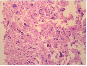
3 31 Glioblastoma multiforme with marked nucear atypia, bizarre nuclei and mitotic figures (H&EX90).
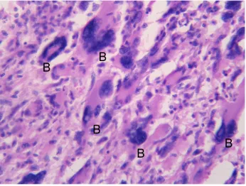
4 32 Glioblastoma multiforme with multinucleated giant cells and bizarre forms (B) (H&EX400).
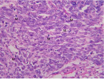
5 33 Glioblastoma multiforrme with numerous mitotic figures (M) (H&EX400).
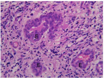
6 34 Vascular (endothelial) proliferation (E) in glioblastoma multiforme (H&EX400).
7 35 Palisading necrosis (N) in glioblastoma multiforme (H&EX400).
8a 37 Nuclear positivity for p53 (P) on immuno histochemistry in cases of glioblastoma multiforme (400X).
8b 38 Nuclear positivity for p53 (P) on immuno
histochemistry in cases of glioblastoma multiforme (400X).
List of Tables
Table Page no Legends
1 8 Comparison of WHO grading of astrocytoma with the St. Anne/Mayo grading system. 2 29 Data of all 58 study patients.
3 41 Data of the 34 patients with follow up details.
4 42 p53 and EGFR expression in patients below 50 years of age (n=37).
5 42 p53 and EGFR expression in patients above 50 years of age (n= 21).
6 43 p53 and EGFR expression in patients with less than 3 months of symptoms (n=33).
7 43 p53 and EGFR expression in patients more than 3 months of symptoms (n = 25). 8 44 p53 and EGFR expression in male patients (n = 44).
9 44 p53 and EGFR expression in female patients (n = 14)
List of graphs
Graph page no Legends
Introduction
Over the last twenty years, with the advances in molecular biology, the understanding of tumorigenesis has reached a stage where novel treatments are being experimented. The present study analyzes the expression pattern of p53 and EGFR in glioblastoma multiforme and correlates this to outcome.
Literature Review
In 1863 Virchow described Glioblastoma multiforme (GBM) as a highly malignant brain tumor of glial origin with the worst prognosis of all the brain tumors17. These tumors are notorious for their rapid invasion
into neighboring structures of brain along the white matter, perivascular space, subependymal plane or subpial plane5, 7. Glioblastoma multiforme accounts for 12-15% of intracranial neoplasms26 and 50-60 % of the astrocytic tumors52. The peak incidence is between 45-70 years52, but they also occur in children9. Males are
more commonly affected than females52. The commonest location is in the cerebral subcortical white matter in
the following order of frequency - temporal lobe, parietal lobe, frontal lobe and occipital lobe. The typical histological features include nuclear atypia, microvascular proliferation, multinucleated giant cells and necrosis17. In the WHO classification it has been allocated a “grade 4” owing to its highly malignant and bizarre
characteristics. WHO classification:
WHO classification 17, 19 is more widely accepted than the previously used Kernohan 16 and Ringertz 37
Table 1: Comparison of WHO grading of Astrocytoma with the St
Anne/Mayo grading system.
In 1940 Scherer described two different types of GBMs, primary GBMs and secondary GBMs38.
Primary GBMs were those with a shorter duration of symptoms usually less than 3 months, seen in adults older than 45 years and with a bad prognosis. Secondary GBMs were those that progressed from a low grade tumor through to a high grade astrocytoma, with a longer duration of symptoms, seen in younger patients usually less than 45 years of age 15, 31.
Ohgaki et al32 in their study of 715 cases of GBM concluded that 38 patients had clinical and
histopathological evidence of progression from a less malignant precursor lesion and these were diagnosed as secondary GBMs. The remaining 677 patients showed either clinical or histopathological evidence of highly malignant lesion and were classified as primary GBMs.
At present it is increasingly accepted that the two types of GBMs have different genetic pathways of evolution18, 32. The primary GBMs are supposed to arise from deregulation of epidermal growth factor
receptor pathway15, 16 and the secondary GBMs are supposed to arise from deregulation of cell cycle
pathway by mutation of p53 gene which is a tumor suppressor gene or over expression of p53 protein48, 49.
Lang et al25 in his study of 65 astrocytic gliomas postulated different genetic pathways leading to GBMs.
One pathway was characterized by 43 astrocytomas with alteration in p53. GBMs with p53 alterations O WHO Grade WHO designation St.Anne/Mayo
designation
Histological criteria
I Pilocytic astrocytoma
II Diffuse astrocytoma Astrocytoma grade 2 One criterion, usually nuclear atypia
III Anaplastic astrocytoma
Astrocytoma grade 3 Two criteria, usually nuclear atypia and mitotic activity IV Glioblastoma
represented tumors that progressed from low grade astrocytoma. This variant was more likely to show loss of chromosome 17p than tumors without p53 alterations (p<0.04). Seventy five percent of tumors with loss of one 17 p allele demonstrated mutation in the p53 gene. Loss of chromosome 10 was associated with progression from anaplastic astrocytoma (13%) to glioblastoma (38%) (p<0.04). Amplification of EGFR gene was a rare (7%) but late event in tumor progression (p<0.03). A second pathway was characterized by six astrocytomas without p53 alterations and represented clinically denovo high grade tumors. These tumors were more likely to show amplification of EGFR gene (83%). 60% of them also showed loss of chromosome 10; loss of chromosome 17p was infrequent in this variant. In addition, there were 16 astrocytomas with none of the known genetic changes. He concluded that GBMs were a heterogeneous group of tumors that probably arose via multiple genetic pathways.
Hayashi et al12, in their study of 70 patients of GBM noted that EGFR amplification and p53
mutation were mutually exclusive (p<0.0001, Fisher exact test). They also noticed that p53 mutation was seen in younger patients and EGFR amplification was seen in older patients and tumors with EGFR amplification behave in a more aggressive manner. Watanabe et al51 in their study also found that p53 and
EGFR mutations were exclusive for secondary and primary GBMs respectively.
Recent reports revealed that the survival after surgery, radiotherapy and chemotherapy is better in secondary GBM compared to primary GBM.
Cell growth and proliferation are enhanced by ‘proto-oncogenes’ such as, the epidermal growth factor receptor gene, whereas genes which restrict abnormal cell division and growth are termed ‘tumor suppressor genes’ such as, p53.
in the absence of cellular stress, although the exact biochemical nature of this latency is under debate. When the cells experience a variety of stress conditions, p53 becomes activated. Activation involves a marked increase in cellular p53, as well as qualitative changes that endow each p53 molecule with improved capabilities to modulate gene expression and alter the cell phenotype. The types of stress that promote p53
activation include many conditions associated with cancer initiation and progression, such as direct DNA damage, chromosomal aberration, illegitimate activation of oncogenes, hypoxia, telomere shortening and more33. p53 expression is studied by quantifying the p53 protein by immunohistochemical studies by biotin
avidin peroxidase method and p53 gene mutation is identified by studying loss of heterozygosity, southern blot analysis28 .
EGFR
Epidermal growth factor receptor (EGFR) is a transmembrane receptor that binds to extracellular ligands such as epidermal growth factor and transforming growth factor alpha and transduces mitotic signals1. It has an external ligand binding site, a trans membrane component and an intracellular
cytoplasmic region which modulates signal transduction pathways by activation of kinase pathway. EGFR amplification has been identified as a genetic hallmark of GBMs. The predictive value of EGFR amplification has been unclear. Layfield et al27, have noted that EGFR identification by
immunohistochemistry correlates well with identification of EGFR gene amplification by fluorescence in situ hybridization (FISH).
Factors affecting glioblastoma multiforme prognosis: Age
Pre operative Karnofsky’s performance score Location of the lesion
c) Oligodendroglial component d) Cystic component di)
Radiation Chemotherapy Genetic factors Age:
Patients with younger age have a better prognosis compared to the older patients. It has also been found that secondary GBMs are seen in younger patients whereas primary GBMs are seen in the older age group. Shrieve et al42, in their study of 78 patients with GBM reported that patients less than 40 years of age
have a median survival time of 48.6 months compared to patients older than 40 years who’s median survival time was 18.2 months (p<0.0001). Korshunov et al21 and Lamborn et al24 also reported an age < 40 years as an
important prognostic indicator of better survival. Ohgaki et al32 in his study also showed by both univariate
and multivariate analyses that old age is a significant predictor of poor survival in GBMs. The cut off age in his series was 50 years, with younger patients (<50 years) having a significantly longer survival (median 8.8 months) than older patients (>=50 years; median, 4.1 months; p<0.0001). Sneed et al45 in their study on 159
glioblastoma multiforme patients noticed that nine patients between 18 and 29.9 years of age had a 3 years survival probability of 78+/- 14%. The 64 patients between 30 and 49.9 years of age had a 3 year survival probability of 29+/-6% and 86 patients >= 50 years had a 3 years survival probability of 6+/-3%. Stark et al46,
in their study of 267 cases of GBM noted that age below 61 years was significantly associated with prolonged survival (p<.001).
Pre Operative Karnofsky’s performance status score:
Karnofsky’s performance score is a good prognostic indicator. Layfield et al27 in their study of 34
patient of glioblastoma multiforme noted that those with a higher Karnofsky’s performance status score survived longer. Stark et al46, in their study of 267 patients of GBM showed that a pre operative Karnofsky’s
study of 416 patients with GBM also reported a better prognosis with good Karnofsky’s performance status score.
Duration of symptoms:
It is believed that patients with primary GBM have a shorter duration of symptoms, usually less than three months, compared to patients with secondary GBM It has also been reported that patients with a shorter duration of symptoms have a worse prognosis2.
Location of lesion:
Superficial lesions have a better prognosis compared to the deep seated lesions. This is due to the wider excision that can be performed on superficial lesions. Lacroix et al23, in their study of 416 patients of
GBM observed that tumors located in the eloquent brain were associated with a shorter duration of survival but this effect was lost in multivariate analysis. He did not find a survival difference between deeply located tumors and superficial tumors.
Extent of excision:
Patients who have a radical excision have a better prognosis compared to the patients who have a partial excision or biopsy.. Lacroix et al23, in their study of 416 patients with GBM found that there was a
significant survival advantage associated with resection of 98% or more of the tumor volume, median survival was 13 months compared with 8.8 months for resection less than 98% (p<0.0001). Kreth et al22, in
their study of 57 patients reported a median survival time for resection followed by radiotherapy group of 39.5 weeks as compared with 32 weeks for biopsy followed by radiotherapy group. This difference was however not statistically significant. Barker et al4, in their study of 301 patients with GBM found that the
extent of resection and the immediate response to radiation therapy correlated with survival both in univariate analysis and multivariate Cox model after correction for age and KPS (p<0.0001 for radiation response and p=0.04 for extent of resection). Stark et al46, in their study of 267 cases of GBM also reported
that total tumor excision was significantly associated with prolonged survival (p=.014). Histopathology of the lesion:
Homma et al13, in their study of 403 patients of GBM showed that GBMs containing > 5 %
multinucleated cells had a poorer survival. However, patients with giant cell glioblastoma had a longer survival (12.4+16.2 months) than those with other forms of GBM (8.4+-7.9 months), but this difference was not significant. (p=0.105). Shinojima et al41, in their study of 113 patients of GBM six patients survived
more than five years. Incidentally three of the six were diagnosed as giant cell GBM. Gemistocytes:
Homma et al13, in their study had found no correlation between the presence of gemistocytes and
prognosis. When present in large numbers, particularly in a patient known to have a pre existing glioma, these cells may represent a lower grade precursor lesion within a secondary glioblastoma. Reis et al36, in
their study mention that gemistocytic variants of low grade astrocytoma are prone for rapid progression to anaplastic astrocytoma and GBM.
Oligodendroglial component:
Oligodendroglial component is present in about 20% of GBMs13. Patients with GBM containing an
oligodendroglial component are significantly younger13. In a study by Homma et al13, the median survival of
GBM patients with an oligodendroglial component was 8.1 months and for those without an oligodendroglial component it was 6 months. Pinto et al34, in their study on 47 cases of GBM also detected
a longer survival in patients with an oligodendroglial component. Vordermark D et al50, in their study of 10
cases of GBM with an oligodendroglial component showed that these patients had a better response to chemotherapy and radiation therapy compared to those without this component.
Cystic component
Maldaun et al29, in their study of 22 cases of cystic GBM found that the median survival time after
Radiation therapy:
At present multimodality therapy is favored for GBM which includes surgery, radiotherapy and chemotherapy. Barker et al3, studied 301 GBM patients and concluded that younger patients (p=0.006), high
pre operative Karnofsky’s performance score (p=0.027) and more extensive surgical resection (p=0.028) predicted better radiation response in univariate analyses. Almost similar responses were noted in multivariate analyses. They had in an earlier study also reported similar findings.27. Fazeny et al10 in their
study of 98 GBM patients noted that the median survival time for patients who underwent only stereotactic biopsy was 9 weeks, 13 weeks for patients who underwent biopsy and radiotherapy and 31 weeks for patient who received radiation and chemotherapy(p<=0.001) following biopsy.
Chemotherapy:
Chemotherapy improves the survival marginally when combined with surgery and radiation. Combs et al8, in their study on 53 patients diagnosed to have GBM concluded that the overall survival is relatively
long for patients who had radiation therapy and chemotherapy compared to the survival times reported for radiation alone. Fine et al11, in a meta analysis of 16 randomized clinical trials, which included more than
3000 patients treated between 1975 and 1989, demonstrated a survival benefits for those patients with malignant glioma treated with radiation and adjuvant chemotherapy compared to those treated with radiation alone. Stupp et al47 in their study have shown that addition of temozolomide to radiation therapy
confers a meaningful survival advantage compared with post operative RT alone in GBM. As mentioned previously Fazeny et al10 has shown that patients with chemotherapy and radiation therapy survived better
compared to patients who underwent biopsy alone and patients who underwent biopsy and radiation therapy (P= 0.001). Mirimanoff et al30 in their study of five hundred and seventy three patients subjected
219 patients for postoperative radiotherapy and chemotherapy with a median survival time of 15 months. 261 patients who underwent radiation therapy alone had a median survival time of 12 months.
Genetic factors:
or over expression. Kleinschmidt et al20, in their study of 38 patients with GBM observed that there was a
shorter patient survival with extensive necrosis, extensive expression of p53 and absence of EGFR amplification. None of these factors however reached statistical significance. Homma et al13 in their study
of 420 cases of GBM correlated key genetic alteration and clinical outcome. EGFR amplification and p16 homozygous deletion were significantly more frequent in small cell glioblastomas than in non small cell glioblastomas. Multivariate analysis with adjustment for age and gender showed that small cell glioblastoma had frequent EGFR amplification and p16 deletion but infrequent PTEN mutations. Simmon et al43, and Smith et al44 showed that there was no association between p53 expression and outcome in
patients with GBM,. Schmidt et al39, in their study of 97 patients suggested that p53 expression was a
favorable prognostic indicator of GBM (p=0.0085). Ohgaki et al32, in their population based study
suggested that p53 mutation was predictive of favorable prognosis in univariate analysis. Birner et al6 in
their study of 114 patients of primary GBM noted that there were 29 patients with p53 protein expression. These patients were significantly younger and had a significantly longer survival in univariate analysis (p=0.0399, log rank test). Reavey-Cantwell et al33 in their study of 32 patients of glioblastoma on the other
hand concluded that there was no prognostic significance of p53 expression.
EGFR amplification has been associated with poorer survival in patients with GBM14, 51. Shinojima
et al40, reported that EGFR amplification was a unfavorable predictor for overall survival in glioblastoma
patients but that the EGFR gene status was a more significant prognostic factor in younger patients (<60 years). Simmons et al43 , also reported that EGFR over expression was associated with poorer survival of
GBM patients younger than the median age and that EGFR over expression was negatively associated with survival in cases without TP 53 mutation. Smith et al44 on the other hand, reported that EGFR amplification
was a predictor of longer survival only in older glioblastoma patients, However, Ohgaki et al32, in their
study on 715 cases of GBM showed that there was no correlation between EGFR amplification and survival at any age. Schmidt et al39 in their study of 97 patients also showed a lack of predictive value of EGFR
Aims and Objectives
1. To determine the expression pattern of p53 and EGFR in glioblastoma multiforme.
2. To determine whether the above two markers predict the biological behavior of glioblastoma multiforme.
Hypotheses
1) Younger patients show p53 over expression and older patients show EGFR over expression (age >= 50 yrs versus < 50yrs).
2) EGFR is over expressed in patients with shorter duration of symptoms and p53 is over expressed in patients with longer duration of
symptoms. (< 3months versus >3 months)
3) Males are more likely to have EGFR overexpression and females are more likely to have p53 overexpression.
4) EGFR expression is associated with poorer survival compared to p53 expression.
Material and methods
In a prospective study, fifty eight patients who were diagnosed with GBM from June 2003 to Sept 2004 were included. Age, gender and duration of symptoms were recorded in the data sheet. The clinical symptoms were grouped as seizures, focal neurological deficits and features of raised intracranial pressure. Pre operative Karnofsky’s performance status score and post operative Karnofsky’s performance status score were noted. All patients had pre operative CT scan or MRI of the brain. The location of the lesion was noted. The size of the lesion was measured in centimeters. The presence of necrosis and mass effect was also noted.
Operative details:
All patients underwent open excision except one who underwent a stereotactic biopsy. The surgery was classified as radical excision, subtotal excision, partial excision and biopsy based on the post operative imaging (contrast CT or contrast MRI). Radical excision was defined as gross total removal of tumor. Sub total excision was defined as excision of 90% of the tumor and partial excision was defined as removal of less than 90% of tumor and in biopsy as those where tumor tissue sampled for histological diagnosis. Treatment protocol:
As mentioned above, all patients underwent open surgery with the exception of one patient who had stereotactic biopsy. This was followed by radiation therapy in the form of conventional radiation or conformal radiation therapy. 180 cGy was given five days a week for a period of 31 days over 6 weeks to a total dose of 5580 cGy. All patients were given chemotherapy with CCNU or Temozolomide or PCV. CCNU was given at a dose of 200 mg/m2 in single dose every six weeks for six cycles and Temozolomide(TMZ) was given at a dose of 75mg/m2 daily concomitantly during RT and adjuvant TMZ was given at 150-250 mg/m2 in gradually increasing doses for 5days every 28 days for 6 cycles. PCV was
Follow-up:
Follow up was obtained either through patient record of direct visits to the clinic or through letters. Follow up was obtained in 34 patients.
Histopathology and immunohistochemistry:
All the cases were reviewed histopathologically and the diagnosis of Glioblastoma multiforme, WHO grade IV was confirmed. A representative section was chosen in each case and immunohistochemistry for p53 protein and EGFR protein was done by the avidin biotin peroxidase method using monoclonal antibodies to these two proteins. p53 (DAKO ppts) and EGFR (DAKO ppts). The protocol used in given in Appendix I. Negative and positive controls were included. p53 was considered positive if there was evidence of nuclear staining and EGFR positivity was seen as diffuse membrane staining.
Statistical method:
The data was analyzed using cross tabulations. Survival analysis was done using Kaplan Meier graphs in patients with follow up.
Results
Fig 1: Shows a gadolinium enhancing lesion of the dominant frontal lobe with extension into the ventricles and the frontal opercula.
Fig 2: Post operative scan of the same patient showing radical excision with no tumor residue. This patient did not have speech deficit post operatively.
TABLE 2. Data of all 58 study patients.
NA - Not Available ; CCNU – Carmustine; PCV – Procarbazine, Carmustine, Vincristine; TMZ – Temozolomide
Histopathology of GBM:
Sno EGFR statusP53 Follow up duration Sex Age Preop KPS Chemo RT dur-symp
1 + + 17 M 56 90 TMZ Y 2
2 + + NA M 42 30 NA NA 2
3 + + 17 M 37 60 NA Y 5
4 + + 20 F 35 80 NA Y 5
5 + + NA M 67 60 NA NA 8
6 + + NA F 28 70 NA NA 24
7 + + 11 M 55 80 TMZ Y 10 days
8 + - NA F 53 60 NA NA 1
9 + - 3 M 55 60 NA Y 2
10 + - 13 M 50 70 CCNU Y 2
11 + - 16 F 51 70 CCNU Y 2
12 + - NA M 46 80 NA NA 2
13 + - NA M 44 50 NA NA 2
14 + - 17 F 30 60 CCNU Y 3
15 + - 18 M 55 70 NA Y 3
16 + - NA M 35 90 CCNU Y 3
17 + - NA M 42 80 NA NA 3
18 + - 24 F 37 80 NA Y 4
19 + - NA M 38 70 NA NA 5
20 + - 4 M 38 80 TMZ Y 6
21 + - 10 F 55 50 TMZ Y 12
22 + - NA M 48 60 NA NA 20 days
23 + - 6 M 54 60 TMZ Y 5days
24 - + 16 M 52 70 TMZ Y 1
25 - + NA M 28 90 NA NA 1
26 - + 5 M 65 70 CCNU Y 2
27 - + 10 M 35 60 CCNU/PCV Y 2
28 - + 23 M 59 80 CCNU Y 2
29 - + 32 M 23 100 CCNU Y 2
30 - + NA M 27 60 NA NA 2
31 - + NA M 37 60 NA NA 2
32 - + NA M 36 70 NA NA 2
33 - + NA F 40 60 NA NA 2
34 - + 9 M 33 90 NA Y 3
35 - + 6 M 28 80 NA Y 3
36 - + 21 M 62 70 CCNU Y 3
37 - + NA M 35 80 NA NA 3
38 - + 4 M 43 60 NA Y 5
39 - + 5 M 60 80 NA Y 5
40 - + 20 F 35 70 CCNU Y 5
41 - + 10 M 38 70 CCNU Y 6
42 - + 16 F 13 90 CCNU Y 12
43 - + NA M 35 60 NA NA 18
44 - + NA F 49 60 CCNU NA 15 days
45 - + 3 M 7 80 CCNU Y 7days
46 - - 3 F 59 80 CCNU Y 1
47 - - 10 M 55 80 TMZ Y 1
48 - - NA M 35 30 NA NA 1
49 - - 22 M 31 80 TMZ/CCNU/PCV Y 2
50 - - NA M 60 80 NA NA 2
51 - - NA F 45 60 NA NA 2
52 - - 24 M 32 70 CCNU/PCV Y 3
53 - - 12 M 45 70 CCNU Y 4
54 - - NA M 17 50 NA NA 6
55 - - 10 M 55 80 NA Y 24
56 - - NA M 68 70 NA NA 12 days
57 - - 5 F 55 60 CCNU Y 14 days
All 58 cases showed the classical histological features of a glioblastoma multiforme, which included mitotic activity, necrosis and endothelial proliferation (Fig 3-7).
Fig 3
[image:21.612.56.420.108.384.2]Fig 4
[image:22.612.57.403.56.318.2]Fig 5
[image:23.612.57.419.56.330.2]Fig 6
[image:24.612.55.411.53.321.2]Fig 7
Palisading necrosis (N) in glioblastoma multiforme (H&E X 400).
[image:25.612.57.412.57.327.2]TABLE 3. Data of the 34 patients with follow up details.
Hypothesis 1:
Younger patients (< 50yrs) show p53 overexpression and older patients (> 50
yrs) show EGFR over expression
Sl no EGFR p53 status Survival (months) Sex Age Preop KPS Chemo RT Duration of symp Death/recurred/no recurrence 1 + + 17 M 37 60NA Y 5 Died
2 + + 20 F 35 80NA Y 5 Died 3 + + 17 M 56 90TMZ Y 2 Died 4 + + 12 M 55 80TMZ Y 10 days Died 5 + - 13 M 50 70CCNU Y 2 Died 6 + - 3 M 55 60 Y 2 Died 7 + - 8 M 54 60TMZ Y 5 days Died
8 + - 17F 30 60CCNU Y 3 Died
9 + - 24 F 37 80NA Y 4 Died 10 + - 10 F 55 50TMZ Y 12 Died 11 + - 16 F 51 70CCNU Y 2 Died 12 + - 4 M 38 80TMZ Y 6 Recurred 13 + - 18 M 55 70NA Y 3 Recurred 14 - + 4 M 65 70CCNU Y 2 Died
15 - + 21M 62 70CCNU Y 3 Died
16 - + 16 M 52 70TMZ Y 1 Died 17 - + 5 M 60 80NA Y 5 Died 18 - + 4 M 43 60NA Y 5 Died
19 - + 20 F 35 70CCNU Y 5 No recurrence 20 - + 23 M 59 80CCNU Y 2 No recurrence 21 - + 16 F 13 90CCNU Y 12 No recurrence 22 - + 10 M 28 80NA Y 3 No recurrence 23 - + 10 M 38 70CCNU Y 6 No recurrence 24 - + 32 M 23 100CCNU Y 2 No recurrence 25 - + 13 M 33 90NA Y 3 Recurred
26 - + 10 M 35 60 CCNU/ PCV Y
2 Recurred
27 - + 3 M 7 80CCNU Y 7 days Recurred 28 - - 3 F 59 80CCNU Y 1 Died
29 - - 10 M 55 80TMZ Y 1 No recurrence
30 - - 24 M 28 70CCNU/PCV Y 3 Recurred 31 - - 10 M 55 80NA Y 24 Recurred
It was found that there was a significant association of p53 overexpression with patients age of < 50 yrs (p=0.01) (Table 4). In older patients of age >50 yrs there was however no significant difference in the expression of p53 or EGFR (p= 0.39) (Table 5).
TABLE 4: p53 and EGFR expression in patients below 50 years of age (n=37).
P53
+
-EGFR + 4 9
- 17 7
[image:31.612.56.429.324.382.2]P= 0.01; Odds ratio- 0.81; CI - 0.03, 0.97
TABLE 5: p53 and EGFR expression in patients above 50 years of age.(n=21)
P53
+
-EGFR
+ 3 7
- 5 6
P= 0.39; Odds ratio – 0.51; CI – 0.06,4.29 Hypothesis 2:
EGFR is over expressed in patients with shorter duration of symptoms (<3months) and p53 is over
expressed in patients with longer duration of symptoms ( >3 months)
The present study suggests that p53 is over expressed in patients with shorter duration of symptoms (p=0.005) (Table 6) There is no significant over expression of p53 or EGFR in patients with longer duration of symptoms (p=0.34). (Table 7)
TABLE 6: p53 and EGFR expression in patients with less than 3 months of symptoms. (n=33)
P53
+
-EGFR + 2 10
- 14 7
[image:31.612.55.428.619.675.2]TABLE 7: p53 and EGFR expression in patients with more than 3 months of symptoms. (n=25)
P53
+
-EGFR + 5 6
- 9 5
P= 0.34; Odds ratio – 0.46; CI – 0.07, 3.05 Hypothesis 3:
Males are more likely to have EGFR over expression and females are more likely
to have p53 overexpression.
[image:32.612.56.423.391.446.2]In this study p53 overexpression was significantly associated with the male patients. (p=0.003) Table 8. The female patients did not have a significant predilection for either p53 or EGFR over expression (p=0.28), Table 9.
TABLE 8: p53 and EGFR expression in male patients (n=44).
P53
+
-EGFR + 5 11
- 18 10
[image:32.612.56.425.524.582.2]P=0.03; Odds ratio – 0.25; CI – 0.05, 1.11
TABLE 9: p53 and EGFR expression in female patients. (n=14)
P53
+
-EGFR + 2 5
- 4 3
P=0.28; Odds ratio – 0.30; CI – 0.02, 4.29.
Hypothesis 4:
EGFR expression is associated with poorer survival compared to p53 expression.
overexpression, this was not statistically significant (p=0.39).
GRAPH 1: Survival pattern of patients with tumors that are only p53 positive or only EGFR positive . Hypothesis 5:
Co
expression of
both p53 and
EGFR are
associated
with poorer
survival as
compared to
lack of
expression of
both p53 and
EGFR.
Although patients with both p53 overexpression and EGFR over expression had a shorter duration of survival this was not statistically significant.(p=0.79).
GRAPH 2: Survival pattern of patients with p53 & EGFR positive and p53 & EGFR negative patients. Survival analysis for P53 only +ve and EGFR only +ve
Survival analysis for EGFR & P53 +ve / EGFR & P53 -ve
Follow-up Duration
30 20
10 0
C
u
m
u
la
tiv
e
S
u
rv
iv
a
l
1.2
1.0
.8
.6
.4
.2
0.0
-.2
POSNEG
Both Negative
Discussion
Glioblastoma multiforme (GBM) is notorious for its rapid growth and local invasion. The main stay of the current treatment modality is radical excision followed by radiation therapy and chemotherapy. With all the available treatment modalities the expected mean survival time is 12 months. The various factors that influence the prognosis of GBM are age of the patient, pre operative Karnofsky’s performance score, location of the lesion, size of the lesion, extent of excision, histological type, radiation therapy and chemotherapy. Further primary GBMs (de novo) has a poorer prognosis compared to secondary GBMs (evolved from low grade astrocytoma).
The concept of primary and secondary GBM was first observed by Scherer, a German pathologist in 194038. Initially it was based on duration of clinical symptoms. As the understanding evolved
there were attempts to confirm this concept with histopathological and genetic correlations. The last two decades saw an exhaustive attempt in identifying the genetic alteration underlying primary and secondary glioblastoma.
There are two different genetic pathways attributed to the development of primary and secondary glioblastomas. The EGFR pathway for primary glioblastomas and p53 pathway for secondary glioblastomas which was proposed by Kleihues et al in 199918 . p53 is a cell cycle regulator protein which directly
regulates cell cycle by activating transcription and EGFR is a epidermal growth factor receptor protein which indirectly regulates cell cycle through the kinase pathway.
In the present study we compared the p53 and EGFR expression with age of the patient, duration of symptoms and gender. We also analyzed the survival pattern of GBM patients in various genetic groups.
Primary GBMs are generally associated with EGFR amplification and are seen in older patients, on the contrary secondary GBMs are seen in younger patients and are more often associated with p53
Of the 58 patients 33 (56.89%) patients had symptoms less than three months and 25 (43.10%) patients had symptoms more than three months. There were 21(36.20%) patients who were more than 50 years of age and 37 (63.79%) patients who were less than 50 years of age.
It is said that primary glioblastoma are seen in patients with a shorter duration of symptoms and secondary glioblastomas are seen in patients with longer duration of symptoms53. If duration of symptom is
considered an indicator of the tumor being a primary or secondary glioblastoma, then in this study the finding of the patients with shorter duration of symptoms being p53 positive, contradicts the general theory that primary GBMs usually associated with EGFR over expression, have a shorter duration of symptoms. There was no association of longer duration of symptoms with p53 and EGFR expression.
In the present study p53 over expression was significantly associated with patient age less than 50 years. However an age of > 50 years was not associated with p53 or EGFR over expression. Watanabe et al51 in their study showed 55 years as the mean age of primary GBMs and 39 years as the mean age of
secondary GBMs. In the present study the mean age of patients with EGFR over expression was 45.2 years and the mean age of patients with p53 over expression was 38.86 years.
Louis DN et al28 have shown that secondary GBMs are more frequently seen in women. In this
study the reverse was found true with significant number of patients with p53 overexpression in the male group.
On comparing the survival pattern for patients with either p53 overexpression or EGFR over expression, although patients with EGFR over expression had a shorter overall survival than patients with p53 overexpression, this was not statistically significant. The mean survival duration was 12 months. Survival analysis of patients who were both p53 and EGFR positive and both p53 and EGFR negative, also showed no significant difference in survival duration, although patients with both p53 overexpression and EGFR over expression had a shorter duration of survival.
The findings in the present study are in keeping with those reported by Simmon et al44, Smith et al44
prognosis. It is however contradictory to that of Schmidt et al39, Ohgaki et al32 and Birner et al6 who found
that p53 over expression is a favorable prognostic indicator.
Similarly, although Hurtt MR et al14, Torp SH et al49, Shinojima et al40 found that EGFR
amplification was associated with poorer prognosis, the result of the present study is similar to the meta analysis of Huncharek M et al15 and Ohgaki et al32 which showed no association between EGFR
amplification and prognosis.
As mentioned above although not statistically significant it was found in this study that patients with both p53 and EGFR over expression survived for a shorter period of time as compared to those with both genetic factors negative. The present study had fifty eight patients with follow up information available on a little over 60% of patients. Further studies on a larger cohort of patients are indicated to conclusively determine whether p53 overexpression and EGFR over expression affect prognosis in patients with glioblastoma multiforme.
Conclusions
1. There was a significant association of p53 overexpression with patient’s age of < 50 yrs. In older patients of age >50 yrs there was, however, no significant difference in the expression of p53 or EGFR.
2. Contrary to the second hypothesis p53 overexpression is seen in patients with a shorter duration of symptoms (< 3 months) and there is no correlation between a longer duration of symptoms (>3months) and p53 or EGFR expression.
3. There were a significant number of male patients who were p53 positive. This contradicts the third hypothesis.
4. There was no significant survival difference between patients with either p53 or EGFR over expression. 5. There was no significant difference in survival between patients with both p53 & EGFR positive compared to patients with both p53 & EGFR negative.
References
1) Arteaga CL. Epidermal growth factor receptor dependence in human tumors: more than just expression? .Oncologist 2002; (suppl 4):31-39. 2) Backlund LM, Nilsson Bo R, Goike HM et al. Short post operative survival for glioblastoma patients with a dysfunctional Rb1 pathway in combination with No Wild-type PTEN. Clin. Cancer Res 2003; 9: 4151-58.
3) Barker FG 2 nd, Chang SM, Larson DA et al. Age and radiation response in glioblastoma multiforme. Neurosurgery 2001; 49: 1288- 98.
4) Barker FG 2 nd, Prados MD, Chang SM, et al. Radiation response and survival in patients with glioblastoma multiforme . J
Neurosurg 1996; 84: 442-48.
5) Bernstein JJ, Woodard CA. Glioblastoma cells do not intravasate into blood vessels. Neurosurgery 1995; 36: 124-32.
6) Birner P, Piribauer M, Fischer I et al. Prognostic relevance of p53 protein expression in glioblastoma. Oncol Rep 2002; 9: 703-07. 7) Burger PC, Heinz ER, Shibata T, et al. Topographic anatomy and CT correlations in the untreated glioblastoma multiforme. J Neurosurg 1988; 68: 698-704.
8) Combs SE, Gutwein S, Schulz-Ertner D, et al. Temozolamide combined with irradiation as post operative treatment of primary glioblastoma multiforme. Phase I/II study. Strahlenther Onkol 2005; 181: 372-77. 9) Dohrmann GJ, Farwell JR, Flannery JT. Glioblastoma multiforme in children. J Neurosurg 1976; 44: 442-48.
10) Fazney-Dorner B, Wenzel C, Veitl M et al. Survival and prognostic factors of patients with unresectable glioblastoma multiforme; Anticancer Drugs 2003; 14:305-12.
11) Fine H, Dear K, Loeffler J et al. Meta-analysis of radiation therapy with or without adjuvant chemotherapy for malignant gliomas in adults. Cancer 1993; 71: 2585-97.
12) Hayashi Y, Ueki K, Waha A, et al. Association of EGFR gene
Amplification and CDKN2(p16/MTS1) Gene deletion in Glioblastoma Multiforme. Brain Pathol ; 7: 871-75.
14) Hurtt MR, Moossy J, Donovan-Peluso M, et al. Amplification of
epidermal growth factor receptor gene in gliomas: histopathology and prognosis. J Neuropathol Exp Neurol 1992; 51: 84-90.
15) Huncharek M, Kupulnick B. Epidermal growth factor receptor gene amplification as a prognostic marker in glioblastoma multiforme. Oncol Res. 2000; 12: 107-12.
16) Kernohan JW, Mabon RF, Svien HJ, et al. A simplified
classification of glioma. Proc Staff Meet Mayo Clin 1949; 24: 71-75 17) Kleihues P, Burger PC Collins VP, et al. Glioblastoma. In: Kleihues P, Cavenee W.K. eds. WHO Classification of Tumours - Pathology and Genetics of Tumours of the Nervous system. Lyon: IARC Press, 2000:29-39 18) Kleihues P, Ohgaki H. Primary and secondary glioblastoma: from
concept to clinical diagnosis. Neuro Oncol 1999; 1: 44-51. 19) Kleihues P, Burger PC, Scheithauer BW. The new WHO classification of brain tumours. Brain Pathol 1993; 3: 255-68. 20) Kleinschmidt- DeMasters BK, Meltesen L, McGavran L et al.
Characterization of glioblastomas in young adults. Brain Pathol 2006; 16: 273-86.
21) Korshunov A, Sycheva R, Golanov A. The prognostic relevance of molecular alterations in glioblastomas for patient age < 50 years. Cancer 2005; 104: 825-32.
22) Kreth FW, Warnke PC, Scheremet R et al. Surgical resection and radiation therapy versus biopsy and radiation therapy in the treatment of glioblastoma multiforme. J Neurosurg 1993; 78: 762-66.
23) Lacroix M, Abi-Said D, Fourney DR, et al. A multivariate analysis of 416 patients with glioblastoma multiforme: prognosis, extent of resection and survival. J Neurosurg 2001; 95: 190-98.
24) Lamborn KR, Chang SM, Prados MD. Prognostic factors of survival of patients with Glioblastoma: recursive partitioning analysis. Neuro Oncol. 2004; 6: 227-35.
25) Lang FF, Miller DC, Koslow M et al. Pathways leading to
glioblastoma multiforme: a molecular analysis of genetic alterations in astrocytic tumors. J Neurosurg 1994; 81: 427-36.
26) Lantos PL, VandenBerg SR, Kleihues P.(1996) Tumours of the Nervous System. In: Graham DI, Lantos PL (eds), Greenfield’s Neuropathology. London: Arnold, pp. 583-879.
factor receptor gene amplification and protein expression in glioblastoma multiforme. Appl Immunohistochem Mol Morphol. 2006; 14: 91-6.
28) Louis DN. The p53 gene and protein in human brain tumors. J of Neuropathol Exp Neurol 1994; 53:11-21.
29) Maldaun MV, Suki D, Lang FF, et al. Cystic glioblastoma multiforme: Survival outcomes in 22 Patients. J Neurosurg 2004; 100: 61-67.
30) Mirimanoff R O, Gorlia.T, Mason W, et al. Radiotherapy and Temozolomide for newly diagnosed glioblastoma: Recursive Partitioning Analysis of the EORTC 26981/22981-NCIC CE3 phase III randomized trail. J of Clin Oncol 2006; 24: 2563-69.
31) Ohgaki H, Watanabe K, Peraud A, et al. A case history of glioma progression. Acta Neuropathol(Berl) 1999; 97: 525-32.
32) Ohgaki H, Dessen P, Jourde B, et al. Genetic pathways of glioblastoma: A populaton based studies. Cancer Res 2004; 64: 6892- 99.
33) Oren M. Decision making by p53: life, death and cancer. Cell Death and Differentiation 2003; 10: 431-42.
34) Pinto LW, Chimelli L. Oligodendroglial and neuronal component in Glioblastomas: A possible relation with prognosis. Neuropsiquiatr. 2004; 62: 1074-78.
35) Reavey-Cantwell JF, Haroun RI, Zahurak M et al. The prognostic value of tumor markers in patients with glioblastoma multiforme: analysis of 32 patients and review of the literature. J Neuro Oncol 2001; 55: 195-204.
36) Reis RM, Hara A, Kleihues P, et al. Genetic evidence of the
neoplastic nature of gemistocytes in astrocytomas. Acta Neuropathol (Berl). 2001; 102: 422-25.
37) Ringertz J. Grading of gliomas. Acta Pathol Microbiol Scand 1950; 27: 51-64.
38) Scherer HJ. The forms of growth in gliomas and their practical significance. Brain 1940; 63: 1-35.
39) Schmidt MC, Antweiler S, Urban N, et al. Impact of genotype and morphology on the prognosis of glioblastoma. J Neuropathol Exp Neurol 2002; 61: 321-28.
Cancer Res 2003; 63: 6962-70.
41) Shinojima N, Kochi M, Hamada J et al. The influence of sex and the presence of giant cells on the post operative long term survival in adult patients with supratentorial glioblastoma multiforme. J Neurosurg 2004; 101: 219-226.
42) Shrieve DC, Alexander E3rd, Black PM, et al. Treatment
of patients with primary glioblastoma post operative radiotherapy and radiosurgical boost. Prognostic factors and long term outcome.
J Neurosurg. 1999; 9: 72-77.
43) Simmons ML, Lamborn KR, Takahashi M, et al. Analysis of complex relationship between age, p53, epidermal growth factor receptor and survival in glioblastoma patient. Cancer Res 2001; 61: 1122-28.
44) Smith JS, Tachibana I, Passe SM et al. PTEN mutation, and EGFR amplification and outcome in patient with anaplasic astrocytoma and glioblastoma multiforme. J Natl Cancer Inst 2001; 93:1246-56
45) Sneed.P.K., Prados,M.D, McDermott MW, et al. Large effect of age on the survival of patients with Glioblastoma treated with
radiotherapy and brachytherapy boost. Neurosurgery 1995; 36: 898- 904.
46) Stark AM, Nabavi A, Mehdorn HM, et al. Glioblastoma multiforme – report of 267 cases treated at a single instituition. Surg Neurol. 2005; 63: 162-69.
47) Stupp R, Mason WP, Van den Bent MJ, et al. Radiotherapy plus concomitant and adjuvant temozolamide for glioblastoma. N Engl J Med 352: 987-96
48) Tohma Y, Gratas C, Biernat W, et al. PTEN (MMAC1) mutations are frequent in primary glioblastomas (denovo) but not in secondary glioblastomas. J Neuropathol Exp Neurol 1998; 57: 684-689. 49) Torp SH, Helseth E, Dalen A et al. Relationship between Ki 67 labelling index, amplification of epidermal growth factor receptor gene and prognosis in human glioblastoma. Acta Neurochir(Wien) 1992; 117: 182-86.
50) Vordermark D, Ruprecht K, Rieckmann P et al. Glioblastoma multiforme with oligodendroglial component(GBMO): favorable outcome after post operative radiotherapy and chemotherapy with nimustine(ACNU) and teniposide (VM 26). BMC Cancer 2005; 18: 247.
1996; 6: 217- 24.
52) Zulch KJ. Brain Tumors. Their Biology and Pathology. 3rd ed,
Appendix 1
Peroxidase conjugate avidin method for paraffin sections.
1) Sections are cut at approximately 5 microns, floated on poly L-lysine coated slides & left at 37 degree overnight.
2) Do not allow slides to dry at any stage of procedure.
3) Carry out the steps of incubation with antibody in AC room. 4) Use appropriate control for each antibodies tested.
Procedure:
1) Dewax section in xylene and bring to water.
2) Transfer those sections which do not need trypsinisation to dish containing TRIS buffered saline (pH7.6) (TBS)
3) Trypsinisation
a) Drain off tap water
b) Transfer into preheated dish containing distilled water in 37 degree water bath. c) Leave for 10 min to attain temperature.
d) Transfer into preheated trypsin solution at 37degree. Allow digestion was 10 min. e) Stop digestion by running tap water for 5min and then transfer to TBS.
4) Rinse twice in TBS – 5 min each 5) Drain and cover slides with
a) Normal human pooled serum diluted1/5 monoclonal b) Normal swine serum diluted for 1/5 for polyclonal. c) Incubate for 10min.
6) Drain and cover section with optimally diluted primary antibody. Incubate for 30 min. 7) Rinse in TBS three times each 5 mins.
a) Polyclonal- biotinylated swine antirabbit-dilution 1/100 b) Monoclonal- biotinylated rabbit antimouse-dilution 1/200 9) Rinse in TBS three times 5 min each
10) Block endogenous peroxidase with 0.5%H2O2 in methanol 11) Rinse in TBS three times –3 times 5mins each
12) Drain and cover slides with peroxidase conjugated avidin 1/200. Incubate for 30 min. 13) Rinse in TBS three times 5 min each.
14) Develop sections with freshly prepared diaminobenzidine solution containing H2O2 for approximately 10 min. Check positive control to ascertain end incubation.