A STUDY OF BACTERIAL AND FUNGAL PATHOGENS WITH
EMPHASIS ON MYCOPLASMA
IN
LOWER RESPIRATORY INFECTIONS
IN
HIV SERO-POSITIVE PATIENTS
Dissertation Submitted in
Partial Fulfillment of the regulations required for the award of
M.D. DEGREE
in
Microbiology – Branch IV
The Tamil Nadu
Dr. M.G.R. Medical University
DECLARATION
I, Dr. S. Nirmala solemnly declare that the dissertation titled “A STUDY OF BACTERIAL AND FUNGAL PATHOGENS WITH EMPHASIS ON MYCOPLASMA IN LOWER RESPIRATORY INFECTIONS IN HIV SERO-POSITIVE PATIENTS” was done by me at Coimbatore Medical College Hospital during the period from January 2005 – August 2006 under the guidance and supervision of Dr. R.K. Geetha, M.D., D.C.P., Professor and Head of the Deparment of Microbiology.
This dissertation is submitted to the Tamilnadu Dr. M.G.R. Medical University towards the partial fulfillment of the requirement for the award of M.D. Degree (Branch – IV) in Microbiology.
I have not submitted this dissertation on any previous occasion to any University for the award of any degree.
Place:
CERTIFICATE
This is to certify that the enclosed work “A STUDY OF BACTERIAL AND FUNGAL PATHOGENS WITH EMPHASIS ON MYCOPLASMA IN LOWER RESPIRATORY INFECTIONS IN HIV SERO-POSITIVE PATIENTS” submitted by Dr. S.Nirmala to the Tamilnadu Dr. M.G.R. Medical University is based on
bonafide cases studied and analysed by the candidate at the Department of
Micobiology, Coimbatore Medical College Hospital during the period from
January 2005-August 2006 under my guidance and supervision and the
conclusions reached in this study are her own.
Dean Signature of the Guide
ACKNOWLEDGEMENT
I express my gratitude to our honourable Dean Dr.T.P.Kalanithi,M.D.,
Coimbatore Medical College,Coimbatore for permitting me to carry out this study. I wish to place on the records my deep sense of gratitude and sincere thanks to Dr.R.K.Geetha, M.D.,D.C.P., Prof & Head of the Department of Microbiology, Coimbatore Medical College, Coimbatore for her constant guidance and
encouragement given to me throughout this study.
I am grateful to the Additional Professors of the Department of Microbiology Dr.C.Rajeswari M.D.,D.G.O., Dr.Anbu N. Aravazhi, M.D. and Dr.K.Rajendran, M.D., for their valuable help and suggestions given to me through out this study. I am thankful to Dr.M.Bhaskar, M.D., Assistant Professor and
Mrs.C.Gandhimathi, M.Sc., Non Medical Assistant Professor in Microbiology for giving valuable support and encouragement.
I am thankful to Dr.G. Yashodhara, M.D., Professor & Head of the Department of Medicine for extending her support and help.
I express my sincere thanks to Dr.Mahadevan, Assistant Professor of Venereology, Dr.Ramkumar, Assistant Professor, Department of Thoracic Medicine and Dr.Lakshmana Bharathi, Medical officer, ART Center for their kind cooperation in selection of cases and collection of samples.
CONTENTS
S. No. Page No.
1. INTRODUCTION 1
2. AIMS AND OBJECTIVES 3
3. REVIEW OF LITERATURE 4
4. MATERIALS AND METHODS 25
5. RESULTS 46
6. DISCUSSION 61
7. SUMMARY 70
8. CONCLUSION 72
9. REFERENCES 10. APPENDICES
(i) REVISED WHO CLINICAL STAGING
FOR HIV OR AIDS (ii) IMMUNOSTAGING
(iii) BRAHMADATHAN ET AL SYSTEM (iv) LIST OF TABLES
(v) LIST OF COLOUR PLATES
LIST OF TABLES
1. BACTERIAL AND FUNGAL ETIOLOGY 2. STAINS USED IN OUR STUDY
3. GRADING OF SPUTUM SMEARS FOR AFB 4. (ISR) IMMUNE STATUS RATIO INTERPRETATION 5. AGE AND SEXWISE DISTRIBUTION OF CASES 6. AGE GROUPS UNDER STUDY
7. PATHOGENS ISOLATED FROM HIV SEROPOSITIVE PATIENTS 8. AGE & SEXWISE DISTRIBUTION OF CULTURE POSITIVE CASES 9. BACTERIAL PATHOGENS ISOLATED FROM HIV
SERO-POSITIVE PATIENTS
10. MYCOBACTERIA ISOLATED FROM HIV SERO-POSITIVE PATIENTS 11. FUNGAL PATHOGENS ISOLATED FROM HIV SERO-POSITIVE
PATIENTS
12. IMMUNOSTAGING IN STUDY CASES AND SPUTUM CULTURE POSITIVE CASES
13. HIV CLINICAL STAGING IN STUDY CASES AND SPUTUM CULTURE POSITIVE CASES
14. AGE AND SEX-WISE DISTRIBUTION OF CULTURE POSITIVE CASES OF MYCOPLASMA PNEUMONIAE IN HIV SERO-POSITIVE PATIENTS
15. COPATHOGENS ISOLATED ALONG WITH THE
MYCOPLASMA PNEUMONIAE CULTURE POSITIVE CASES 16. ANTIMICROBIAL SUSCEPTIBILITY PATTERN OF BACTERIAL
ISOLATES
17. CD4 CELL COUNT AND HIV STAGING IN CULTURE POSITIVE CASES OF MYCOPLASMA PNEUMONIAE
18. MYCOPLASMA PNEUMONIAE IgM ELISA POSITIVE CASES WITH CULTURE POSITIVE CASES
LIST OF ABBREVIATIONS
1. HIV Human Immuno Deficiency Virus 2. LRTI Lower Respiratory Tract Infection
3. AIDS Acquired Immuno Deficiency Syndrome 4. Mp Mycoplasma pnemoniae
5. SDA Sabouraud’s Dextrose Agar 6. LJ Lowenstein Jensen’s
7. CLSI Clinical laboratory Standards Institute 8. TBO Toluidine blue O
9 AFB Acid fast bacilli
10. PPLO Pleuropneumonia like organisms 11. SP4 Soy Peptone
12. ELISA Enzyme linked immunosorbent assay 13. PCR Polyamerase Chain Reaction
LIST OF COLOUR PLATES
1. GRAM STAINED SMEAR SHOWING GRAM NEGATIVE BACILLI 2. KLEBSIELLA PNEUMONIAE ON MAC CONKEY AGAR
3. MYCOBACTERIUM TUBERCULOSIS &ATYPICAL MYCOBACTERIUM
4. CANDIDA AND CRYPTOCOCCUS SP ON SDA 5. ASPERGILLUS NIGER ON SDA
6. PENICILLIUM MARNEFFEI/RED PIGMENT ON REVERSE 7. PPLO AGAR BASE AND HORSE SERUM
8. PPLO AGAR PLATE-UNINOCULATED 9. PPLO DIPHASIC MEDIUM
10. FRIED EGG COLONIES OF MYCOPLASMA PNEUMONIAE 11. POSITIVE HAEMADSORPTION TEST IN COLONY OF
INTRODUCTION
Human immunodeficiency virus (HIV) infection / Acquired immune deficiency syndrome (AIDS) is a global pandemic 1 and is the most important public health
problem of the 21 st century2,3. Though HIV made a delayed entry into India in
1986 its spread has been very rapid and at present is in an advanced stage of the pandemic in some states of our country 4,5.
India has a population of one billion with HIV infection spreading among them at an increasing rate. The spread of HIV in India has been diverse with much of India having a low rate of infection and the epidemic being most extreme in the southern half of the country and in the far north-east. The highest HIV prevalence rates are found in Maharashtra, Andhra Pradesh, Karnataka, Nagaland, Gujarat, Tamil Nadu, Manipur and West Bengal.
The Indian National AIDS control organization (NACO) estimates that 5.21 million people were living with HIV in 2005 giving an adult prevalence of 0.91%. It also estimates that 111,608 people were living with AIDS at the end of July 2005 6 and the average HIV prevalence among women attending antenatal
clinics in India is 0.88% and in Tamil Nadu is 0.5%7 .
infections particularly respiratory infections due to various bacterial and fungal pathogens8 which account for 70% of AIDS defining illness9-11
Lower respiratory tract infections are an important cause of morbidity and mortality in all age groups in HIV patients12. In India it has been reported that
respiratory infections account for 20 – 40% of outpatient and 12 – 35% of inpatient attendance in a general hospital in HIV patients13,14. Since the etiologic
agents of lower respiratory tract infections cannot be determined clinically microbiological investigations are critical for both treatment and epidemiological purposes as lower respiratory tract infections form an important part of opportunistic infections in HIV infected persons15-17.
In our hospital we have also noticed an increasing incidence of lower respiratory tract infections in HIV infected patients. This led us to conduct a prospective study in order to record clinical and microbiological observations and to find out the bacterial and fungal etiology of lower respiratory tract infections in HIV seropositive patients.
AIMS AND OBJECTIVES
1. To evaluate the bacterial and fungal etiology of lower respiratory tract infections in the HIV sero-positive patients.
2. To study the incidence of Mycoplasma pneumoniae among the bacterial isolates
3. To ascertain the significance of serology in diagnosis of Mycoplasma pneumoniae infection in HIV sero-positive patients.
4. To study the various haematological and clinico-radiological findings in lower respiratory tract infections in HIV sero-positive patients.
REVIEW OF LITERATURE
Pulmonary disease is the first clinical presentation and most frequent complication of HIV infection and the most common manifestation of pulmonary disease is the lower respiratory tract infection particularly pneumoniaof bacterial etiology. The incidence and severity of lower respiratory tract infections(LRTI) increase with degree of immunosuppression1. At the acquired immuno
deficiency syndrome stage the responsible bacteria and clinical presentation may be atypical18,19. Bacterial and fungal pneumonias may be fatal particularly in
AIDS and its occurrence is predictive of a reduced survival time.
HIV associated respiratory infections include upper respiratory tract infections i.e. acute or chronic sinusitis, pharyngitis and lower respiratory tract infections – acute or chronic bronchitis, pneumonias1. These are prevalent during all stages
of HIV infection. Many cases of LRTI in HIV patients present with high frequency of bacteremia, unusual radiographic abnormalities, high rate of pleural effusions and bacterial pneumonias due to opportunistic bacteria and hence these cases have to be studied.20
Risk factors for LRTI
The most important risk factor is the degree of immunosuppression as reflected by the CD4 + T lymphocyte count. Acute bronchitis is highly prevalent during all stages of HIV disease whereas pneumonias were clearly related to decreased CD4 count.21,22 A similar relationship between a decreased CD4
Hirschtick et al 25showed that the risk was greater in intra venous drug users
than in homosexual male or female partners. In the same study tobacco was equally shown to be an independent risk factor in the subgroup of HIV sero-positive subjects with < 200 CD4 lymphocytes/mm3.
Another risk factor is neutropenia which may result from direct retroviral infection, the use of antiretroviral and other drug therapy, systemic opportunistic infections and autoimmune mechanisms. 26Splenectomy,27,28 previous pneumonia
whatever the cause 29 and smoking illicit drugs have also been associated with
bacterial pneumonia.
Regarding specifically nosocomial pneumonia, advanced HIV infection, CNS diseases have been shown to be risk factors in the case control study of Tumbarello et al30.
Bacterial and fungal etiology of LRTI
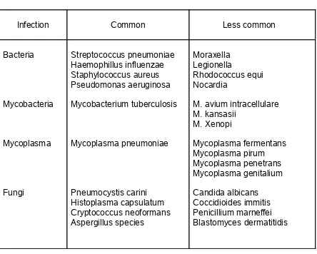
The following pyogenic bacteria and fungi have been recognized to be a major cause of lower respiratory tract infection10,11 in HIV infected patients whatever
TABLE 1 - Bacterial and fungal etiology of LRTI
Infection Common Less common
Bacteria Mycobacteria Mycoplasma Fungi Streptococcus pneumoniae Haemophillus influenzae Staphylococcus aureus Pseudomonas aeruginosa Mycobacterium tuberculosis Mycoplasma pneumoniae Pneumocystis carini Histoplasma capsulatum Cryptococcus neoformans Aspergillus species Moraxella Legionella Rhodococcus equi Nocardia
M. avium intracellulare M. kansasii M. Xenopi Mycoplasma fermentans Mycoplasma pirum Mycoplasma penetrans Mycoplasma genitalium Candida albicans Coccidioides immitis Penicillium marneffei Blastomyces dermatitidis
Incidence of bacterial Lower respiratory tract infection
The true incidence of LRTI in HIV infected patients is difficult to assess and varies with the population surveyed9. Acute bronchitis and bacterial and fungal
In intra venous drug users the incidence of pneumonia per 100 person years was 1.93 in HIV sero-positive and 0.45 in HIV sero-negative subjects29. Boschini
et al23 reported 149 episodes of community acquired pneumonia( CAP) among
HIV sero-positive patients and 61 among HIV sero-negative subjects with incidence rates per 1,000 person years of 90.5 and 14.2 respectively. The incidence rates of pneumonia per 1,000 person years were 38.6 in HIV sero-positive females and 3.7 in HIV sero-negative females in Nairobi, Kenya among female sex workers. More precisely, the incidence rates of invasive pneumococcal disease and pneumococcal bacteraemia per 1,000 person years were 42.5 and 23.8 in HIV positeve females and 3.7 and 0 in HIV sero-negative females respectively18. The relative risk of development of invasive
pneumococcal disease with underlying HIV infection was 17.8 (95% confidence interval 2.5 – 126.5). Pyogenic bacteria as well as Mycobacterium tuberculosis were recognized as major cause of respiratory diseases in HIV infected Africans30-32.
The first series investigating bacterial pneumonia in patients with AIDS or ARC have shown the predominant role of Streptococcus pneumoniae and to a lesser degree, Haemophilus influenzae, in adults as well as children33 in
developing countries 32-34 and developed contries35-38. Subsequently other series
with greater number of patients followed up over a long period have emphasized that other bacteria have also been the cause in HIV infected patients.
S. pneumoniae and H. influenzae were the cause of 52 and 16% of bacteriologically confirmed pneumonia respectively39. Hirschtick et al 25,found S.
pneumoniae and H. influenzae in 52 and 15% of patients with confirmed pneumonia respectively. In the African cohort study by Gilks et al.34, 91% of
pneumococcalserotypes incriminated in HIV-seropositive adults were 1, 3, 5, 7, 19 and 23.
Klebsiella pneumoniae, other members of Enterobacteriaceae family and Pseudomonas aeruginosa were present in 13,10 and 8% of cases respectively of confirmed pneumonia in a study by Hirschtick et al25. Pseudomonas aeruginosa
and S.aureus were cause of 25,9% of CAP in a study by Afessa et al40. Levin et
al41 recovered S.aureus in 23% of sputum cultures in 129 HIV infected
patients.Nocardia species42,Rhodococcus equi43,Streptomyces sp44 were found to
be responsible for chronic pneumonia in AIDS. Pasteurella multocida45,Bordetella
bronchiseptica46,Neisseria sp47,Rocholimea sp48,Corynebacterium
pseudodiphtheriticum49,Legionella50 were equally the cause of pneumonia in HIV
patients.Casedo et al51 showed lung involvement in AIDS patient with Salmonella
septicaemia.
Tuberculosis (TB) ranks as the most common infection seen in the developing countries. About 55 to 89% of AIDS patients in India were found to be suffering from extensive pulmonary tuberculosis52,53. It is estimated that world wide, nearly
Incidence of Fungal lower respiratory infection
Pneumocystis jiroveci previously known as Pneumocystis carinii is the most common opportunistic infection in the West (>60%) and is found in patients with profound immunosuppression (CD 4 count < 200 cells/mm3). Unlike in the west, the incidence of PCP is negligible in India (about 12%)11. This is possibly due to
poor index of suspicion and extensive use of cotrimoxazole for prophylaxis of PCP in HIV54.
Cryptococcus neoformans, an encapsulated yeast is the most common deep seated fungal infection in HIV patients. Though the cryptococcal meningitis is the commonest presentation, lungs is the primary site of infection. In a study
from Rwanda, Cryptococcus neoformans was isolated from sputum specimen of 37 HIV infected patients55.
Meyohas in his study on AIDS patients has isolated Aspergillus species from patients on anti-retroviral and steroid therapy56. Shivananda in his study in 1992
first reported 15.3% of isolates to be Aspergillus species57. Geetha Lakshmi from
Chennai has documented pulmonary aspergillosis in 36 HIV infected patients58.
Though pulmonary candidiasis is documented to be a very rare disease
occurring in late stages of AIDS, oral and esophageal candidiasis is reported as second most common (58%) of opportunistic infection among HIV patients from India54.
Histoplasmosis, Coccidioidomycosis, Blastomycosis are not seen very frequently in India11.
Incidence of mycoplasma in HIV
Mycoplasmas are commensals causing self-limiting and clinically unimportant infections in human beings. Recent isolation of these organisms from adults with AIDS suggests that mycoplasmas might act as cofactors in patients infected with human immunodeficiency virus (HIV)59.
The five species of mycoplasmas identified as being associated withAIDS include Mycoplasma pneumoniae,M. fermentans, M. pirum, M. penetrans and M. genitalium60-61.
Teel et al 65 have reported that mycoplasmas colonized the respiratory tracts
of 28 per cent of HIV-positive and 10.5 per cent of HIVnegative patients.
A detection rate of 12.5 per cent of mycoplasmas has been documented from bronchoalveolar lavage specimens of HIV infected patients which indicated that AIDS patients might be more often colonized or infected by mycoplasmas than HIV-negative patients or other immunocompromised persons 64.
Hjordis 62 isolated M. pneumoniae from respiratory specimens from AIDS
patients by culture. Ainsworth et al 63 have detected the presence of M.
fermentans in the respiratory tract of 27 per cent of the HIV population.
Mycoplasma as cofactor in hiv progression66-71
Luc Montagnier,co-discoverer of AIDS virus proposed that HIV may not work alone and mycoplasmas could be playing the key role in HIV progression68.
Historical background
• The first mycoplasma to be isolated in culture was the bovine
pleuropneumonia agent now known as Mycoplasma mycoides subsp.
mycoides, which was described initially by Nocard andRoux in 1898 72
• In the 1930s Klieneberger introduced the concept that mycoplasmas were “L-forms” of bacteria lacking cell walls and living symbiotically with other, walled bacteria 73.
• Dienes and Edsall detected the first mycoplasma isolated from humans in a Bartholin’s gland abscess in 1937 74. This
mycoplasma was probably the organism we now know as Mycoplasma hominis.
• The organism eventually known to be Mycoplasma pneumoniae
was first isolated in tissue culture from the sputum of a patient with primary atypical pneumonia by Eaton et al in 1944, and thereafter it became known as the Eaton agent 75.
• In 1961 Marmion and Goodburn postulated that the Eaton agent was a PPLO and not a virus76,77.
Mollicute taxonomy and classification
The term “mycoplasma” (Greek; “mykes” - fungus and“plasma” - formed) emerged in the 1950s 78 and replaced the older PPLO terminology. In the 1960s,
mycoplasmas were designated members of a class named Mollicutes,which derives from Latin words meaning soft (“mollis”) and skin (“cutis”). Members of the class Mollicutes are characterized by their small genomes consisting of a single circular chromosome containing 0.58 to 2.2 Mbp, a low G+C content (23 to 40 mol%), and the permanent lack of a cell wall 80.
M. pneumoniae is a member of the family Mycoplasmataceae and order
Mycoplasmatales. Studies of 16S rRNA sequences suggest that mycoplasmas are most closely related to the gram-positive eubacterial subgroup that includes the bacilli, streptococci, and lactobacilli79.
Cell biology
Mycoplasmas represent the smallest self-replicating organisms, in both cellular dimensions and genome size, that are capable of cell-free existence81.
Individual spindle-shaped cells of M. pneumoniae are 1 to 2 µm long and 0.1 to 0.2 µm wide, compared with a typical bacillus of 1 to 4 µm in length and 0.5 to 1.0 µm in width. Accordingly, the M. pneumoniae cell volume is less than 5% of that of a typical bacillus. Cells may either divide by binary fission or first elongate to multinucleate filaments, which subsequently breakup to coccoid bodies
microscopy, and they do not produce visible turbidity in liquid growth media. Typical colonies of M. pneumoniae, show fried egg appearance, rarely exceed 100µm in diameter when cultivated on enriched medium and require examination under a stereomicroscope to visualize their morphological features. The genome of M. pneumoniae was completely sequenced in 1996 and shown to consist of 816,394 bp with 687 genes. Mollicutes have no ability to synthesize peptidoglycan cell walls, since the genes responsible for these processes are not present in the genome. The lack of a rigid cell wall confers pleomorphism on the cells and makes them unable to be classified as cocci or bacilli in the manner of conventional eubacteria. Sterols are necessary components of the triple-layered mycoplasmal cell membrane that provide some structural support to the osmotically fragile mycoplasma.Maintenance of osmotic stability is especially important in mollicutes due to the lack of a rigid cell wall. Another structural component of the M.pneumoniae cell that is important for extracellular survival is aprotein network that provides a cytoskeleton to support thecell membrane
M. pneumoniae has been shown to bind on glass and other solid surfaces, with the organism moving with the attachment organelle at the leading end. Neithergenomic analysis nor electron microscopy of M. pneumoniae
Pathogenesis
M.pneumoniae is primarily an extracellular pathogen that depends on close association with host cells to survive, it has evolved a complex and specialized attachment organelle to facilitate its parasitic existence .This attachment organelle consists of a specialized tip structure with a central core of a dense rodlike central filament surrounded by a lucent space that is enveloped by an extension of the organism’s cell membrane83,84. The tip structure is actually a
network of adhesins, interactive proteins, and adherence accessory proteins that cooperate structurally and functionally to mobilize and concentrate adhesions at the tip of the organism.
The P1 adhesin85,86 is a 170-kDa protein concentrated in the attachment tip
that is now known to be the major structure responsible for interaction of M. pneumoniae with host cells. P30 87 is one of several additional proteins that have
been implicated in the adherence process, based on the knowledge that antibodies developed against P30 can block M. pneumoniae hemadsorption Other structures produced by M. pneumoniae that have been studied as mediators in cytadherence in M. pneumoniae include proteins 88 HMW1, HMW2,
Hydrogen peroxide and superoxide radicals are synthesized as a result of a flavin-terminated electron transport chain M.pneumoniae act in concert with endogenous toxic oxygen molecules generated by host cells to induce oxidative stress in the respiratory epithelium89. Once M. pneumoniae reaches the lower
respiratory tract, the organism may be opsonized by complement or antibodies macrophages become activated, begin phagocytosis, and undergo chemotactic migration to the site of infection90. High percentages of neutrophils and
lymphocytes are present in alveolar fluid. Lymphocyte proliferation, production of immunoglobulins, and release of tumor necrosis factoralpha (TNF-α), gamma interferon (IFN-gamma), and various interleukins(including interleukin-1 [1], IL-2, IL-4, IL-5,IL-6, IL-8, IL-10, and IL-18) occurs91
Immune Response and Immunomodulatory Effects82
Following an initial infection, the normal immune system responds by rapidly producing antibodies that peak after 3 to 6 weeks, followed by a gradual decline over months to years.
Elevation of M. pneumoniae-specific IgM alone can often be interpreted as evidence of acute infection, since this antibody typically appears within 1 week of the initial infection and approximately 2 weeks before IgG antibody.
IgA, while often overlooked as a diagnostic antibody class, may actually be a better indicator of recent infections in all age groups. IgA antibodies are produced early in the course of disease, rise quickly to peak levels, and decrease earlier than IgM or IgG.
In addition to M. pneumoniae-specific antibodies, a variety of cross-reactive antibodies may develop in association with M. pneumoniae infection. The extensive sequence homology of the M. pneumoniae adhesin proteins and glycolipids of the cell membrane with mammalian tissues is a well-known example of molecular mimicry that may trigger autoimmune disorders that involve multiple organ systems through formation of antibodies against substances such as myosin, keratin, fibrinogen, brain, liver, kidney, smooth muscle, and lung tissues.
Antigenic Variation
High-frequency phase and antigenic variation of surface adhesin proteins made possible by DNA rearrangements in truncated and sequence-related copies of the P1 adhesin genes that are dispersed throughout the genome has been described for M. pneumoniae82. Recombinational events among the repetitive
Clinical significance
M.pneumoniae is one of the many causes of a pneumonic process called atypical pneumonia along with several bacterial (Chlamydia pneumoniae, Legionella pneumophila) and fungal (Pneumocystis carinii) pathogens. About 10-30% of CAP are caused by M.pneumoniae infection and these cases represent only 3-10% of M.pneumoniae infection since tracheobronchitis or URT develop in most individuals or remain asymptomatic. Infection usually occurs in school age children >5 yrs, adolescents, young adults in normal population. Most infection in these are minor and include pharyngitis, tracheobronchitis, bronchiolitis and croup. Severe infection develops in immuno compromised HIV patients and elderly requiring hospitalization and death may occur in some92.
Incubation period is 2-3 weeks. Clinical presentation is usually insidious with the gradual onset of constitutional and pneumonic symptoms. Fever of 101-1020F with chills, malaise, head ache, sore throat, nasal congestion, dry non
bronchopneumonia generally involving multiple lobes of the lung without consolidation.X ray pattern may vary widely and may show peribronchial infiltrates, atelectasis and hilar lymphadenopathy. Radiologic findings reveal more than the physical examinations of patients. Other pleuro-pulmonary complications like lung abscess and pneumothorax are also uncommon. Abnormalities on chest films resolve more slowly in 10 days to 6 weeks. Recurrence and relapses of pneumonia despite appropriate antimicrobial therapy can also occur in HIV patients.
Complications
Dermatologic, cardiovascular, musculoskeletal, neurologic, urologic, hepatobiliary or ocular complications can occur. Humoral and cellular Immunodeficiency states in HIV patients predispose than to serious disease with M pneumonia.These patients may suffer repeated bouts of M pneumonia and have difficulty in eliminating the organism from respiratory tract despite adequate therapy.These patients often have upper and lower respiratory tract symptoms with few or no infiltration observed on chest X-ray and have significant complications. Fulminant dessiminated infection with multi system involvement is rare. But it has been reported.
Media for culture of Mycoplasmas:
of about 7.8. The optimum temperature for mycoplasmal growth is 35-370C.
M.pneumoniae grows well in air or in an atmosphere of 95% N and 5% CO2
Several types of media have been described for the cultivation of M. pneumoniae.The medium recommended by Center for disease control for isolation of M. pneumoniae is Methylene blue glucose diphasic medium also known as PPLO diphasic medium. This medium contains PPLO (Pleuropneumonia like organism) broth and agar, yeast extract, serum supplements along with glucose, Methylene blue and phenol red. The methylene blue in the medium inhibits the growth of other human mycoplasmas that may be found in respiratory tract making the medium selective for M. pneumoniae. During growth of M. pneumoniae the medium becomes more acidic and the phenol red turns colour from salmon to yellow. At the same time the organisms reduce the methylene blue and turn it from blue to colourless. Therefore the colour of the broth phase changes from purple to green or yellow green while the agar phase turns from purple colour to yellow or yellow orange. This medium is used in conjunction with mycoplasma glucose agar(PPLO agar) medium. Colonies recovered either directly on this medium or from subcultures of positive broth organ subjected to inspection and identification procedures. Inorder to avoid overgrowth by other pathogens a broad spectrum beta lactum antibiotic and an antifungal agent should be added to the medium.
Another medium that is recommended for isolation for Mycoplasma pneumoniae from clinical specimens is Soy peptone (SP4 ) Broth and SP4 agar
Isolation and identification
The growth of M. pneumoniae from clinical specimens is detected by the ability of these organisms to produce acid from glucose. Methylene blue glucose diphasic medium is inoculated with 0.2 ml of specimens and broth cultures are incubated at 350C with caps tightened. Tubes are inspected daily for colour
changes and turbidity for 4 weeks. A slight, gradual shift in the pH indicator over 8 to 15 days without gross turbidity suggests true positive cultures. As soon as the colour changes are apparent, the broth is sub-cultured onto agar medium and incubated for 5 to 7 days at 350C. Agar plates are sealed with air permeable
cellophane tapes to prevent agar from drying out. Inspection of agar under low power of microscope will reveal fried egg colonies. In the absence of obvious colour change a blind subculture to agar media should be performed after 1 and 3 weeks of incubation. A general scheme for isolation of M. pneumoniae is shown in chart.
Human Mycoplasmas can be divided into three groups on the basis of utilization of three substrates – glucose, arginine and urea. An enriched peptone basal medium containing yeast extract and serum supplemented with one of the three substrates with a pH indicator is used. Mycoplasma pneumoniae metobolises glucose to produce lactic acid resulting in a shift to acidic pH.
Serology
Serology is an important tool for the diagnosis of M. pneumoniae infection. This is due to the ease of specimen collection and wide spread availability of serological test82. Before the availability of more advanced serologic techniques,
detection of cold agglutinins was a valuable tool for M. pneumoniae diagnosis96.
The formation of cold agglutinins is the first humoral response to M. pneumoniae. However cold agglutinins are not reliable indicators as they are elevated in only 50 to 60 % of patients. Cross reactions may be induced by Ebstein-Barr virus, Cytomegalo virus and Klebsiella pneumoniae, malignancies of lymphoid cells and auto-immune diseases93. Complement fixation test(CF) once considered as
Molecular techniques :
Hybridisation assays provide a similar sensitivity as ELISA tests for antigen detection. The Genprobe rapid system which involves a I 125 labelled DNA probe
to an rRNA sequence specific for M.pneumoniae was widely used before the application of PCR82,96.
Nucleic acid amplification by PCR was first applied for M.pneumoniae diagnosis in 1989 by Bernet et al. the main advantage of PCR is its superior sensitivity. The sensitivity may be increased by nested PCR which may be required for detection of M.pneumoniae from extrapulmonary sites. Multiplex PCR assays were designed for simultaneous detection of M. pneumoniae and other respiratory pathogens. RNA amplification techniques are used for their high sensitivity and detection of this nucleic acid is more indicative of viable mycoplasmas in clinical samples.
Antimicrobial susceptibilities and chemotherapy
Infections caused by M.pneumoniae are generally treated with tetracycline or
erythromycin. Because of the difficulty in culturing the organism, its slow growth rate
and the lack of a readily available method, antimicrobial susceptibility testing of
M.pneumoniae is neither necessary nor appropriate. Antimicrobial susceptibility testing
of M.pneumoniae strains indicate that this organism is susceptible to a wide variety of
antimicrobial agents including the quinolones that is cipfloxacin , levofloxacin, ofloxacin,
gemifloxacin, moxifloxacin, clindamycin, lincomycin, tetracycline, minocycline,
doxycycline, erythromycin, streptomycin. Some of the newer macrolide antibiotics such
as clarithromycin, azithromycin, flurithromycin are also highly active against
MATERIALS AND METHODS
This prospective study was conducted on 100 HIV sero-positive patients attending the outpatient department or admitted in the Medical, Venereology and Thoracic Medicine Departments with signs of lower respiratory infection at Coimbatore Medical College Hospital, Coimbatore. Specimens for the study were collected over a period of one year and eight months from January 2005 to August 2006 .
Approval was obtained from the ethical committee prior to conducting the study and Informed consent from all patients under study was also obtained.
• Inclusion criteria:
One hundred consecutive HIV sero-positive patients with symptoms and signs suggestive of lower respiratory tract infection and who have not received antibiotics for minimum of 2 weeks prior to this study.
• Exclusion criteria:
Pediatric HIV sero-positive patients were excluded.
The name, age, sex, address, date of admission, inpatient number, clinical history of the patient were noted. A thorough general examination and systemic examination of the patient was also done.
HIV seropositive patients were classified under different clinical stages95
according to the world health organization [WHO] guidelines as mentioned in appendix i.
Basic investigations such as Hb%, TC, DC, LFT, RFT, Blood Sugar, Urine complete examination, chest x-ray were also done.
CD4 cell counts assays done by flow-cytometry and immune status grading of the patients was done according to the WHO 95 guidelines as mentioned in
appendix ii.
Sputum specimens were collected for bacterial, fungal and mycobacterial cultures.
Blood cultures were done for all patients to find out any bacteraemia. Specimen collection and transport:
Samples collected: Expectorated sputum Induced sputum Blood
Collection of expectorated sputum sample:
The patient was instructed to cough out deeply and early morning sputum samples on three consecutive days were collected in sterile wide mouthed containers fitted with screw capped lids.
Collection of induced sputum sample:
Collection of blood sample:
After cleansing the site for venepuncture with betadine and 70% alcohol about 7 ml of blood was collected and 5 ml was added to 50 ml of sterile brain heart infusion broth in blood culture bottles and remaining 2 ml of blood was used for serological tests.
All the specimens were sent to the laboratory immediately after collection. Safety precautions
• All sputum samples were potentially infectious and leak proof containers were used for collection and transportation of the samples.
• Biological safety cabinet level III was used for carrying out all procedures involving sputum and protective wears like mask, gloves etc was used. • Disinfection of the sputum cups/ containers by treating with freshly
prepared 1-2% sodium hypochlorite solution or autoclaving was followed. Processing of sputum samples:
Induced sputum was used for detection of trophozoites and cysts of P. jiroveci.
Expectorated sputum samples were used for isolation of bacterial, mycobacterial and other fungal pathogens.
The quality of expectorated sputum was assessed by macroscopic examination and microscopic examination. Any sample that was thin, watery and with no purulent matter was considered unsuitable for further processing. Gram stain was done and Brahmadhatan et al94 system was used for assessing the
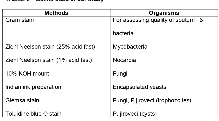
The following staining methods were carried out for detection of various organisms as described in the table below:
TABLE 2 – Stains used in our study
Methods Organisms
Gram stain
Ziehl Neelson stain (25% acid fast) Ziehl Neelson stain (1% acid fast) 10% KOH mount
Indian ink preparation Giemsa stain
Toluidine blue O stain
For assessing quality of sputum & bacteria.
Mycobacteria Nocardia Fungi
Encapsulated yeasts
Fungi, P.jiroveci (trophozoites) P. jiroveci (cysts)
Preparation and procedure for staining
Gram’s stain Procedure
1. Appropriate smear is made on a clean glass slide.
The smear is fixed by passing the slide over flame 2-3 times quickly. Cover the slide with crystal violet solution and allow to act for about 30 seconds.
2. Pour off stain and holding the slide at an angle downwards pour on the iodine solution on the slide so that it washes away the crystal violet. Cover the slide with fresh iodine solution and allow to act for 1 minute.
as to cover its whole surface. Decolorization is very rapid and is usually complete in 2-3 seconds. After this period of contact, wash thoroughly with water under a running tap
4.Apply the counterstain (0.5% safranine) for 30 seconds. 5. Wash with water and blot dry.
6. Examine the smear under oil immersion microscopy. Ziehl Neelsen Staining Procedure
Reagents
– Carbol fuchsin ---- 1% – Sulphuric acid ---- 25% – Methylene blue --- 0.1% Method
1. Select a new, unscratched slide and label the slide with a laboratory serial number.
2. Make a smear from yellow purulent portion of the sputum using the jagged end side of a bamboo stick. A good smear is spread evenly, 2cms x 3cms in size and is neither too thick nor too thin. The optimum thickness of the smear can be assessed by placing the smear on a printed matter, the print should be just readable through the smear.
3. Let the smear air-dry for 15-30 mins.
4. Fix the smear by passing the slide over the flame 3-5 times for 3-4 seconds
5. Place the fixed slide on the staining rack with the smeared side facing upwards.
6. Pour filtered 1% carbol fuchsin over the slide so as to cover the entire slide. Do not leave the carbol fuchsin on the slide for a long time (not more than 5 mins.)
7. Heat the slide underneath until vapours start rising. Do not let carbol fuchsin to boil or the slide to dry. Continue the process up to five minutes.
8. Allow the slide to cool for 5-7 minutes.
9. Gently rinse the slide with tap water to remove the excess carbol fuchsin stain. At this point, the smear on the slide looks red in colour.
10. Decolour the stained slide by pouring 25% sulphuric acid on the slide and leaving the acid for 2-4 mins.
11. Lightly wash away the free stain. Tip the slide to drain off the water. If the slide is still red, reapply sulphuric acid for 1-3 mins, and rinse
gently with tap water.
12. Counter stain the slide by pouring 0.1% methylene blue solution on to slide and let it stand for one min.
13. Gently rinse the slide with the tap water and tip the slide to drain off the water.
14. Place the slide in the slide tray and allow it to dry.
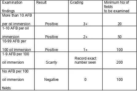
a drop of immersion oil for the characteristic acid fast bacilli. At least 100 oil immersion fields should be examined before declaring a smear as negative. In case of the scanty result, examine another 100 oil immersion fields.
TABLE 3 - Grading of sputum smears for AFB was done as follows :
Examination findings
Result Grading Minimum No of
fields
to be examined More than 10 AFB
per oil immersion Positive 3+ 20
1-10 AFB per oil
immersion Positive 2+ 50
10-99 AFB per
100 oil immersion Positive 1+ 100
1-9 AFB per 100
oil immersion Scanty
Record exact
number seen 200 No AFB per 100
oil immersion fields
Negative 0 100
Giemsa stain
Preparation and procedure for staining94 Preparation:
Giemsa powder 0.3 gm Glycerine 25.0 ml Acetone free methanol 25.0 ml
The stock solution is diluted before use by adding 1 ml of stain to 10 ml of distilled water.
Procedure for staining: 1. Air dry thin films.
2. Fix in methanol for 1 minute
3. Wash in tap water and flood the slide with Giemsa diluted 1in 10 with buffered distilled water (pH 7.2). The diluted stain must be freshly prepared each time.
4. Stain for 25 – 30 minutes.
5. Run tap water on to the slide to float off the stain and to prevent deposition of precipitate on the film.
6. Examine the film using X 100 objective. Toluidine blue O stain
Preparation and procedure for staining94
Preparation:
Absolute alcohol 40.0 ml Distilled water 60.0 ml
Dissolve the stain in absolute alcohol. Add 60 ml water and finally add 2 ml of conc. HCl.
Procedure for staining
1. Fix the smear in sulfation reagent ( 4.5 ml glacial acetic acid + 1.5 ml concentrated sulfurinc acid) for 10 minutes. Mix the reagent with a glass rod immediately and 5 minutes after.
2. Wash in a gentle stream of running tap water for 5 minutes. 3. Dip the slide in Toludine blue O stain, for 10 minutes.
4. Decolourise in 95% alcohol / absolute alcohol ( by taking in and out) 5 secongs X 2.
5. Dip in xylene for cleaning – 10 seconds. 6. Mount with mounting fluid.
7. Observe with X 20 and X 40 objectives.
The cyst wall stains violet to purple. Thickenings and folds in cyst wall stain darker violet to purple. Trophozoites and intracystic bodies are not stained.
Culture of sputum specimen
The sputum samples were inoculated onto blood agar with 10% sheep blood, chocolate agar with 10% sheep blood, Mac Conkey agar. Inoculated blood agar and chocolate agar plates were incubated at 37oC with 5-10% CO
2 and Mac
The sputum was inoculated onto Sabouraud’s Dextrose agar(SDA) with antibiotics and SDA without antibiotics in duplicate ( incubated at 37oC and
25oC).
A portion of sputum specimen was decontaminated as per modified Petroff’s method as described94 and inoculated in Lowenstein Jensen’s (LJ) medium for
culturing mycobacteria. Inoculated LJ medium bottles were incubated at 37oC for
6-8 weeks and observed for any growth.
All the bacterial and fungal pathogens isolated were identified as per standard protocol92. Antimicrobial susceptibility test of the bacterial isolates to various
antibiotics was also done by Kirby Bauer’s disc diffusion method and antibiotic sensitivity pattern studied according to clinical laboratory standards institute(CLSI).
Processing of sputum for culture of M. pneumoniae:
Homogenisation of expectorated sputum specimen was done by repeatedly drawing through a needle and syringe as chemical treatments for sputum liquefaction are toxic for mycoplasma.
Isolation and identification of M. pneumoniae was performed as described by Koneman et al92.
Inoculation and Isolation: About 0.2 ml of homogenized sputum specimen was inoculated in a tube of Mycoplasma selective diphasic culture medium. The test tube was then loosely sealed and incubated at 35oC for upto 4 weeks and
change in the colour of the medium with no increase in turbidity several drops of broth culture were subcultured onto a mycoplasma selective agar medium plate and was incubated at 35oC with 5% CO
2 for another 7 days. The agar surface
observed for colonies after the fifth day under 10 X and 40 X magnification had the appearance of fried eggs.
Blind subcultures of broth to agar medium was done in the absence of obvious colour change after 1 and 3 weeks of incubation.
Commercially available PPLO broth and agar base from Hi Media was used and PPLO diphasic medium and agar was prepared for selective isolation of M. pneumoniae and selective supplements added as follows:
Procedure for preparation of methylene blue glucose diphasic medium and
agar PPLO diphasic medium and agar.
(I) Base reagents for preparation of complete media
A. Mycoplasma base agar: This medium may be prepared using commercial PPLO agar base according to the package instruction.Basal ingredients in PPLO agar base are as follows:
Beef heart infusion 250 gm
Peptone 10 gm
NaCl 5 gm
Mix together and melt the agar in a boiling water bath. Dispense in 70 ml aliquots and autoclave at 15 lbs for 15 minutes. Store at 4oC.
B. Yeast extract:
1. Purchase commercial 25% yeast extract or prepare as follows – weigh out 250 gm of active baker’s yeast and place in 1 ltr of distilled water. Heat to boiling, cool and filter to remove particular matter. Adjust the pH to 8.0 and filter – sterilize.
2. Yeast extract ( 20ml) is then mixed with uninactivated horse serum (10ml) in a 2:1 ratio and frozen in 30 ml aliquots at -20oC.
C. Phenol red solution (0.4%): Phenol Red (1 g) is dissolved in 3 ml of 1 N NaOH and then 247 ml of distilled water is added. Solution is filtered – sterilized and stored at 4 to 8oC.
D. Methylene Blue solution (1%): Methylene blue (1gm) is dissolved in 100 ml distilled water and autoclaved for 15 minutes.
E. Thallium acetate solution(10%): Thallium acetate (10g) is dissolved in 100 ml of distilled water and filter sterilized.
F. Glucose solution: Glucose (50 gm) is dissolved in 100 ml distilled water and filter sterilized.
G. Penicillin solution: Penicillin powder is dissolved in distilled water to achieve a concentration of 1,00,000 units / ml.
(II) Methylene Blue – Glucose Diphasic Medium:
1. Melt 70 ml of mycoplasma agar in a boiling water bath. Cool to 50oC and add
2. Add the following:
Methylene blue solution (1%) 0.1 ml Phenol red solution (0.4%) 0.5 ml
Glucose solution 2.0 ml
Thallium acetate solution (10%) 0.25 ml Penicillin solution 3.0 ml 3. Adjust the pH to 7.8 with sterile 1 N NaOH.
4. Dispense agar in 1 ml aliquots into sterile 13 X 100 mm screw capped tubes. 5. Mix 140 ml of mycoplasma broth (A, above, without agar) with 60 ml of yeast extract serum. Add the following to this mixture
Methylene blue solution (1%) 0.2 ml Phenol red solution 1.0 ml Glucose solution 4.0 ml Thallium acetate solution (10%) 6.0 ml Pencillin solution 0.5ml
6. Adjust the pH to 7.8 with sterile 1 N NaOH. Dispense 2.0 ml into each of the tubes containing the solidified agar medium.
(III) Mycoplasma glucose agar medium
1. Melt 70 ml of mycoplasma agar in a boiling water bath. Cool to 50oC.
2. Prewarm 30 ml of yeast extract serum and mix with the agar medium 3. Aseptically add the following reagents:
Thallium acetate solution (10%) 0.25 ml Penicillin solution 3.0 ml 4. Adjust the pH to 7.8 with sterile 1 N NaOH.
5. Dispense 6 ml of medium into 60 X 15 mm Petri dishes. Allow to cool. Store media at 4oC.
Identification: Presumptive identification of colonies of mycoplasma
pneumoniae was done by the following methods:
1.Diene’s stain, 2.Haemadsorption, 3.Glucose fermentation, 4.Hydrolysis of urea, 5.Arginine hydrolysis.
Diene’s stain procedure for identification of mycoplasmas:
I. Principle: Mycoplasma colonies from are easily identified by observing typical colonies on agar medium. Visualization of colony morphology is facilitated by application of Diene’s stain directly to agar surface.
Diene’s stain is a nonspecific stain that imparts a contrasting appearance of mycoplasma colonies on agar, allowing easier visualization of colony morphology and characteristics.
II. Reagents: A. Modified Diene’s stain ( stock solution) Methylene blue 2.5 gms
Azure blue 1.2 gm
Maltose 10.0 gm
Na2CO3 0.25 gm
Diene’s stain working solution is prepared by diluting an aliquot of the stock solution 1:3 with distilled water.
(III) Procedure:
1. Flood an agar plate containing mycoplasma growth with 1 ml of Diene’s stain working solution.
2. Immediately rinse the agar surface with distilled water to remove the stain. 3. Decolorize the medium by adding 1 ml of 95% ethanol. Leave in contact
with the agar for 1 minute, then remove. Repeat the wash step a second time.
4. Rinse the plate with distilled water and allow to dry.
5. Observe for colonies under the low power of a microscope ( 50 to100X). IV Results: A. Interpretation:
Mycoplasmas with the ‘fried – egg’ colony morphology will stain with a dark blue centre and light blue periphery and will appear highly granular. The agar background will be clear or slightly violet.
Hemadsorption test for identification of mycoplasma pneumoniae
(I) Principle: Among the respiratory Mycoplasmas, M. pneumoniae is the only species that will specifically absorb red blood cells. This property,therefore, provides a method for the presumptive identification of M. pneumoniae.
When colony growth is noted on mycoplasma isolation media inoculated with respiratory tract specimens a suspension of guinea pig erythrocytes is placed on the agar surface for a given time and then washed off. M. pneumoniae colonies will adsorb some of the red cells to the colony surface.
(II) Reagents:
A. Mycoplasma glucose agar with suspicious colonies present
B. Washed guinea pig erythrocytes ( 0.2 – 0.4%) suspended in mycoplasma broth medium.
(III) Procedure:
1. Flood the surface of the agar plate with 2 ml of red cell suspension.
2. Incubate the plate at 35oC for 30 minutes and rotate the plate occasionally
to prevent the red cells from settling out.
3. Wash the surface of the plate three time with 3 ml of mycoplasma broth by gently rotating the plate. Remove wash fluid by aspiration with a pipet. 4. Examine the colonies at 50 to 100X magnification under a dissecting
(IV) Results: A. Interpretation:
1. Positive test: Colonies with red cells adsorbed onto the surface – M. pneumoniae
2. Negative test: Colonies with no red cells adsorbed – Mycoplasma species, not M. pneumoniae.
Glucose fermentation:
Sterile PPLO broth with 0.5% glucose with pH 7.8 prepared as described above and about 2 ml of medium was dispersed in 13 X100 mm sterile screen capped tubes.
0.25 ml of positive broth culture was inoculated and incubated aerobically at 37oC for 10 days. Fermentation status was checked daily. A change in colour
from purple to yellow was noted. Arginine hydrolysis
Sterile screw capped tubes containing 2 ml of Arginine broth with 0.25% arginine at pH 7.0 was inoculated with 0.25 ml of broth culture of the isolate and incubated aerobically at 37oC for 10 days. The result of hydrolysis was checked
daily by observing a colour change from purple to red. Urea hydrolysis
culture of the isolate and incubated aerobically at 35oC for 10 days. Urea
breakdown was checked based on observing colour change from purple to red. Serological tests
About 2 ml of blood was centrifuged and serum was separated and commercially available Mycoplasma pneumoniae IgM TMB ELISA (Bio Rad Laboratories) was done according to manufacturer’s instructions supplied in the kit.
Principle : When antigens bound to the solid phase are brought into contact with a patient's serum, antigen specific antibody, if present, will bind to the antigen on the solid phase forming antigen-antibody complexes. Excess antibody is removed by washing. This is followed by the addition of goat anti-human IgM globulin conjugated with horseradish peroxidase, which then binds to the antibody-antigen complexes. The excess conjugate is removed by washing, followed by the addition of substrate and chromogen, Tetramethylbenzidine (TMB). If specific antibody to the antigen is present in the patient's serum, a blue color develops. When the enzymatic reaction is stopped with 1N H2SO4, the contents of the wells turn yellow. The color, which is proportional to the concentration of antibody in the serum, is read on ELISA microwell plate reader at 450nm.Serological response of the patients against mycoplasma antigen coated to microtitre wells is thus determined.
day to day fluctuations in temperature.Horse radish peroxidase conjugate containing goat antihuman IgM and tetra methyl benzidine substrate were used.
Assay Procedure
1. Place the desired number of strips into a microwell frame. Allow six (6) Control/Cutoff Calibrator determinations (one Negative Control, three Cutoff Calibrators, one High Positive Control, and one Low Positive Control) per run. A reagent blank (RB) should be run on each assay. 2. Dilute test sera, Cutoff Calibrator and Control sera 1:81 (e.g., 10 µL + 800
µL) in Serum Diluent Plus
3. To individual wells add 100 µL of diluted patient sera, Cutoff Calibrator and Control sera. Add 100 µL of Serum Diluent Plus to the reagent blank well.
4. Incubate each well at room temperature (21° to 25° C) for 30 minutes + 2 minutes.
5. Using automated washing equipment add 250-300 µL of diluted Wash Buffer to each well. Repeat the wash procedure four times (for a total of five washes) for automated equipment
6. Add 100 µL Conjugate to each well, including the reagent blank well. 7. Incubate each well 30 minutes + 2 minutes at room temperature (21° to
25° C).
9. Add 100 µL Chromogen/Substrate solution (TMB) solution to each well, including reagent blank well10. Incubate each well 10 minutes + 2 minutes at room temperature (21° to 25° C).
11. Stop reaction by addition of 100 µL of Stop Solution (1N H2SO4) following the same order as Chromogen/Substate addition, including reagent blank well. Tap the plate gently along the outsides to mix contents of the wells. The plate may be held up to one hour after addition of the Stop Solution before reading.
12. The developed color should be read on an ELISA plate reader equipped with a 450 nm filter.
Calculations
1. Mean Cutoff Calibrator O.D. (Optical Density) - Calculate the mean O.D. value for the Cutoff Calibrator from the three Calibrator determinations. 2. Correction Factor - To account for day-to-day fluctuations in assay activity
due to room temperature and timing, a Correction Factor is determined for each lot of kits. The Correction Factor is printed on the Calibrator vial. 3. Cutoff Calibrator Value - The Cutoff Calibrator Value for each assay is
determined by multiplying the Correction Factor by the mean Cutoff Calibrator O.D.
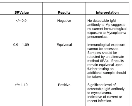
4. ISR Value - Immune Status Ratio (ISR) for each specimen is calculated by dividing the specimen O.D. Value by the Cutoff Calibrator Value
TABLE 4 -(ISR) Immune Status Ratio Interpretation:
ISRValue Results Interpretation
</= 0.9
0.9 – 1.09
>/= 1.10
Negative
Equivocal
Positive
No detectable IgM antibody to Mp suggests no current immunological exposure to Mycoplasma pneumoniae.
Immunological exposure cannot be assessed. Samples should be retested by an alternate method (IFA). If results remain equivocal upon further testing an
additional sample should be taken.
Significant level of detectable IgM antibody to mycoplasma.
[image:52.612.86.538.92.457.2]RESULTS
[image:53.612.84.531.173.339.2]This study included 100 HIV seropositive patients with lower respiratory tract infections.
TABLE 5 - AGE & SEX-WISE DISTRIBUTION OF CASES
Sex No. of
patients
Age in years
Range Mean Median Percentage
Male 71 27 – 55 37 38 71
Female 29 28 – 65 36 35 29
Among the 100 cases, 71 were males with range of age from 27 – 55 years and mean age 37.6 and 29 were females with range of age from 28 – 65 years and mean age of 36.2.
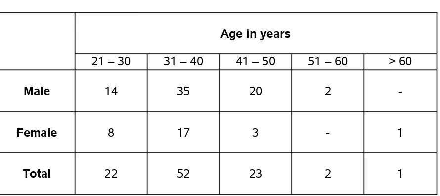
TABLE 6 - AGE GROUPS UNDER STUDY
Age in years
21 – 30 31 – 40 41 – 50 51 – 60 > 60
Male 14 35 20 2
-Female 8 17 3 - 1
[image:53.612.86.531.467.665.2]Among the 71 males,14 were in 21 -30 year age group, 35 were in 31 – 40 year age group, 2 were in 51-60 year age group and among the females, 8 were in 21 – 30 year age group, 17 were in 31 -40 year age group and 2 were in 40 – 50 year and 1 female was 65 years of age.
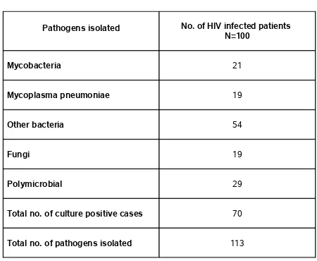
TABLE 7 - PATHOGENS ISOLATED FROM HIV SEROPOSITIVE PATIENTS
Pathogens isolated No. of HIV infected patients N=100
Mycobacteria 21
Mycoplasma pneumoniae 19
Other bacteria 54
Fungi 19
Polymicrobial 29
Total no. of culture positive cases 70
Total no. of pathogens isolated 113
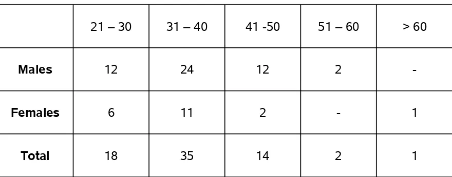
TABLE 8 - AGE & SEXWISE DISTRIBUTION OF CULTURE POSITIVE CASES
IS AS FOLLOWS
21 – 30 31 – 40 41 -50 51 – 60 > 60
Males 12 24 12 2
-Females 6 11 2 - 1
Total 18 35 14 2 1
Etiological agent could be identified in more number of patients in 21 – 30 and 31 – 40 year age groups.
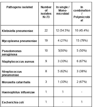
TABLE 9 - BACTERIAL PATHOGENS ISOLATED FROM HIV
SERO-POSITIVE PATIENTS
Pathogens isolated Number of isolates
N=73
In single / Mono-microbial
In combination
/ Polymicrobi
al
Klebsiella pneumoniae 22 12 (54.5%) 10 (45.4%)
Mycoplasma pneumoniae 19 4 (21%) 15 (79%)
Pseudomonas aeruginosa
10 5(50%) 5 (50%)
Staphylococcus aureus 9 3 (33%) 6 (67%)
Streptococcus pneumoniae
8 5 (62%) 3 (38%)
Moraxella catarrhalis 3 1 (33%) 2 (67%)
Haemophilus influenzae 1 1
-Escherichia coli 1 - 1
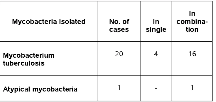
TABLE 10 - MYCOBACTERIA ISOLATED FROM HIV SERO-POSITIVE
PATIENTS
Mycobacteria isolated No. of cases
In single
In
combina-tion
Mycobacterium tuberculosis
20 4 16
Atypical mycobacteria 1 - 1
21 Mycobacteria were isolated from our study. Most of the organisms (16) were polymicrobial isolations. One atypical mycobacterium was isolated from a patient with stage III disease and CD4 count of 115 cells/mm3. The atypical
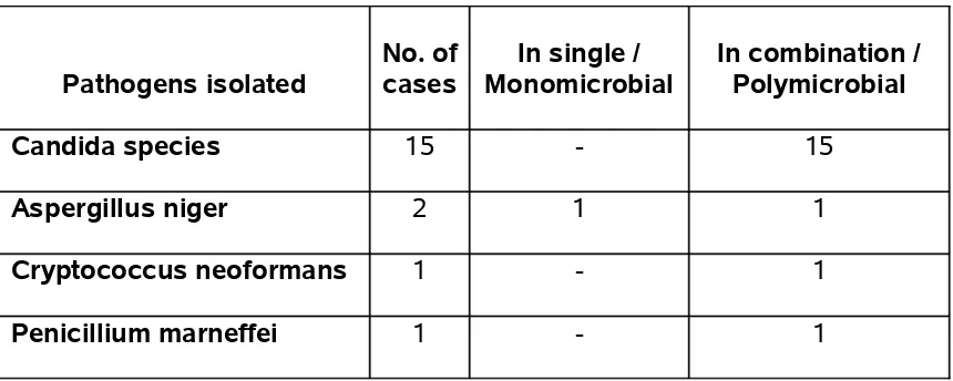
TABLE 11 - FUNGAL PATHOGENS ISOLATED FROM HIV SERO-POSITIVE
PATIENTS
Pathogens isolated
No. of cases
In single / Monomicrobial
In combination / Polymicrobial
Candida species 15 - 15
Aspergillus niger 2 1 1
Cryptococcus neoformans 1 - 1
Penicillium marneffei 1 - 1
19 pathogenic fungi were isolated in our study, out of which, 15 were Candida species. Gram stained smears of patients showing pseudohyphae and yeast cells were only considered as pathogenic isolates. Sputum examination was done for 3 consecutive days to confirm the fungal isolates.
Cryptococcus neoformans was isolated in a case diagnosed to have pulmonary tuberculosis on anti tuberculous treatment and that patient succumbed to death. This patient had CD4 cell count of 120 cells / mm3.
Penicillium marneffei was also isolated in a patient with CD4 cell count of 125 cells/mm3.
Aspergillus niger was isolated in two cases and both cases had CD 4 counts < 200 cells /mm3. This was confirmed by repeated sputum culture on 3
TABLE 12 - IMMUNOSTAGING IN STUDY CASES AND SPUTUM CULTURE
POSITIVE CASES
Levels of Immuno-suppression
CD4 count
Cells/ mm3 Total No. of cases
N=100
No. of culture positive cases
N=70
Severe IS <200 43 38
Advanced IS 200 – 349 52 29
Mild IS 350 – 499 5 3
No significant IS > 500 0 0
TABLE 13 - HIV CLINICAL STAGING IN STUDY CASES AND SPUTUM
CULTURE POSITIVE CASES
HIV stage Total No. of cases N=100
No. of culture positive cases N=70
I 2 0
II 43 30
III 51 36
IV 4 4
[image:59.612.85.529.430.657.2]culture positive cases were in stages of advanced and severe immunosuppression with CD4 counts of 200-349 and <200 cells /mm3
respectively.
[image:60.612.86.530.310.568.2]30 sputum culture positive cases were in clinigal stage II and 36 cases were in clinical stage III and 4 cases were in stage IV and this constituted a total of 94% in clinical stages II and III.
TABLE 14 - AGE AND SEX-WISE DISTRIBUTION OF CULTURE POSITIVE CASES OF MYCOPLASMA PNEUMONIAE IN HIV SERO-POSITIVE
PATIENTS
Age group in years
No. of males N=71 Culture +ive for Mycoplasma pneumoniae No. of females N=29 Culture +ive for Mycoplasma pneumoniae No. N=14 % No. N=5 %
21 – 30 14 3 21.4 8 1 12.5
31 – 40 35 7 20 17 3 17.6
41 – 50 20 4 20 3 1 33.3
>50 2 - - 1 -
TABLE 15 – CO-PATHOGENS ISOLATED ALONG WITH MYCOPLASMA PNEUMONIAE
Pathogens isolated
Mycoplasma pneumoniae positive cases
N = 19
Mycoplasma pneumoniae negative
cases N = 81
Klebsiella pneumoniae 5 ( 26% ) 17 (20.9%)
Pseudomonas aeruginosa 3 ( 16% ) 7 (8.6%)
Staphylococcus aureus 3 ( 16% ) 6 (7.4%)
Moraxella catarrhalis 0 ( 0% ) 3 (3.7%)
Streptococcus pneumoniae 1 ( 5% ) 7 ( 8.6%)
Mycobacteria 6 ( 32% ) 15 ( 18.5%)
Aspergillus niger 1 ( 5% ) 1 ( 1.2% )
Candida species 5 ( 26% ) 10 ( 12.3% )
TABLE 16 - ANTIMICROBIAL SUSCEPTIBILITY PATTERN OF BACTERIAL ISOLATES Pathogens G e n t a m i c i n 10µg A m i k a c i n 30µg C i p r o f l o x a c i n 5µg O f l o x a c i n 5µg C e f o t a x i m e 30µg C e f t r i o x a z o n e 30µg C o t r i m o x a z o l e 25µg o x a c i l l i n 1µg A m o x i c i l l i n 20µg V a n c o m y c i n 30µg E r y t h m y c i n 15µg Klebsiella pneumonia (N=22)
10 14 14 15 14 15 5 - - -
-Pseudomonas
aeroginosa (N=10) 4 5 6 7 4 5 3 - - -
-Haemophillus influenzae (N=1)
1 1 1 1 1 1 1 - - -
-Escherichia coli
(N=1) 1 1 1 1 1 1 0 - - -
-Staphylococcus aureus (N=9)
- 5 5 6 6 8 4 5 4 9 4
Streptococcus
pneumoniae (N=8) - 7 5 6 7 8 6 7 7 8 7
Moraxella catarrhalis (N=3)
- 2 2 2 2 2 1 2 1 2 1