A DISSERTATION ON
CLINICAL, LABORATORY AND
RADIOLOGICAL PROFILE OF URINARY
TRACT INFECTION IN CHILDREN
M.D (BRANCH VII)
PAEDIATRIC MEDICINE
APRIL 2011
THE TAMILNADU
ACKNOWLEDGEMENT
I thank the
Dean Dr.A. Edwin Joe
M.D.,
Madurai Medical College and Hospital Superintendent
Dr. S.M.Sivakumar, M.S.,
Government Rajaji Hospital Madurai
for permitting me to perform this study.
I express my sincere thanks and gratitude to
Prof. Dr. G. Mathevan
,
M.D.,D.C.H.,
Professor and Head of the
Department of Pediatrics for his guidance , encouragement
,valuable suggestions and support during this study.
I am greatly indebted to my chief, guide and
teacher,
Prof. Dr. Chitra Ayyappan
,
M.D.,D.C.H.,
FRCP.,(Glasg) FRCPCH, (Lond)
Professor of Pediatrics for her
guidance, supervision, critical review, constant encouragement and
support throughout this study.
I wish to express my sincere thanks to my
Kuppusamy and Dr. Saravanan
for their guidance, supervision,
valuable suggestions and support throughout this study.
I also thank all the members of the Dissertation Committee
for their guidance during the study.
I also express my gratitude to my fellow Post graduates for
their kind cooperation in carrying out this study.
I thank all the parents and children without whom this study
would not have been possible.
CONTENTS
S.NO. TOPIC PAGE NO.
1. INTRODUCTION 1
2. REVIEW OF LITERATURE 3
3. AIM OF THE STUDY 23
4. MATERIALS AND METHODS 24
5. OBSERVATION, ANALYSIS & RESULTS 28
6. DISCUSSION 43
7. CONCLUSION 53
8. LIMITATIONS 54
9. RECOMMENDATIONS 55 BIBLIOGRAPHY
PROFORMA
ABBREVIATIONS
MASTER CHART
INTRODUCTION
Urinary tract infection (UTI) is a common problem in
children1 .The incidence varies according to age, race and sex of
children2,3. UTI occurs in about 1% of boys and 3-5% of girls 4. In girls
the first UTI occurs by the age of 5 years with peaks during infancy and
toilet training. After the first UTI 60-80% 0f the girls will develop a
second UTI within 18 months. In boys most UTIs occur during the first
year of life .UTI is much more common in uncircumcised males. The
prevalence of UTI varies with age. During the 1st year of life; the male
to female ratio is 2.8-5.4:1.Beyond 1-2 years there is a striking female
preponderance , with a male to female ratio of 1:10.
Three to five percent of febrile children are found to have
UTI 6. Symptoms of UTI may be minimal and non-specific in infants
and small children 7. In most cases the first episode of UTI occurs in the
1st year of life and it is believed that young growing kidneys are more
vulnerable to renal parenchymal damage. UTI may lead to life
threatening complications like sepsis and renal scarring. Renal scarring
is the most common cause of hypertension in later childhood and renal
Recognition of UTI in children should be made as early as
possible to prevent these complications 7. Therefore, investigations for
early diagnosis of UTI are of outmost importance 5. In the pediatric
population the recurrence rates for UTI are very high.
Within 1 year of a first infection, approximately 30% of
boys and 40% of girls will develop a repeat UTI 8. .After the first
episode, children can expect a recurrence rate of 30%. This rate will
double for each subsequent infection 9.Anatomic obstruction (posterior
urethral valves, ureteropelvic junction obstruction, ureterovesical
obstruction, and ureterocele) as an etiology for UTI is seen in 2% to
10% 10 and 30% to 50% will have vesicoureteral reflux 11.
This study was conducted to analyse the common
presenting features, laboratory characteristics and radiological
REVIEW OF LITERATURE
Literature on pediatric Urinary tract infection is
extensive and studies have been performed worldwide on the clinical
characteristics, microbiological analysis and imaging for UTI.
In the study by Thaer al Momani et al 12, Urinary tract
infection was more common in the 1-5 years age group (49%) and least
common in 0-1 year age group
N.Choudari et al 13 in his study showed that UTI was
more common in male children (53.6%) when compared to the female
children (46.4%).
In Anis-ur-Rehman et al 14 study on the frequency and
clinical presentation of UTI , almost half of them 48.5% were less than 3
years old, fever was the commonest presentation 91%, followed by
dysuria in 65%,Failure to thrive in 40%, vomiting in 28%, abdominal
pain in 22%, poor stream in 15% and previous episodes of UTI in 30 %.
Azhar Munir Qureshi et al 15, study on the clinical
presentation of UTI ,UTI was more common in the female children
presentation accounting to 98% followed by fever 92%, recurrent UTI in
28%, vomiting in 26%, poor stream in 20% and failure to thrive in 31%.
Malla K K et al 16, on the clinical profile, bacterial isolates
and antibiotic susceptibility patterns, dysuria was found in 23.8%, fever
86.9%, abdominal pain in 46.4%, vomiting in 39.2%, diarrhea in
14.28% and febrile seizures in 13% and culture positivity in 57%.
S.Fouzas et al 17 in his study has shown that significant
pyuria was found only in 32.7% of the children with febrile UTI.
G.K.Rai 18 et al on causative agents of UTI in children, found significant
bacteriuria in 28.6% of children with no significant differences in
growth positive rates between male 51.7% and female 48.3%.
Many studies have been done on the urine culture
pattern in UTI and Escherichia coli is the most common organism
grown and the growth pattern of other organisms varies from study to
study.
In the study by Neelam Taneja et al 19, pyuria was
detected in 53.6% and Escherichia coli was the commonest organism
grown in urine culture 47.15%, followed by Klebsiella species 15.6%,
Enterococcus Fecalis 8.7%, Pseudomonas aeruginosa 5.9% and Candida
Fakhrossadat Mortazavi et al 20 found Esherichia coli in 63% cultures, Klebsiella pneumoniae in 19.4%, Proteus mirabilis in
3.9%,Stapylococcus aureus in 0.4%, Providencia in 0.4%.,Enterococcus
fecalis in 2.2%, Pseudomonas aeruginosa in 4.7% and Enterobacter in
6% of children with UTI.
Abdollah Karimi et al 21 study shows Citrobacter in
1.9% and other organisms including Morganella, Acinetobacter,
Providentia, Edwardsiella and Corynebacterium accounting for 2.1%.
Nasim Kashef et al 22 on the antimicrobial susceptibility
patterns found maximum sensitivity of E.Coli to Nitrofurantoin,
Klebsiella to Norfloxacin, Staphylococcus to Gentamycin and
Vancomycin ,Pseudomonas to Ceftazidime, Proteus to Ciprofloxacin,
Citrobacter to Fluroquinolones, Nalidixic acid,Gentamycin and
cotrimoxazole, Enterococcus to Nitrofurantoin and Enterobacter to
Fluroquinolones and cotrimoxazole.
Sharifian et al 21 study reveals maximum E.Coli
susceptibility to Ceftriaxone, Staphylococcus to Cephalexin,
Pseudomonas and Klebsiella to Ciprofloxacin.
In the study by Malla et al 16, 100% susceptibility
were found for E.coli, Klebsiella, Proteus, Citrobacter, Enterobacter and
susceptibility for Klebsiella, Proteus, Enterobacter and Staphylococcus
and Pseudomonas to Ciprofloxacin. Pseudomonas also showed 100%
susceptibility to Ceftriaxone, Ceftazidime and Cefazolin.
P Senguttuvan et al 23 on Infections encountered in
childhood nephrotics. found Urinary Tract Infection as the commonest
infection in children with Nephrotic syndrome amounting to 22.4% of
male and 24.1% of female nephrotics.
Alejandro Hoberman et al 24 on imaging studies in
UTI found normal ultrasonogram in 88%, Vesicoureteric reflux 39% in
micturiting cystourethrogram and 9.5% renal scarring in DMSA scan.
Ali Ahmedzadeh et al 25 found vesicoureteric reflux
39.6% as the commonest abnormality in imaging in UTI, followed by
calculus in 8% and PUJ obstruction in 6.3%
L.Pead et al 26 found newly diagnosed urinary tract
abnormalities in 114 children with Urinary tract infection , 48 boys
(21% of those investigated) and 66 girls (12% of those investigated),
Vesico Ureteric Reflux in 40%, Pelvi ureteric junction obstruction in
DEFINITIONS
SIMPLE UTI
Significant UTI with resolution on treatment without recurrences
and renal scarring.
COMPLICATED UTI: Definition 1:
The presence of fever >39° C, marked toxicity, persistent
vomiting, dehydration and renal angle tenderness suggests complicated
UTI.
Definition 2:
UTI occurring in a patient with functional or structural
abnormalities of the genitourinary tract.
SYMPTOMATIC UTI:
Significant bacteriuria in a child with symptoms like dysuria,
frequency, urgency, vomiting, fever, flank pain.
LEUKOCYTURIA:
Presence of >5 white cells/high power field in a centrifuged urine
SIGNIFICANT BACTERIURIA
A colony count of more than 10^5 of a single organism in a mid
stream urine sample.
Depending on the method of urine sample collection,
METHOD COLONY COUNT (CFU/ml)
Mid stream clean catch >10^5
Catheter sample >5*10^4
Suprapubic aspiration Any number
FALSE POSITIVE CULTURES
Inadequate sample
Procedure delay
Vaginal or foreskin contamination
FALSE NEGATIVE CULTURES
pH< 5
Urinary dilution
Bacteriostatic agents for genitalia asepsis
COLLECTION OF URINE FOR CULTURE 41
The specimen for urine culture should be obtained carefully to
prevent contamination by commensal flora, especially in females. A
clean-catch specimen is most widely used in toilet-trained older
children. Early morning urine samples harbor greater bacterial counts.
In neonates and infants, the best technique for obtaining an
uncontaminated specimen is suprapubic aspiration from the bladder. It
can be performed safely using a 21gauge needle, 1-2 cm above the pubic
symphysis. Occasionally parents may succeed in collecting a clean catch
sample in a male infant, the sensitivity and specificity of which
approaches that of suprapubic aspiration. Urine specimen can also be
collected by temporary transurethral catheterization or from a bag
applied to the perineum.Bag specimens have unacceptably high
contamination rate, even with thorough cleaning of the prepuce or
perineum, and are not recommended. In children with indwelling
catheters urine can be aspirated from the catheter using a sterile needle
and syringe. Urine specimen from patients with ureterostomy or
CLASSIFICATION
41) PYELONEPHRITIS:
Infection of renal parenchyma and renal pelvis presenting with
abdominal or flank pain, fever, malaise, nausea, vomiting and diarrhea.
Results in pyelonephritic scarring.
PYELITIS:
Infection of renal pelvis alone.
ACUTE LOBAR NEPHRONIA:
Localised renal bacterial infection involving more than one lobe.
2)CYSTITIS
Indicates bladder involvement and symptoms include dysuria,
urgency, frequency, suprapubic pain, incontinence and malodorous
urine. Does not result in renal injury.
Acute hemorrhagic cystitis
Eosinophilic cystitis
Interstitial cystitis
3) ASYMPTOMATIC BACTERIURIA:
Positive urine culture without any manifestation of infection. This
ETIOLOGY
29GRAM NEGATIVE BACTERIA
Escherichia Coli
Klebsiella pneumoniae
Proteus mirabilis
Enterobacter aerogenes
Pseudomonas aeruginosa
Serratia marcescens
GRAM POSITIVE BACTERIA
Staphylococcus epidermidis
Staphylococcus aureus
Staphylococcus saprophyticus
Enterococcus
OTHERS
Adenovirus 11 & 12
Influenza A
Polyomavirus BK
Herpes simplex, Herpes zoster
Candida albicans
PREDISPOSING FACTORS
4Female gender
Uncircumcised male
Vesicoureteric reflux
Toilet training
Voiding dysfunction
Obstructive uropathy
Urethral instrumentation
Wiping from back to front in female
Tight clothing
Constipation
Anatomical abnormalities
Neuropathic bladder
CLINICAL FEATURES
29LOWER URINARY TRACT
CLASSIC NON SPECIFIC
Frequency Poor appetite
Urgency Irritability
Dysuria Lethargy
Hematuria Vomiting
Incomplete emptying Diarrhoea
Dribbling Abdominal distension
UPPER URINARY TRACT
CLASSIC NON SPECIFIC
Fever Poor appetite
Flank pain Irritability
Dysuria Lethargy
Hematuria Vomiting
Frequency Diarrhoea
INVESTIGATIONS
URINE ANALYSIS
1) Urine microscopy
2) Urine culture
BLOOD INVESTIGATIONS:
1) Urea
2) Creatinine
3) Total and differential white cell count
4) Erythrocyte Sedimentation rate
5) C- Reactive protein
6) Blood culture ( in complicated UTI)
IMAGING
1) X ray KUB and lumbar spine
2) Ultrasound examination of the abdomen
3) Intravenous urography
4) Voiding cystourethrogram
5) Radionuclide scans- DMSA, DTPA
6) CT abdomen, MRI
IMAGING PROTOCOLS FOLLOWING
FIRST UTI
41RECOMMENDATIONS
USG MCU DMSA scan
Indian pediatric nephrology
group 2001
All <2 years <5 years
American academy of pediatrics
1999
<2 years <2 years Not
recommended
Royal college of physicians
London 1991
VESICOURETERAL REFLUX4
Retrograde flow of urine from the bladder to the ureter
and renal pelvis is referred to as vesicoureteral reflux.
Reflux severity is graded using the International study
classification of I to V based on the appearance of the urinary tract on a
contrast voiding cystourethrogram.
The higher the reflux grade, the greater the likelihood
of renal injury. Reflux severity is an indirect indication of the degree of
abnormality of the ureterovesical junction.
CLASSIFICATION OF VESICOURETERAL REFLUX
1) Primary
2) Primary associated with other malformations of the uretero vesical
junction
3) Secondary to increased intravesical pressure
4) Secondary to inflammatory processes
5) Secondary to surgical procedures involving the ureterovesical
GRADING OF VESICOURETERAL REFLUX 4
GRADE I
Reflux into a nondilated ureter
GRADE II
Reflux into the upper collecting system without dilatation
GRADE III
Reflux into dilated ureter and/or blunting of calyceal fornices
GRADE IV
Reflux into a grossly dilated ureter
GRADE V
Massive reflux with significant ureteral dilatation and tortuousity
and loss of the papillary impression.
RESOLUTION OF REFLUX
For grades 1 and 2 reflux, the likelihood of resolution is similar
regardless of the age at diagnosis and whether it is unilateral or bilateral.
For grade 3, a younger age at diagnosis and unilateral reflux
usually are associated with a higher degree of spontaneous resolution.
Bilateral grade 4 reflux is much less likely to resolve than is
unilateral grade 4 reflux.
Grade 5 reflux rarely resolves.
Children with high grade reflux who acquire a UTI are at
significant risk for pyelonephritis and renal scarring.
POSTERIOR URETHRAL VALVES
Posterior urethral valves (PUV) are the most frequent cause of
congenital bladder outlet obstruction. PUV are obstructing, membranous
folds within the lumen of the prostatic urethra and only occur in boys.
Antenatal ultrasound showing a distended, thick-walled fetal bladder
and bilateral hydronephrosis is suggestive of PUV. Clinical presentation
after birth includes respiratory difficulty, sepsis, renal failure, and a
distended bladder. Less affected boys can present with recurrent UTI
or urinary incontinence. One half to one-third of boys with PUV also
TREATMENT
UNCOMPLICATED UTI
Oral drugs for a duration of 7 to 10 days
DRUGS AND DOSAGE
CEPHALEXIN : 30-50 mg / kg /day in 3 divided doses
CEFIXIME : 10 mg / kg /day in 2 divided doses
AMOXICILLIN : 30-40 mg / kg /day in 3divided doses
CEFADROXIL : 30-40 mg / kg /day in 2 divided doses
CIPROFLOXACIN: 10-20 mg / kg /day in 2 divided doses
COMPLICATED UTI
Parenteral antibiotics for 10-14 days
DRUGS AND DOSAGE
AMIKACIN : 15-20 mg / kg /day in 1-2 divided doses
GENTAMICIN : 5-6 mg / kg /day in 2 divided doses
CEFOTAXIME : 100 mg / kg /day in 3 divided doses
CEFTRIAXONE : 75-100 mg / kg / day in 1-2 divided doses
AMPICILLIN : 100 mg / kg / day in 1-2 divided doses
DRUG PROPHYLAXIS INDICATIONS:
Recurrent UTI
UTI with VUR
DMSA scan showing scars
DURATION: 6months to 2 years or till the child reaches 5 years
DRUG DOSAGE
COTRIMOXAZOLE: 1-2mg / kg /day
NITROFURANTOIN: 1-2 mg / kg /day
CEPHALEXIN: 10 mg / kg / day
CEFIXIME: 2 mg / kg / day
CEFADROXIL: 3- 5 mg / kg / day
TREATMENT OF CYSTITIS Acute hemorrhagic cystitis:
Self limiting
No treatment needed.
Eosinophilic cystitis:
Antihistamines
Non Steroidal Anti Inflammatory Drugs
Intravesical dimethyl Sulphoxide
Interstitial cystitis:
Bladder hydrodistension
TREATMENT OF VESICOURETERIC REFLUX 4
GRADE AGE SCARRING INITIAL TREATMENT FOLLOW
UP
I-II Any Yes/No Antibiotic prophylaxis No consensus
III-IV 0-5 Yes/No Antibiotic prophylaxis Surgery
III-IV 6-10 Yes/No Unilateral: Antibiotic prophylaxis
Bilateral: Surgery
Surgery
V 1-5 No Unilateral: Antibiotic prophylaxis
Bilateral: Surgery
Surgery
V 1-5 Yes Surgery
SURGERY FOR VUR4
• Open surgical management
Politano-Leadbetter
Cohen transtrigonal
Glenn-Anderson
• Extra vesical approach
Lich-Gregoir detrusorrhaphy
• Endoscopic repair of reflux:
Deflux (subureteral injection)
• Nephrectomy
• Nephroureterectomy
• Vesicoscopic reflux correction
SURGERY FOR PUV
• Transurethral ablation of the valve leaflets
AIM OF THE STUDY
•
To study the clinical presentation, laboratory
characteristics and radiological profile of Urinary Tract
Infection in hospitalized children between 1 month and 12
MATERIALS AND METHODS
STUDY CENTER:
The study was conducted at the Institute of Child Health and
research Center, Government Rajaji Hospital, Madurai
STUDY PERIOD:
The study was conducted prospectively from February 2009 to
September 2010.
STUDY DESIGN:
Prospective observational study
STUDY POPULATION
Children admitted in Govt. Rajaji Hospital, Madurai Medical
College , Madurai
SAMPLE SIZE:
INCLUSION CRITERIA:
Children between 1 month to 12 years presenting with features of
urinary tract infection- dysuria, dribbling, lower abdominal pain/supra
pubic pain, diarrhoea, fever, febrile convulsions ,vomiting, poor stream
and recurrent UTI.
EXCLUSION CRITERIA:
1) Age <1 month and >12 years
2) Pre existing urinary tract anomalies
CONFLICT OF INTEREST : Nil
FINANCIAL SUPPORT : Nil
METHODOLOGY
For all children admitted with the above mentioned criteria, a
detailed history, general examination and system examination were
done. Children with pre existing urinary tract anomalies were excluded
after history taking. Measurement of weight and blood pressure was
done. Urine analysis by microscopy and culture of the clean catch mid
stream urine specimen was done and the growth pattern of organisms
and their susceptibility pattern to antibiotics determined. Based on
history, examination and laboratory results the comorbidites were
identified. All children were subjected to Ultrasound examination of the
abdomen and micturiting cystourethrogram, and antibiotic prophylaxis
was started for those with mild degrees of vesicoureteral reflux and
those with abnormalities requiring surgical management were
transferred to pediatric surgical department and necessary treatment
STATISTICAL METHODS
The information collected regarding the selected cases were
recorded in a Master chart. Data analysis was done with the help of
computer using SPSS software. Using this software, frequencies,
percentages, mean and standard deviation, Chi square, ‘p’ and
coefficient of correlation were calculated. A ‘p’ value less than 0.05 is
taken to denote significant relationship. If the coefficient of correlation
( r ) is more than 0.5 then the two variables are taken to be correlated.
ANALYSIS:
Data will be spread in excel spread sheet and analysed using
RESULTS
The study population consisted of 246 children with urinary tract
infection.
TABLE – 1 AGE GROUP
Age group CASES %
<1 year 15 6.1
1-4 years 100 40.7
5-8 years 75 30.5
9-12 years 56 22.7
TOTAL 246 100
15 were less than 1 year of age, 100 between 1-4 years, 75
SEX DISTRIBUTION
[image:33.612.201.427.281.423.2]Of the 246 children, 143 were male and 103 were female
TABLE- 2
SEX NUMBER %
Male 143 58.1
Female 103 41.9
AGE AND SEX DISTRIBUTION TABLE 3
AGE MALE % FEMALE %
<1 year 12 8.5 3 2.9
1-4 years 67 46.7 33 32
5-8 years 40 28.3 35 34
9-12 years 24 16.5 32 31.1
TOTAL 143 100 103 100
In the < 1 year and 1-4 years age group UTI was more common in
male children. After 5 years UTI is more common in the female
CLINICAL PROFILE TABLE 4
CLINICAL FEATURES
NUMBER %
Dysuria 220 89.4
Fever 208 84.5
Vomiting 56 22.7
Abdominal pain 53 21.5
Diarrhea 61 24.8
Febrile seizures 16 6
Dehydration 8 3.3
Flank pain 2 0.8
Recurrence 15 6.1
Dribbling 2 0.8
Poor stream 15 6.1
Dysuria was the commonest symptom followed by fever,
diarrhea, vomiting and abdominal pain. Febrile seizures was present in
16 children. 8 children had dehydration, 2 children had flank pain.
Recurrence of UTI was found in 15, dribbling in 2 and poor stream in 15
CLINICAL PROFLIE- SEX DISTRIBUTION TABLE 5
MALE % FEMALE %
Dysuria 128 90 92 89
Fever 125 87.2 83 80.8
Vomiting 27 19.2 29 28.2
Abdominal pain 22 15.1 31 29.8
Diarrhea 38 26.5 23 22.5
Febrile seizures 9 6.3 7 6.8
Dehydration 3 2.1 5 5.4
Flank pain 2 1.4 0 0
Recurrence 9 6.3 6 5.8
Dribbling 2 1.39 0 0
Poor stream 15 10.4 0 0
Dysuria and fever were the commonest symptoms in both sexes
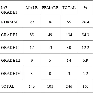
MALNUTITION AND UTI TABLE 6
IAP GRADES
MALE FEMALE TOTAL %
NORMAL 29 36 65 26.4
GRADE I 85 49 134 54.3
GRADE II 17 13 30 12.2
GRADE III 9 5 14 5.9
GRADE IV 3 0 3 1.2
TOTAL 143 103 246 100
UTI was found in 26.4% of children with normal weight while the
remaining 73.6% of children were malnourished. Majority of the
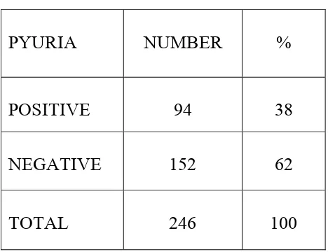
SIGNIFICANT PYURIA TABLE 7
PYURIA NUMBER %
POSITIVE 94 38
NEGATIVE 152 62
TOTAL 246 100
Significant pyuria defined as more than 10 pus cells in
uncentrifuged urine was found in 94 children while 152 children had
SIGNIFICANT PYURIA TABLE 8
PYURIA MALE % FEMALE %
Positive 51 35.6 43 41.7
Negative 92 64.4 60 58.3
Total 143 100 103 100
SIGNIFICANT BACTERIURIA
TABLE 9
CULTURE NUMBER %
POSITIVE 146 59.3
NEGATIVE 100 40.7
TOTAL 246 100
Significant growth of bacteria was seen in 146 children and
SIGNIFICANT BACTERIURIA
TABLE 10
CULTURE MALE % FEMALE %
POSITIVE 84 58.3 62 60.2
NEGATIVE 59 41.7 41 39.8
TOTAL 143 100 103 100
58.3% of male children and 60.2% of the female children had
BACTERIOLOGICAL GROWTH PROFILE
TABLE 11
ORGANISM NUMBER %
Escherichia coli 80 54.7
Klebsiella pneumoniae 10 6.8
Klebsiella oxytoca 8 5.4
Staphylococcus aureus 22 15
CONS 8 5.4
Proteus mirabilis 2 1.3
Proteus vulgaris 2 1.3
Pseudomonas aeruginosa 3 2
Enterococcus fecalis 4 2.7
Acinetobacter 2 1.3
Citrobacter 4 2.7
Providencia 1 1.4
TOTAL 146 100
E.Coli was grown in 80 children, Klebsiella species in 18,
Staphylococcal group in 30, Proteus species in 4, Pseudomonas in
BACTERIOLOGICAL GROWTH PROFILE
[image:43.612.137.499.175.561.2]SEX DISTRIBUTION
TABLE 12
E.coli was the commonest organism grown in 44 male children
and 36 of female children. Proteus mirabilis and Providencia was seen in
male children only.
ORGANISM MALE % FEMALE %
Escherichia coli 44 52.4 36 58.1
Klebsiella pneumoniae 7 8.3 3 4.8
Klebsiella oxytoca 4 4.7 4 6.5
Staphylococcus aureus 13 15.5 9 14.6
CONS 5 5.9 3 4.8
Proteus mirabilis 2 2.4 0 0
Proteus vulgaris 1 1.2 1 1.6
Pseudomonas aeruginosa 2 2.4 1 1.6
Enterococcus fecalis 2 2.4 2 3.2
Acinetobacter 1 1.2 1 1.6
Citrobacter 2 2.4 2 3.2
CULTURE AND SENSITIVITY PATTERN TABLE 13
ORGANISM SENSITIVITY
PATTERN
NUMBER %
Escherichia coli Amikacin 51 88
Klebsiella pneumoniae Ciprofloxacin 7 72
Klebsiella oxytoca Norfloxacin 5 69
Staphylococcus aureus Ciprofloxacin 17 80
CONS Ofloxacin 7 86
Proteus mirabilis Amikacin 1 50
Proteus vulgaris Ofloxacin 2 100
Pseudomonas aeruginosa Ceftazidime 2 83
Enterococcus fecalis Cotrimoxazole 3 76
Acinetobacter Amikacin 1 50
Citrobacter Norfloxacin 2 42
Providencia Ciprofloxacin/Oflo xacin/ Norfloxacin
RADIOLOGICAL PROFILE
TABLE 14
DIAGNOSIS MALE FEMALE TOTAL %
Cystitis 55 12 67 60.9
Hydroureteronephrosis 6 4 10 9.2
PUJ obstruction 4 2 6 5.4
Posterior urethral
valve 5 0 5 4.5
Vesico ureteric reflux 9 5 14 12.8
Calculus 4 0 4 3.6
Pyelonephritis 2 0 2 1.8
Medical renal disease 0 2 2 1.8
TOTAL 85 25 110 100
110 of the 246 children had radiological abnormalities in
Ultrasonogram. Micturiting cystouretherogram was done in 142 children
COMORBIDITY IN UTI
TABLE 15
DISEASE MALE FEMALE TOTAL %
Enteric fever 1 5 6 2.4
Nephrotic syndrome 5 3 8 3.2
Heart disease 0 2 2 0.8
Hypertension 1 1 2 0.8
6 children had coexistent Enteric fever, 8 were known nephrotic
syndrome patients, while 1 child had Patent ductus arteriosus and 1 child
DISCUSSION
This study included 246 children in the age group between 1
month to 12 years. 15 (6.1%) were less than 1 year, 100 (40.7%) were
between 1 to 4 years, 75 (30.5%) were between 5 to 8 years and 56
(22.7%) between 9 to 12 years of age. Urinary tract infection was more
common in the 1 to 4 years age group in this study and this is supported
by other studies by Thaer-Al-Momani et al 12 where 49 % were in the 1
to 4 years age group and Neelam et al 19where 38.7% belonged to the
same age group.
Of the 246 children 143 (58.1%) were males and 103 (41.9%)
were females. The male to female ratio in this study is 1.3:1. This is in
contrast to the other studies (15, 16) which show a female
preponderance. The study by Malla et al 16also shows a male to female
ratio of 1:2, 67.2% being females. N.Choudhuri et al13 study on
community acquired UTI ,with a male preponderance (53.6%) when
compared to the females(46.4%) supports our study .Another study by
Under the less than 1 year age group UTI was more
common in the male children (8.5%) as reported in other studies 17. In
children more than 5 years UTI was common among the females (34%)
and this is supported by other studies 4,18.
CLINICAL PROFILE
The clinical profile of UTI is varied. Symptoms of UTI may be
minimal and non-specific in infants and small children. Febrile children
not suspected of having UTI are as likely to have UTI as those who are
suspected of having UTI. Many studies have been done to study the
clinical characteristics of UTI in children and fever is the most common
symptom in younger children while dysuria and fever is more common
in older children .
In this study dysuria is the commonest symptom found in 89.4%
followed by fever accounting for 84.5% diarrhea in 24.8%, vomiting in
22.7%, abdominal pain in 21.5%, febrile seizures in 6%, dehydration in
3.3%, flank pain in 0.4%, recurrences in 6.1% dribbling in 0.8% and
poor urinary stream in 6.1%.
Azhar Munir Qureshi et al 15study shows dysuria in 98%, fever
in 92%, recurrence in 28%, vomiting in 26%,poor stream in 20% and
Anis- ur- Rahman et al 14 study on the clinical presentation of UTI, fever was seen in 91% dysuria in 65% previous
episodes in 30%,vomiting in 28%, abdominal pain in 22% poor urinary
stream in 15%.
Malla et al 16 study shows fever in 86.9%, dysuria in
40%, pain abdomen in 46.4% vomiting in 39.2%, diarrhea in 14.28% .
Dysuria is the most common clinical presentation in
this study in contrast to the above 2 studies which show fever as the
most common clinical feature followed by dysuria.
In this study diarrhea formed a feature in 24.8% and
the study by M.H.Fallahzadeh et al 30 supports this with diarrhea found
in 25% of children with UTI. The association of diarrhea with urinary
tract infection has also been shown in other studies by Thakar et al 31 and
Pryles C V et al 32.
Febrile seizures with UTI in this study is 6% while
other studies by Mc Intyre et al 33 shows 2.6%, Lee P et al 34 study shows
3.9% and Malla et al 16 shows 13% occurrence of febrile seizures.
Recurrence if found in 6.1% in this study. There have
been many studies on the causes for recurrence in UTI in children but
C .Mingin 35 on the factors predictive of UTI recurrence , shows a recurrence rate of 24% which is very high when compared to this study.
Schlager et al study 9 shows that recurrence is more
common in girls regardless of the presence or absence of urinary tract
abnormality. It also states that among the males one third have recurrent
UTI.
In this study recurrence was found in 15 children - 9
male (6.3%) and 6 female (5.8%) and urinary tract abnormalities was
found in majority of them in the form of Posterior urethral valves
,vesico ureteric reflux and cystitis.
On analysis, normal weight was found only in 26.4% of
the children with UTI while the remaining 73.6 % fall in the
malnourished group with 54.3% in grade I, 12.2% in grade II, 5.9% in
grade III and 1.2% in grade IV malnourishment of the IAP grading.
Similar results are found in other studies.
In a study on the frequency and clinical presentation of
UTI 15 failure to thrive was found in 40% of the children. Dayal et al36
and Brooke et al 37 studies show failure to thrive in 26% and 9.5%
LABORATORY PROFILE
Significant pyuria defined as more than 10 pus cells in uncentrifuged urine sample was found in 38% of children. S.Fouzas et
al 17 study shows significant pyuria in 67.3%.Another study 19 shows
significant pyuria 53.6%.
Significant bacteriuria was found in 146 (59.3%) and the
urine culture was negative or showed insignificant bacteriuria in
100(40.7%) children. This is supported by a study 16 where culture
positivity was found in 57%. But another study18 showed positive
culture in 28.6% .
62(60.2%) of the females and 84 (58.3%) of the males
showed significant growth in urine culture. This is similar to the study
by G.K.Rai et al 18 which showed no significant difference in growth
positive rates in 2 genders (Male 51.7% and female 48.3%).
The commonest pathogen grown in culture in this study
was Escherichia coli 54.7% .
Klebsiella pneumoniae was found in 6.8%, Klebsiella
oxytoca in 5.4%, Staphylococcus aureus in 15%, Coagulase negative
staphylococcus in 5.4%, Proteus mirabilis in 1.3%, Proteus vulgaris in
Acinetobacter in 1.3%,Citrobacter sp. in 2.7%,Providencia species in
1.4%.
Another study 17 shows culture pattern as follows:
E.coli 47.1%, Klebsiella pneumoniae 15.6% Klebsiella oxytoca 1%,
Proteus mirabilis 5.9%, Proteus vulgaris in 0.3%, Staphylococcus aureus
in 1.7%, Enterococcus fecalis 8.7%, Acinetobacter 2.8% and Citrobacter
1.4%
E.coli was grown in 93.3%, Klebsiella in 1.5%
Proteus in 2.3%, Citrobacter in 0.7%, Staphylcoccus aureus in 0.7%,
Pseudomonas in 0.7%, Salmonella in 0.2% and Enterobacter in 0.6% in
study by Rai et al.
Fakhrossadat et al 20 studies showed E.coli in 63%,
Klebsiella in 19.4%, Enterobacter 6%, Pseudomonas 4.7%, Proteus
mirabilis in 3.9%, Enterococcus fecalis in 2.2%, Staphlyococcus aureus
in 0.4%, Providentia in 0.4% .
One study showed growth of Coagulase negative
staphylococcus in 1.1%.
On analysis Escherichia coli was the commonest
organism in most of the studies and ranged from 47% to 93% supporting
While Klebsiella species forms the second most common
organism in other studies ranging from 1.5% to 16%, Staphylococcus
aureus forms the second most common urinary pathogen in this study
(14.6%)
Staphylococcus aureus growth ranged from 0.4% to 10% in other
studies. S.K.Abdulhadi et al 38 study showed Staphylococcus aureus in
10% of children with UTI.
The drug sensitivity pattern of the urinary pathogens are as
follows : E.Coli sensitive to Amikacin in 88%, Klebsiella pneumoniae
to Ciprofloxacin in 72%, Klebsiella oxytoca to Norfloxacin in 69%,
Staphylococcus aureus to Ciprofloxacin in 80% ,Pseudomonas to
Ceftazidime in 83% and ciprofloxacin in 70% ,Proteus mirabilis was
sensitive to amikacin in 50%,Proteus vulgaris to Ofloxacin in 100%
CONS sensitive to Ofloxacin in 86%,Enterococcus fecalis showed
maximum sensitivity to Cotrimoxazole in 76%,Citrobacter to
Norfloxacin in 42%, Acinetobacter to Amikacin in 50% and Providencia
to ciprofloxacin, Ofloxacin and Norfloxacin in 100% .
Nasim Kashef et al 22 study on the sensitivity pattern of urinary
pathogens to drugs showed maximum susceptibility of E.Coli to
Norfloxacin (91.7%), Staphylococcus aureus to Gentamicin (100%),
Vancomycin (100%), Nitrofurantoin (75%) and Cotrimoxazole ( 75%).
Proteus was sensitive to Ciprofloxacin (71.2%), Pseudomonas to
Ceftazidime (83.3%) and Norfloxacin (80%) ,CONS to Norfloxacin,
Genatmicin, Nitrofurantoin and Cotrimoxazole in 88.9%, Citrobacter
was 100% sensitive to Ciprofloxacin, Norfloxacin, Nalidixic acid,
Gentamicin, Nitrofurantoin and Cotrimoxazole.
Fakhrossadat et al 20 study showed E.Coli sensitivity
to Nitrofurantoin in 90.4% and Amikacin in 84.2% and Klebsiella to
Ciprofloxacin in 77% and Nalidixic acid in 64%.
Sharifian et al study 21 shows maximum sensitivity of
E.Coli to Ceftriaxone (97.8%) and Cefotaxime (95.2%), Staphylococcus
to Cephalexin in 90.5% and Ciprofloxacin in 89.7%, Pseudomonas to
Ciprofloxacin in 94.7% and Amikacin in 83.9%, Klebsiella to
RADIOLOGICAL PROFILE
Ultrasonogram was done in all patients admitted with
UTI and micturiting cystourethrogram was done in 142 children.
140(55%) children had normal ultrasound while 110 (45%) had
radiological abnormalities. Cystitis was the commonest radiological
feature found in 67 (60.9%) children (55 male and 12 female).
Hydroureteronephrosis in 10 (9.2%),6 male and 4 female children, Pelvi
ureteric junction abnormalities in 6(5.4%), Posterior urethral valve in
5(4.5%), Vesicoureteric reflux in 14 (12.8%) ,calculus in 4(3.6%),
Pyelonephritis in 2(1.8%) and Medical renal disease in 2(1.8%). were
the other radiological abnormalities found. Grade I VUR was seen in 9
children grade II in 2 grade IV in 1 and grade V in 2 children. Children
with grade I and II VUR were given antibiotic prophylaxis and children
in grade IV and V were referred for surgical management. Anatomical
abnormalities were more commonly found in the male children than the
female children.
Jothilakshmi et al 39 in her study on the radiological
evaluation of urinary tract in children with urinary infection found
Pui Meng Mok et al 40 found normal radiology in 63.9% and
abnormalities in 36.1%.Neelam et al 19 in her studies found VUR in
19.9% and PUV in 27.6% of children with UTI.
In a study by Ali Ahmedzadeh 25, VUR was seen in 40%,
Calculus in 8%, PUJ obstruction in 6.3%, PUV in 2 boys and double
collecting system in 2 girls.
COMORBIDITIES
Nephrotic syndrome was the commonest comorbidity in this study
found in 8 children, enteric fever in 6 children, congenital heart disease
CONCLUSIONS
• Urinary tract infection was more common in children between 1
to 4 years.
• In children less than 5 year UTI is common in male children and
more common in female children in the 5 to 12 years age group.
• Dysuria was the most common clinical presentation followed by
fever.
• 54.3% of children fell into the Grade I malnutrition group.
• Significant pyuria was found in 38%.
• 59.3% showed significant bacteriuria.
• Escherichia Coli was the commonest urinary pathogen and
showed maximum sensitivity to Amikacin.
• Radiological abnormalities were found in 43% of the children.
• Cystitis was the commonest radiological finding.
• Vesicoureteric reflux was documented in 13.2%.
LIMITATIONS OF THIS STUDY
•
Follow up could not be done for all children.
•
Sensitivity pattern of the cultured organism varied from time
to time during the study period due to differences in the
sensitivity discs used in the microbiology lab.
•
MCU was not done in all children due to technical difficulties
and loss of follow up.
•
DMSA scan was not done due to non availability of the
investigation in the institution and financial constraints.
RECOMMENDATIONS
1)
Urine culture should be sent to all infants with fever and non
specific symptoms.
2)
Micturiting cystouretherogram to be done in children less
than 5 years of age.
3)
Micturiting cystouretherogram to be done to all children with
BIBLIOGRAPHY
1) Gulati S, Kher V. Urinary tract infection. Indian Pediatr 1996;33:
212-7.
2) Bickerton MW, Ducket JW. Urinary tract infection in pediatric
patients. American Urological Association, Houston, Texas 1985.
3) Shaw KN, Gorelick M, Mcgowan KL, Yakscore NM, Schwartz
JS. Prevalence of urinary tract infection in febrile young children
in the emergency department.Pediatr 1998; 102: 16-21
4) Elder JS. Urinary tract infections. In: Kliegman RM, Behrman
RE, Jenson HB, Stanton BE, editors. Nelson Textbook of
Pediatrics. Philadelphia: Saunders 2007. 2223-8.
5) Watson AR. Disorders of the urinary system. In: Campbell AG,
McIntosh N, editors. Forfar and Arneil’s Textbook of Pediatrics.
Churchill Livinstone 1998: 949-56.
6) Bachur R, Harper MB. Reliability of the urine analysis for
predicting urinary tract infections in young febrile children. Arch
Pediatr Adoles Med2001; 155: 60-5.
7) Chon CH, Lai FC, Shorthffe LM. Pediatric urinary tract
8) Winberg J, Andersen HJ, and Bergstrom T, et al: Epidemiology of
symptomatic urinary tract infection in childhood. Acta Paediatr
Scand 63 (suppl 252): 1-20. 1974.
9) Schlager TA, and Loher JA: Urinary tract infection in outpatient
febrile infants and children younger than 5 years of age. Pediatr
Ann 22: 505-509.1993.
10) Spenser JR, and Schaeffer AJ: Pediatric urinary tract infections.
Urol Clin North Am 13: 661-672. 1986.
11) Weiss R, Tamminen-Mobius T, Koskinties O, et al: International
reflux study in children. J Urol 148: 1644-1734. 1992.
12) Dr.Thaer- al- Momani; Microbiological study of UTI in children
at Princess Haya hospital in South Jordan.Middle East Journal of
Family Medicine Mar 2006 Vol 14
13) N.Choudhuri T.K.Chatterjee K.Nayak , T.N. Ghosh, M.S.Akhtar ;
A study of bacteriological investigation of community acquired
UTI in children attending a tertiary care hospital.
14) Anis-ur-Rehman, Muhamad Jahanzeb, Tahir Saeed Siddiqui,
Muhammad Idris;Frequency and clinical presentation of UTI
among children of Hazara division, Pakistan J Ayub Med Coll
15) Azhar Munir Qureshi; Clinical presentation and organisms
causing urinary tract infection in pediatric patients at Ayub
teaching hospital Abbottabad; Deparment of Pediatrics , Ayub
medical college Abbottabad
16) Malla KK, Sarma MS, Malla T, Thapalial A:Clinical profile,
bacterial isolates and antibiotic susceptibility patterns in Urinary
Tract Infection in children – hospital based study , Department of
Pediatrics ,Manipal college of medical sciences and teaching
hospital, Nepal
17) S.Fouzas, L.Mantagou, N.Fotiadis, A.Filias, S.Mantagos : UTI
without significant pyuria in children; Archives of Disease in
childhood. 2008;93
18) GK.Rai, HC Upreti, SK Rai,K P Shah, R M Shrestha ;Causative
agents of UTI in children and their antibiotic sensitivity pattern ;
a hospital based study .Nepal Med Coll J 2008; 10(2): 86-90
19) Neelam Taneja, Shiv Sekhar Chatterjee, Meenakshi Singh, Surjit
Singh& Meera Sharma ; Pediatric urinary tract infections in a
tertiary care center from north India, Ind J Med Res 131, Jan
20) Fakhrossadat Mortazavi, Narges Shahin: Changing patterns in
sensitivity of bacterial uropathogens to antibiotics in children
:Pak J Med Sci October –December 2009 (Part-I)Vol. 25 No.
5 ,801-805
21) Sharifian M, Karimi A, Tabatabaei SR, Anvaripour N. Microbial
sensitivity pattern in urinary tract infections in children: A single
center experience of 1177 urine cultures. Jpn J Infect Dis
2006;59:380-2
22) Nasim Kashef, Gholamreza Esmaeeli Djavid, Sahba Shahbazi:
Antimicrobial susceptibility patterns of community acquired
uropathogens in Tehran, Iran, Department of microbiology,
University of Tehran, Iran
23) P Senguttuvan, K Ravanan, N Prabhu, V Tamilarasi: Infections
encountered in childhood nephrotics in a pediatric renal unit
:Indian J Nephrol 2004;14: 85-88
24) Alejandro Hoberman , Martin Charron, Robert W Hickey, Marc
Baskin,Diana H Kearney, Ellen P Wald ; Imaging studies after a
first febrile UTI in young children, The New England Journal of
25) Ali Ahmedzadeh, Shahnam Askarpour; Association of urinary
tract abnormalities in children with first urinary tract infection;
Pakistan journal of medical sciences Jan 2007 Vol 23
26) L.Pead, R.Maskell; Study of urinary tract infection in children in
one health district BMJ 1994 ;309: 631-634
27) Farrell DJ, Morrissey I, Rubeis D, Robbins M, Felmingham D. A
UK multicentre study of the antimicrobial susceptibility of
bacterial pathogens causing urinary tract infection. J Infect
2003;46(2):94-100.
28) Pape L, Gunzer F, Ziesing S, Pape A, Offner G, Ehrich JH.
Bacterial pathogens, resistance patterns and treatment options in
community acquired pediatric urinary tract infection.Klin Paediatr
2004.216(2):83-6.26)
29) Danielo V P. Antelo MD:Med students homepage
30) M.H.Fallahzadeh F.Ghane; Urinary tract infection in infants and
children with diarrhea ; Shiraz university of medical sciences
31) Thakar R et al. Urinary tract infection in infants and young
children with Diarrhea. Indian pediatrics, 2000, 37(8): 886–9.
32) Pryles CV, Luders D. The bacteriology of the urine in infants and
33) McIntyre PB, Gray SV and Vance JC. Unsuspected bacterial
infections in febrile convulsions. Medical Journal of Australia.
1990; 152(4):183-6
34) Lee P and Verrier-Jones K. Urinary Tract Infection in Febrile
Convulsions. Archives of Disease in Childhood. 1991;
66(11):1287-90
35) Gerald C Mingin, Angie Hinds,Hiep T Nguyen, Laurence
S.Baskin;Factors predictive of UTI recurrence; Dept of urology
and pediatrics , San Francisco Childrens Hospital University of
California
36) Dayal RS, Luthra UK, Kalra K, Lall JC,Pal R. Renal biopsy in
malnourished children. Indian Pediatr 1970,7: 596-604.
37) Brooke OG, Kerr DS. The importance of routine urine culture in
malnourished children. Environ Child Health 1973, 19:348-349
38) S. K. Abdulhadi, A. H. Yashua and A/ Uba :Organisms causing
Urinary Tract Infection in paediatric patients at Murtala
Muhammad Specialist Hospital, Kano, Nigeria ; International J of
Biomed and health sci vol 4, No 4 Dec 2008
39) K. Jothilakshmi , Bhoopathy Vijayaraghavan , Sarah Paul and
John Matthai :Radiological Evaluation of the Urinary Tract in
Radiology, PSG institute of Medical Sciences and Research,
India.
40) Pui Meng Mok ,Paul .R.White; The value of radiological
investigation in pediatric UTI. Princess Mary Hospital,
NewZealand, Aust.Radiol.1979.23
41) Bulletin of the Indian pediatric Nephrology group: IAP speciality
chapter on Nephrology : March 2004, vol.5 No.1
PROFORMA
GOVT RAJAJI HOSPITAL, MADURAI
CLINICAL, LABORATORY AND RADIOLOGICAL PROFILE OF URINARY TRACT INFECTION IN CHILDREN
NAME : AGE/SEX :
IP NO : UNIT :
ADDRESS :
SYMPTOMS:
DYSURIA □ FEVER □
VOMITING □ DIARRHOEA □
ABDOMINAL PAIN□ FLANK PAIN □
HEADACHE □ CONVULSIONS □
DRIBBLING □ POOR STREAM □
PREVIOUS EPISODES :
WEIGHT :
BLOOD PRESSURE :
LAB PROFILE :
URINE ALBUMIN :
DEPOSITS :
CULTURE AND SENSITIVITY:
USG ABDOMEN :
MCU :
COMORBIDITIES :
ABBREVIATIONS
UTI - URINARY TRACT INFECTION
USG - ULTRASONOGRAM
MCU - MICTURITING CYSTOURETHROGRAM
CT - COMPUTERISED TOMOGRAPHY
MRI - MAGNETIC RESONANCE IMAGING
PEM - PROTEIN ENERGRY MALNUTRITION
DMSA - DI-MERCAPTO SUCCINIC ACID
E.Coli - ESCHERICHIA COLI
CONS - COAGULASE NEGATIVE
STAPHYLOCOCCUS
MASTER CHART ABBREVIATIONS
SEX
1 - MALE
2 - FEMALE
FEVER, DYSURIA, ABDOMINAL PAIN, VOMITING, DIARRHOEA
1 - PRESENT
POOR STEAM, DRIBBLING, FLANK PAIN, SEIZURES
1 - PRESENT
2 - ABSENT
DEHYDRATION, RECURRENCE
1 - PRESENT
2 - ABSENT
WEIGHT
1 - NORMAL
2 - GRADE I PEM
3 - GRADE II PEM
4 - GRADE III PEM
5 - GRADE IV PEM
PYURIA
1 - SIGNIFICANT
2 - INSIGNIFICANT
ORGANISM
1 - E.COLI
2 - KLEBSIELLA PNEUMONIAE
3 - KLEBSIELLA OXYTOCA
5 - PROTEUS MIRABILIS
6 - PROTEUS VULGARIS
7 - PSEUDOMONAS AERUGINOSA
8 - ENTEROCOCCUS FECALIS
9 - ACINETOBACTER
10 - PROVIDENCIA
11 - COAGULASE NEGATIVE STAPHYLOCOCCUS AUREUS
12 - CITROBACTER
13 - NEGATIVE OR NO SIGNIFICANT GROWTH
AMINOGLYCOSIDES
1 - AMIKACIN
2 - GENTAMICIN
FLUOROQUINOLONES
1 - CIPROFLOXACIN
2 - NORFLOXACIN
3 - OFLOXACIN
CEPHALOSPORINS
1 - CEFTRIAXONE
2 - CEFOTAXIME
3 - CEPHALEXIN
OTHERS
1 - COTRIMOXAZOLE
2 - VANCOMYCIN
3 - NALIDIXIC ACID
COMORBIDITY
1 - ENTERIC FEVER
2 - HEART DISEASE
3 - HYPERTENSION
4 - NEPHROTIC SYNDROME
USG/MCU
1 - NORMAL
2 - CYSTITIS
3 - VESICOURETERIC REFLUX
4 - PYELONEPHRITIS
5 - MEDICAL RENAL DISEASE
6 - HYDROURETERONEPHROSIS
7 - PUJ OBSTRUCTION
8 - POSTERIOR URETHRAL VALVE
AGE DISTRIBUTION
6%
31% 22%
41%
< 1 YEAR 1-4 YEARS 5-8YEARS 9-12YEARS
SEX DISTRIBUTION
MALE
58%
FEMALE
42%
CLINICAL PROFILE
220 208 56 53 61 16 8 2 15 2 150 50 100 150 200 250
Dysuria Fever Vomiting Abd.pain Diarrhoea Febrile seizures Dehydration Flank pain Recurrence Dribbling Poor stream
SIGNIFICANT BACTERIURIA
146, 59% 100, 41%
BACTERIOLOGICAL GROWTH PROFILE 1 4 2 4 3 2 2 8 8 22 10 80
0 10 20 30 40 50 60 70 80 90
E.Coli
K.pneumoniae
K.oxytoca
Staph.aureus
CONS
Proteus
mirabilis
Proteus
vulgaris
Pseudomonas
E.Fecalis
Acinetobacter
Citrobacter
Providencia
NORMAL MCU